Index
Abstract
and
Introduction
pag.1
Abstract pag. 2
Introduction pag. 4
Erythropoietin
and
Stem
Cell
Factor pag.8
Role of Erythropoietin and Stem Cell Factor: pag. 9
Erythropoietin. pag. 9
The Erythropoietin Receptor (EpoR) pag. 10
Stem Cell Factor pag. 11
Kit: The Stem Cell Factor Receptor pag. 12
Mechanism of cooperation between c-Kit and erythropoietin receptor pag.14
Role of SCF and Epo in therapy pag. 14
Erythropoietin and Stem Cell Factor in cancer pag. 16
Material
and
Methods
pag. 19
Isolation, characterization and culture of primary and immortalized tumor cells. pag. 20
Flow cytometric analysis. pag. 20
Confocal Microscopy pag. 20
Western blotting pag. 21
Cell Growth pag. 21
Detection of Apoptosis pag. 21
Adult peripheral blood human progenitor cell (HPC) purification and culture. pag. 22
Mice pag. 22
Stem Cells Factor and Cytotoxic Treatment pag. 22
Statistical Analysis pag. 23
Results
pag. 24c-Kit and EpoR expression in solid tumors pag. 25
chemotherapy-induced apoptosis in MCF-7 breast cancer cell line. Pag. 26 Epo increases the expansion and protects both primary differentiated
and undifferentiated breast cancer cells from drug-induced cell death. pag. 28 Stem Cell Factor protects the hematopoietic system from drug induced cell death pag. 30
Discussion
pag. 35Abstract
The ability of erythropoietin (Epo) to promote the production of red cells is currently exploited to treat chemotherapy-induced anemia. However, the expression of Epo receptor (EpoR) in a variety of cancer cells suggests that Epo-based supportive therapy can negatively affect the clinical outcome. In line with this hypothesis, some clinical trials have questioned the benefit of Epo administration in patients affected by different tumors, including breast cancer. In this study we directly determined the effect of Epo on cancer mammospheres, whose tumorigenic activity was validated through the establishment of xenografts in immunocompromized SCID mice.
Our preliminary data showed that EpoR was expressed in both, undifferentiated mammospheres and in differentiated primary breast cancer cells.
The presence of Epo increased the expansion and survival of tumor mammospheres and differentiated primary breast cancer cells. More importantly, Epo was able to considerably protect both, differentiated and undifferentiated breast cancer cells, from death induced by many anti-neoplastic drugs. Accordingly, we observed that Epo increased the expression of its receptor, induced activation of AKT/PKB and MAPKs and increased the expression of Bcl-xL in breast cancer cells. Thus, the use of Epo may promote the survival and growth of tumorigenic breast cancer cells by counteracting the cytotoxic effects of chemotherapy suggesting the need for alternative therapeutic options in cancer patients.
In a comprehensive investigation, 81/120 tumor types examined did not yield any sample positive for c-kit expression, suggesting that the use of Stem Cell Factor (SCF) should be safe in many of the most common malignancies. 1
To determine the possible oncogenic effect of SCF, we compared the pro-tumor activity of Epo and SCF on breast cancer, the major cancer type in women. Among this, we tested the potential protective effects of SCF in preventing hematopoietic cell death during chemotherapy in vivo.
Our data are showing that Epo increased the expansion and survival of tumor mammospheres and differentiated primary breast cancer cells. More importantly, Epo was able to considerably protect both, differentiated and undifferentiated breast cancer cells, from death induced by many anti-neoplastic drugs possibly through increased expression of the anti-apoptotic protein Bcl-xL.
SCF, on the contrary, can not exert any pro-tumor activity, since the majority of cancer cells tested, particularly breast cancer, resulted negative for c-kit expression.
In in vitro experiments performed on primary human erythroid progenitors we found that SCF is able to prevent apoptosis of erythroid progenitors induced by promising new anticancer agents and in vivo SCF restores the density of bone marrow cells to the level of controls in mice treated with Cisplatin or 5-Fluorouracil (5-FU).
In peripheral blood analysis we observe an increase in the levels of all mature blood cells upon SCF administration. Therefore, taken together our experiments demonstrate that SCF protects the hematopoietic system from chemotherapy-induced damage in vivo and outline a protocol for a potential clinical application of SCF to prevent chemotherapy-induced cytotoxicity.
Introduction
Erytropoiesis is a dynamic and complex process that maintains the number of circulating erythrocytes within a relatively narrow range under changing physiologic conditions in normal individuals 2,3. A reduction of this number it is known as anemia. In clinical oncology, anemia affects the majority of patients undergoing chemotherapy and it is related manly to inhibition of erythoid cell maturation in the bone marrow, tumor-associated bleeding, hemolysis, marrow damage from metastases or myelodysplasia and toxicities associated with chemotherapy and radiation therapy 4.In most cases, cancer-related anemia is thought also to be caused by a complex interaction between the tumor cell population and the immune system, which ultimately disrupts the normal erythropoiesis manly by reducing the production of Erythropoietin (Epo) from the kidney 2,3,5,6.Epo is the primary regulator of erythropoiesis, stimulating growth, preventing apoptosis and promoting differentiation of red blood cell progenitors by binding to its cognate cell surface receptor (EpoR). Therefore, in therapy it is widely used for the prevention and treatment of chemotherapy-associated anemia. Epo significantly increases haemoglobin (Hb) levels, reduces transfusion requirements, and improves quality of life, particularly by relieving fatigue4 .Recent studies however, have shown that Epo has biological functions aside from regulating erythropoiesis and that many solid tumors express EpoR, raising concerns about the fact that Epo could stimulate the growth of indolent tumors and interfere with the action of antitumor therapies, either by enhancing tumor proliferation rates or interfering with apoptotic cell death (Fig. 1). Epo responsiveness has also been identified in capillary endothelial cells, indicating that Epo may have a role in tumor angiogenesis 7-12 .This drawback has raised doubts on the opportunity to use erythropoietin in the supportive care of cancer patients 13, 14 suggesting the need for alternative therapeutic options.
Erythropoietin
EpoR EpoR
Red Blood Cells Tumor
Figure 1.Possible mechanism of Erythropoietin. Epo stimulates red blood cell production by attaching to receptors on those cells. But there is mounting evidence that some nonhematopoietic cells also carry Epo receptors (EpoR). Susan Wolsborn.
Recently, the presence of c-kit, the receptor of an other essential factor for erythropoiesis, the Stem Cell Factor (SCF), has been extensively investigated in cancers of various origin. In a comprehensive investigation 1, 81/120 tumor types examined did not yield any sample positive for c-kit expression. C-kit negative tumors included both solid and hematologic tumors such as hormone-refractory prostate carcinoma, medulloblastoma, tubular breast carcinoma, Kaposi’s sarcoma, non-Hodgkin’s lymphomas and chronic myelogenous leukaemia.
Other investigators15 reported a complete absence of c-kit expression in malignant brain tumors, breast and ovarian cancer, suggesting that the use of SCF should be safe in many of the most common malignancies.
Although SCF has not been used in clinic for the expansion of erythroid progenitors; studies have shown that SCF can cooperate with other cytokines such as G-CSF in long term cultures of human primitive hematopoitic cells as well as for ex vivo expansion of cord blood cells for transplantation16 and in clinical trials in human with multiple myeloma, breast cancer and lymphoma SCF has been used successfully with G-CSF to mobilize stem and progenitors cells 17,18. The c-kit receptor can be expressed by both normal and neoplastic tissues, where its stimulation by the natural ligand SCF can generate proliferative and survival stimuli. Therefore it is extremely important to avoid the use of SCF as a hemoprotective factor in oncologic patients expressing functional c-kit, as one could not rule out the possibility that it would enhance the growth and survival of the tumor itself. The risks related to the use of hematopoietic cytokines in the supportive care of cancer patients must also be carefully evaluated at the light of the recent discovery of cancer stem cells. The existence of a undifferentiated subpopulation of tumorigenic cells responsible for tumor maintenance, growth and spreading was known since several years in leukemias, but has been now demonstrated to occur also in solid tumors such as breast and brain cancers 19, 20. The possibility that cancer stem cells may use hematopoietic growth factors to proliferate and resist to apoptotic stimuli poses an additional caveat for the administration of cytokines to cancer patients and requires a careful assessment of the presence of cytokine receptors on the surface of both stem and differentiated cancer cells. In our laboratory, we have obtained cancer stem cells from several solid tumors including breast, thyroid, colon, and lung carcinomas etc.
We analysed the expression of EpoR and c-kit in a variety of cancer types and we found that the large majority of stem and differentiated cancer cells tested resulted negative for c-kit expression and positive for EpoR expression. Above this, to determine the possible oncogenic effect of SCF, we compared the pro-tumor activity of Epo and SCF on breast cancer, the major cancer type in women.
Breast cancer cells, undifferentiated and differentiated, express EpoR, but do not express c-Kit, indicating that SCF can not have a pro-tumor effect on this type of cancer. In these cells SCF does not interfered with chemotherapy-induced toxicity. On the contrary the presence of Epo increased the expansion and survival of tumor mammospheres and differentiated primary breast cancer cells. More importantly, Epo was able to considerably protect both, differentiated and undifferentiated breast cancer cells, from death induced by many anti-neoplastic drugs possibly through increased expression of the anti-apoptotic protein Bcl-xL.
Thus, the use of Epo may promote the survival and growth of tumorigenic breast cancer cells while SCF should be safe in many of the most common malignancies and particularly in breast cancer. Cancer related anemia occurs primarily due to progenitor cells loss.
In the normal hematopoietic system c-kit is expressed primarily by stem cells and progenitor cells and its expression decreases along with maturation in nearly all blood cells (with the exception of mast cells). Therefore, hematopoietic stem and progenitor cells represent the primary target of SCF, which acts by stimulating proliferation and inhibiting chemotherapy-induced cell death.
Therefore, we tested in vitro and in vivo the ability of SCF to act as an hemoprotective factor during chemotherapy treatment.
In in vitro experiments performed on primary human erythroid progenitors we found that SCF is able to prevent apoptosis of erythroid progenitors induced by CD95/Fas ligand and TRAIL, one of the most promising new anticancer agent, the death receptor ligand TRAIL (now entering phase II clinical trials). Moreover in vivo, by analysing bone marrow histological sections obtained from TRAIL-treated mice, we have found that TRAIL induces a moderate bone marrow toxicity (80% bone marrow cellularity of TRAIL-treated animals compared to 100% cellularity of control animals) and that simultaneous treatment with SCF restores the density of bone marrow cells to the level of controls. Thus, SCF may be useful to prevent blood cell depletion induced by TRAIL and possibly by other novel apoptosis-based anticancer agents.
In a different set of experiments, histological sections from mice treated with Cisplatin or 5-Fluorouracil (5-FU) with or without SCF show that mice treated with cisplatin or 5-FU display strong marrow hypoplasia with myelofibrosis and clusters of megakaryocytes. Treatment of mice with SCF resulted in both cases in a strong myeloprotection, as shown by high bone marrow cellularity and the almost complete disappearance of dysplastic cells.
Moreover, as a consequence of apoptosis inhibition of stem and progenitor cells exposed to chemotherapy, we observe an increase in the levels of all mature blood cells upon SCF administration.
Therefore, our experiments demonstrate that SCF protects the hematopoietic system from chemotherapy-induced damage in vivo and outline a protocol for a potential clinical application of SCF to prevent chemotherapy-induced cytotoxicity.
Role of Erythropoietin and Stem Cell Factor:
Erytropoiesis is a dynamic and complex process and its appropriate regulation is essential for embryonic development, adult red cell production and suppression of carcinogenesis.
Survival and apoptosis of hematopoietic stem/progenitor cells are crucial in maintaining the homeostasis of blood cell production and hematopoietic growth factors are crucial for controlling the balance between survival and apoptosis. It is well know that , among a number of growth factor, Stem Cell Factor (SCF) and Erythropoietin (Epo) are the two essential factor for Erythropoiesis 21-25
Erythropoietin.
Erythropoietin is a glycoprotein hormone that serves as the primary regulator of erythropoiesis by stimulating growth, preventing apoptosis and inducing differentiation of red blood cell precursor 26. Clinically, these actions translate into increased levels of haemoglobin, which has lead to the widespread use of recombinant human Epo (rHuEpo) in the treatment of patients with anemia due to renal failure , cancer or cancer therapy.
In humans, Epo mRNA encodes a protein 193 amino acids (aa) and loss of the C-terminal arginine during post-translation modification result in a 165-aa structure that comprises the mature protein (Fig.1).
The Epo molecules contains two structure-stabilizing disulfide bounds between aa 7 and 161 and 29-33, the reduction of which results in loss of bioactivity.
Additionally, the Epo molecules possesses three N-linked sugars, at position 24, 38 and 83, and one O-linked sugar at position 126. The O-linked sugar has no important function, but the N-linked sugars are necessary for stability of the Epo molecule in the circulation 27,28.
Figure 1. Structure of erythropoietin. Reprinted from Mulcahy.
The Erythropoietin Receptor (EpoR).
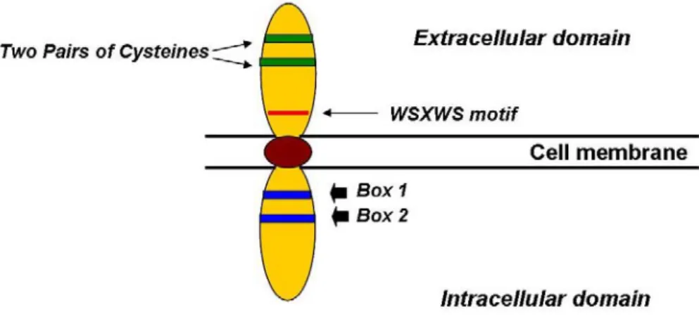
The Epo receptor belongs to the cytokine receptor superfamily 29. Included in this family are receptors for other hematopoietic growth factors, including growth hormone, prolactin, G-CSF, GM-CSF, thrombopoietin, oncostatin M, and several interleukins. Receptors in this family share several distinct features, including an extracellular ligand-binding domain with two pairs of conservedcysteine residues and a conserved motif, WSXWS, located closeto the transmembrane domain; a single transmembrane domain;and an intracellular domain lacking catalytic activity (Fig. 2).
EPO exerts its effects by inducing homodimerization of two moleculesof the Epo receptors on the cell surface, which initiates theJanus kinase (JAK)/signal transducer and activator of transcription (STAT) signal transduction cascade that regulates cell proliferationand differentiation .
Unlike many other receptors, the EPO receptor hasno intrinsic tyrosine kinase activity to activate receptor signaling.Rather, signaling appears to be mediated by JAK2, a cytoplasmictyrosine kinase constitutively associated with the intracellular domain of the EPO receptor. JAK2 molecules associated with each of the individual EPO receptors are brought into close proximity, inducing their transphosphorylation and subsequent activation(Fig. 3). Activated JAK2 then phosphorylates several intracellularproteins, including the EPO receptor itself. The phosphorylatedtyrosines act as docking sites for various intracellular proteins containing Src (tyrosine kinase) homology 2 domains, for example, one pathway activated is the JAK2/STAT5 pathway. Althoughthe precise role of STAT5 in erythroid differentiation is not yet fully understood, JAK2-mediated STAT phosphorylation results in the formation of stable STAT dimers, which in turn translocateto the
Figure 2. Schematic representation of The Epo receptor. The extracellular domains are anchored in the cell membrane. Mulcahy 2001.
nucleus where they bind to specific regulatory sequences and activate the transcription of target genes resulting in erythroid differentiation 28, 30, 31, in particular, STAT5 appears to mediate the induction of Bcl-xL by Epo. In addition to the STAT5 pathway, other signaling pathways,including RAS and PI3K, can be activated by EPO. PI3K signalingis believed to result in the activation of AKT and p70s6K, whichplay a key role in transcription and cell-cycle progression. This PI3K−Akt pathway also leads to upregulation of Bcl-xL and inhibition of apoptosis. A further mechanism could be represented by NF-kB that is also a target of the PI3K-Akt pathway and mediates antiapoptotic signaling by platelet-derived growth factor EPO signaling may also activate many nonreceptor tyrosine kinases(e.g., c-fos/fes, p72syk, and hematopoietic progenitor kinase-1),as well as proliferation-stimulating tyrosine phosphatases SHP2, SH2 inositol 5'-phosphatase, and other signal(Fig.3).
Erythropoietin Signaling
Stem Cell Factor
Stem cell factor is widely expressed during embryogenesis and can be detected in brain, endothelium, gametes, heart, kidney, lung, melanocytes, skin, and the stromal cells of the bone marrow, liver, and thymus 32. SCF exists as membrane-anchored and soluble isoforms that arise from alternative RNA splicing and proteolytic processing; both isoforms contain initially an extracellular domain, a transmembrane segment, and an intracellular component 33.
Figure 3.Summary of demonstrated EPO-signaling, Binding of EPO to its receptor leads to phosphorylation of janus kinase . This subsequently activates multiple cascades recruiting PI3-K, Stat5 and MAPKinase. Further, NF-kB is reported to be dually activated by JAK-2 and by Akt. The net effect is a reduction in the proapoptotic protein BAD and probability of mitochondrial leakage of cytochrome C ,an increased production of antiapoptotic proteins of the Bcl-x family and ultimate preservation of the DNA integrity. To the extent cytochrome C leakage is not prevented, caspase activation also occurs, inducing DNA degradation and the externalization of phosphatidyl serine on the cell membrane promoting the activation of the inflammatory cells. Solid lines indicate activation; dashed indicate inhibition
The precursor for isoform 1, from which the soluble factor is derived, contains 273 amino acids. Residues 1–25 comprise the signal sequence, residues 26–214 make up the extracellular domain, residues 215–237 represent the transmembrane segment, and residues 238–273 constitute the intracellular component (the residue numbers in this article correspond to those of human proteins). Following the removal of the signal sequence, additional processing leads to the generation of the soluble form of SCF (residues 26–189). The enzyme that catalyzes the release of soluble SCF from isoform 1 is most likely matrix metalloprotease-9 34. Isoform 2 contains 28 fewer amino acids because exon 6 is omitted as a consequence of alternative splicing. In humans, isoform 2 lacks the metalloprotease-9 cleavage site and is chiefly membrane anchored.
Kit: The Stem Cell Factor Receptor.
Kit is a type III receptor protein-tyrosine kinase 35, 36 .The type III class also includes the platelet-derived growth factor (PDGF) receptor (α- and β-chains), the macrophage colony-stimulating-factor receptor (CSF-1), and the Fl cytokine receptor (Flt3). Receptor protein-tyrosine kinases all share the same topology: an extracellular ligand-binding domain, a single transmembrane segment, and a cytoplasmic kinase domain. The class III receptors are characterized by the presence of five immunoglobulin-like domains in their extracellular portion. Stem cell factor (SCF) binds to the second and third immunoglobulin domains while the fourth domain plays a role in receptor dimerization 33 . The structure of the class III receptors differs from that of other receptor tyrosyl kinases by the insertion of 70–100 amino acids near the middle of the kinase domain. In human Kit, the kinase insert is about 80 residues in length (Fig.4); this domain undergoes phosphorylation and serves as a docking site for a few pivotal signal transduction proteins.
Figure 4. Organization of Kit. The relative length of the domains is to scale. The location of Kit gain-of-function mutations is indicated by the residue numbers on the right hand side of the figure. Ig, immunoglobulin; AL, activation loop.
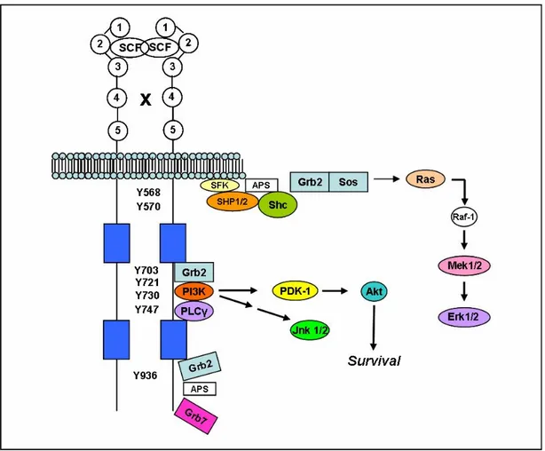
Kit has the potential to participate in multiple signal transduction pathways as a result of interacting with several enzymes and adaptor proteins 37. The adaptor protein APS, Src family kinases, and Shp2 tyrosyl phosphatase bind to phosphotyrosine 568. Shp1 tyrosyl phosphatase and the adaptor protein Shc bind to phosphotyrosine 570. C-terminal Src kinase homologous kinase (Chk) and the adaptor Shc bind to both phosphotyrosines 568 and 570. These residues occur in the juxtamembrane domain of Kit. Three residues in the kinase insert domain are phosphorylated and attract: (a) the adaptor protein Grb2 (Tyr703), (b) phosphatidylinositol 3-kinase (Tyr721), and (c) phospholipase Cγ (Tyr730). Phosphotyrosine 900 in the distal kinase domain binds phosphatidylinositol 3-kinase that in turn binds the adaptor protein Crk. Phosphotyrosine 936, also in the distal kinase domain, binds the adaptor proteins APS, Grb2, and Grb7 37.The numerous Kit interactions cited above lead to activation of several signal transduction pathways (Fig.5) For example, phosphatidylinositol 3-kinase leads to the activation of Akt. Akt (protein 3-kinase B), a protein-serine/threonine 3-kinase, promotes cell survival 38.One substrate of Akt is Bad (Bcl2 antagonist of cell death), a pro-apoptotic protein that promotes cell death. Following phosphorylation, Bad no longer promotes apoptosis. Activation of the phosphatidylinositol 3-kinase/Akt pathway may explain in part how activating mutations of Kit participate in neoplastic transformation. Other downstream effectors of Kit include the Ras/mitogen-activated protein kinases and the Janus kinase/signal transducers and activators of transcription (Jak/STAT) pathways 39.
Figure 5. Schematic illustration of Kit interacting proteins. This figure summarizing signalling proteins activated by Kit and interaction sites on the receptor.
Mechanism of cooperation between c-Kit and erythropoietin receptor
Although SCF has been shown to induce some growth and survival of erythroid progenitors, its response is profoundly amplified in combination with Epo. Biochemical studies have provided evidence for physical association between c-Kit and Epo-R via the box 2 region cytoplasmic domain of the Epo-R 40. Wu et al., utilizing various truncation mutants of Epo-R have demonstrated that c-Kit stimulation by SCF does not activate the Epo-R by inducing its dimerization, but by phosphorylating tyrosine residues in the cytoplasmic domain of the Epo-R 40. Tan et al. have demonstrated that tyrosine residues 567 and 569 in the c-Kit receptor may play an essential role in regulating the phosphorylation of Epo-R as well as the synergy between c-Kit and Epo-R in erythroid cells 41.Specifically These authors demonstrated that SCF and Epo synergistically activate MAP kinase (Erk1/2), which correlates with cell growth and thus may be responsible for the synergistic effects observed in response to SCF and Epo co-stimulation in erythroid cells. Moreover, Kapur and Zhang utilizing an erythroid progenitor cell line G1E-ER2 cells demonstrated that c-Kit stimulation by SCF may play an essential role in the maintenance of Epo-R and Stat5 protein expression, which leads to increased expression of Bcl-xL and survival of erythroid progenitors in response to Epo stimulation 42. In line with these observations, Sato et al. have shown an increase in the Epo-R mRNA in response to stimulation of HML/SE cells with SCF 43. More recently, Boer et al. have shown that SCF can enhance Epo-mediated transactivation of Stat5 via the PKA/CREB pathway 44. They showed that Epo induces transactivation of Stat5, which is enhanced by SCF treatment. SCF pre-treatment prior to Epo stimulation leads to a significant increase in Stat5 transactivation, however SCF stimulation alone did not affect Stat5 transactivation. The increase in Stat5 transactivation upon SCF pre-treatment was dependent on the PKA pathway. Since pre-treating the cells with the PKA inhibitors abrogated SCFs co-stimulatory effect. Biochemically, the downstream target of PKA, CREB showed increased activation after co-stimulation with SCF and Epo. Taken together, the results point to several distinct mechanisms of synergy between c-Kit and Epo-R in regulating normal erythroid cell development, with a major role for Stat5, Src family kinases and MAP kinases (Erk1/2). Whether all these mechanisms are operational in primary erythroid progenitors or whether the mechanisms are cell line specific remains to be determined.
Role of SCF and Epo in therapy
Anemia is a common symptom associated with most cancer patients, and appears in all patients with hematological malignancies 4.Cancer related anemia occurs primarily due to blood loss, bone marrow tumor infiltration, hemolysis, and folic acid deficiency 4.
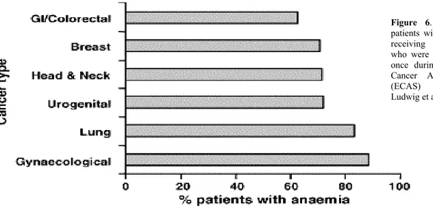
In a recent European survey evaluating anaemia in over 13,000 patients with malignancies, anaemia was observed in 68% of patients at some time during the 6-month survey 45. The frequency varied according to type of malignancy and treatment. Of patients with solid tumours receiving chemotherapy, the frequency of anaemia ranged from 62% in patients with gastrointestinal/colorectal tumours to 88% in patients with gynaecological tumours (Fig.6).
The manifestations of anaemia, including fatigue, dizziness, headache, shortness of breath, chest pain and depression, impact on the overall quality of life (QoL) of anaemic patients with cancer. For more than a decade, recombinant human Epo(rhEpo) has been used as a therapeutic agent to treat anemia in adults with cancer. This type of treatment results in increased hemoglobin production, improves the quality of life and greatly reduces the need for blood transfusions 4, 46 . In addition to its important role in the treatment of anemia associated with various diseases including cancer, human recombinant Epo has been shown to confer neutroprotective effects as well. In recent clinical studies, recombinant human Epo was shown to minimize tissue damage in patients with stroke.
The expression of Epo-R on cells of nervous system explains its role in neuroprotection functions 47, 48. Recent studies suggest that a better understanding of the mechanism of action of Epo in the nervous system could allow the use of human recombinant Epo for treating patients with neurological disorders 49, 50.
Unlike Epo, SCF is not used in clinic for the expansion of erythroid progenitors although studies have shown that SCF can cooperate with other cytokines such as G-CSF in long-term cultures of human primitive hematopoietic cells as well as for ex vivo expansion of cord blood cells for transplantation 16.
Figure 6. Percentage of patients with solid tumours receiving chemotherapy who were anaemic at least once during the European Cancer Anaemia Survey (ECAS) (adapted from Ludwig et al.
In clinical trials in humans with multiple myeloma, breast cancer and lymphoma SCF has been used successfully with G-CSF to mobilize stem and progenitor cells 17, 18.
In some cases, co-administration of G-CSF and SCF compared to G-CSF alone in breast cancer patients has been shown to result in reduced level of tumor cell contamination and in vitro introduction of c-kit into a breast cancer cell line MCF-7 mediate inhibitory signals for the growth of breast cancer cells 51.
Mobilization of erythroid progenitors population is a major issue in cancer related anemia , in the normal hematopoietic system c-kit is expressed primarily by stem cells and progenitor cells and its expression decreases along with maturation in nearly all blood cells, with the exception of mast cells, Horsfall et al. 52 have demonstrated that the combination of G-CSF and SCF mobilizes the highest number of progenitor cells.
Recognition of SCF biological activity might therefore open new possibilities in the use of this factor in clinic.
Erythropoietin and Stem Cell Factor in cancer
Cytokines have assumed increasing importance in cancer biology with the demonstration that many can be produced by tumor cells and can influence the malignant process both positively and negatively. Cytokines may act on the cancer cells in an autocrine manner or on the supporting tissues, such as fibroblast and blood vessels, to produce an environment conducive to cancer growth 53. More over, they can modulate several process involved in tumor progression and metastasis, for example, angiogenesis and the production of metalloproteinases.
The cellular receptor for SCF, c-kit, is detected in several normal and cancer tissues, but shows a restricted tissue distribution. In certain tumors, such as melanoma and breast cancer, the expression of c-kit diminishes as cell acquire a malignant phenotype 54, whereas small-cell lung cancer express c-kit rather frequently, often in combination with SCF 55.
A ligand-dependent activation of c-kit has been observed in gastrointestinal stromal tumors (GIST) as a consequence of mutations involving manly exon 11 56-58 and, more rarely, exon 9 and 13 58, 59. The expression of c-kit in maintained only in some tumor histotypes, such as prostate cancer 60 ovarian and other gynaecological tumors 61-63 , gliomas 64, 65, neuroblastomas 66, small cell cancer and about half of non small cell lung cancers 67-70.
Conversely, its expression is frequently diminished or absent in cutaneous melanoma 71 and in breast 72-75and thyroid cancers 76.
In particular, in breast tissue, some studies have shown that c-kit, widely expressed in normal epithelium, progressively decreases during malignant transformation and is present at low levels or
disappears in primary tumors and metastatic lesions 77. Nishida et coll. showed that introduction of c-kit into a breast cancer cell line, MCF-7, mediate inhibitory signals for the growth of breast cancer cells 51. c-kit then is expressed in a variety of normal cells and tissues, often in concomitance with its ligand, alternatively known as stem cell factor, mast cell growth factor, steel ligand, suggesting a role of these factors in the maintenance of a variety of fully differentiated tissues 78. The interaction of c-kit with its ligand is essential in hematopoiesis, embryogenesis, proliferation and cellular migration, however since many studies have shown how c-kit expression progressively decreases during malignant transformation many authors are suggesting its role in maintaining normal growth rather then malignant transformation.
On the contrary, many studies have reported expression of EpoR in tumor cell lines as well as primary cancer suggesting that Epo might have pro-tumor effect by promoting growth and inhibiting apoptosis. Furthermore, the expression of EpoR in vascular endothelium in tumors has suggested potential effects of Epo on the tumor microenvironment, such as the stimulation of tumor angiogenesis. There is an accumulating body of experimental evidence for the presence of functional endogenus Epo-EpoR signalling in tumors from studies that used strategies to block Epo signalling pathways.
Yashuda et al. 79 reported that Epo signalling contributes to the growth and angiogenesis of female reproductive tract tumors. Blockade of Epo signalling with local soluble EpoR or anti-Epo antibody resulted in tumor cell destruction and reduction of vascularity in ovarian and uterine cancer xenografts, associated with an increased apoptotic death of both, tumor cells and vascular endothelial cells. It was also shown that injection of an EpoR antagonist blocked Stat5 phosphorylation and inhibited melanoma and stomach tumor cell survival and angiogenesis 80. In another study, Arcasoy et al. 8 found that the administration of Epo-EpoR inhibitors in rat mammary adenocarcinoma tumors, resulted in significant tumors growth delay. These preclinical studies, taken together, suggest that the exploration of strategies to block Epo-EpoR function to target tumor growth and angiogenesis may be warranted.
Several studies reported Epo-modulation of tumor cell sensitivity to apoptosis induced by chemotherapy in vitro and in vivo. Batra et al. 11 showed increased expression of antiapoptotic genes (Bcl-XL, Bcl-2 and Mcl-1) as well as increase in Nuclear Factor-kB DNA binding activity in sarcoma and neuroblastoma cell line in response to rEpo. In human melanoma cells, incubation rEpo increased tumor cell resistance to hypoxia-induced cell death, moreover , rEpo increased cell viability during treatment with varying concentration of Cisplatin 81.
In august 2003 the Breast Cancer Erythropoietin Trial (BEST) was terminated early when researchers discovered a higher mortality rate in the Epo group than in those taking the placebo,
while Henke et al. demonstrated that in head and neck cancer Erythropoietin used to treat patients with anaemia was increasing tumor growth 82.
Taken together, these data suggest therefore that Epo may protect cancer cells against the effects of chemotherapy.
Isolation, characterization and culture of primary and immortalized tumor cells. Primary culture
of differentiated and undifferentiated cells from breast, ovary, lung, colon, kidney and thyroid carcinomas were established from specimens obtained from consenting patients undergoing surgery. Samples were obtained from Sant’Andrea Hospital (Rome) after approval by the local ethical committee. All the specimens were received within 2 hours from the surgery.
The tumorswere cut up with scissors into small pieces and subsequently disrupted in the presence of Hanks' balanced salt solution (HBSS) and 150 mg/ml Collagenase I (Gibco) at 37° C for 1 -2 hours. Cell suspension was recovered, passed through 100 µm nylon cell strainer and subjected to Ficoll gradient centrifugation. Cells were washed twice with HBSS and finally cultured in DMEM medium supplemented with b-FGF (20 ng/ml), EGF (20 ng/ml) and Insulin (5 ng/ml).
For primary culture of differentiated cells, very low percentage (1%) of FBS was added to the medium.The immortalized cell lines MCF-7, SKOV, HT29 and H460 were cultivated in DMEM/F12 supplemented with 10% FBS, 100 U/ml penicillin, 100 U/ml streptomycin and 2 mmol/l L-glutamine and maintained in 5% CO2 a 37° C.
TF-1 cell line was culture in Iscove’s modified Dulbecco’s medium (IMDM) supplemented with 10% FBS.
HBSS, DMEM, DMEM/F12, FBS, penicillin, streptomycin and L-glutamine were all purchased from Life Technology Inc. (Grand Island, NY). IMDM was purchased from Euroclone (West York, UK). b-FGF, EGF and Insulin were purchased from Peprotech Inc. (Rocky Hill, NJ).
Flow cytometric analysis. To determine the level of expression of EpoR and c-kit one hundred
thousand cells were used for flow cytometric analysis. Cells were washed with cold PBS and incubated with control or specific antibodies. Mouse anti-human EpoRantibody (R&D Systems, Minneapolis, MN), goat anti–human SCF Receptor/c-kit antibody (R&D Systems, Minneapolis, MN), phycoerythrin-conjugated anti-goat secondary antibodies (Chemicon, Temecula, CA), and FITC-conjugated anti-mouse antibodies (Molecular Probes, Eugene,OR) were used. Labeled cells were washed twice with PBS and fluorescence intensity was evaluated by FACScan (Becton Dickinson,San Jose, CA).
Confocal Microscopy. Primary differentiated and undifferentiated breast cancer cells, MCF-7 and
TF-1 cell lines were fixed in 4% paraformaldehyde, permeabilized with PBS plus 0.1% of TRITON-X, washed 3 x with PBS and incubated O.N. at 4° C with primary antibodies anti-EpoR 0.2 µg/ml (BD Pharmingen), c-kit 0.2 µg/ml (R&D Systems, Minneapolis, MN),and anti-NF.kB 0.2 µg/ml (Santa Cruz, CA), then washed with PBS 3 x, and incubated with secondary Abs. FITC-conjugated anti-mouse antibody (Molecular Probes, Eugene,OR) and Alexa Fluor® 488 goat
anti-rabbit IgG antibody (Molecular Probes, Leiden, Netherlands). were used. To detect Bcl-XL expression, undifferentiated breast cancer cells were expose to Epo 3U/ml for 24 h , fixed and permeabilized as described above and incubated with Anti-Bcl-XL from Santa Cruz Biotechnology (Santa Cruz, CA) for 1 h RT. The cells were washed 3x and incubated with FITC-conjugated anti-mouse antibodies (Molecular Probes, Eugene, OR). Nuclei were stained in blue with Hoechst 33342 (Molecular Probes, Eugene, OR). Fluorescence images were taken by means of an Olympus FV-500 laser scanning confocal inverted microscope equipped with Argon ions, Green and Red Helium-Neon lasers and with PlanApo 40X dry, 60X and 100X oil Olympus objectives (numerical aperture 0.85, 1.4 and 1.35 respectively). Emission at different wavelengths was collected using the proper filters and overriding signal was subtracted. Digital zooming was applied where specified. Images were assembled with the Canvas 8 software (Deneba Systems Inc.).
Western blotting. For detection of EpoR (R&D Systems, Minneapolis, MN), phospho-ERKs (Cell
Signaling Technology Inc., Beverly, MA), phospho-AKT (Cell Signaling Technology Inc., Beverly, MA), and Bcl-XL (Santa Cruz, CA), MCF-7 cells were synchronized in PRF-SFM for 24h and subsequently exposed to Epo 3U/ml for other 24h. Protein extracts were prepared by resuspending cell pellets in 1% NP40 lysis buffer (20 mM Tris/HCl pH 7.2, 200 mM NaCl, 1% NP40) supplemented with Protease Inhibitor Cocktail and Phosphatase Inhibitor Cocktails I and II (all from Sigma-Aldrich). The concentration of lysates was determined by the Bradford assay (Bio-Rad Laboratories, Richmond, CA) and 30 µg amounts of proteins were used for SDS-PAGE. Samples were analyzed by standard immunoblot procedure and visualized by chemiluminescence (Super Signal West Pico Pierce, Rockford, IL). Anti-tubulin antibody was purchased from Sigma-Aldrich Inc. (Saint Louis, MO).
Cell Growth. To determine the pro-tumor activity of Epo and SCF on breast cancer, 20.000
differentiated breast cancer cells/well were seated in the presence and in the absence of 3 U/ml Hr-Epo and 100 ng/ml of HrSCF. Both factors were added every 48 h and for three weeks in the medium. Cell growth was evaluated by cell counting every week. Human recombinant Erythropoietin (Hr-Epo) and Human recombinant Stem Cell Factor (Hr-SCF) were purchased from Peprotech Inc. (Rocky Hill, NJ).
Detection of Apoptosis. 70% confluent MCF-7 cells were shifted in PRF-SFM for 24 h. Cells were
counted and 5000 cell/well were cultured for additional 24 h in the presence or the absence of Epo 3U/ml. 5-FluoroUracil (5-FU, 25 µg/ml), Methotrexate (MTX, 10 µM) and Cyclophosphamide (CPA,500 µg/ml) were subsequently added for 48h. 10.000 primary human differentiated and
undifferentiated breast cancer cells, obtained from four different patients, pre-cultured with or without Epo 3 U/ml and SCF 100 gn/ml for 24 h were exposed for 48h to Cisplatin (Cis, 3mg/ml), Doxorubicin (Doxo, 5mM), Vincristine (VCR, 1µM), Methotrexate (MTX, 10 µM) or Paclitaxel (150 ng/ml). Primary erythroid progenitor cells were seeded in a 96 well plate (10000 cells/well) and treated for 36 hours with 200 ng/ml recombinant TRAIL (LZ-TRAIL, kindly provided by Dr. Hening Walczak, Heidelberg, Germany) or Fas ligand (FasL ) in the presence or absence of 100 ng/ml recombinant SCF added two hours before the treatment.. cell death was evaluated by Cell Titer 96 assay (Promega, Madison, WI). Colorimetric assay was analyzed by Victor 2 plate reader (Wallac, Turku, Finland). All chemotherapeutic drugs were purchased from Sigma-Aldrich Inc. (Saint Louis, MO).
Adult peripheral blood human progenitor cell (HPC) purification and culture. Adult peripheral
blood was obtained from male donors after their informed consent and approval by the institutional Committee for Human Studies. Human CD34+ precursor cells were purified from peripheral blood by positive selection using the midi-MACS immunomagnetic separation system (Miltenyi Biotec,
Bergisch Gladbach, Germany) according to the manufacturer's instructions. CD34+ cells were
cultured in serum-free medium prepared as follows: Iscove’s modified Dulbecco’s medium (Euroclone, West York, UK) was supplemented with delipidated bovine serum albumin (10 mg/ml), pure human transferrin (0.7 mg/ml), human low-density lipoprotein (40 µg/ml), insulin (10 µg/ml), sodium pyruvate (10-4 mol/L), L-glutamine (2 x 10⁻³ mol/L), rare inorganic elements supplemented with iron sulphate (4 x 10⁻⁸ mol/L) and nucleosides (10 µg/ml each).
Mice. Four weeks old C57Bl/6 female mice weighing approximately 20g were purchased from
Jackson Laboratory (Bar Harbor, ME). All were maintained with food and water ad libitum for the duration of the studies. Typically, experimental groups consisted of five mice each. Mice were observed daily and animals appearing moribund were humanely destroyed.
Stem Cells Factor and Cytotoxic Treatment. Recombinant murine SCF (Peprotech Inc., Rocky
Hill, NJ) diluted in phosphate buffer saline (PBS) was injected subcutaneously at 50µg/Kg four hours before and four hours after the chemotherapeutic drugs on day 1. From day two to day 4 or 7, SCF was injected subcutaneously twice a day every 8 h. Control group received only vehicle (PBS) injection, twice a day, beginning with SCF. 5 mg/kg of Cisplatin (Teva Pharma BV, Mijdrecht, Olanda) and 100 mg/kg 5-FluoroUracile (Teva Pharma BV, Mijdrecht, Olanda) were administrated in a single intraperitoneal injection at day 1. After 4 or 7 days, blood (200 µl) was withdrawn from
the retroorbital plexum. Blood was dripped directly after removal into tubes containing 0.5M of EDTA. Analysis of peripheral blood parameter was conducted by a contract laboratory (AppiaLab, Rome, Italy) within two hours from bleeding.
After blood sampling, mice were humanely killed, femora were removed, fixed in buffered paraformaldehyde 10% for 24 h, washed and exposed to decalcificating solution (EDTA 0.05M, NaOH 5 N). For in situ apoptosis detection , terminal deoxy-nucleotidyl transferase-mediated dUTP nick and labelling (TUNEL) reaction was performed on 6 µm-thick paraffin-embedded sections. Briefly, sample were deparaffinised and hydrated, In situ Cell Death Detection AP kit (Boehringer Mannheim, Indianapolis, IN) was used according to manufacturer’s instructions.
To evaluate bone marrow cellularity, histological sections were stained with Hematoxylin/Eosin.
To evaluate single bone marrow population, from mice sacrificed on day 7, femora were harvested and marrow flushed with a 23G (0.45x10mm) syringe needle to collected single cell suspensions.
Cells were spun on a glass slide and bone marrow population evaluated by May-Grünwald-Giemsa staining and cytologic analysis. Stained cells were observed through a Nikon Eclipse E1000 transmitted light right microscope equipped with PlanFluor 40X dry and oil objectives (numerical aperture 0.75 and 1.3 respectively) and with PlanApo 60X and 100X oil objectives (numerical aperture 1.4 both). All objectives were from Nikon (Melville, NY).
c-Kit and EpoR expression in solid tumors: The risks related to the use of hematopoietic
cytokines in the supportive care of cancer patients must be carefully evaluated at the light of the recent discovery of cancer stem cells. Therefore, to determine the possible oncogenic effect of SCF and Epo, we first analyzed c-kit and EpoR expression on several differentiated and undifferentiated solid tumor populations, including ovary, lung, colon, kidney, thyroid, carcinomas and breast cancer.
We found that most cancer cells, undifferentiated and differentiated, were negative for c-kit expression, whereas EpoR positivity was found among, ovary, lung ,colon and kidney cancer (Table 1 A and B).
As shown in Fig. 1, breast cancer cells, undifferentiated (mammopheres) and differentiated, express EpoR, but do not express c-Kit (Fig. 2), indicating that SCF can not have a pro-tumor effect on breast cancer. Both receptors were absent in thyroid cancer cells, while c-kit was readily detectable in the TF-1 cell line used as positive control (Fig. 2).
Table 1. Erythropoietin Receptor and c-Kit Expression in Solid Tumors Cytofluorimetric analysis of EpoR (A) and c-kit (B) expression in stem cell clones and differentiated cells obtained from human tumors of different origin. Tumor stem cells were obtained as described in Material and Methods and validated by the ability to reproduce the tumor of origin in immunodeficient mice. Differentiated cells were directly derived from surgical specimens As control immortalized human tumor cell lines were used. NC: Not Classified.
Erythropoietin activates EpoR signalling and inhibits chemotherapy-induced apoptosis in MCF-7 breast cancer cell line. Next step was to evaluate if Epo was able to counteract the cytotoxic effects
of antineoplastic drugs and eventually understand which was the molecular mechanism responsible. First we examined the influence of exposure to a pharmacologically relevant concentration of recombinant human Epo (3U/ml) on cell signalling of MCF-7 breast cancer cell line.
In MCF-7 cells , Epo was able to increase the expression of its receptor (Fig.3 A) and NF-kB, to induce activation of AKT/PKB and MAPKs and finally increase the expression of the antiapoptotic protein Bcl-XL(Fig.3 B).
Further we observed that in the presence of antitumor agent such as 5-FluoroUracil, Methotrexate and Cyclophosphamide, Epo was able to protect MCF-7 cells from drug-induced cell death (Fig.4)
Figure 1. Erythropoietin Receptor Expression in Breast cancer Confocal microscopy analysis of breast cancer undifferentiated (A) and differentiated cells (B). After taking phase contrast images cells were fixed, cyto-spinned on glass slides for immunofluorescence microscopy and visualized with 40x objective lens. DICT states for Differential Interference Contrast with Transmitted Light.
A
B
Figure 2. C-Kit expression in Breast Cancer c-kit on primary undifferentiated (A) and differentiated (B) breast cancer cells as evaluated by immunofluorescence and microscopic analysis. The hematopoietic cell line TF-1 (C) was used as positive control for c-Kit. Nuclei are in blue. One representative of five independent experiments with cells from different patients is shown.
EpoR MCF-7 + - Epo 3U/ml A NF-kB B p-AKT P-ERKs Bcl-xl Tubulina Figure 3 Erythropoietin activates EpoR signaling in
MCF-7 breast cancer cells and inhibits chemotherapy-induced apoptosis. In MCF-7 breast cancer cells, Epo is able to increase the expression of its receptor (A) and NF-kB, to induce activation of AKT/PKB and MAPKs and increase the expression of Bcl-xL (B). 70% confluent MCF-7 cells were synchronized in PRF-SFM and treated with Epo 3 U/ml for 24 h. The cellular levels of EpoR, phospho AKT, phospho ERKs, Bcl-XL and Tubulin were tested in 50 µg of total lysates using specific antibodies. The expression of NF.kB was evaluated through confocal analysis using a specific immunofluorescence antibody as described in Material and Methods
Cell Deat h ( % ) 5-FU 0 20 40 60 80 100 NT MTX CPA
-
Epo 3 U/mlFigure 4 In breast cancer cell line Erythropoietin inhibits chemotherapy-induced apoptosis MCF-7 cells, previously synchronized in PRF-SFM were cultured with or without Epo 3U/ml for 24 h and subsequently exposed to chemotherapeutic drugs, such as 5-FluoroUracil (5-FU, 25 µg/ml), Methotrexate (MTX, 10 µM) and Cyclophosphamide (CPA,500 µg/ml) for 48h. Cell Death was evaluated by Cell Titer 96 assay as described in Material and Methods.The results are the mean ± SD of three independent experiments.
Epo increases the expansion and protects both primary differentiated and undifferentiated breast cancer cells from drug-induced cell death. In line with the absent c-kit expression in breast
cancer cells, we did not observe any significant proliferative effect of SCF on primary breast cancer cultures (Fig. 5), nor the SCF interfered with chemotherapy-induced toxicity in both differentiated and indifferentiated cells (Fig.6 A and B). In contrast, the presence of Epo in the culture medium significantly increased breast cancer cell proliferation (Fig. 5) while inducing substantial cell death protection (Fig.6 A and B). These data clearly indicate that, differently from Epo, SCF is devoid of any pro-tumor effect in breast cancer cells.
Epo 3U/ml NT 0 20 40 60 80 100 0 1 2 3 Control Epo SCF Weeks Cell Num b er x 10 3
Figure 5. Epo increases the expansion of primary differentiated breast cancer cells Picture (upper panel ) and cell growth curve of differentiated breast cancer cells cultivated for three weeks in the presence or absence of Epo 3 U/ml and SCF 100 ng/ml in medium containing low percentage of serum supplemented with low doses of EGF (20ng/ml), b-FGF (20 ng/ml) and Insulin (5 ng/ml) . Cell growth was evaluated by cell counting every week as indicated. The results are a representative of similar independent expreriments with cells from three different patients.
Finally, as showed in Figure 7, the presence of Epo in the culture medium was unequivocally increasing the expression of Bcl-XL, indicating this as the molecular mechanism responsible of the cytoprotective effect exert by Epo during chemotherapy treatment .
Figure 6. Epo protects cancer primary differentiated breast canecr cells and mammospheres from drug-induced cell death Primary human differentiated (A) and undifferentiated (B) breast cancer cells pre-cultured with or without Epo 3 U/ml for 24 h and then exposed for 48h to Cisplatin (Cis, 3mg/ml), Doxorubicin (Doxo, 5mM), Vincristine (VCR, 1µM), Methotrexate (MTX, 10 µM) or Paclitaxel (150 ng/ml). Cell death was determined by both MTS assay and trypan blue exclusion. The results are the mean ± SD of three independent experiments with differentiated breast cancer cells obtained from 3 different patients.
Stem Cell Factor protects the hematopoietic system from drug induced cell death. The c-kit
receptor can be expressed by both normal and neoplastic tissues, where its stimulation by the natural ligand SCF can generate proliferative and survival stimuli. Our results showed clearly that tumorigenic breast cancer cells do not express c-kit therefore SCF can not protect them from cytotoxic effects of antineoplastic drugs. Next, considering that drug-induced apoptosis of immature (c-kit-expressing) hematopoietic cells is the primary cause of hematopoietic cell depletion in cancer patients undergoing chemotherapy (Zeuner A. et al, Blood 2003 and manuscript in preparation), we wanted to test the ability of SCF to act as a hemoprotective factor in vitro and in vivo in the presence of antitumoral agents.
In in vitro experiments performed on primary human erythroid progenitors we found that SCF is able to prevent apoptosis of erythroid progenitors induced by CD95/Fas ligand and TRAIL (Fig.8), one of the most promising new anticancer agent now entering phase II clinical trials, which is selectively able to induce apoptosis in tumor cells while sparing the large majority of normal cells.
Figure 7. Epo protects cancer mammospheres from drug-induced cell death thought increased Bcl-xL expression Confocal analysis of Bcl-xL in undifferentiated breast cancer cells. As described in material and Methods, cells were exposed to Epo 3U/ml for 24 h, fixed and cyto-spinned on glass slides for immunofluorescence microscopy and visualized with 100x objective lens.
Next we implemented the evidence that SCF can protect the hematopoietic system from chemotherapy-induced damage by analysing the effects of SCF in vivo in experimental models of chemotherapy-induced cytotoxicity. C57BL/6 mice were subdivided in groups of four and treated with saline solution (PBS), with Cisplatin or 5-FluoroUracile (injected intraperitoneally), with SCF alone (injected subcutaneously) and with cisplatin + SCF or 5-FluoroUracile + SCF.
SCF was administered two hours before the chemotherapeutic agent and every day for the following 4 days and 7 days. Blood cell values were assessed 4 and 7 days after chemotherapic administration and representative results are shown in Table 2.
Figure 8. SCF prevents primary human erythroid progenitors cell death Percentage of cell death of primary erythroid cells treated for 36 hours with 200 ng/ml recombinant TRAIL or Fas ligand (FasL) in the presence (SCF) or absence (untreated) of 100 ng/ml recombinant SCF added two hours before the treatment.
Cell Death (%)
50 0 10 20 30 Control TRAIL 40 FasLUntreated
SCF
Table 2. Blood values of mice treated with saline solution (PBS), with cisplatin (5 mg/kg of weight), with SCF (100 micrograms/kg/day), injected subcutaneously (SC) or with SCF + cisplatin (SCF + Cis). Values refer to a retro-orbital bleeding done 7 days after cisplatin administration. RBC = red blood cells, Hb = haemoglobin, WBC = white blood cells, PLT = platelets. . The results are a representative of 5 independent expreriments .
In a different set of experiments, mice treated with Cisplatin or 5-Fluorouracil (5-FU) in the presence or in the absence of SCF were sacrificed 4 and 7 days after the chemotherapic insult to obtain femoral bone marrow sections. Histological sections for TUNEL assay show in Fig. 9 presence of apopototic cells in mice treated with only Cisplatin while Stem Cell Factor in mice treated with both was able to prevent cell depletion.
In the second set of experiment mice treated with cisplatin or 5-FU display strong marrow hypoplasia with myelofibrosis and clusters of megakaryocytes. Treatment of mice with SCF consecutively for a week resulted, in both cases, in a strong myeloprotection, as shown by high bone marrow cellularity and the almost complete disappearance of dysplastic cells (Fig.10 A). The relative percentages of bone marrow cellularity are shown in Fig.10 B.
Figure 9. SCF prevents Apoptosis in vivo. Tunel Assay on bone marrow histological sections of mice treated with PBS, Cisplatin (5 mg/Kg), SCF (100 µg/Kg/day) and the combination of both for 4 days. Chemotherapeutic agents was administered on day one intraperitoneally, whereas SCF was injected subcutaneously twice a day, from day 1 to day 4 as described in Material and Methods.
In our expreriments, we also analyze the entire bone marrow population, and as shown in Figure 11 we observe an increase in all bone marrow cells of mice treated with SCF and chemotherapeutic agents compared to those treated with the drug alone.
A
B
Figure 10. Bone Marrow cellularity in C57Bl6 mices. A) Bone marrow histological sections stained with Hematoxylin/Eosin obtained from mice injected intraperitoneally with PBS (Control), with cisplatin (Cis, 5 mg/kg of weight) or with 5-Fluorouracil (5-FU, 100 mg/kg), with or without SCF (100 micrograms/kg/day). Chemotherapeutic agents were administered on day1 whereas SCF was given twice a day, from day 1 to day 7 as described in Material and Methods. B) Bone marrow cellularity of samples treated with chemotherapeutic agents with or without SCF as above. Panel B shows the means and standard deviations of results obtained from three independent experiments with chemotherapy.
The experiments described above demonstrate that SCF protects the hematopoietic system from chemotherapy-induced damage in vivo and outline a protocol for a potential clinical application of SCF to prevent chemotherapy-induced cytotoxicity, thus considerably reducing the amount of experimentation needed for a future use of SCF in cancer patients.
Figure 11. SCF acts an hemoprotective agent on the entire bone marrow population. Chemotherapeutic agent was administered on day 1 whereas SCF was given twice a day, from day 1 to day 7 as described in Material and Methods. After a week, mices were sacrified and and bone marrow from the femurus flashed to obtained cell population. Cells were washed, counted and spun on a glass slide for May-Grünwald- Giemsa- staining. Results are the mean of three separate experiments (each group represents the data from 15 anumals).
Anaemia is a common symptom associated with most cancer patients, and appears in all patients with hematological malignancies 4.In a recent European survey evaluating anaemia in over 13,000 patients with malignancies, anaemia was observed in 68% of patients at some time during the 6-month survey 45. The manifestations of anaemia, including fatigue, dizziness, headache, shortness of breath, chest pain and depression, impact on the overall quality of life (QoL) of anaemic patients with cancer.
The ability of erythropoietin (Epo) to promote the production of red cells is currently exploited to treat chemotherapy-induced anemia. Epo significantly increases haemoglobin (Hb) levels, reduces transfusion requirements, and improves quality of life, particularly by relieving fatigue4 .
However, recent studies have shown that Epo has biological functions aside from regulating erythropoiesis and that many solid tumors express EpoR, raising concerns about the fact that Epo could stimulate the growth of indolent tumors and interfere with the action of antitumor therapies, either by enhancing tumor proliferation rates or interfering with apoptotic cell death, therefore the expression of Epo receptor (EpoR) in a variety of cancer cells suggests that Epo-based supportive therapy can negatively affect the clinical outcome.
Recently, the presence of c-kit, the receptor of an other essential factor for erythropoiesis, the Stem Cell Factor (SCF), has been extensively investigated in cancers of various origin as this molecule represents a target for the tyrosine kinase inhibitor imatinib mesylate (Gleevec), thus rendering available an unprecedented array of data on c-kit expression in solid and haematological tumors. In a comprehensive investigation 1,15, 81/120 tumor types examined did not yield any sample positive for c-kit expression. C-kit negative tumors included both solid and hematologic tumors, suggesting that the use of SCF should be safe in many of the most common malignancies.
More over, the risks related to the use of hematopoietic cytokines in the supportive care of cancer patients must also be carefully evaluated at the light of the recent discovery of cancer stem cells. The existence of a undifferentiated subpopulation of tumorigenic cells responsible for tumor maintenance, growth and spreading was known since several years in leukemias, but has been now demonstrated to occur also in solid tumors such as breast and brain cancers 19, 20.
The possibility that cancer stem cells may use hematopoietic growth factors to proliferate and resist to apoptotic stimuli poses an additional caveat for the administration of cytokines to cancer patients and requires a careful assessment of the presence of cytokine receptors on the surface of both stem and differentiated cancer cells.
In our laboratory, we have obtained cancer stem and differentiated cells from several solid tumors including breast, thyroid, colon, and lung carcinomas etc.
We analysed the expression of EpoR and c-kit in a variety of cancer types and we found that the large majority of stem and differentiated cancer cells tested resulted negative for c-kit expression and positive for EpoR expression. In breast cancer EpoR and c-kit have a different expression patterns. Ulivi et al 77 demonstrated that c-kit expression is downregulated toward a more aggressive phenotype, while EpoR expression is upregulated .
The aim of this study was to evaluate the possible oncogenic effect of SCF on differentiated and undifferentiated breast cancer cells and eventually outline a protocol for a potential clinical application of SCF to prevent chemotherapy-induced anemia in this type of cancer. Our data showed that breast cancer cells, undifferentiated and differentiated, express EpoR, but do not express c-Kit, indicating that SCF can not have a pro-tumor effect on breast cancer. In line with the absent c-kit expression in breast cancer cells, we did not observe any significant proliferative effect of SCF on primary breast cancer cultures, nor the SCF interfered with chemotherapy-induced toxicity in both differentiated and undifferentiated cells.
In contrast, the presence of Epo in the culture medium significantly increased breast cancer cell proliferation while inducing substantial cell death protection. The presence of Epo in the culture medium was unequivocally increasing the expression of the antiapoptotic protein Bcl-XL, indicating this as the molecular mechanism responsible of the cytoprotective effect exert by Epo during chemotherapy treatment. These data clearly indicate that, differently from Epo, SCF is devoid of any pro-tumor effect in breast cancer cells.
In clinic, drug-induced apoptosis of immature hematopoietic cells is the primary cause of hematopoietic cell depletion in cancer patients undergoing chemotherapy, more over mobilization of erythroid progenitors population is a major issue in cancer related anemia .
Horsfall et al. 52 have demonstrated that the combination of G-CSF and SCF mobilizes the highest number of progenitor cells and some clinical trials in humans with multiple myeloma, breast cancer and lymphoma SCF has been used successfully to mobilize stem and progenitor cells 17, 18 . In some cases, co-administration of G-CSF and SCF compared to G-CSF alone in breast cancer patients has been shown to result in reduced level of tumor cell contamination and in vitro introduction of c-kit into a breast cancer cell line MCF-7 mediate inhibitory signals for the growth of breast cancer cells51. In the second part of our studies, we wanted to test the ability of SCF to act as a hemoprotective factor in vitro and in vivo in the presence of antitumoral agents.
We implemented the evidence that SCF can protect the hematopoietic system from chemotherapy-induced damage in in vitro experiments performed on primary human erythroid progenitors, SCF was able to prevent apoptosis of erythroid progenitors induced by CD95/Fas ligand and TRAIL.
In in vivo in experimental models of chemotherapy-induced cytotoxicity we analyze the entire bone marrow population, and we observe an increase in all bone marrow cells of mice treated with SCF and chemotherapeutic agents compared to those treated with the drug alone. Treatment of mice with SCF resulted in a strong myeloprotection as demonstrated by TUNEL Assay.
Analysis of peripheral blood values showed and increase in cells number after stem cell factor administration, indicating the ability of SCF to restore a normal erythropoiesis.
In summary, we clearly showed that SCF effectively protects normal cells from cytotoxic stimuli both in vitro and in vivo. Moreover, we provide clear evidence that many tumors do not express the c-kit receptor, even at cancer stem cell level, particularly in breast cancer, the Stem Cell Factor does not have any pro-tumor activity, as a further confirmation of the safety of SCF administration and outline a protocol for a potential clinical application of SCF to prevent chemotherapy-induced cytotoxicity, thus considerably reducing the amount of experimentation needed for a future use of SCF in cancer patients.
1) Went PT, Dirnhofer S, Bundi M, Mirlacher M, Schraml P, Mangialaio S, Dimitrijevic S, Kononen J, Lugli A, Simon R, Sauter G. Prevalence of KIT expression in human tumors. J Clin Oncol. 2004 Nov 15;22(22):4514-22.
2) Mercadante S, Gebbia V, Marrazzo A, Filosto S. Anaemia in cancer: pathophysiology and treatment.Cancer Treat Rev. 2000 Aug;26(4):303-11. Review
3) Ludwig H, Fritz E.Anemia in cancer patients. Semin Oncol. 1998 Jun;25(3 Suppl 7):2-6 4) Ferrario E, Ferrari L, Bidoli P, De Candis D, Del Vecchio M, De Dosso S, Buzzoni R,
Bajetta E. Treatment of cancer-related anemia with epoetin alfa: a review.Cancer Treat Rev. 2004 Oct;30(6):563-75. Review.
5) Nowrousian MR, Waschke S, Bojko P, Welt A, Schuett P, Ebeling P, Flasshove M, Moritz T, Schuette J, Seeber S. Impact of chemotherapy regimen and hematopoietic growth factor on mobilization and collection of peripheral blood stem cells in cancer patients. Ann Oncol. 2003;14 Suppl 1:i29-36.
6) Faquin WC, Schneider TJ, Goldberg MA.Effect of inflammatory cytokines on hypoxia-induced erythropoietin production. Blood. 1992 Apr 15;79(8):1987-94.
7) Dagnon K, Pacary E, Commo F, Antoine M, Bernaudin M, Bernaudin JF, Callard P. Expression of erythropoietin and erythropoietin receptor in non-small cell lung carcinomas. Clin. Cancer Res. 2005 Feb 1;11(3):993-9.
8) Arcasoy MO, Amin K, Karayal AF, Chou SC, Raleigh JA, Varia MA, Haroon ZA. Functional significance of erythropoietin receptor expression in breast cancer. Lab. Invest. 2002 Jul;82(7):911-8.
9) Arcasoy MO, Amin K, Vollmer RT, Jiang X, Demark-Wahnefried W, Haroon ZA. Erythropoietin and erythropoietin receptor expression in human prostate cancer. Mod. Pathol. 2005 Mar;18(3):421-30.
10) Acs G, Xu X, Chu C, Acs P, Verma A .Prognostic significance of erythropoietin expression in human endometrial carcinoma. Cancer. 2004 Jun 1;100(11):2376-86.
11) Batra S, Perelman N, Luck LR, Shimada H, Malik P. Pediatric tumor cells express erythropoietin and a functional erythropoietin receptor that promotes angiogenesis and tumor cell survival. Lab. Invest. 2003 Oct;83(10):1477-87.
12) Arcasoy MO, Amin K, Chou SC, Haroon ZA, Varia M, Raleigh JA. Erythropoietin and erythropoietin receptor expression in head and neck cancer: relationship to tumor hypoxia. Clin. Cancer Res. 2005 Jan 1;11(1):20-7.
13) Brower V. Erythropoietin may impair, not improve, cancer survival. Nat. Med. 2003 Dec;9(12):1439.
14) Pajonk F, Weil A, Sommer A, Suwinski R, Henke M. The erythropoietin-receptor pathway modulates survival of cancer cells. Oncogene. 2004 Nov 25;23(55):8987-91.
15) Singer CF, Hudelist G, Lamm W, Mueller R, Czerwenka K, Kubista E Expression of tyrosine kinases in human malignancies as potential targets for kinase-specific inhibitors. Endocr. Relat Cancer. 2004 Dec;11(4):861-9.
16) Yao CL, Chu IM, Hsieh TB, Hwang SM.A systematic strategy to optimize ex vivo expansion medium for human hematopoietic stem cells derived from umbilical cord blood mononuclear cells.Exp Hematol. 2004 Aug;32(8):720-7.
17) Chin-Yee IH, Keeney M, Stewart AK, Belch A, Bence-Buckler I, Couban S, Howson-Jan K, Rubinger M, Stewart D, Sutherland R, Paragamian V, Bhatia M, Foley R. Optimising parameters for peripheral blood leukapheresis after metHuG-CSF (filgrastim) and r-metHuSCF (ancestim) in patients with multiple myeloma: a temporal analysis of CD34(+) absolute counts and subsets.Bone Marrow Transplant. 2002 Dec;30(12):851-60.
18) Menedez P, Prosper F, Bueno C, Arbona C, San Miguel JF,Garcia-Conde J, Sola C, Hornedo J, Cortes-Funes H, Orfao A. Sequential analysis of CD34+ and CD34- cell subsets in peripheral blood and leukapheresis products from breast cancer patients mobilized with SCF plus G-CSF and cyclophosphamide. Leukemia. 2001 Mar;15(3):430-9.