UNIVERSITY OF PISA
School of Graduate Studies
“Scienza del Farmaco e delle Sostanze
Bioattive
”
PhD THESIS
2006-2008
“Pharmacological Ischaemic Preconditioning:
Design and Synthesis of mito-K
ATPChannel Openers and
Aldose Reductase Inhibitors”
Mariaelisa Manganaro
DIRECTOR OF THE SCHOOL Prof. Claudia Martini
INDEX
1 POTASSIUM CHANNELS 7
1.1 Introduction 7
1.1.1 ATP-sensitive potassium channels 8
1.1.2 Protein architecture of KATP channels 8
1.1.3 Nucleotide gating KATP channels 9
1.1.4 KATPchannels diversity 10
1.1.5 Potential therapeutic 10
1.1.6 Asthma 11
1.1.7 Urinary incontinence 12
1.1.8 Nervous System 13
1.1.8.1 Peripheral Nervous System 13
1.1.8.2 Central Nervous System 13
1.1.9 Skeletal Muscle 14
1.1.10 Myocardial Ischaemia 15
2.1. Ischaemia-Reperfusion(I/R) Injury 21
2.2. Preconditioning 24
2.1.1. MPTP and its role in cell death 25
2.1.2. Signal trusduction in IPC 27
2.1.3. Cardiac KATPchannels 30
2.1.4. Sarcolemmal KATP channels 32
2.1.5. Mitochondrial KATPchannels 33
2.1.6. Impaired glycolitic flux 38
2.3. Aldose Reductase 39
2.4.References 42
3 KCOs and ARIs 53
3.1. Potassium channels openers 53
3.1.1. Chemical Classification 53
3.1.2. Benzopyrans 53
3.1.3. Cyanoguanidines 58
3.1.4. Thioformamides 60
3.1.5. Benzo- and Pyridothiadiazines 62
3.1.6. Pyridyl nitrates 64
3.1.7. Benzopyranyl-Cyanoguanidine 64
3.1.8. Diaminocyclobutenediones 66
3.1.10. Tertiary Carbinols 69
3.1.11. Potassium channel blockers 71
3.2. Aldose Reductase Inhibitors 74
3.2.1. Chemical classification 77
3.2.2. Cyclic imides 77
3.2.3. Carboxilic acid Derivatives 80
3.2.4. Phenolic Compounds 82
3.2.5. Aryl Sulfonyl Nitromethanes 83
3.2.6. Amino Acids 83
3.2.7. Other Compounds 83
4 INTRODUCTION TO THE EXPERIMENTAL SECTION 93
4.1. New mito-KATPchannel openers 93
4.2. Influence of the position of the benzylic substituent 96 4.3. Replacement of the benzylic moiety and
modification of the heterocyclic core 100
4.4. Importance of the stereochemistry 105
4.4.1. Enantiomeric Resolution 105
4.4.2. Pharmacological Study 110
4.5. New aldose reductase inhibitors 114
4.6 References 129 5 EXPERIMENTAL SECTION 131 5.1. Chemistry 131 5.2. Enantiomeric resolution 175 5.3. Pharacological procedures 177 5.4. Elemental composition 185
1 POTASSIUM CHANNELS 1.1 Introduction
Potassium channels are an ubiquitous group of ion channels involving in a multitude of physiological functions. They are multimeric integral membrane proteins forming transmembrane aqueous pores through which K+ specifically permeates at a rate of 106-108 ions/s.1 Many molecular subfamilies of K+ channels are known and these correspond to the physiological signals by which pore opening is controlled such as voltage; Ca2+; ATP; G proteins or polyamines, but a unique set of functional determinants characterize this superfamily of membrane proteins. These include (i) a water-filled permeation pathway (pore) that allows K+ ions to flow across the cell membrane, (ii) a selectivity filter that specifies K+ as permeant ion species, and (iii) a gating mechanism that serves to switch between open and closed channel conformations.1
K+ channels are classified on the basis of primary amino acid sequence of the pore containing unit (α subunit) into three major families: the voltage gated (Kv) containing six-transmembrane-regions with a single pore; the inward rectifier (Kir) containing only two-transmembrane-regions and a single pore; the two-pore tandem K+ channels containing four transmembranes with two pores. A tripeptide sequence Gly-Tyr(Phe)-Gly is common to the pore of all K+ channels and constitutes the signature motif for determining K+ ion selectivity. The “slowpoke” Ca2+-activated K+ channels are architecturally similar to Kv subtypes and present an extra transmembrane segment near the amino terminus, while the twin-pore K+ (K2P) channels are weak inward rectifier with four putative transmembrane domains and two pore domains.2
1.1.1 ATP-sensitive potassium channels
ATP-sensitive potassium (KATP) channels, discovered in the early 1980s in cardiac muscle and pancreatic β-cells, 3-6 are a weakly inward-rectifying K+ channels that are inhibited by the non-hydrolytic binding of ATP, but activated by interactions with Mg2+-bound nucleotides (Mg-nucleotides) at separate sites. KATP channels are postulated to act as sensors of intracellular metabolism, tuning the potassium permeability, and therefore the electrical activity, of a cell to its energetic balance7. The inhibitory effect dominates, and channels are closed, when cellular phosphorylation potential is high, but as metabolism decreases, the activating effect wins out and channels open. This metabolic sensing role is involved in many physiological processes, for example, it plays a role in the cascade linking insulin secretion to glycaemia in the pancreatic β-cell and participates in ischaemic pre-conditioning and thereby cardio-protection during heart failure.6,8,9
1.1.2 Protein architecture of KATP channels
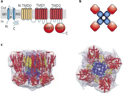
KATP channels are formed by the combination of two types of proteins:10 one, Kir6, is a ~50 kDa protein belonging to the inwardly rectifying potassium (Kir) channel family, the other, SUR (for sulphonylurea receptor), is a ~140-180 kDa member of the ATP-binding cassette (ABC) transporter family. The channel is a functional octamer of four Kir6 subunits assemble to form the pore, and each subunit is associated to four SUR subunits generating the regulatory site.11,12
Figure 1.2Protein architecture of KATP channels. a) Inward rectifier K+ channel Kir6 subunits generate the channel pore and sulphonylurea receptor (SUR) subunits generate the regulatory subunit. TMD, transmembrane domain; NBF, nucleotide-binding fold; M1, M2, transmembrane helices; P, pore. b) The channel is a functional octamer of four Kir6 subunits, and each subunit is associated with four SUR subunits. c) Images at 18 Å resolution of the entire KATP complex viewed in the plane of the membrane (left) or from above the membrane (right).
Kir6 consisting of two transmembrane helices (M1 and M2) bridged by an extracellular loop that generates the narrow portion of the pore (H5) and control ion selectivity. Two members of the Kir6 family are known, Kir6.1 and Kir6.2 encoded by KNCJ8 and KNCJ11 genes respectively, which have about 70% aminoacid identity. SUR has strong homologies with other members of subfamily C of ABC proteins and are encoded by two genes, SUR1 and SUR2 with 70% homology. The SUR2 gene has two major splice variants, SUR2A and SUR2B, which differ by 42 aminoacids in the C-terminus. SURs possess the core domain composed of two transmembrane domains TMD1 and TMD2, with six transmembrane regions, and two cytoplasmic nucleotide binding folds NBF1 and NBF2.12 In addition, it has a supplementary N-terminal domain TMD0 composed of five transmembrane helices which is linked to the core ABC domain by a cytosolic loop L0. Both Kir6, in the C-terminus, and SUR, in the sixth intracellular loop, have endoplasmatic reticulum (ER) retention sequences which prevent trafficking to the cell surface in the absence of the other subunit13. There is evidence14 that TMD0 is crucial for trafficking Kir6.2 subunits to the surface membrane, and its role in controlling the gating of Kir6.215,16 suggests an intimate relationship with the poreforming subunit. In recombinant cells, truncation of the C-terminus of Kir6.2 (Kir6.2∆C) removes its retention sequence (Arg-Lys-Arg), allowing it to form functional channel without SUR subunits17 and indicating that channel inhibition by ATP is a property of Kir6 subunit. However, the ATP-sensitivity of Kir6.2∆C is 10-20 fold lower than that of Kir6.2/SUR1 channels.
1.1.3 Nucleotide gating KATP channels
Several ligands affect KATP channel activity. These channels are inhibited by ATP (with or without Mg2+) and activated by phosphatidylinositol-4,5-bisphosphate (PIP2) by a direct interaction with Kir6.2 subunits of the channels. Sulphonylureas inhibit, and potassium channel-opener drugs activate, by interaction with the SUR subunit. In addition, in the presence of Mg2+, ATP and ADP can activate the channel through interaction with the NBFs of SUR. Inhibition by ATP binding to Kir6.2 and activation by Mg-nucleotides is almost certainly the primary physiological regulatory mechanism. The metabolically controlled gate of the channel is located at the cytoplasmic end of the inner cavity. PIP2 interaction provides an energetic pull to open channels, and ATP binding provides the energetic push to close the ligand-operated gate, perhaps through the physical link provided by a “slide helix”, that lies along the plane of the membrane and physically connects the N terminus, which in Kir6.2 forms part of the ATP-binding site, to the cytoplasmic end of M1. MgATP binds to each of the ATP-binding sites (ABSs) that are formed at the interface between nucleotide-binding domains NBF1 and NBF2 in each sulphonylurea receptor subunit. ATP hydrolysis at the second site results in a conformational “activated” state that is transduced to an “override” of ATP inhibition at the
Kir6.2 subunit. The “activated” state persists through MgADP dissociation, and can be maintained by MgADP rebinding7.
1.1.4 KATP channels diversity
KATP channels were first described by Noma3 in cardiac ventricular myocytes, but they are also expressed in several tissue types including kidney, brain, skeletal muscle, heart, pancreatic β-cells and smooth muscle18-21 and have been clearly identified in both sarcolemmal and mitochondrial membranes (sarc- and mito-KATP, respectively). Their functional role in many tissues occurs in response to metabolic changes: in the pancreas KATP channels underlie insulin secretion in response to an increase blood glucose concentration; in the heart they have a protective function in response to hypoxia or ischaemia. In the brain KATP channels have a similar protective function and are also involved in sensing the level of blood glucose. Opening of smooth muscle KATP channels causes relaxation, and in particular activation of vascular KATP channels suggest a role in contributing to the increase of blood flow in response to increased tissue metabolic demand. In skeletal muscle, KATP channels may play roles both in fatigue and in glucose uptake.22
KATP channels in different tissues are formed by different combination of Kir and SUR subunits, accounting for the tissue-specific properties of the channels. For example, the pancreatic β-cells are made up of SUR1/Kir6.223,24; while SUR2A is associated with Kir6.2 in the cardiac sarcolemmal channel25,26; Kir6.1 and SUR2B probably constitute the major KATP channel in the vascular smooth muscle27, while the channels in non-vascular smooth muscle are formed by SUR2B and Kir6.1 or Kir6.2. Different SUR subtypes respond differently to intracellular nucleotides, sulfonylureas, and potassium channel openers (KCOs), while Kir6.1 and Kir6.2 have similar ATP sensitivities. Therefore, the metabolic sensitivities and the pharmacological properties of a KATP channel are largely determined by its constituent SUR.
1.1.5 Potential therapeutic
The efflux of K+ ions following opening of potassium channels is a mechanism for recovering (repolarization), maintaining (clamping) and/or enhancing (hyperpolarization) the resting state of the cell. Thus, the opening of potassium channels is a fundamental physiological means for reversal or prevention of depolarizing activity of the membrane and the role of these ion channels in cellular control and their opening by endogenous mediators, such as neurotrasmitters and hormones, is recognized as an inhibitory mechanism. Potassium channels are a diverse and ubiquitous group of ion channel exhibiting a wide range of physiological roles in cell control processes, so drugs that decrease cell excitability by opening these channels could have a broad clinical potential.
In particular, considering the unique role that KATP play in the maintenance of the cellular homeostasis, potassium channel openers (KCOs) add to existent pharmacotherapy with potential in promoting cellular protection under conditions of metabolic stress, for example, preclinical evidences28-35 have shown their potential therapeutic in asthma, urinary incontinence, hypertension, cardiac ischaemia, CNS desease.
The therapeutic usefulness of KCOs ultimately depends on their tissue selectivity which was majorly not achieved with first generation compounds. Thus, only four KCOs were introduced to clinical practice32 including nicorandil (angina, hypertension), diazoxide (refractory hypertension), minoxidil sulfate (alopecia, refractory hypertension), and pinacidil (hypertension).
1.1.6 Asthma
Therapeutic potential of KCOs in lung disease primarly refers to asthma and chronic obstructive pulmonary disease (COPD)36-41. Bronchial asthma is characterized by wide variations over short periods of time in the resistance to flow in intrapulmonary airways. Accordingly, this disease is commonly treated with bronchodilators. Nowadays asthma is no longer viewed simply as a reversible airways obstruction, but is considered as an inflammatory illness resulting in bronchial hyperreactivity and bronchospasm. Thus, pharmacological treatment of asthma deserves anti-inflammatory and bronchodilator drugs. Recent trends in the development of new anti-asthmatic include PDE inhibitors, thromboxane A2 antagonists as well as KCOs40.
Evidence for a bronchodilator potential KCOs dates back to 1986, when Allen
et al.42 showed cromakalim to reduce spontaneous, and to a lesser extent,
cholinergic and histaminergic tone in preparations of guinea-pig trachealis. Effective relaxion of tone by KCOs was also shown in bovine43 and in human airways44. KCOs exhibit stronger spasmolytic rather than anti-spasmogenic activity which resembles other classes of smooth muscle relaxants36. Corresponding to their spasmolytic efficacy in vitro, KCOs relax established bronchocostriction in vivo. However, in contrast to in vitro findings these drugs exhibit pronounced protective effects in vivo also when given prior to challenge. In vivo studies preferentially addressed the anti-spasmogenic activity of KCOs. Orally administration inhibited dyspnoea evoked by histamine in conscious guinea-pigs45 while intravenously or intraduodenal administration inhibits bronchocostriction elicited by histamine, serotonin and bombesin.45-49 Bronchoconstriction often involves a reflex parasympathetic component, thus, neural effects of KCOs might be relevant for their therapeutic utility in asthma. Several studies show KCOs to potently inhibit the neurotransmitter release from cholinergic and non-adrenergic, non-cholinergic (NANC) neurons. KCOs more effectively inhibit Ach- or NANC-mediated bronchoconstriction because of stimulated neurotransmitter release as compared to bronchoconstriction because
of an exogenous supply of Ach or NANC neurotransmitters such as substance P or neurokinin A.50,51 Inhibitory effects are probably mediated by hyperpolarizaion of sensory nerve endings via activation of prejunctional K+ -channels32. Beyond their effects on bronchoconstriction NANC neurotransmitters stimulate mucus secretion and the leakage of plasma from post-capillary venules, both of which are important in the pathology of asthma. These inflammatory effects of NANC neurotransmitters seem to be primarly due to the action of substance P.52
Airways hyper-responsiveness (AHR) to a plethora of physiological and pharmacological stimuli is a commonly observed characteristic of asthmatic patients. KCOs acutely reverse AHR by a direct effect on airways smooth muscle and they are able to prevent the development of AHR.
Molecular mechanism underlying the prevention of AHR by KCOs remain to be clarified.
1.1.7 Urinary incontinence
Urinary incontinence is classified in four major types: 1) stress incontinence; 2) urge incontinence; 3) reflex incontinence; 4) overflow incontinence (Table 1.1).
Table 1. 1 Subtypes of urinary incontinence53 Stress
incontinence Involuntary loss of urine during coughing, sneezing or physical activities; it occurs when intravescical pressure is higher than maximum urethral pressure but in the absence of detrusor activity
Urge
incontinence Characterized by abnormal spontaneous detrusor contraction unrelated to bladder volume; broadest category of urinary incontinence
Reflex
incontinence Involuntary loss of urine because of abnormal reflex activity in the spinal corde Overflow
incontinence
Involuntary loss of urine when the intravescical pressure exceeds the maximum urethral pressure because of an elevation of intravescical pressure associated with bladder distention but in the absence of detrusor activity
The etiology is multivariant and includes neurogenic and myogenic instabilities. Most incontinence is because of failure to store urine (storage dysfunction), only overflow incontinence is because of failure to empty urine (emptying dysfunction).
Pharmacological treatment of storage dysfunction should be aimed at decreasing detrusor overactivity, increasing bladder capacity, and/or increasing outlet resistance.
Pharmacological treatment of emptying dysfunction should be aimed at increasing detrusor contractility and/or decreasing outlet resistance53-57. Most widely used in pharmacological treatment are anti-muscarinic agents, which effectively inhibit bladder hyper-reflexia, but suffer from main side-effects, such as the inhibition of normal bladder smooth muscle contractility and the blockade of muscarinic receptors in other organs.
As an alternative to anti-muscarinics, KCOs should be able to decrease the bladder overactivity. Several KCOs were shown in vitro to relax the bladder smooth muscle in various species including man58-61.
1.1.8 Nervous System
1.1.8.1 Peripheral Nervous System
The KCOs interfere with neurotransmission in peripheral parasympathetic neurones in the airways and the gastrointestinal tract62. This has led to suggestions of a presynaptic site of action, whereby the KCOs control the release of neurotransmitter. In contrast, cromakalim, nicorandil, and pinacidil failed to exert an inhibitory effect on noradrenaline release in rat isolated mesenteric artery63. The presynaptic inhibitory role of KCOs, therefore, appears to be selective for parasympathetic (cholinergic) innervation. Interestingly, cromakalim and pinacidil inhibited nicotinic acetylcholine receptor-mediated and voltage-dependent catecholamine secretion from bovine adrenal chromaffin cells64. Thus, KATP channels could be involved in regulation of catecholamine secretion mainly indirectly through voltage operated channels (VOCs).
1.1.8.2 Central Nervous System
Potassium channels play a pivotal role in the control of neuronal excitability, action potential, and neurotransmitter release within the CNS65,66. Activation of a variety of receptors (e.g., opioid, 5-HT3, somatostatin, α2-adrenoceptors) by the appropriate neurotransmitter alters the flux of K+ ions from neurons67. Because of this role in normal CNS physiology, derangements in the function of K+ channels may underlie several CNS diseases. Localization in the cerebral cortex and hippocampus would suggest that this channel may have a role in the processing of memory. Studies in vitro and in animal models indicate the potential clinical utility of KCOs for diseases of the CNS.
The genesis and propagation of non physiologic electrical impulses are the hallmark of epilepsy. Thus, the hyperpolarization (and restraining) of excitable cells through the opening of K+ channels could demonstrate therapeutic benefit in this setting. Opioids exert their analgesic effects by binding to opiate receptors, which leads to opening of K+ channels and neuronal hyperpolarization67. Morphine induced antinociception in mice tail-flick tests is mediated by the opening of KATP channels68. These observations would suggest a potential role for KCOs as analgesics. During anoxic conditions, neuronal
depolarization is due, at least, to the release of large concentrations of excitatory amino acids, such as glutamate, which may be involved in long-term ischaemia-induced damage to the brain69. In in vitro experiments, diazoxide and somatostatin were shown to prevent anoxia-induced depolarization of CA3 hippocampal neurons following the opening of K+ channels70; these effects were inhibited by pretreatment with glyburide. The authors proposed that KCOs may prevent anoxia-induced damage to hippocampal neurons by inhibiting the release of excitatory amino acids. Schmid-Antomarchi et al. 71 noted that the order of potency of the KCOs (levcromakalim > nicorandil > cromakalim > diazoxide > pinacidil) was found to be different from that in either the pancreatic β-cell or in smooth muscle, possibly indicating a difference between the target K+ channel in this brain region and that in other tissues. The KATP channel in neuronal tissue is not the classical (Type 1) channel found in pancreatic β -cells or heart, but a large-conductance non-rectifying version (Type 2) 72.
1.1.9 Skeletal Muscle
In skeletal muscle, KATP channel activity has been shown to increase upon intracellular acidification73. Falls in intracellular pH reduce the inhibitory effect of ATP on K+ channels in frog skeletal muscle. This could mean that during increased muscle exercise and consequent lowering of pH, KATP channel-induced hyperpolarization could compensate for a decrease in electrical excitability and prevent spontaneous contractions from occurring. KCOs, however, increased open probability of an sensitive and an ATP-insensitive K+ channel in human skeletal muscle74; the effect of the KCOs on both channels were blocked by glibenclamide. Ischaemia-induced damage in a rat skeletal muscle, like cardiac and neuronal ischaemia, has been shown to be prevented by cromakalim75. This result, and the observation that cromakalim restores the membrane potential of depolarized human skeletal muscle fibres76, indicate that KCOs may be useful for the treatment of peripheral vascular disease. In rat skeletal muscle, Angersbach and Nicholson77 demonstrated that KCOs, but not Ca2+ antagonists or hydralazine, selectively increase blood flow to collateral vessels in a previously ischemic limb. Weselcouch et al. 78, however, suggested that KCOs would not be beneficial in treatment of skeletal muscle ischaemia in vivo, but may be useful in preserving skeletal muscle function in cases of ischaemia followed by reperfusion. Studies in human muscle have implicated SKCa, (apaminsensitive) channels in the condition myotonic muscular dystrophy, characterised by muscle stiffness79. Although K+ channel subtypes other than KATP channels exist in skeletal muscle (SKCa, BKCa, delayed rectifier K+ channel)80, however, selective openers are awaited to determine their role and the therapeutic potential of activation.
1.1.10 Myocardial Ischaemia
A beneficial role of KCOs in myocardial ischaemia81-84 was first suggested by investigations on nicorandil85. Later, Grover et al. 86 studied the anti-ischaemic effects of KCOs in a model of ischaemia and reperfusion in which complicating vasodilator effects were avoided and thus direct cardioprotective activity could be verified. These authors showed pinacidil and cromakalim to protect isolated rat heart subjected to 25 min global ischaemia followed by 30 min reperfusion; concentrations needed to increase the time to ischaemic contracture by 25% were used as indicators of anti-ischaemic potency. Cardioprotective properties of other KCOs including bimakalim and aprikalim could be shown in similar models87-92. Putative involvement of KATP channels with the mechanism of myocardial preconditioning further increased the interest in these channels. KCOs mimic preconditioning in the absence of ischaemia, whereas KATP blockers such as glibenclamide93 and 5-hydroxydecanoate (5-HD)94 were shown to antagonize preconditioning. The original hypothesis to explain these observations involved sarcolemmal KATP channels; shortening of action potential duration (APD) was viewed as a putative mechanism of the cardioprotection by KCOs. However, KCOs exert their cardioprotective effects without a significant cardiodepression indicating a lack of importance of sarcolemmal KATP channels to mediate cardioprotection. This view is supported by the abolition of cardioprotective effect of KCOs in the presence of 5-HD94 that is a selective blocker of mitochondrial KATP channels95, while the sarcolemmal KATP channels blocker HMR1883 failed to abolish preconditioning96.
1.2 References
(1) Hille, B. Ionic channels of excitable membranes; 2nd ed.; Sinauer Associates: Sunderland, MA, 1992.
(2) Ketchum, K. A.; Joiner, W. J.; Sellers, A. J.; Kaczmarek, L. K.; Goldstein, S. A. N. Nature 1995, 376, 690-695; Brown, D. A. Curr. Biol. 2000, 10, R456-459.
(3) Noma, A. Nature 1983, 305, 147-148.
(4) Trube, G.; Hescheler, J. Pflügers Arch. 1984, 401, 178-84. (5) Cook, D. L.; Hales, C. N. Nature 1984, 311, 271-273.
(6) Ashcroft, F. M.; Harrison, D. E.; Ashcroft S. J. Nature 1984, 312, 446-448. (7) Nichols, G. C. Nature 2006, 440, 470-476.
(8) Jahangir, A. & Terzic, A. J. Mol. Cell. Cardiol. 2005, 39, 99–112.
(9) Yamada, S.; Kane G. C.; Behfar, A.; Liu, X.K.; Dyer, R.B.; Faustino, R. S.
et al. J. Physiol. 2006, 577, 1053–1065.
(10) Bryan, J.; Vila-Carriles, W. H.; Zhao, G.; Babenko, A. P. & Aguilar-Bryan, L. Diabetes 2004, 53, S104–S112.
(11) Clement, J. P.; Kunjilwar, K.; Gonzalez, G.; Schwanstecher, M.; Panten, U.; Aguilar-Bryan, L. et al. Neuron 1997, 188, 27–838.
(12) Moreau, C.; Prost, A. L.; Derand, R. & Vivaudou, M. J. Mol. Cell. Cardiol.
2005b, 38, 951–963.
(13) Zerangue, N,; Schawappach, B.; Jan, Y. N.; Jan, L. Y. Neurons, 1999, 22, 537-548.
(14) Chan, K. W.; Zhang, H.; Mishahi, T. & Logothetis, D. E. Biophys. J. 2002,
82, 590a.
(15) Babenko, A. P. & Bryan, J. J. Biol. Chem. 2002, 277, 43997–44004. (16) Babenko, A. P. & Bryan, J. J. Biol. Chem. 2003, 278, 41577–41580.
(17) Tucker, S. J.; Gribble F. M.; Zhao, C.; Trapp, S.; Ashcroft F. M. Nature
1997, 387, 179-183.
(18) Edwards, G.; Weston, A. Annu. Rev. Pharmacol. Toxicol 1993, 33, 597– 637.
(19) Spruce, A.; Standen, N.; Stanfield, P. Nature 1985, 316, 736–738.
(20) De Weille, J.; Schimd-Antonmarchi, H.; Fosset, M.; Lazdunski, M. Proc
Natl Acad Sci USA 1998, 85, 1312–1316.
(21) Treherne, J.; Ashford, M. Neuroscience 1991, 40, 523–531.
(22) Rodrigo, G. C.; Standen, N.B. Curr. Pharm. Des. 2005, 11, 1915-1940. (23) Aschcroft, F. M.; Gribble, F. M. Trens Neurosci. 1998, 21, 288-294.
(24) Inagaki, N.; Gonoi, T.; Clement, J. P.; Namba, N.; Inazawa, J.; Gonzalez, G. et al. Science, 1995, 270, 1166-1170.
(25) Inagaki, N.; Gonoi, T.; Clement, J. P.; Wang, C. Z.; Aguilar-Bryan, L.; Bryan J. et al. Neurons, 1996, 16, 1011-1017.
(26) Babenko, A. P.; Gonzalez, G.; Aguilar-Bryan, L.; Bryan J. Circ. Res..
(27) Yamada, M.; Isomoto, S.; Matsumoto, S.; Kondo, C.; Scindo, T.; Horio, Y.
et al. J. Physiol. 1997, 499, 715-720.
(28) Longman, S. D.; Hamilton, T. C. Med. Res. Rev. 1992, 12, 73-148. (29) Poyser R. H.; Hamilton, T.C. Drugs Fut. 1994, 19, 39-47
(30) Lawson, K. Clin. Sci. 1996, 91, 651-663.
(31) Colatsky, T. J.; Hamilton, T. C. Potassium channel modulators: clinical experiences and future prospects. In: Evans J. M.; Hamilton, T. C.; Longman, S. D.; Stemp, G. editors. Potassium channels and their modulators. London: Taylor & Francis, 1996, pp383-411.
(32) Lawson K. Kidney Int. 2000, 57, 838-845.
(33) Lawson, K. Expert. Opin. Invest. Drugs, 2000, 9, 2269-2280.
(34) Jahangir, A,; Terzic, A.; Shen, W. K. Expert. Opin. Pharmacother. 2001, 2, 1995-2010.
(35) Lawson, K.; Dunne, M. J. Expert. Opin. Investig. Drugs, 2001, 10, 1345-1359.
(36) Raeburn, D.; Karlsson, J-A. Progr. Drug Res. 1991, 37, 161-180. (37) Morley, J. TiPS, 1994, 15, 463-468.
(38) Arch, J. R. S. Potassium channel activators: Airway pharmacology and bronchial asthma. In: Evans J. M.; Hamilton, T. C.; Longman, S. D.; Stemp, G. editors. Potassium channels and their modulators. London: Taylor & Francis, 1996, pp275-302.
(39) Bucheit, K. H.; Fozard, J. R. Pulm. Pharmacol. Ther. 1999, 12, 103-105. (40) Prasad, M. R.; Bahekar, R. H.; Rao, A. R. Parmazie, 2000, 55, 475-482. (41) Pelaia, G.; Gallelli, L.; Vatrella, A.; Grembiale, R.D.; Maselli, R.; De
Sarro, G. B.; Marsico, S. A. Life Sci. 2002, 70, 977-990.
(42) Allen, S. L.; Boyle, J. P.; Cortigo, J.; Foster, R. W.; Morgan, G. P.; Small, R.C. Br. J. Pharmacol. 1986, 89, 395-405.
(43) Longmore, J.; Bray, K. M.; Weston, A.H.; Br. J. Pharmacol 1991, 102, 979-985.
(44) Buckle, D. R.; Arch, J. R. S.; Bowring N. E.; Foster, K. A.; Taylor J. F.; Taylor, S.G.; Shaw, D. J. Pulm. Pharmacol 1993, 6, 77-86.
(45) Bowring N. E.; Arch, J. R. S.;Buckle, D. R.; Taylor, J. F. Br. J. Pharmacol
1993, 109, 1133-1139.
(46) Arch, J. R. S.; Buckle, D. R.; Taylor, J.F.; Taylor, S. G. Br. J. Pharmacol.
1988, 95, 763-770.
(47) Englert, H. C.; Wirth, K.; Gehring, D.; Fürt, U.; Albus, U.; Scholz, W.; Rossenkranz, B.; Schölkens, B. A. Eur. J. Pharmacol. 1992, 210, 69-75. (48) Champman, I. D.; Kristersson, A.; Mathelin, G.; Schaeublin, E.; Mazzoni,
L.; Boubekeur, K.; Murphy, N.; Morley, J. Br. J. Pharmacol. 1992, 106, 423-429.
(49) Taylor, S. G.; Arch, J. R. S.; Bond, J.; Buckle, D. R.; Shaw, D. J.; Taylor, J. S. J. Pharmacol. Exp. Ther. 1992, 261, 429-437.
(50) Yamamoto, A.; Shikada, K.; Tanaka, S. Jap. J. Pharmacol. 1992, 59, 129-132.
(51) Clapham, J. C.; Bowring N. E.; Trail, B. K.; Fuller, D. A.; Good D. M.
Pulm. Phamacol. 1993, 6, 201-208.
(52) Small, R. C.; Berry, J. L.; Cook, S. J.; Foster, R. W.; Green, K. A.; Murray, M. A. Potassium channels in airways. In: Chung K. F.; Bames, P. J. editors. Pharmacology of the respiratory tract: Experimental and clinical research. New York, NY: Marcel Dekker, 1993, pp 137-176.
(53) Hattori, T. Drug of Today, 1998, 34, 125-138.
(54) Brading, A. F.; Turner, W. H. Potassium channels and their modulation in urogenital smooth muscles. In: Evans J. M.; Hamilton, T. C.; Longman, S. D.; Stemp, G. editors. Potassium channels and their modulators. London: Taylor & Francis, 1996, pp335-359.
(55) Andersson, K-E. Exp. Physiol, 1999, 84, 195-213.
(56) Sellers, D. J.; Chapple, C.R.; Chess-Williams, R. World. J. Urol. 2001, 19, 307-311.
(57) Wein, A. J. Expert. Opin. Investig. Drugs 2001, 10, 65-83.
(58) Zografos, P.; Li, J. H.; Kau, S. T. Pharmacology, 1992, 45, 216-230.
(59) Martin, S. W.; Radley, S. C.; Chess-Williams, R.; Korstanje, C.; Chapple, C.R. Br. J. Urol. 1997, 80, 405-413.
(60) Chess-Williams, R.; Martin, S. W.; Korstanje, C.; Chapple, C.R. Br. J.
Urol. 1999, 83, 1050-1054.
(61) Wojdan, A.; Freeden, C.; Woods, M.; Oshiro, G.; Spinelli, W.; Colatsky, T. J.; Sheldon, J. H.; Norton, N. W.; Warga, D.; Antane, M. M.; Antane, S. A.; Butera, J. A.; Argenteri, T. M. J. Pharmacol. Exp. Ther. 1999, 289, 1410-1418.
(62) Longman, S. D.; Hamilton, T. C. Med. Res. Rev. 1992, 12, 73-148. (63) Fabiani, M. E.; Story, D. F. J. Auton. Pharmacol.1994, 14, 87-98.
(64) Masuda, Y.; Yoshizumi, M.; Ishimura, Y.; Katch, I.; Oka, M. Biochem.
Pharmacol. 1994, 47, 1751-1758.
(65) Hille, B. Ionic Channels of Excitable Membranes. Sinauer Associates, Sunderland. 1984.
(66) Cook, N. S. Potassium Channels. Structure, Classification, Function and Therapeutic Potential. Ellis Horwood Ltd., Chichester. 1990.
(67) North, R. A. Br. J. Pharmacol. 1989, 98, 13-28.
(68) Ocana, M.; Del Pozo, E.; Baeyens, J. M. Eur. J. Pharmacol. 1993, 230, 203-207.
(69) Miller, R. J. Trends Neurosci. 1990, 13, 197-199.
(70) Ben-Ari, Y.; Krnjevic, K.; Crdepel, V. Neuroscience 1990, 37, 55-60. (71) Schmid-Antomarchi, H.; Amoroso, S.; Fosset, M.; Lazdunski, M. Proc.
Natl. Acad. Sci. USA 1990, 87, 3489-3492.
(73) Davies, N. W. Modulation of ATP-sensitive K channels in skeletal muscle by intracellular protons, Nature 1990, 343, 375-377.
(74) Quasthoff, S.; Spuler, A.; Spittelmeister, W.; Lehmann-Horn, E; Grafe, P.
Eur. J. Pharmacol. 1990, 186, 125-128.
(75) Hatton, R.; Heys, C.; Todd, M. H.; Downing, 0. A.; Wilson, K. A. Br. J.
Pharmacol. Proc. 1991, Suppl. 102, 28OP.
(76) Spuler, A.; Lehmann-Horn, F.; Grafe, P. Naunyn Schmeidebergs Arch.
Pharmacol. 1989, 339, 3277331.
(77) Angersbach, D.; Nicholson, C. D. Naunyn Schmiedebergs Arch.
Pharmacol. 1988, 337, 341-346.
(78) Weselcouch, E. O., Sargent, C., Wilde, M. W. and Smith, M. A. J.
Pharmacol. Exp. Ther. 1993, 267, 410-416.
(79) Renaud, J. F.; Desmuelle, C.; Schmid-Antomarchi, H.; Hugues, M.; Serratice, G.; Lazdunski, M. Nature 1986, 319, 678-680.
(80) Wareham, A. C. Skeletal muscle potassium channels and their relevance to muscle disease. In: Potassium Channel Modulators: Pharmacological Molecular and Clinical Aspects, 1992, pp. 237-271, Weston, A. H. and Hamilton, T. C. (eds.) Blackwell Scientific, Oxford.
(81) Atwal, K.S. Curr. Med. Chem. 1996, 3, 227-238.
(82) Duncker, D. J.; Verdouw P. D. Cardiovasc. Drugs Ther. 2000, 14, 7-16. (83) Rubino, A.; Yellon, D.M. TiPS 2000, 21, 225-230.
(84) Gomma, A. H.; Pucell, H. J.; Fox, K. M. Drugs, 2001, 61, 1705-1710. (85) Lamping, K.; Gross, G. J. J. Cardiovasc. Pharmacol, 1985, 7, 158-166. (86) Grover, G. J.; McCullough J. R.; Henry, D. E.; Conder M. L.; Sleph, P. G.
J. Pharmacol. Exp. Ther, 1989, 251, 98-104.
(87) Grover, G. J.; Dzwonezyk, S.; Parham C.; Sleph, P.G. Cardiovasc. Drugs
Ther., 1990, 4, 465-474.
(88) Grover, G. J.; Newburger, J.; Sleph, P. G.; Dzwonezyk, S.; Taylor, S.; Ahmed, S.; Atwal, K. S. J. Pharmacol. Exp. Ther., 1991, 257, 156-162. (89) Grover, G. J.; Dzwonezyk, S.; Sleph, P. G. Eur. J. Pharmacol., 1990, 191,
11-19.
(90) Ohta, H.; Jinno, Y.; Harada K.; Ogawa, N.;Fukushima, H.; Nishikori, K.
Eur. J. pharmacol.,1991, 204, 171-177.
(91) Galinanes, M.; Shattock, M.; Hearse, D. Cardiovasc. Res., 1992, 26, 1063-1068.
(92) Tan, H.; Mazón, P.; Verberne, H.; Sleeswijk, M.; coronel, R.; Opthof, T, Janse, M. Cardiovasc. Res., 1993, 27, 644-651.
(93) Gross, G. J.; Auchampach J. A. Circ. Res.., 1992, 70, 223-233.
(94) Auchampach J. A.; Grover, G. J.; Gross, G. J. Cardiovasc. Res., 1992, 26, 1054-1062.
(95) McCullough, J. R.; Normandin, D. E.; Conder, M. L.; Sleph, P. G.; Dzwonezyk, S.; Grover, G. J. Cir. Res., 1991, 69, 949-958.
(96) Linz, W.; Jung, W.; Schölkens, B. A.; Englert, H. C. J. Mol. Cell. Cardiol.,
2 MYOCARDIAL ISCHAEMIC PRECONDITIONING (IPC) 2.1 Ischaemia-Reperfusion (I/R) Injury
I/R injury of the heart is characterized by reversible contractile dysfunction, known as stunning, and irreversible injury leading to cardiomyocytes death and myocardial infarction. Myocardial stunning, is defined by the mechanical dysfunction that persists after reperfusion, despite the absence of irreversible damage and despite restoration of normal coronary flow.1
Two major hypotheses have been proposed to explain the mechanism of myocardial stunning:2 1) the oxyradical hypothesis, in which it is caused by the generation of ROS; 2) the Ca2+ hypothesis, in which it is caused by a transient Ca2+ overload on reperfusion. The final lesion responsible for the contractile depression appears to be a decreased responsiveness of contractile filaments to Ca2+. Recent evidence suggests that Ca2+ overload may activate calpains, Ca2+ -dependent neutral proteases, resulting in selective proteolysis of myofibrils. The time required for resynthesise damaged proteins would explain in part the delayed recovery of function in stunned myocardium. The ROS and Ca2+ hypotheses are not mutually exclusive and are likely to represent different component of the same pathophysiologic cascade. For example, increased ROS formation could cause cellular Ca2+ overload, which would damage the contractile apparatus of the cardiomyocytes. ROS generation could also directly alter contractile filaments in a manner that renders them less responsive to Ca2+ (e.g., oxidation of critical thiol groups). An important implication of the phenomenon of myocardial stunning is that so-called chronic hibernation may be the result of repetitive episodes of stunning, which have a cumulative effect and cause protracted post-ischaemic dysfunction. Besides direct effects of ROS and Ca2+ on Ca2+-regulatory proteins and contractile machinery, ROS and intracellular Ca2+ overload modulate cardiomyocyte contractility through phosphorylation of contractile proteins. A variety of protein kinases are activated in response to oxidative stress, and this process is pivotal in regulating the contractility. Particularly, PKC and p38 mitogen-activated protein kinase (MAPK) have been implicated in the pathogenesis of ischemic contractile dysfunction.3,4
Paradoxically, reperfusion can exacerbate the damage occurring during the ischaemic period. This is known as reperfusion injury and is accompanied by enzyme release and morphological changes characteristic of necrosis.5-7
In addition to the necrotic cell death that represents the major damage to the reperfused heart there is also evidence that some myocytes around the periphery of the infarct die by apoptosis.8,9 The myocardial cell appears to be primed by a sequence of events during the ischaemic period that “sets up” the cell for an irreversible injury, which is then triggered by reperfusion.
During ischaemia the intracellular acidosis that results from anaerobic glycolysis drives a series of coupled exchanges in which the Na+/H+ exchanger removes H+ and accumulates Na+ in the cell, which is then coupled to Ca2+ -influx by reverse-mode Na+/Ca2+ exchange activity, resulting in a Ca2+ accumulation. Moreover, during ischaemia, the intracellular ATP concentration falls with consequent reduction in activity of the Na+/K+ ATPase and increase in [Na+]i. Additional effect of ischaemia includes inihibition of the sarcoplasmic reticulum Ca2+-release channel (RyR). At the moment of reperfusion, the cell is characterized by high levels of intracellular Na+, Ca2+ and possibly mitochondrial reducing power (NADH, FADH2) and the sarcoplasmic reticulum is loaded with Ca2+. Reperfusion triggers an injury cascade that combines reactivation of electron transport and oxidative phosphorilation, which increases the availability of ATP, with the existing Ca2+ load to produce a strong hypercontracture, which in the intact tissue leads to mechanical damage. There is also a secondary Ca2+ influx in the Na+/Ca2+ exchanger driven by a reperfusion induced influx of Na+ via the Na+/H+ exchanger stimulated by the washout of extracellular acidosis. In addition to these metabolic effects, re-energization of the mitochondria products a burst of reactive oxygen species and rapid mitochondrial repolarization which leads to Ca2+ accumulation and opening of mitochondrial permeability transition pores (MPTP) and thus the initiation of cell death at reperfusion.
Understanding the causes of reperfusion injury and devising ways of preventing it is of major clinical importance in cardiac surgery and the treatment of coronary thrombosis. There is increasing evidence that mitochondrial dysfunction plays a central role in mediating both the necrotic and apoptotic components of reperfusion injury, and that one of the most effective ways of protecting hearts from such injury is to subject them to brief ischaemic periods with intervening recovery periods before the prolonged period of ischaemia is initiated. This phenomenon is known as ischaemic preconditioning (IPC).5,7,10-13 Some authors have also hypothesized that functional recovery after ischaemia is predicated on the ability of the myocyte to maintain sufficient substrate metabolism during ischaemia. This hypothesis starts from the observation that glucose metabolism via the polyol pathway, and consequently aldose reductase (AR) activity, is increased in ischemic hearts14-17 and recently, it was demonstrated that inhibition of AR protects rat hearts from ischemic injury and improves functional recovery upon reperfusion.
During ischaemia, lack of oxygen leads to rapid stimulation of glucose uptake, glycogenolysis and glycolytic flux.18,19 Moderate reduction in blood flow and oxygen supply result in increased glucose extraction and glycolysis with no changes in glucose uptake, whereas severe reductions result in increased myocardial glucose uptake, glucose extraction and significantly increased glycolysis. The induction of ischaemia is followed by recruitment of the glucose transporters GLUT-4 and GLUT-1 from the intracellular stores to the
plasma membrane.20-22 Stimulation of glucose transport during ischaemia is reflected by the increased glycolytic flux.18-22 However, prolonged ischaemia results in inhibition of glycolysis due to the increased levels of lactate, protons and decreased availability of NAD+.16,17,19 Recent studies have shown that ischaemia increases aldose reductase activity and NAD+ use via this pathway. As a result glycolysis is impaired due to shortage of NAD+ for glyceraldehyde-3-phosphate dehydrogenase (GAPDH).14,16,17
Sustaining glycolysis during ischaemia is an important source of ATP for maintaining the activities of sarcolemmal Na+/K+ATPase and the sarcoendoplasmic Ca2+-ATPase.23-25 There is evidence to suggest that glycolysis preferentially supplies ATP for these critical ion pumps24,25 and thus, even if glycolytic metabolism does not provide sufficient energy for to provide enough ATP to maintain cardiac contraction, it may provide adequate energy to maintain the ionic homeostasis and thus preserve viability during the ischaemic insult.
Fatty acids have been shown to be detrimental to ischaemic myocardium. Accumulation of long-chain fatty acids may adversely affect intermediary metabolism by increasing NADH/NAD+ ratio; increasing acetyl-CoA/CoASH with consequent increases in citrate levels and inhibition of PFK; inhibiting pyruvate carboxylase and increasing the amounts of long chain acyl carnitine (LCA).
Numerous experimental studies have convincingly demonstrated that maintenance of glycolytic metabolism by the ischaemic myocardium can preserve viability,16,17,26-28 can delay ischaemic contracture and can prevent reversible cell damage.29 Glycolysis is required to support membrane function and in particular, maintain sodium and calcium homeostasis.23-25,30 It has been shown repetely in numerous studies that protection myocardium during ischaemia by favourably modifying metabolism reduces ischaemic injury and improves functional recovery upon reperfusion.17,28,31-34 Thus, there is excellent experimental and clinical data to support the concept that increased glycolytic flux, induced by increased exogenous glucose and reduced serum fatty acids, assists in protecting the ischaemic myocardium and improving survival and function after acute ischaemic episodes. Moreover, recent studies have shown that targeting aldose reductase may provide a novel approach to protecting ischaemic myocardium.35The reduction in ischemic injury due to aldose reductase inhibition was associated with attenuation of the rise in cytosolic redox (NADH/NAD+ ratio) and improved glycolysis in ischemic myocardium.16,34,36
Furthermore, AR inhibition improved myocardial glucose oxidation upon reperfusion34and beneficial changes in ATP and ion homeostasis.16,34
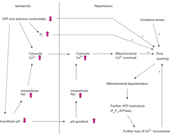
Figure 2.1 Involvement of MPTP in I/R induced cell death. ATP dissipation during ischaemia
leads to rises in resting cytosolic free [Ca2+] and P
i. Reperfusion leads to excessive
mitochondrial Ca2+ uptake. Mitochondrial Ca2+ overload together with oxidative stress and the prevailing high Pi and low ATP provoke MPTP opening.
2.2 Preconditioning
Ischaemic Preconditioning (IPC) has been exploited as a powerful endogenous form of cardioprotection. IPC was first discovered by Murry37 and associates, who demonstrated that a brief period of repetitive cardiac I/R exerts a protective effect against subsequent lethal periods of ischaemia, resulting in a marked reduction in infarct size, the extent of stunning and the incidence of cardiac arrhythmias.38,39 This “self-defence” mechanism also induces an improvement of the post-ischaemic recovery40 and a protection of the coronary endothelium.41
The development of IPC shows two distinct phases: an early phase (or classic IPC) that lasts for 1 to 3 hours following the preconditioning stimulus and a delayed one (or second window of IPC) that lasts for 24 to 96 hours. The initial early IPC does not appear to depend on new protein synthesis because of the rapid onset and because the inhibition of protein synthesis does not block this phase, while the second window of IPC involves up-regulation of genes rather than post-traslactional modification of proteins.42-44
The exact mechanisms involved in preconditioning are still being debated, but several processes have been implicated. In the longer-term effects, probably caused by stimulation of the transcription of specific genes, perhaps through a mechanism activated by free radicals and stress-activated protein kinases, of particular interest may be the up-regulation of heat shock proteins since recent data have shown that heat shock specifically up-regulates the expression of liver mitochondrial Hsp25 and this is associated with desensitisation of mitochondrial permeability transition pore (MPTP) opening to Ca2+and HgCl2 in isolated mitochondria.45 It is also known that heart mitochondria from mice in which Hsp25 is down-regulated are more sensitive to MPTP opening and also exhibit hallmarks of oxidative stress including oxidatively damaged adenine-nucleotide traslocase (ANT).46 The mechanisms responsible for the short-term effects of preconditioning include the activation of protein kinase C (PKC). This may be mediated either by reactive oxygen species (ROS) released during the short intervening reperfusion periods, or by factors released during the brief ischaemic periods such as adenosine, bradykinin, noradrenaline, and opioids. Thus, PKC inhibitors and free radical scavengers antagonise IPC, whilst adenosine agonists and PKC activators mimic the effect.47-49 The ultimate target of these kinases is unknown, although it may be significant that activation of PKCε and its translocation to mitochondria has been reported to be important for preconditioning.50,51 There is also evidence for an involvement of sulphonylurea-sensitive KATP channels, since KATP channel openers such as
diazoxide can mimic IPC whilst blockers such as glibencamide inhibit.52,53 Furthermore, PKC-dependent activation of plasma membrane KATP channels by
IPC has been demonstrated.54 The mechanism of protection from cardiomyocyte apoptosis by IPC appears to be due to protection of mitochondria. It has been shown that IPC reverses many aspects of mitochondrial dysfunction induced by I/R, including loss in the activity of the redox-sensitive Krebs cycle enzyme α-ketoglutarate dehydrogenase, declines in NADH-linked ADP-dependent mitochondrial respiration, insertion of the pro-apoptotic Bcl-2 protein Bax into the mitochondrial membrane, and release of cytochrome c into the cytosol.55
This protection of mitochondria by IPC is mediated by activation of survival signaling that converges on prevention of the MPTP opening.56-58
The survival signalling includes the PKCε–mitochondrial KATP (mito-KATP) channel pathway,59 the phosphatidylinositol 3-kinase (PI3K)/ Akt pathway,60 the Janus kinase (JAK)/signal transducers, and activators of transcription (STAT) pathway.61
2.2.1 MPTP and its role in cell death
Mitochondria play critical roles in both the life and death of cells. In healthy cardiac myocytes, their primary function is the provision of ATP through oxidative phosphorylation to meet the high energy demands of the beating
heart. Glycolysis alone is unable to meet these demands even in the resting state and inhibition of oxidative phosphorylation, as occurs in anoxia or ischaemia, leads to impairment or cessation of normal heart function. However, latent within mitochondria, there are mechanisms that, once activated, convert the mitochondria from organelles that support the life of the cell to those that actively induce both apoptotic and necrotic cell death. The switch in roles is mediated by the opening of a nonspecific pore in the mitochondrial inner membrane, known as the mitochondrial permeability transition pore (MPTP). This normally remains closed, but can open under conditions of cellular stress with dire consequences.
The release of proapoptotic molecules, such as cytochrome c or apoptosis-inducing factor, from mitochondria triggers the activation of caspases that finally leads to cellular destruction.62 This process is associated with opening of the MPTP, a ‘‘megachannel’’ spanning both the inner and the outer mitochondrial membrane. It can exist in various subconductance states. In the fully opened state, it allows molecules up to a molecular weight of 1.5 kDa to pass.63 Accordingly, opening of the MPTP leads to a breakdown of the mitochondrial membrane potential, causing failure of oxidative phosphorylation and ATP depletion.64 The pore is formed from a complex of the voltage dependent anion channel (VDAC), the adenine-nucleotide traslocase (ANT) and cyclophilin-D (CyP-D) at contact sites between the mitochondrial outer and inner membrane. Ca2+is the major physiological activator of the MPTP, other triggers are ROS and nitrogen species, mitochondrial depolarization and high levels of inorganic phosphate (Pi). In contrast, hyperpolarization of the
mitochondrial membrane as well as high levels of ATP and adenosine diphosphate (ADP) are known to inhibit the pore.63
The MPTP opens under condition of high matrix Ca2+, ROS, high NADH, depletion of adenine nucleotides and loss of membrane potential (∆ψ),65-67 conditions that occur during ischaemia-reperfusion.68,69
Low pH, as occurs during ischaemia, inhibits MPTP, but intracellular pH is rapidly restored on reperfusion, thus allowing activation of the pore. Furthermore, the reduced ∆ψ during ischaemia would limit uptake of mitochondrial Ca2+, but the reintroduction of oxygen on reperfusion would reconstitute ∆ψ (stimulating Ca2+ uptake into the mitochondria) and the introduction of oxygen will also allow generation of ROS. Thus the conditions that exist right at the start of reperfusion are ideal to stimulate opening of the MPTP.
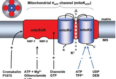
Recent data shown that mito-KATP channels interact with MPTP. In particular, it has been shown that activation of the mito-KATP channel leads to a depolarization of mitochondria by increasing K+ influx, thus reducing the Ca2+ influx into mitochondria.70 A reduced intramitochondrial Ca2+ concentration is known to prevent opening of the MPTP. Accordingly, inhibition of mito-KATP channel might hyperpolarize mitochondria by decreasing K+ input, thus
increasing the Ca2+ influx and facilitating MPTP opening. On the other hand, hyperpolarization of mitochondria is commonly considered to be a MPTP-inhibiting factor.71 A fall in mitochondrial potassium concentration due to inhibition of the mito-KATP channel should alkalize the mitochondria matrix because of a reduced activity of the K+/H+exchanger.
A rise in pH is known to increase the open probability of the MPTP.72. Furthermore, an alkaline pH elevates the activity of the Pi/OH-antiporter of the inner mitochondrial membrane and, consequently, matrix phosphate concentrations. Phosphate is a physiological modulator of the MPTP as high phosphate increases the open probability. A different explanation of the interaction between the mito-KATPchannel and the MPTP has been suggested by Costa et al.,73 who reported that inhibition of the MPTP by mito-KATP channel opening can be abolished by inhibitors of protein kinase C (PKCε) as well as by a scavenger of reactive oxygen species. They proposed that mito-KATPchannel activation increases production of H2O2, which in turn blocks the MPTP by activating PKCε. The mito-KATP channel is able to modulate the opening probability of the MPTP74 that is related to both apoptotic and necrotic cell death (e.g., in ischaemia), so modulation of this mitochondrial ‘‘megachannel’’ by controlling the state of the mito-KATPchannel may offer therapeutic benefit for various diseases.
2.2.2 Signal trusduction in IPC
IPC involves variuos signaling as triggers and/or end effectors and an important role is undoubtely played by the activation of cardiac KATP.
The triggers of IPC can be classified as dependent and receptor-independent: the former are activators that elicit their action by specific receptor interaction, such as adenosine, bradykinin and opioids which bind to G protein-coupled receptors (GPCRs); the latter are endogenous substances that do not need a selective binding with the receptor such as nitric oxide (NO), free radicals and Ca2+.
Receptor-dependent Triggers
GPCRs are coupled with Gi/o, wich consists of heterotrimeric G protein Gαβγ, or with a Gqfamily, which consists of the heterotrimeric G protein, Gαq. Activation of Gi/o-coupled receptors induces dissociation of Gα from Gβγ which activates phospholipase C (PLC) and a resultant generation of the second messengers D-myoinositol 1,4,5-triphosphate and diacylglycerol, leading to activation of PKC, an essential step for mediating IPC.75 Conversely, signaling pathways linked with activation of Gαq also converge at the level of PLC.76 General agreement exists that adenosine is a major GPCR in triggering IPC.77 Cardiac adenosine levels are increased by brief periods of ischaemia in rats, dogs and pigs78-82 and improve the oxygen supply-demand balance through the dilatation of resistance vessels and the reduction of the cardiac inotropism. These effects
are mediated by A1 and A3 receptors, while the A2 receptor subtypes in the IPC seems to be excluded.83,84
Like other Gq-coupled receptors, the activation by autacoid in preconditioned tissue triggers a multiple kinase cascade through phosphorylation reactions, leading to the rapid post-translational modifications of proteins that regulate cellular resistance to I/R injury.85,86 Among the autacoids, kinins have been implicated in myocardial IPC.87 The most abundant of these, bradykinin, play an important role, during a short IPC in pigs82 or a single cycle of ischaemia-reperfusion in rabbits, and it has been suggested that adenosine and bradykinin can act synergistically.88
In addition to GPCR coupled agonists, it was found that growth-factor receptor (GFR) agonists that are coupled with receptor tyrosine kinase (RTK) are equally effective in cardioprotection. The mechanism of GFR-mediated preconditioning effects is not well understood but may share common intracellular signaling pathways to GPCRs. These pathways ultimately converge on activation of end-effector systems because GFR activation is also known to be coupled with activation of PLC and resultant activation of PKC.89 Moreover, recent studies suggest that GPCR activation induces transactivation of GFRs via a redox-sensitive mechanism.90,91 Thus, it is conceivable that coordinated activation of GPCR and GFRs provokes efficient cardioprotective signal transduction in IPC.
Opioid receptors are expressed in rat cardiac myocytes and their activation is involved in the reduction of infarct size.92,93 Several experimental data demonstrate that the δ opioid receptor type is involved in IPC.94,95 In particular, the δ1 subtype stimulation can induce a delayed cardioprotection96, while a possible role of the κ opioid receptor in IPC is controversial.97
The JAK-STAT pathway is a stress-responsive mechanism that transduces signals from the cell surface to the nucleus, thereby modulating gene expression. Recent studies have demonstrated that myocardial I/R induces various members of the cytokine superfamily, such as interleukin (IL)-698 and tumor necrosis factor (TNF)-α, and induces rapid activation of the JAK-STAT pathway.99 Although an immediate effect of JAK/STAT signaling during ischaemia and reperfusion may be detrimental to cardiomyocyte survival and cardiac function,100 activation of this signaling pathway ultimately promotes cytoprotection.101 Emerging evidence suggests that JAK/STAT signaling plays an important role in the development of the cardioprotective phenotype associated with IPC.
A potential role of other triggers, such as prostaglandins, noradrenaline, angiotensin and endothelin is still debated.102-106
Receptor-independent Triggers
Nitric oxide (NO), whose cardiac levels are increased during ischaemia, has been indicated as an important endogenous factor involved in cardioprotection.
Data from several laboratories have implicated activation of cyclic GMP dependent protein kinase (PKG), perhaps by nitric oxide, in the signalling pathway for IPC. Thus it has been reported that exogenous NO-donors and PKG activators can induce preconditioning whilst NO scavengers and PKG inhibitors prevent preconditioning.107-109 Pharmacological studies from Garlid's laboratory have led them to conclude that PGK and PKCε work in concert to induce IPC through an effect on the mito-KATP. They propose that cGMP activates PKG localized at the cytosolic surface of the mitochondrial outer membrane and that this phosphorylates some target proteins which in turn can somehow activate PKCε residing in the intermembrane space of mitochondria. This PKCε then would phosphorylate the mito-KATP channel to mediate preconditioning.108
A growing body of evidence suggests that ROS play a crucial role in signal transduction mediated by IPC. Vanden Hoek and associates48 first demonstrated the loss of preconditioning protection with antioxidants in cardiomyocytes.
Mediators and end-effectors in IPC
PKC has been indicated as one of the main intracellular mediators of IPC since inhibition of PKC has been shown to block the protection afforded by IPC and pharmacological activators of PKC are cardioprotective.110 There remains some controversy over which of the many PKC isoforms may be involved in IPC, whether they translocate to the particulate fraction and how they exert their effects.110,111 Nevertheless, there is a large body of evidence to implicate PKCε a novel PKC isoform that has consistently been implicated in the cardioprotective signal transduction,112,113 as an important player in IPC.110 Thus hearts from PKCε knockout mice do not exhibit IPC110,114 whereas transgenic mice with cardiac specific over-expression of PKCε or expression of an activator of PKCε are protected from reperfusion injury.110,115-117
Although a role for PKCε in IPC seems established, the mechanism(s) by which it exerts its effects are less clear, some studies have reported PKCε translocation to the particulate fraction, including mitochondria110,118-120 and have suggested a direct inhibition of the MPTP by PKCε involving phosphorylation of components of the MPTP such as VDAC.110,119-121
It is also uncertain how ischaemic preconditioning activates PKCε, although several pathways may be involved. Factors released during the brief ischaemic periods such as adenosine, bradykinin, noradrenaline and opioids may bind to their G protein-coupled receptors to stimulate phospholipase C, producing
diacylglycerol that activates PKC. Indeed, all of these factors can pharmacologically precondition the heart.110,112
Accumulating evidence supports a role for the modest increase in ROS that occurs during IPC protocol in the activation of PKC.122,123 Thus ROS are known to activate PKC in the isolated heart123-125 and IPC can be prevented if free radical scavengers are present during the preconditioning phase.110,123,126-128 Oxidation of critical cysteine residues on PKC isoforms is known to cause their activation110,129,130 and thus provides a mechanism by which ROS could activate PKC.
The unique feature of IPC is the memory of cardioprotection, which lasts for up to 2 h after the discontinuation of the preconditioning stimulus.131 The underlying mechanisms for memory of cardioprotection generated by IPC have been a subject of extensive research for many years. It has been suggested that mito-KATP channels and PKC create a self-perpetuating cycle during the memory
of IPC.132 Recent studies133,134 raised the hypothesis that ROS generated through the activation of mito-KATP channels play a pivotal role in the memory
of cardioprotection. In line with this hypothesis, Juhaszova and associates134 suggested that the characteristic memory of IPC is mediated by moderate, reversible, and sustained mitochondrial swelling, which causes increased generation of ROS and consequent redox activation of PKCε, A growing body of evidence indicates that PKCε and PI3K play a crucial role in cardioprotective
signal transduction mediated by IPC.135 Important cross-talk between PKCε and PI3K exists in mediating the memory of cardioprotective signal transduction in IPC. It has been demonstrated that PI3K exists upstream of PKC in cardioprotection mediated by IPC in the isolated and perfused rat heart model.136 However, more recent study137 suggests that PKCε and PI3K exist in
parallel positions, and the activities are regulated at least in part by each other’s kinase.
Although the end- effector phenomena of IPC have not yet been completely identified, several observation report that in IPC there is a reduced accumulation of catabolites, such as lactate.138 Furthermore, IPC reduces intracellular acidification, an indication of decreased anaerobic glycolysis. Other putative end-effector include: Na+/H+ axchanger, cytoskeleton changes down regulation of TNFα and the activation of cardiac, and in particular, mitochondrial KATP channels.139,140
2.2.3 Cardiac KATP channels
KATP channels exist in high density in the sarcolemmal membrane (sarc-KATP) as well as the mitochondrial membrane (mito-KATP) of cardiomyocytes. Both sarc- and mito-KATP channels in the cardiovascular system might have a physiological role in modulating cardiac function, particularly under conditions
of metabolic stress, such as hypoxia, ischaemia, and metabolic inhibition when intracellular ATP stores are reduced. Under normoxic conditions, the KATP channel exists mainly in a closed, inactive form. However during myocardial ischaemia, as the intracellular ATP concentration falls and ischemic metabolites (ADP, lactate, H+) accumulate, the probability of the channel being open increases. This results in an enhanced outward repolarizing flow of K+ and cell membrane hyperpolarization. Consequently, the myocardial action potential duration (APD) is shortened, the voltage-dependent calcium current and myocardial contractility are decreased thereby leading to ATP preservation during ischaemia. Thus, it is thought that KATP channels exert a protective property in myocardial ischemic diseases.141
Anti-ischaemic properties of cardiac KATP
It is well known that the plateau phase of the cardiac action potential (AP) shortens markedly and contractions decrease during metabolic depression induced by ischaemia, hypoxia, anoxia. However, the mechanisms responsible for these functional alterations and their significance to myocardial survival during ischaemia remain to be fully understood. The more rapid repolarization of the AP could be due to:
1) a decrease in inward current; 2) an increase in outward current, or 3) a combination of these changes.
Voltage-clamp studies imply that the primary alteration in membrane currents during metabolic poisoning, anoxia, and hypoxia is the development of a very large time-independent outward K+ conductance,142,143 resulting from the activation of KATP.144-147
Noma148 postulated that KATP activation and a fall in AP duration might lead to a preservation of cellular ATP and prevention of irreversible ischemic injury. The mechanism by which a KATP-induced decline in AP duration protects the myocardium during ischaemia remains to be defined, but two possible mechanisms have been suggested. The first is a marked reduction of the time for Ca2+ influx via voltage sensitive Ca2+ channels, and the second is an increased window of time during which the Na+-Ca2+ exchanger may operate in forward mode activity to extrude Ca2+. Decreased Ca2+ influx could directly improve the ability of cells to maintain appropriate [Ca2+]i levels by reducing the load on the Ca2+ pump and the Na+-Ca2+ exchanger at a time when both of these Ca2+ extrusion mechanisms are very likely depressed: the former because of reduced energy supply and the latter because of the marked depolarization that occurs during ischaemia as a result of the shift in the K+ equilibrium potential (EK).
The presence of a short AP also means that the cell will spend a greater period of time at membrane potentials negative to the reversal potential of the Na+ -Ca2+ exchanger and favourable for Ca2+ extrusion via forward mode activity. A
short AP and reduced Ca2+ influx may also indirectly reduce the consumption of high energy phosphate stores. This energy would be used in contractile activity and, to a lesser extent, for the maintenance of [Ca2+]i. Noma148 originally postulated that since contraction would be indirectly depressed by a decline in AP duration, KATP activation would indirectly inhibit a major site of cellular energy consumption and reduce the rate of decline in [ATP]i during the no-flow period. It is clear that contractile failure during anoxia146 and metabolic poisoning147 is principally the result of activation of KATP, Ca2+ current, Ca2+ release from the sarcoplasmic reticulum.
The ability of KATP to alter AP duration may be the most important mechanism by which these channels contribute to myocardial preservation during ischaemia.149
2.2.4 Sarcolemmal KATP channels
It was initially believed that opening of sarcolemmal KATP channels (sarc-KATP)
was protective because it shortened action potential duration (APD), thereby reducing Ca2+ entry to the cytosol.149
Sarcolemmal KATP in cardiac muscle are formed from Kir6.2 and SUR2A subunits and have a unitary conductance of 70-90pS. They open in the absence of ATP and are closed by micromolar concentrations of ATP ( Ki ~23-30 µM). However, while free ATP4+ as well as MgATP are able to close the channel, maintenance of channel activity relies upon MgATP through channel phosphorylation, which may explain the transient nature of channel opening observed during prolonged metabolic inhibition. The ATP-sensitivity of the channel is decreased by nucleotide diphosphates (ADP, GDP, UDP) and acidosis, conditions likely to exist in the ischaemic myocardial cell. Sarc-KATP are also regulated by phosphorylation with PKC, which increases channel activity in the presence of millimolar levels of ATP. Sarc-KATP coupling to mitochondrial function through phosphotransfer system, so that changes in mitochondrial production of ATP are rapidly translated to changes in subsarcolemmal nucleotide levels, it could be of particular relevance when the mitochondrial membrane potential is depolarized, as occurs in metabolic inhibition or ischeamia. Under this conditions, mitochondrial F0/F1 ATPase and phosphotransfer systems can lead to a rapid depletion of ATP at the cell surface membrane and so sarc-KATP activation.150
The activation of sarc-KATP channel, due to hypoxia, ischaemia or pharmacological agents, accelerates the repolarisation of the myocardiocyte membrane, shortens the APD and prevents the reversal of the Na+/K+ exchanger, with a consequent inhibition of Ca2+ entry into the cell and Ca2+ overload, increased cell viability, readjusting the balance between energy supply and energy demand. These events can account for an increased resistance against ischaemic injury due to the activation of sarc-KATP channels; however several features then arose that raised doubts about this mechanism. In