Scuola di Dottorato in Neuroscienze e Scienze
endocrino-metaboliche
Programma dottorale: Esplorazione molecolare metabolica
e funzionale del sistema nervoso e degli organi di senso
Presidente Riccardo Zucchi
TESI DI DOTTORATO
“SPECT exploration of the dopamine transporter in
Parkinson Disease and in Parkinsonisms”
Relatori:
Prof. Gabriele Siciliano Prof. Ubaldo Bonuccelli Dott. Roberto Ceravolo
Candidato: Dott. Carlo Rossi
INDEX
Chapter 1
Introduction - Dopaminergic SPECT in parkinsonisms 4Chapter 2
Introduction - The Dopamine Transporter 23Chapter 3
‘‘Parkinson-dementia’’ diseases: A comparison by double tracer SPECTstudies 30
Chapter 4
Differences in nigro-striatal impairment in clinical variants of early Parkinson’sdisease: evidence from a FP-CIT SPECT study 47
Chapter 5
Dopamine Transporter SPECT Imaging in Corticobasal Syndrome 64Chapter 6
Evidence of delayed nigrostriatal dysfunction in Corticobasal Syndrome: aSPECT follow-up study 93
Chapter 7
Dopamine Transporter expression in patients chronically treated with Rotigotine: results from a preliminary prospective SPECT study 106In this thesis I will describe the results of some studies conducted during my Ph.D regarding the exploration of some Parkinson Disease and Parkinsonisms’ features by mean of Dopamine transporter SPECT. SPECT investigation represents a valid instrument to study the extensive field of movement disorders giving also important information in regard to the pathophysiologic pathways underlying these diseases.
In Chapter 1 a short review on clinical application of SPECT methodology will explain its usefulness in the differential diagnosis of Movement disorders. Chapter 2 will focus on the complex regulation of the Dopamine transporter Proteome. In Chapter 3 and 4 the results of two recently published works regarding some motor and non motor aspects of Parkinson disease will be discussed in detail. Dementia represents a frequent non motor symptom with poor clinical outcome in PD but the pathological mechanisms are still debated. Similarly, tremor represents the most popular Parkinsonian symptom but also the less related to dopaminergic degeneration. The results of a multicenter cross over and of a longitudinal SPECT study in Corticobasal Syndrome will be discussed respectively in Chapter 5 and 6. This rare parkinsonism represents a challenge in movement disorder diagnostic tool and the actual clinical criteria have still a too low accuracy. Lastly, chapter 7 focus on a very “hot topic” in functional neuroimaging studies: the preliminary results of an ongoing trial shows a possible interaction between FP-CIT and a new dopaminergic treatment with intriguing consequences in aspects such as neuroprotection/neurotoxicity, disease modifying or tolerance mechanisms to medical treatment.
Chapter I
INTRODUCTION
D
OPAMINERGICSPECT
INP
ARKINSONISMSParkinson disease (PD) is clinically diagnosed based on the presence of the classical motor features (in details bradykinesia, rigidity and rest tremor), the presence of supportive features (such as asymmetry of motor presentation or a good improvement to dopaminergic treatment) and the absence of exclusion criteria (anti-dopaminergic drugs use, vascular lesions in basal ganglia, tumours, etc). In most of the cases the diagnosis is straightforward, and no ancillary tests are required.
A clinicopathological study published in the early 1990s found an accuracy of clinical diagnosis of PD of 76%. A more recent study of the same group in a tertiary referral centre in the UK, using ascertainment and methodology comparable with the previous one, has shown an improvement in this diagnostic accuracy, with a positive predictive value for those fulfilling pre-established diagnostic criteria of 90%1. Assessment of the clinical features suggests that an accuracy of 90% may be the highest that can be expected using current diagnostic criteria. Accurate identification of parkinsonism involving presynaptic degeneration is important for patient management, because the disease course, therapy and prognosis differ substantially from non-degenerative diseases.
In recent years, a number of ancillary tests have been developed to improve diagnostic accuracy of parkinsonism with focus on early stages. These tests include testing for dopaminergic responsiveness using Ldopa or
apomorphine, autonomic function tests, brain magnetic resonance (MR), positron emission tomography (PET) and single photon emission computed tomography (SPECT) imaging techniques.
For SPECT studies, dopamine transporter (DAT) ligands (FP-CIT, [β-CIT, TRODAT) have become very popular. In the early 1990s, the cocaine analogue β-CIT has been developed and has proven to have a high affinity to DAT and serotonin transporter (SERT).
Pharmacological characterisation of tracer uptake in primate brains has shown that striatal activity is associated mainly with DAT, whereas midbrain activity is mainly associated with SERT2. One of the main problems with the use of β-CIT is that the uptake in human striatum is characterised by slow kinetics, with an increase in striatal activity for 15– 20 hours after injection, which requires a delay between injection and scan of 24 hours. The flouropropyl derivate of β-CIT, FP-CIT has been developed and has proven to be selective and reliable to measure human brain striatal DAT with SPECT cameras3.
FP-CIT SPECT has the main advantage of faster striatal kinetics, which allow imaging 3–6 h after injection. The popularity of this method in the differential diagnosis of Parkinson disease is explained by the exclusive localisation of DAT in dopamine synthesising neurons4, by a very high sensitivity to detect presynaptic dopaminergic dysfunction and by the widespread availability of SPECT scanners.
In clinical practice, modifying factors on DAT density like gender, smoking and age are of low importance. One great advantage is that dopaminergic therapy can be continued because it has only a minor or no influence on DAT binding. However, dopaminergic CNS stimulants are known to have a significant influence on DaTSCAN and therefore need to
Minor effect on DAT SPECT Significant effect on DAT SPECT
Citalopram, Fluoxetine, Paroxetine, Venlafaxine, Duloxetine, Escitalopram,
Fluvoxamine, Sertraline, Imipramine, Clomipramine, Pimozide, Ziprasidone, Memantine, Amantadine, Budipine, Ephedrine, epinephrine, Phenylephrine,
Pseudoephedrine, Xylometazoline
Cocaine, Amfetamine, Methylamfetamine, Methylphenidate, Methylphenidate, Dexamfetamine, Mazindol, Phentermine, Modafinil, Bupropion or amfebutamone,
Benzatropine
Table 1. Relevant drug interaction with dopamine transporter single photon emission computed tomography (DAT-SPECT)
1.1 PARKINSONISMS
1.1.1 Parkinson disease
Although clinical diagnosis of Parkinson disease is often straightforward and robust in cases with classic presentation of cardinal clinical signs and symptoms, 10–20% of patients diagnosed as having PD have an alternative diagnosis when compared with a pathological gold standard5 underlining the need for additional tests especially in clinically mild or uncertain cases, to improve diagnostic accuracy.
Several studies have demonstrated the contribution of DAT-SPECT in early diagnosis of parkinsonism6-9. However, sensitivity of DAT-SPECT imaging to detect presynaptic dopaminergic deficit is reported to be less than 100%. In a study with 38 patients with a clinical diagnosis of probable PD according to step 1 of the UK PDS Brain Bank Criteria, Benamer and colleagues9 found normal FPCIT SPECT in five patients (13%) presenting with unilateral rest tremor and variable degrees of bradykinesia. Although diagnosis was changed in one patient during follow-up, 10% of clinical PD were not detected by DAT-SPECT imaging in this study. This number is also reflected in the large drug trials using DAT-SPECT or [18F]dopa PET imaging as a surrogate marker of disease progression where 5.7–14.7% of the cases clinically diagnosed as early PD had ‘‘scans without evidence of
dopaminergic deficit’’ (SWEEDs)10-12. Follow-up scans of those SWEDDs after 2 years in the REAL-PET12 study ([18F]dopa PET) and after 4 years in the ELLDOPA study (β-CIT SPECT) remained normal13, 14. Taking into account that a reduced DAT density can be observed already in preclinical stages (bilaterally reduced DAT density in hemi-parkinsonism6, abnormal DATSPECT in REM Sleep Behaviour Disorder15, and patients with an abnormal smell16) and that the first motor symptoms of Parkinson disease occur after 80% of striatal and 50% of nigral dopamine cells are lost, it is unlikely that DAT-SPECT, which mirrors the decline in striatal dopamine levels, is normal even in the early stages of Parkinson disease17.
In a recent report, Schneider and colleagues report 12 patients with SWEDD who were initially diagnosed and treated as PD18. In those patients, DaTSCAN has been done because of atypical features (atypical clinical presentation, unresponsiveness to levodopa or unusual slow disease progression). Asymmetric tremor at rest and on action was accompanied with subtle signs of dystonia in most of the cases, and reduced arm swing or even mild hypomimia was present in some of them. It is very likely that the reported lack of sensitivity to detect presynaptic dopaminergic degeneration in PD using DAT-SPECT is mainly due to the fact that some patients with dystonic tremor are misdiagnosed as PD.
DAT-SPECT has also been used as a potential biomarker for disease progression in PD and has shown a progressive decline of striatal DAT binding with the duration of PD and with increasing disease severity also in agreement with Fdopa PET studies19. Of interest is the lack of correlation between tremor scores and striatal DAT binding, whereas bradykinesia, rigidity as well as gait, posture, facial expression and speech explained a significant part of the variability in striatal FP-CIT binding20-22. In
in striatal DAT uptake is between 6 and 13% in PD patients versus 0–2.5% in healthy controls8, 21.
In conclusion, DAT-SPECT is a highly sensitive (nearly 100%) method to detect presynaptic dopaminergic deficit in PD. A normal DAT-SPECT in a symptomatic patient is very strong evidence against PD and favours alternative diagnoses than neurodegenerative parkinsonism. An abnormal DAT-SPECT supports the diagnosis of PD or other neurodegenerative parkinsonism in early disease or uncertain or incomplete parkinsonian syndromes.
1.1.2 Inconclusive parkinsonism
One study23 addressed the impact of DAT-SPECT on the diagnosis and changes in therapeutic management in patients with inconclusive parkinsonism (patients with uncertainty between PD and MSA or PSP were excluded). DAT imaging led to a change in planned management in 72% of cases; the most common change was the initiation of a new therapy (35%) and the change in follow-up appointments, and in more than half of the patients, diagnosis was changed after a review of the SPECT image. After imaging, however, patients were classified as inconclusive, which suggests a significant diagnostic impact of DAT-SPECT. After 2 years’ follow-up, DAT-SPECT images showed a high rate of agreement with clinical diagnosis, and the follow-up DAT-SPECT after 2 years helped to establish a diagnosis in 87.5% with a previously inconclusive diagnosis24.
1.1.3 Genetic parkinsonism
Patients with PARK2 (Parkin gene-related) have a more severe and more symmetrical loss of striatal DAT density25 but a slower progression compared with PD26, 27, which is in keeping with Fdopa PET findings. The
findings of presynaptic dopaminergic imaging described in PARK2 are paralleled with the findings in PARK6 (pink1- gene-related) of another autosomal-recessive form of young onset parkinsonism with a widespread, rather symmetrical and severe nigrostriatal dysfunction also present in asymptomatic carriers of one single mutation28. One DAT-SPECT study in PARK6 found presynaptic dopaminergic dysfunction similar to PD in patients with symptoms but normal DAT density in asymptomatic carriers of only one mutant gene29. Patients with symptoms with PARK7 (DJ-1 gene-related) have also been found to have abnormal DAT imaging in the range of PD30. Autosomal dominant PD caused by mutations in the alphasynuclein gene (PARK1) as well as the much more common Lrrk2 gene (PARK8) showed a similar degree and pattern of nigrostriatal dopaminergic dysfunction using Fdopa PET as found in PD31, 32. Spino-cerebellar ataxia 2 and 3 which can present with predominant parkinsonism have nigro-striatal dysfunction in Fdopa PET and DAT-SPECT as seen in idiopathic PD33, 34. Dopa-responsive dystonia (DRD) and juvenile Parkinson disease (JPD) can be difficult to distinguish just on clinical grounds, and both have an excellent response to levodopa. Because levodopa causes early and severe motor complications in JPD but not in DRD, it is crucial that JPD is excluded before long-term treatment with levodopa. In this regard, DAT-SPECT is very useful for separating these two conditions because it is normal in DRD35.
1.1.4 Atypical parkinsonism (MSA, PSP, CBD)
The differentiation of atypical parkinsonian disorders from PD and between each other can raise considerable difficulties, particularly in early disease stages. MSA, especially the parkinsonian subtype (MSA-P), can initially be
pronounced autonomic involvement, laryngeal stridor or lack of response to dopaminergic therapy occur. The same is true for the parkinsonian type of PSP (PSP-P) in which the more disease specific signs and symptoms such as supranuclear vertical gaze palsy and imbalance with falls occur. Also, corticobasal degeneration (CBD) can initially easily be mistaken as PD because of its marked asymmetrical akinetic-rigid syndrome before apraxia, myoclonus and cognitive problems occur. A faster disease progression and a poor responsiveness to levodopa are common features in atypical forms and is explained by the pre- and postsynaptic dopaminergic degeneration. However, some responsiveness to levodopa is not uncommon in early MSA-P or PSP-P. It has been shown that DATSPECT is sensitive in detecting presynaptic nigrostriatal degeneration in PD and atypical PD but not useful in the differential diagnosis of PD and atypical PD36-39. PET and DATSPECT studies have shown that even clinically pure forms of C have some decrease in DAT binding but less compared with MSA-P or MSA-PD40. This finding could be of some diagnostic impact in the differential diagnosis of MSA-C to idiopathic late onset cerebellar ataxia (ILOCA). For separating MSA from PD, other techniques such as combined DAT/D2 receptor SPECT can provide more information, although D2 receptor binding imaging methods are influenced by dopaminergic therapy and are therefore most useful in drug-naïve patients. In drug-naïve PD, D2 binding exceeds normal levels because of D2 receptor up-regulation, whereas D2 binding is reduced in MSA early on because of postsynaptic degeneration. Striatal metabolic studies using FDG have shown to be of value in the differential diagnosis of atypical parkinsonism with hypermetoabolism in the dorsolateral putamen in PD, bilateral hypometabolism in the putamen in MSA and hypometabolism of the brainstem and the middle frontal cortex in PSP41. In CBD, unlike PSP
or PD, unilateral balanced (caudate/putamen) reduction in tracer uptake has been observed42.
In conclusion, DAT-SPECT imaging does not help to differentiate between the neurodegenerative parkinsonian disorders. Hence, in clinical practice, DAT-SPECTs are not useful in differentiating between PD and atypical parkinsonian syndromes (MSA, PSP, CBD).
1.1.5 Dementia with Lewy bodies
In dementia with Lewy bodies (DLB), the extent of DAT loss in the striatum is in the range of PD43 and therefore not useful in the differential of PD and atypical PD. Neuropathological data suggest that 50–60% of dementia in people aged 65 or older is due to Alzheimer disease, with a further 10–20% each attributable to DLB or vascular cognitive impairment. Distinguishing Alzheimer disease from DLB is clinically relevant in terms of prognosis and appropriate treatment. A striking biological difference between DLB and Alzheimer disease is the severe nigrostriatal degeneration and consequent DAT loss that occurs in DLB, but not to any significant extent in Alzheimer disease44. Several imaging studies have shown that DAT imaging improves diagnostic accuracy with a sensitivity of 78% and a specificity of up to 94% in the separation between DLB and AD45, 46. Most of these studies have used clinical diagnosis as the gold standard, and the results have to be taken with some caution. One study with 20 cases with pathologically proven dementias (DLB/non-DLB) and with an FP-CIT SPECT at initial clinical workup showed that the DAT imaging substantially enhanced the accuracy of diagnosis of DLB by comparison with clinical criteria alone47. Abnormal DAT imaging has therefore also been included as a suggestive feature in the DLB consensus
1.1.6 Vascular parkinsonism
The diagnosis of vascular Parkinsonism (VP) often causes problems in the daily clinical work not only for general neurologists but also for movement disorders specialists and has remained a controversial clinical concept. The classical sudden-onset lower-body parkinsonism is present only in the minority of cases and often appears slowly progressive as seen in neurodegenerative causes of parkinsonism. Furthermore, vascular lesions are a common incidental finding in pathologically confirmed PD. Thus, a large proportion of patients with late onset PD have some white-matter changes on CT/MRI brain scans. Hence, the diagnosis of VP cannot be reliably confirmed on the basis of clinical features or anatomical imaging modalities alone, although the introduction of clinical diagnostic criteria for VP49 has improved diagnostic accuracy. The criteria for VP include (1) bradykinesia, (2) cerebrovascular disease visualised by CT or MRI and (3) a temporal relationship between the location of vascular lesions and the appearance of parkinsonian symptoms or the presence of extensive subcortical white-matter lesions and bilateral symptoms at onset. The value of DATSPECT has been explored in several studies with different results. One study included 20 patients with lower body parkinsonism and cerebrovascular disease but without lesions affecting the basal ganglia. Abnormal DAT-SPECT has been found in more than half of the patients50. Half of the patients with abnormal DAT-SPECT had a good response to levodopa. However, some response to levodopa does not exclude VP because in a series of pathologically proven VP, a substantial number had a good response to levodopa, especially those with lesions in or close to the nigrostriatal pathway51. Another study found a significantly lower
[123I]FP-CIT uptake compared with controls and in the range of PD52, and only the rather symmetrical FP-CIT uptake in VP was different to PD. In summary, one can conclude that a normal or mild and symmetrical reduction in [123I]FP-CIT uptake supports the diagnosis of VP if clinical criteria are fulfilled, and marked cerebrovascular disease or strategic infarction is present on MRI/CT. Strictly unilateral reduced uptake in the region of a defined vascular lesion on the MRI/CT can also be considered as VP. However, PD can only be excluded if DAT-SPECT is normal because there is considerable overlap in striatal FP-CIT uptake between VP and PD.
1.1.7 Drug-induced parkinsonism
Drug-induced Parkinsonism (DIP) is a common, underdiagnosed and serious health problem accounting for 24–35% of the causes of parkinsonism53 and for up to 50% of hospital admissions due to parkinsonism. Common offending drugs are neuroleptics and calcium-channel blockers (flunarizine and cinnarizine). All neuroleptics, the so-called atypical included, can cause DIP, although clozapine followed by quetiapine carries the lowest risk. Neuroleptics for nausea, vomiting or vertigo, such as prochlorperazine, thiethylperazine and metocloparmide are frequently responsible for DIP and commonly overlooked.
Neuroleptic-induced parkinsonism is due to a blockade of postsynaptic D2 receptors, whereas calcium-channel blockers and tetrabenazine have additional presynaptic effects. The mechanism whereby tetrabenazine causes only parkinsonism but not tardive dyskinesias is not entirely known. DIP invariably develops if more than 80% of striatal dopamine postsynaptic receptors are blocked54. There is evidence from patients with
PD. After withdrawal of the offending drug, there is the possibility that parkinsonism resolves to reoccur later on or that some parkinsonism persists or even worsens. DIP sometimes can be diagnosed quite easily if parkinsonism develops quickly after introduction of an antidopaminergic drug and resolves within weeks after drug withdrawal or if other movement disorders like oro-lingual dyskinesias, stereotypies or akathisia coexist. In a patient with prolonged neuroleptic therapy for psychosis who gradually develops parkinsonism, the question often arises as to whether this is true PD or whether it is DIP. In these situations DAT-SPECT can be particularly helpful because although neuroleptics have a marked effect on the brain dopaminergic system, they have a negligible affinity for DAT55, do not change DAT density56 and therefore have a negligible effect on DAT-SPECT.
1.1.8 Psychogenic parkinsonism
Psychogenic movement disorders can present with a whole variety of movements seen in movement disorders of organic origin (tremor, dystonia, chorea, bradykinesia, myoclonus, tics, athetosis, ballism, incoordination) and can affect speech and gait. The estimated frequency in movement disorders clinics is 2–3%. Psychogenic parkinsonism (PsyP) is a rare syndrome, accounting for 0.17–0.5% of all parkinsonism cases57, 58. The clinical characteristics of PsyP are atypical variable tremor, which lessens with distraction or concentration, in contrast with the usual enhancement seen in typical parkinsonian tremor. Onset is usually abrupt with a precipitating event, and the progression to maximum symptom severity and disability is usually fast. Early and accurate diagnosis is important to provide an adequate and potentially effective treatment but also to avoid unnecessary, and potentially harmful, diagnostic or
therapeutic procedures. Although data are limited, normal DAT-SPECT in PsyP is the rule, and a decreased striatal tracer uptake strongly suggests degenerative parkinsonism.
1.2 TREMOR
Tremor is a rhythmical, involuntary oscillatory movement of a one or more body parts and is produced by alternating and sometimes also synchronous contractions of agonist and antagonistic muscles. Physiological tremor has a frequency of 8–13 Hz on outstretched fingers and is barely visible to the naked eye. If the same postural tremor is easily visible but only present with endogenous or exogenous factors (stress, intoxication, metabolic factors) it is classified as enhanced physiological tremor.
1.2.1 Essential tremor
Classical essential tremor (ET) mainly affects both hands with no or only mild asymmetry, is classically present on posture and is not strikingly made worse during visually guided movements. Rarely and in more advanced cases, tremor can be present at rest. The tremor can also affect the chin and lips, neck and voice. According to the accepted definition, ‘‘essential’’ tremor should have no associated features. Hence, true parkinsonism or dystonia should not be seen in typical ET. However, some authors describe an association with dystonia (cervical dystonia, spasmodic dysphonia or writer’s cramp) in up to 47% of their patients with ET59, whereas others exclude patients with concomitant dystonia60. Another debate is whether patients with ET are at increased risk of PD. This link is supported by the observation that in some PD patients, a longstanding postural tremor
DAT imaging was abnormal in some patients diagnosed as having ET38. Because different diagnostic criteria have been applied, ET is probably a heterogeneous disorder with differences in sex distribution, rate of progression and anatomic distribution of tremor as well as the associated features. Cases fulfilling ET criteria and without overlapping clinical features have invariably been found to have normal DATSPECT39. It can be concluded that abnormal DAT-SPECT can be considered as an exclusion criteria for ET.
1.2.2 Dystonic tremor and tremor associated with dystonia
According to the current MDS consensus criteria on tremor, dystonic tremor (DT) is defined as a low-frequency (usually less than 7 Hz) and low-amplitude irregular position or task-specific tremor in an extremity or body part that is affected by dystonia. A more heterogeneous entity is tremor associated with dystonia, which is defined as tremor in a body part not affected by dystonia, but dystonia is present elsewhere. Uni- or bilateral position-/action tremor of the hands is a common feature for example in patients with cervical dystonia. It should be recognised that patients with DT often have a rest tremor, sometimes even with classical pill-rolling, jaw tremor, facial hypomimia and impaired arm swing in the affected arm—all features that may suggest PD to the unwary18. In addition, some dystonia patients have increased limb tone (distinct from cogwheeling alone in a tremulous limb). This does not necessarily equate with parkinsonian rigidity but may be difficult to distinguish. What such patients do not have is true akinesia, as defined by progressive fatiguing and decrement of alternating repetitive movements. Patients with DT or tremor associated with dystonia have normal DAT-SPECT. As discussed before, DT can sometimes mimic PD (SWEDDs)18.
1.2.3 Orthostatic tremor
Orthostatic tremor (OT) is characterised by a subjective feeling of unsteadiness during stance but only in severe cases during gait. No such problems are present when seated. Tremor is barely visible but usually palpable by touching the quadriceps muscle. The frequency is fast with a typical 13–18 Hz pattern which can be confirmed only by EMG62. One DAT-SPECT study of 11 patients with pure OT showed a significant reduction in mean striatal FP-CIT uptake in OT compared with controls63. A study of 41 patients with OT found that 25% had additional neurological features (OT-plus) such as parkinsonism, restless legs syndrome, VP or DIP64. One DAT-SPECT study of 11 patients with pure OT showed a significant reduction in mean striatal FP-CIT uptake in OT compared with controls63. In summary, OT is an heterogenous condition. Patients with pure OT may show a minor but significant reduction on DATSPECT, however in the individual case, in the clinical setting, images would be reported as normal or near normal. On the other hand, DAT-SPECT could be abnormal in patients with OT and additional VP or PD (OT-plus).
REFERENCES
1. Hughes AJ, Daniel SE, Lees AJ. Improved accuracy of clinical diagnosis of Lewy body Parkinson’s disease. Neurology 2001;57:1497–9.
2. Laruelle M, Baldwin RM, Malison RT, et al. SPECT imaging of dopamine and serotonin transporters with [123I]beta-CIT: pharmacological characterization of brain uptake in nonhuman primates. Synapse 1993;13:295–309.
3. Abi-Dargham A, Gandelman MS, DeErausquin GA, et al. SPECT imaging of dopamine transporters in human brain with iodine-123-fluoroalkyl analogs of beta-CIT. J Nucl Med 1996;37:1129–33.
4. Ciliax BJ, Heilman C, Demchyshyn LL, et al. The dopamine transporter: immunochemical characterization and localization in brain. J Neurosci 1995;15:1714– 23.
5. Hughes AJ, Daniel SE, Kilford L, et al. Accuracy of clinical diagnosis of idiopathic Parkinson’s disease: a clinico-pathological study of 100 cases. J Neurol Neurosurg Psychiatry 1992;55:181–4.
6. Marek KL, Seibyl JP, Zoghbi SS, et al. [123I] beta-CIT/SPECT imaging demonstrates bilateral loss of dopamine transporters in hemi-Parkinson’s disease. Neurology 1996;46:231–7.
7. Marshall VL, Reininger CB, Marquardt M, et al. Parkinson’s disease is overdiagnosed clinically at baseline in diagnostically uncertain cases: A 3-year European multicenter study with repeat [(123)I]FP-CIT SPECT. Mov Disord 2009;24:500–8.
8. Pirker W, Djamshidian S, Asenbaum S, et al. Progression of dopaminergic degeneration in Parkinson’s disease and atypical parkinsonism: a longitudinal beta-CIT SPECT study. Mov Disord 2002;17:45–53.
9. Benamer HT, Oertel WH, Patterson J, et al. Prospective study of presynaptic dopaminergic imaging in patients with mild parkinsonism and tremor disorders: part1. Baseline and 3-month observations. Mov Disord 2003;18:977–84.
10. Parkinson Study Group. Dopamine transporter brain imaging to assess the effects of pramipexole vs levodopa on Parkinson disease progression. JAMA 2002;287:1653–61. 11. Fahn S, Oakes D, Shoulson I, et al. Levodopa and the progression of Parkinson’s
disease. N Engl J Med 2004;351:2498–508.
12. Whone AL, Watts RL, Stoessl AJ, et al. Slower progression of Parkinson’s disease with ropinirole versus levodopa: the REAL-PET study. Ann Neurol 2003;54:93–101. 13. Marek K, Jennings D, Seibyl J. Long-term follow-up of patients with scans without
evidence of dopaminergic deficit (SWEDD) in the ELLDOPA study. Neurology 2005;64(1 Suppl):A274.
14. Marek K, Seibyl J, Parkinson’s Study Group. B-CIT scans without evidence of dopaminergic deficit (SWEDD) in the ELLDOPA-CIT and CALM-CIT study: longterm imaging assessment. Neurology 2003;60(1 Suppl):A298.
15. Stiasny-Kolster K, Doerr Y, Moller JC, et al. Combination of ‘‘idiopathic’’ REM sleep behaviour disorder and olfactory dysfunction as possible indicator for {alpha}-synucleinopathy demonstrated by dopamine transporter FP-CIT-SPECT. Brain 2005;128:126–37.
16. Sommer U, Hummel T, Cormann K, et al. Detection of presymptomatic Parkinson’s disease: combining smell tests, transcranial sonography, and SPECT. Mov Disord 2004;19:1196–202.
17. Bezard E, Dovero S, Prunier C, et al. Relationship between the appearance of symptoms and the level of nigrostriatal degeneration in a progressive 1-methyl-4-phenyl-1,2,3,6-tetrahydropyridine-lesioned macaque model of Parkinson’s disease. J Neurosci 2001;21:6853–61.
18. Schneider SA, Edwards MJ, Mir P, et al. Patients with adult-onset dystonic tremor resembling parkinsonian tremor have scans without evidence of dopaminergic deficit (SWEDDs). Mov Disord 2007;22:2210–5.
19. Morrish PK, Sawle GV, Brooks DJ. An [18F]dopa-PET and clinical study of the rate of progression in Parkinson’s disease. Brain 1996;119:585–91.
20. Seibyl JP, Marek KL, Quinlan D, et al. Decreased single-photon emission computed tomographic [123I]beta-CIT striatal uptake correlates with symptom severity in Parkinson’s disease. Ann Neurol 1995;38:589–98.
21. Benamer HT, Patterson J, Wyper DJ, et al. Correlation of Parkinson’s disease severity and duration with 123I-FP-CIT SPECT striatal uptake. Mov Disord 2000;15:692–8. 22. Pirker W. Correlation of dopamine transporter imaging with parkinsonian motor
handicap: how close is it? Mov Disord 2003;18(7 Suppl):43–51S.
23. Catafau AM, Tolosa E. Impact of dopamine transporter SPECT using 123I-Ioflupane on diagnosis and management of patients with clinically uncertain Parkinsonian syndromes. Mov Disord 2004;19:1175–82.
24. Tolosa E, Borght TV, Moreno E. Accuracy of DaTSCAN (123I-Ioflupane) SPECT in diagnosis of patients with clinically uncertain parkinsonism: 2-year follow-up of an open-label study. Mov Disord 2007;22:2346–51.
25. Varrone A, Pellecchia MT, Amboni M, et al. Imaging of dopaminergic dysfunction with [123I]FP-CIT SPECT in early-onset parkin disease. Neurology 2004;63:2097–103.
27. Pellecchia MT, Varrone A, Annesi G, et al. Parkinsonism and essential tremor in a family with pseudo-dominant inheritance of PARK2: an FP-CIT SPECT study. Mov Disord 2007;22:559–63.
28. Khan NL, Valente EM, Bentivoglio AR, et al. Clinical and subclinical dopaminergic dysfunction in PARK6-linked parkinsonism: an 18F-dopa PET study. Ann Neurol 2002;52:849–53.
29. Kessler KR, Hamscho N, Morales B, et al. Dopaminergic function in a family with the PARK6 form of autosomal recessive Parkinson’s syndrome. J Neural Transm 2005;112:1345–53.
30. Hedrich K, Djarmati A, Schafer N, et al. DJ-1 (PARK7) mutations are less frequent than Parkin (PARK2) mutations in early-onset Parkinson disease. Neurology 2004;62:389–94.
31. Samii A, Markopoulou K, Wszolek ZK, et al. PET studies of parkinsonism associated with mutation in the {alpha}-synuclein gene. Neurology 1999;53:2097.
32. Adams JR, van Netten H, Schulzer M, et al. PET in LRRK2 mutations: comparison to sporadic Parkinson’s disease and evidence for presymptomatic compensation. Brain 2005;128:2777–85.
33. Furtado S, Farrer M, Tsuboi Y, et al. SCA-2 presenting as parkinsonism in an Alberta family: Clinical, genetic, and PET findings. Neurology 2002;59:1625–7.
34. Yen TC, Tzen KY, Chen MC, et al. Dopamine transporter concentration is reduced in asymptomatic Machado–Joseph disease gene carriers. J Nucl Med 2002;43:153–9. 35. Jeon BS, Jeong JM, Park SS, et al. Dopamine transporter density measured by
[123I]beta-CIT single-photon emission computed tomography is normal in doparesponsive dystonia. Ann Neurol 1998;43:792–800.
36. Pirker W, Asenbaum S, Bencsits G, et al. [123I]beta-CIT SPECT in multiple system atrophy, progressive supranuclear palsy, and corticobasal degeneration. Mov Disord 2000;15:1158–67.
37. Varrone A, Marek KL, Jennings D, et al. [(123)I]beta-CIT SPECT imaging demonstrates reduced density of striatal dopamine transporters in Parkinson’s disease and multiple system atrophy. Mov Disord 2001;16:1023–32.
38. Parkinson Study Group. A multicenter assessment of dopamine transporter imaging with DOPASCAN/SPECT in parkinsonism. Neurology 2000;55:1540–7.
39. Benamer TS, Patterson J, Grosset DG, et al. Accurate differentiation of parkinsonism and essential tremor using visual assessment of [123I]-FP-CIT SPECT imaging: the [123I]-FP-CIT study group. Mov Disord 2000;15:503–10.
40. Rinne JO, Burn DJ, Mathias CJ, et al. Positron emission tomography studies on the dopaminergic system and striatal opioid binding in the olivopontocerebellar atrophy variant of multiple system atrophy. Ann Neurol 1995;37:568–73.
41. Eckert T, Barnes A, Dhawan V, et al. FDG PET in the differential diagnosis of parkinsonian disorders. NeuroImage 2005;26:912–21.
42. Sawle GV, Brooks DJ, Marsden CD, et al. Corticobasal degeneration: a unique pattern of regional cortical oxygen hypometabolism and striatal fluorodopa uptake demonstrated by positron emission tomography. Brain 1991;114:541–56.
43. Ransmayr G, Seppi K, Donnemiller E, et al. Striatal dopamine transporter function in dementia with Lewy bodies and Parkinson’s disease. Eur J Nucl Med 2001;28:1523–8. 44. Piggott MA, Marshall EF, Thomas N, et al. Striatal dopaminergic markers in dementia with Lewy bodies, Alzheimer’s and Parkinson’s diseases: rostrocaudal distribution. Brain 1999;122:1449–68.
45. McKeith I, O’Brien J, Walker Z, et al. Sensitivity and specificity of dopamine transporter imaging with 123I-FP-CIT SPECT in dementia with Lewy bodies: a phase III, multicentre study. Lancet Neurol 2007;6:305–13.
46. O’Brien JT, Colloby S, Fenwick J, et al. Dopamine transporter loss visualized with FP-CIT SPECT in the differential diagnosis of dementia with Lewy bodies. Arch Neurol 2004;61:919–25.
47. Walker Z, Jaros E, Walker RWH, et al. Dementia with Lewy bodies: a comparison of clinical diagnosis, FP-CIT single photon emission computed tomography imaging and autopsy. J Neurol Neurosurg Psychiatry 2007;78:1176–81.
48. McKeith IG, Dickson DW, Lowe J, et al. Diagnosis and management of dementia with Lewy bodies: Third report of the DLB consortium. Neurology 2005;65:1863–72. 49. Zijlmans JC, Daniel SE, Hughes AJ, et al. Clinicopathological investigation of vascular
parkinsonism, including clinical criteria for diagnosis. Mov Disord 2004;19:630–40. 50. Lorberboym M, Djaldetti R, Melamed E, et al. 123I-FP-CIT SPECT imaging of
dopamine transporters in patients with cerebrovascular disease and clinical diagnosis of vascular Parkinsonism. J Nucl Med 2004;45:1688–93.
51. Zijlmans JCM, Katzenschlager R, Daniel SE, et al. The L-dopa response in vascular parkinsonism. J Neurol Neurosurg Psychiatry 2004;75:545–7.
52. Zijlmans J, Evans A, Fontes F, et al. [(123)I] FP-CIT spect study in vascular parkinsonism and Parkinson’s disease. Mov Disord 2007;22:1278–85.
54. Farde L, Wiesel FA, Halldin C, et al. Central D2-dopamine receptor occupancy in schizophrenic patients treated with antipsychotic drugs. Arch Gen Psychiatry 1988;45:71–6.
55. Valchar M, Hanbauer I. Comparison of [3H]WIN 35,428 binding, a marker for dopamine transporter, in embryonic mesencephalic neuronal cultures with striatal membranes of adult rats. J Neurochem 1993;60:469–76.
56. Reader TA, Ase AR, Huang N, et al. Neuroleptics and dopamine transporters. Neurochem Res 1998;23:73–80.
57. Factor SA, Podskalny GD, Molho ES. Psychogenic movement disorders: frequency, clinical profile, and characteristics. J Neurol Neurosurg Psychiatry 1995;59:406–12. 58. Lang AE, Koller WC, Fahn S. Psychogenic parkinsonism. Arch Neurol 1995;52:802–
10.
59. Lou JS, Jankovic J. Essential tremor: clinical correlates in 350 patients. Neurology 1991;41:234–8.
60. Bain PG, Findley LJ, Thompson PD, et al. A study of hereditary essential tremor. Brain 1994;117:805–24.
61. Chaudhuri KR, Buxton-Thomas M, Dhawan V, et al. Long duration asymmetrical postural tremor is likely to predict development of Parkinson’s disease and not essential tremor: clinical follow up study of 13 cases. J Neurol Neurosurg Psychiatry 2005;76:115–17.
62. Gerschlager W, Munchau A, Katzenschlager R, et al. Natural history and syndromic associations of orthostatic tremor: a review of 41 patients. Mov Disord 2004;19:788– 95.
63. Marshall V, Grosset DG. Role of dopamine transporter imaging in the diagnosis of atypical tremor disorders. Mov Disord 2003;18(7 Suppl):22–7S.
64. Katzenschlager R, Costa D, Gerschlager W, et al. [123I]-FP-CIT-SPECT demonstrates dopaminergic deficit in orthostatic tremor. Ann Neurol 2003;53:489–96.
Chapter II
INTRODUCTION
The Dopamine Transporter
PD and several other parkinsonian syndromes are characterized neuropathologically by degeneration of dopaminergic cells, resulting in a loss of dopamine transporters (DATs) in the striatum. Several radiotracers for positron emission tomography (PET) and single photon emission computed tomography (SPECT) have been developed successfully to assess the integrity of presynaptic dopaminergic neurons and postsynaptic receptors in humans (Figure 1).
On the presynaptic side, potential markers for imaging of the integrity of dopaminergic neurons are:
· FDOPA (PET): provides a measure of the structural and biochemical integrity of the dopaminergic neurons [3]. The radiotracer is taken up in the dopaminergic neuron via an amine acid transporter and is then decarboxylated to fluorodopamine by L-aromatic acid decarboxylase and temporarily stored in vesicles within the nerve terminals.
· 11C-DTBZ (PET): is a commonly used marker for the vesicular transporter [vesicular monoaminergic transporter-2 (VMAT-2)] in humans.
· IBZM (SPECT), 11C-raclopride (PET), 18F-fallypride (PET): commonly used radiotracers for D2/3 receptors
Figure 1. PET and SPECT ligands for pre and post-synaptic dopaminergic study. From Booij J and Kemp P1
Nowadays, DAT-SPECT is used extensively in clinical practice for the investigation of parkinsonian patients, as well as in experimental studies, and it is reasonable to expect that the use of this radiotracer will increase even further in the near future.
In the Central Nervous System the neurotransmitter DA mediates a wide array of physiological functions including regulation of locomotor activity, cognitive processes, neuroendocrine secretion, and the control of motivated behaviours including, emotion, affect and reward mechanisms2, 3. A key step that determines the intensity and duration of DA signalling at synapses is re-uptake of the transmitter back into nerve terminals by the
plasma membrane DA transporter (DAT). Re-uptake through the DAT is the primary mechanism for the regulation of synaptic DA concentrations and thus the most effective way of terminating DA actions at postsynaptic and presynaptic receptors4. In addition, DAT molecules are high-affinity targets for cocaine and amphetamine, both of which are highly addictive and major substances of abuse worldwide5-7. Psycho-stimulants and therapeutic agents bind to the DAT, alter transporter function, and thereby prolong the intensity and duration of DA in the brain.
Several proteins have been shown to interact with the DAT and regulate its function8-10. The DAT cannot be considered an isolated molecule working to pump DA inside the nerve terminal. Instead, the identification of interacting partners for this transporter suggests a highly regulated multiprotein complex formed via a coordinated network of protein–protein interactions. These interactions may be involved in the regulation of cellular processes associated with synthesis, assembly, targeting and trafficking, and/or may reveal novel mechanisms associated with the function of the transporter.
DAT belongs to the Na+/Cl–-dependent family of neurotransmitter transporters, which also includes transporters for the related biogenic amines norepinephrine and serotonin (NET and SERT respectively), as well as for the inhibitory neurotransmitters GABA and glycine11. The topological arrangement of these transporters consists of 12 transmembrane domains (TMs), a large glycosylated loop between TMs 3 and 4, and intracellular amino and carboxy terminal domains (Figure 2).
Figure 2. DAT Proteome. From Torres GE22
The DAT can be considered a specific marker for dopaminergic neurons because it is expressed exclusively in neurons that synthesize DA as a neurotransmitter. There is increasing evidence suggesting that the DAT exists as an oligomeric complex in cells. The data are consistent with a model in which oligomerization is necessary for the efficient exit of the DAT from the endoplasmic reticulum12, 13. To carry out its physiological role, the DAT must be targeted to specialized domains near sites of presynaptic DA release. DAT molecules are not located at sites where neurotransmitter release takes place, but are confined to peri-synaptic areas14, which implies that specific targeting mechanisms must exist for the proper localization of this transporter. DAT proteins are associated with intracellular compartments, particularly membranes of tubulo-vesicular structures and DAT molecules are strategically targeted to peri-synaptic sites in nerve terminals and a recycling mechanism has been postulated to shuttle the transporter proteins between intracellular compartments and the plasma membrane.
At the cell membrane, the function of the DAT can be regulated by multiple second messenger systems, including protein kinase A, protein kinase C (PKC), protein kinase G, tyrosine kinases, phosphatases, calcium and calmodulin dependent kinases, and arachidonic acid16-18. The best
characterized effect is the down-regulation of transporter activity by PKC activators, which has been described extensively in several systems, including striatal synaptosomes and heterologous cells. PKC activation down-regulates the DAT by decreasing transporter Vmax with little change in substrate affinity. This modulation occurs largely through redistribution of the transporter protein at the cell surface rather than changes in intrinsic transport activity. More recent studies suggest that trafficking of the DAT can also be regulated by substrates and inhibitors of the transporter. DA and amphetamine induce internalization of the DAT in heterologous cells19, whereas cocaine increases cell surface expression of the transporter20, 21. Together, these results suggest that rapid DAT endocytic trafficking mechanisms are established at neuronal synapses.
REFERENCES
1. Booij J, Kemp P. Dopamine transporter imaging with [123I]FP-CIT SPECT: potential effects of drugs. Eur J Nucl Med Mol Imaging 2008; 35:424–438.
2. Carlsson A. (1987) Perspectives on the discovery of central monoaminergic neurotransmission. Annu Rev Neurosci 1987;10:19–40.
3. Greengard P. (2001) The neurobiology of slow synaptic transmission. Science 2001; 294:1024-1030.
4. Gainetdinov RR, Caron MG. Monoamine transporters: from genes to behaviour. Annu Rev Pharmacol Toxicol 2003; 43:261–284.
5. Ritz MC, Lamb RJ, Goldberg SR, Kuhar MJ. Cocaine receptors on dopamine transporters are related to self-administration of cocaine. Science 1987;237:1219–1223. 6. Amara SG, Kuhar MJ. Neurotransmitter transporters: recent progress. Annu Rev
Neurosci 1993;16:73–93.
7. Giros B, Caron MG. Molecular characterization of the dopamine transporter. Trends Pharmacol Sci 1993;14:43–49.
8. Bauman AL, Apparsundaram S, Ramamoorthy S, Wadzinski BE, Vaughan RA, Blakely RD. Cocaine and antidepressant-sensitive biogenic amine transporters exist in regulated complexes with protein phosphatase 2A. J Neurosci 2000;20:7571–7578.
9. Torres GE, Yao WD, Mohn AR, et al. Functional interaction between monoamine plasma membrane transporters and the synaptic PDZ. domain-containing protein PICK1. Neuron 2001;30:121–134.
10. Lee KH, Kim MY, Kim DH, Lee YS. (2004) Syntaxin 1A and receptor for activated C kinase interact with the N-terminal region of human dopamine transporter. Neurochem Res 2004;29:1405–1409.
11. Masson J, Sagne C, Hamon M, El Mestikawy S. Neurotransmitter transporters in the central nervous system. Pharmacol Rev 1999:51, 439–464.
12. Sorkina T, Doolen S, Galperin E, Zahniser NR, Sorkin A. Oligomerization of dopamine transporters visualized in living cells by fluorescence resonance energy transfer microscopy. J Biol Chem 2003; 278:28274–28283.
13. Torres GE, Carneiro A, Seamans K, et al. Oligomerization and trafficking of the human dopamine transporter. Mutational analysis identifies critical domains important for the functional expression of the transporter. J Biol Chem 2003;278:2731–2739.
14. Nirenberg MJ, Vaughan RA, Uhl GR, Kuhar MJ, Pickel VM. The dopamine transporter is localized to dendritic and axonal plasma membranes of nigrostriatal dopaminergic neurons. J Neurosci 1996; 16:436–447.
15. Doolen S, Zahniser N. Chronic and acute regulation of Na+/Cl–-dependent neurotransmitter transporters: drugs, substrates, presynaptic receptors, and signaling systems. Pharmacol Ther 2001;92:21–55.
16. Mortensen OV, Amara SG. Dynamic regulation of the dopamine transporter. Eur J Pharmacol 2003;479:159–170.
17. Melikian HE. Neurotransmitter transporter trafficking: endocytosis, recycling, and regulation. Pharmacol Ther 2004;104:17–27.
18. Vaughan RA. Phosphorylation and regulation of psychostimulant-sensitive neurotransmitter transporters. J Pharmacol Exp Ther 2004;310:1–7.
19. Saunders C, Ferrer JV, Shi L, et al. Amphetamine-induced loss of human dopamine transporter activity: an internalization-dependent and cocaine-sensitive mechanism. Proc Natl Acad Sci USA 2000;97:6850–6855.
20. Daws LC, Callaghan PD, Moron JA, et al. Cocaine increases dopamine uptake and cell surface expression of dopamine transporters. Biochem Biophys Res Commun 2002;290: 1545–1550.
21. Little KY, Elmer LW, Zhong H, Scheys JO, Zhang L. Cocaine induction of dopamine transporter trafficking to the plasma membrane. Mol Pharmacol 2002;61:436–445. 22. Torres GE. The dopamine transporter proteome. Journal of Neurochemistry 2006;97
Chapter III
‘‘Parkinson-dementia’’ diseases: A comparison by
double tracer SPECT studies
Carlo Rossi a, Duccio Volterrani b, Valentina Nicoletti a, Gianpiero Manca
b
, Daniela Frosini a, Lorenzo Kiferle a, Elisa Unti a, Paola De Feo a, Ubaldo Bonuccelli a, Roberto Ceravolo a
a Department of Neuroscience, Neurology Section, University of Pisa, Via Roma, 67, 56126 Pisa, Italy b Nuclear Medicine Service, University of Pisa, Via Roma, 67, 56126 Pisa, Italy
Abstract – We performed 123I-FP-CIT/SPECT and ECD/SPECT in 30
patients with Parkinson’s disease with dementia (PDD) and 30 patients with dementia with Lewy bodies (DLB) to evaluate whether presynaptic nigrostriatal function and/or cerebral perfusional pattern is different in these diseases. The striatal uptake of DAT tracer was statistically significantly lower in PDD and DLB with respect to control data (p < 0.0005), however no significant difference was found between PDD and DLB. Patients with PDD and DLB showed a significant reduction of rCBF (p < 0.001) in parieto-occipital and frontal areas, with respect to controls, but the comparison between the two groups did not result in any significant difference by SPM analysis. Finally no correlation was found between any regional perfusional changes and nigro-striatal dysfunction. We conclude that neither studies with 123I-FP-CIT nor ECD/SPECT were able to discriminate between DLB and PDD in vivo.
1. Introduction
Parkinson’s disease (PD) is a neurodegenerative disorder characterized by tremor, rigidity and slowness of movements. Dementia is common and occurs in 40–70 percent of patients with PD during the course of their illness1. Risk factors for dementia in PD patients include advanced age, treatment-induced visual hallucinations, and more severe motor symptoms2. The term Parkinson’s disease Dementia (PDD) refers to dementia that develops at least one year after the diagnosis of PD whereas if the cognitive impairment develops before or within one year of the onset of motor symptoms then the criteria are met for a diagnosis of dementia with Lewy bodies (DLB)3. Although clinical and neurobiologic changes are similar in DLB and PDD4, subtle differences have been reported. More executive dysfunction and more frequent psychoses have been found in DLB than in PDD5, as well as differences in the pattern and severity of parkinsonism6, and Ldopa responsivity7. Morphologic studies have indicated that Alzheimer-type and Lewy body pathology are more pronounced in DLB than in PDD8. In one study comparing structural MRI in DLB and PDD by means of voxel based morphometry, DLB and PDD differed from Alzheimer’s disease (AD) but no differences between DLB and PDD were shown9, whereas in a recent report despite a similar severity of dementia, DLB subjects had more cortical atrophy than PDD patients10.
Several cross-sectional studies using SPECT and 123I-2b-carbomethoxy-3b-(4-iodophenyl) tropane (b-CIT) and 123I-n-fluoropropyl- 2b-carbomethoxy-3b-(4-iodophenyl)nor tropane (FP-CIT) demonstrated dopamine transporter loss in PD, PDD and DLB11, 12. Most of the extrapyramidal motor features of these pathologies are in fact associated with a progressive loss of dopaminergic neurons in the substantia nigra. Previous SPECT comparison studies between PD, PDD and DLB
demonstrated a relatively greater caudate involvement in DLB and PDD with respect to PD, with no differences however between such two diseases characterized by dementia and parkinsonism11.
Previous studies on cerebral metabolic profiles have focused separately on patients with either DLB or PDD and have compared these patients with those suffering from AD13, 14. The few reports directly comparing perfusion profiles in patients with DLB and PDD using SPECT have shown that these diseases produce similar perfusion deficits in the frontal and lateral parieto-occipital regions and similar results were obtained in a recent study carried out with FDG PET15. However, so far no direct relationship between regional cerebral perfusional patterns and severity of nigro-striatal dopaminergic presynaptic dysfunction has been investigated.
We conducted a study using SPECT and double tracer in DLB and PDD patients in order to test the following hypotheses:
1. there are no significant differences between DLB and PDD regarding either nigro-striatal dopaminergic dysfunction or cerebral perfusional pattern;
2. there are no significant correlations between changes in cerebral regional perfusion and nigro-striatal function as evaluated by FP-CIT SPECT and clinical assessment.
2. Patients and methods 2.1. Patients
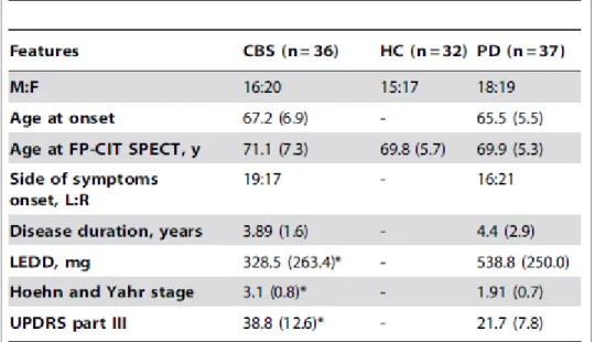
We studied 60 patients (30 with PDD, 30 with DLB) referred to the Movement Disorders Center of Department of Neuroscience – University of Pisa. The diagnosis of PD was made according to UK Brain Bank criteria and dementia according to DSM IV whereas for DLB patients McKeith criteria were met3. Subjects underwent neurological and psychiatric examination and CT/MRI brain scan. The two groups of subjects were matched for sex and age (male/female PDD: 17/13; DLB: 16/14; age: PDD 75.3±3.5; DLB 77.6±4.2). PDD differed from DLB in the disease duration (PDD 9.5±2.3 years; DLB 3.4±1.3 years). All patients were on levodopa therapy (PDD 730 mg/day±240; DLB 650 mg/day± 330). The severity of patients’ motor impairment was assessed by Unified Parkinson Disease Rating Scale (UPDRS) and Hoehn–Yahr staging whereas cognitive functions were evaluated by a full neuropsychological battery and measured by the Mini Mental State Examination (MMSE). Twenty AD patients (9 male/11 female, age 72.3±4.9) were recruited as controls for the perfusional SPECT study since previous investigations performed with perfusional SPECT had compared DLB with AD16. Ten healthy elderly (4 women, 6 men, mean age 65.5±9.3 years) with MMSE score >27 were included in the study as controls in both SPECT studies. Extrapyramidal signs were excluded in the healthy control group.
All PDD and DLB patients were on levodopa therapy and 11 PDD patients were taking dopamine agonists. Levodopa has been demonstrated not to modify FP-CIT uptake17. However all patients were off medication from the night before each SPECT study. No patient was taking any treatment known to interfere with FP-CIT SPECT scan.
All participants gave informant consent. The study was approved by the local research ethics committee.
2.2. FP-CIT SPECT scanning
SPECT studies were carried out according to standard procedure. Scanning took place between 3 and 4 h after the i.v. injection of 185 MBq of 123I-FP-CIT (DaTSCAN®, GE Healthcare, UK). All subjects were scanned with a dual-head gamma camera (Optima NT, GE Healthcare, Milwaukee, WI, USA) equipped with low energy high resolution collimators (system resolution at 10 cm ¼ 8.3 mm). One hundred and twenty-eight projections for each detector were acquired on a 128 x 128 matrix (3 mm pixel size) over a circular 360° orbit; Radius of rotation was less than 16 cm and the overall scanning time for each patient was 30–45 min. Image reconstruction was performed by filtered back projection using a Butterworth prefilter (cut-off 0.4 cycles/cm, order 8); uniform Chang attenuation correction was applied and transaxial slices were reoriented along the fronto-occipital axis.
2.2.1. ROI analysis
Semiquantitative analysis was performed by using a software developed by GE Healthcare on a GE Xeleris workstation. The software used five predefined fixed ROIs which were manually positioned over the caudates, the putamen nuclei and the occipital cortex on reoriented axial data which were summed in order to obtain an overall slice of 15 mm thick, including almost the whole striatum nuclei. Mean counts calculated within caudate and putamen nuclei were divided by mean occipital counts obtaining striatal/to non-specific binding ratios for each nucleus. Mann–Whitney
signed rank U test was used in order to make the following comparisons: PDD group versus DLB group, PDD group versus healthy controls, DLB group versus healthy controls.
2.3. Perfusional SPECT scanning
On different days (at most 10 days apart) patients underwent perfusional SPECT. 99mTc-ethyl cysteinate dimer (Neurolite®, Bristol-Myers Squibb) (900 MBq) was injected with the patient lying supine in a quiet room. About 30 min after injection, SPECT data acquisition was carried out by using the same gamma camera and acquisition parameters used for FP-CIT SPECT. Filtered back projection with a Butterworth prefilter (0.55 cycles/cm, order 10) was used for reconstruction, applying uniform attenuation correction by the Chang’s method (0.12 cm-1 attenuation coefficient). After reconstruction, transaxial images were reoriented along the fronto-occipital line and converted to DICOM 3.0 format. All subsequent image manipulation and data analysis were performed on a Windows-based computer (Microsoft, Redmond, WA). The software for image manipulation included the statistical parametric mapping software SPM2. DICOM files were converted to Analyze format to make them compatible with SPM by using a freeware software. Assessment of regional Cerebral Blood Flow (rCBF) differences between PDD, DLB and AD was made by SPM analysis. Finally the correlation between perfusional changes and nigrostriatal impairment was performed by SPM analysis using as covariates the striatal binding data as semi-quantitative measures obtained from ROI analysis and UPDRS measures (single-subject covariate model).
3. Results 3.1. Subjects
The motor impairment of the two groups (DLB and PDD) did not differ significantly. The mean UPDRS score in the off state was 42.3±6.2 in PDD patients and 39.1±5.4 in DLB group and the Hoehn–Yahr stage was 3.1±0.9 in the PDD group and 2.9±1.1 in the DLB group. The cognitive profile was similar with a mean MMSE of 17.3±2.3 in PDD and 16.9±3.2 in DLB. Twenty-four PDD patients presented with visual hallucinations (and 3 of them with delusions and irritability) whereas 28 DLB patients presented with visual hallucinations, and 7 of them delusions that were severe enough to require a decrease in levodopa dosage. The AD group had a mean MMSE of 17.9±2.1 and none of these patients had visual hallucinations.
3.2. FP-CIT SPECT scanning
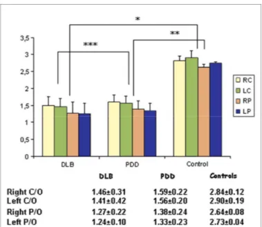
The images obtained from patients with PDD and DLB showed a specific/non-specific uptake ratio significantly lower (p<0.0005) with respect to the control group. The statistical analysis between the two groups showed no significant difference as regards putamen or caudate uptake (right caudate DLB 1,46±0.31 vs PDD 1.59±0.22; left caudate DLB 1.41±0.42 vs PDD 1.56±0.20; right putamen DLB 1.27±0.22 vs PDD 1.38±0.24; left putamen DLB 1.24±0.10 vs PDD 1.33±0.23) (Figure 1 and
2). The analysis of the index of asymmetry showed no difference between
Figure 1 123FP-CIT uptake values, expressed in terms of
semi-quantitative measures as striatal/non-specific (occipital) binding ratios, in patients affected by Parkinson’s Disease with Dementia (PDD), Lewy Body Dementia (DLB) and healthy controls. *PD vs controls p < 0.005; **DLB vs controls p < 0.005; ***PDD vs DLB ns RC/LC: Right/Left Caudate RP/LP: Right/Left Putamen
Figure 2 123I- FP-CIT SPECT images obtained in single cases of Lewy Body Dementia (DLB), Parkinson’s Disease with Dementia (PDD) and (right) healthy control.
3.3. Perfusional SPECT scanning
Patients with PDD and DLB showed a significant reduction of rCBF (p<0.001) in parieto-occipital and frontal areas with respect to controls (Figure 3). The comparison between the two groups did not result in any
significant difference. Both DLB and PDD groups when compared to AD group showed a significant decrease of rCBF (p<0.001) in the occipital regions (Figure 3). No correlation was observed between rCBF changes and caudate/putamen FP-CIT binding ratios as well as the UPDRS scores.
Figure 3 MIP (maximum intensity projection) images reporting brain areas with significant reduction of rCBF (p<0.001) (top) in parieto-occipital and frontal areas in DLB and PDD with respect to controls, (middle) in occipital regions in PDD and DLB with respect to Alzheimer’s Disease (AD) patients, (bottom) in temporo-parietal regions in AD with respect to PDD and DLB.
4. Discussion
In the present study significant reductions in uptake of a DAT ligand bilaterally in the caudate and putamen were demonstrated in patients with DLB and PDD compared to normal controls consistently with previous studies11, 12, 18. The level of dopamine transporter binding in DLB was indistinguishable from PDD. This finding is entirely in keeping with previous studies11, 12. In detail, Walker et al. found a similar impairment of the basal ganglia in both disorders with a symmetric loss in dopamine transporter more pronounced in putamen than in the caudate and more severe in the posterior part than in the anterior part of the putamen12. Similarly, in our study the loss of dopamine transporter was more pronounced in putamen than in the caudate and it was symmetric in both PDD and DLB with no significant differences. The recruitment method of our study population used strict criteria in order to avoid any bias due to potentially confounding clinical factors, in particular both the cognitive status and the motor impairment was very similar in both groups of patients. DLB and PDD are the most common dementias associated with deposition of Lewy bodies, and they are distinguished by a differential sequence of parkinsonism and dementia. Neuropathological findings suggest that DLB and PDD differ most in the selective vulnerability of the neuronal populations early in the disease process, and that cortical Lewy bodies along with pathological aging and Alzheimer-type pathology are the most common pathologic substrate for dementia in both disorders19. At autopsy both DLB and PDD have shown concomitant AD pathology with amyloid plaques and neurofibrillary tangles. Amyloid load has been measured in vivo with [11C]PIB PET and recent studies have shown indeed that the majority of DLB had PIB levels in the AD range whereas
most PDD patients had normal PIB uptake. These findings suggest that the amyloid load could contribute to an acceleration of the dementia process whereas Lewy body pathology alone leads to a slower dementing process in PDD20. Our data, confirming some diverse earlier findings, could suggest the lack of any difference as regards the presynaptic dopaminergic nigro-striatal function as assessed in vivo by FP-CIT SPECT between DLB and PDD.
We found that the pattern of cerebral regional perfusion is similar in DLB and PDD, which is different from AD. In particular there was greater evidence of hypoperfusion in the lateral parts of the parieto-occipital cortex in DLB and PDD than AD. In addition the comparison between DLB and PDD using a high statistical threshold did not demonstrate any significant difference. Parietal regions are involved in visuo-spatial processing, which is impaired in PD, PDD and DLB groups3 and dysfunctions in occipito-parietal regions as assessed by in vivo neuroimaging in PD have been reported to well correlate with visuo-spatial performance21. Some authors have linked the findings of the occipito-parietal hypoperfusion/hypometabolism to the presence of the visual hallucinations, but these findings are controversial. Indeed, in a post-mortem study of patients with PD and DLB, visual hallucinations have been shown to correlate with Lewy bodies deposition in medial and inferior temporal areas including amygdala, parahippocampus and inferior temporal cortices whereas no correlation was found with occipital impairment22. Moreover, a recent FDG PET study has suggested the role of frontal regions in association with the hallucinations occurring in PD23. Similarly, in a functional MRI study, hallucinating PD patients showed an increased activation in the superior and inferior frontal gyrus and a decreased