R E S E A R C H A R T I C L E
Open Access
Adiposity, metabolites, and colorectal
cancer risk: Mendelian randomization study
Caroline J. Bull
1,2,3*†, Joshua A. Bell
1,2†, Neil Murphy
4, Eleanor Sanderson
1,2, George Davey Smith
1,2,
Nicholas J. Timpson
1,2, Barbara L. Banbury
5, Demetrius Albanes
6, Sonja I. Berndt
6, Stéphane Bézieau
7,
D. Timothy Bishop
8, Hermann Brenner
9,10,11, Daniel D. Buchanan
12,13,14, Andrea Burnett-Hartman
15,
Graham Casey
16, Sergi Castellví-Bel
17, Andrew T. Chan
18,19,20,21, Jenny Chang-Claude
22,23, Amanda J. Cross
24,
Albert de la Chapelle
25, Jane C. Figueiredo
26,27, Steven J. Gallinger
28, Susan M. Gapstur
29, Graham G. Giles
30,31,32,
Stephen B. Gruber
33, Andrea Gsur
34, Jochen Hampe
35, Heather Hampel
36, Tabitha A. Harrison
5,
Michael Hoffmeister
9, Li Hsu
5,37, Wen-Yi Huang
6, Jeroen R. Huyghe
5, Mark A. Jenkins
31, Corinne E. Joshu
38,
Temitope O. Keku
39, Tilman Kühn
22, Sun-Seog Kweon
40,41, Loic Le Marchand
42, Christopher I. Li
5, Li Li
43,
Annika Lindblom
44,45, Vicente Martín
46,47, Anne M. May
48, Roger L. Milne
30,31,32, Victor Moreno
46,49,50,51,
Polly A. Newcomb
5,52, Kenneth Offit
53,54, Shuji Ogino
55,56,57,58, Amanda I. Phipps
5,59, Elizabeth A. Platz
38,
John D. Potter
5,60,61,62, Conghui Qu
5, J. Ramón Quirós
63, Gad Rennert
64,65,66, Elio Riboli
67, Lori C. Sakoda
5,68,
Clemens Schafmayer
69, Robert E. Schoen
70, Martha L. Slattery
71, Catherine M. Tangen
72, Kostas K. Tsilidis
67,73,
Cornelia M. Ulrich
74, Fränzel J. B. van Duijnhoven
75, Bethany van Guelpen
76,77, Kala Visvanathan
38,
Pavel Vodicka
78,79,80, Ludmila Vodickova
78,79,80, Hansong Wang
42, Emily White
5,81, Alicja Wolk
82,
Michael O. Woods
83, Anna H. Wu
84, Peter T. Campbell
85, Wei Zheng
86, Ulrike Peters
5, Emma E. Vincent
1,2,3†and
Marc J. Gunter
4†Abstract
Background: Higher adiposity increases the risk of colorectal cancer (CRC), but whether this relationship varies by
anatomical sub-site or by sex is unclear. Further, the metabolic alterations mediating the effects of adiposity on CRC
are not fully understood.
(Continued on next page)
© The Author(s). 2020 Open Access This article is licensed under a Creative Commons Attribution 4.0 International License, which permits use, sharing, adaptation, distribution and reproduction in any medium or format, as long as you give appropriate credit to the original author(s) and the source, provide a link to the Creative Commons licence, and indicate if changes were made. The images or other third party material in this article are included in the article's Creative Commons licence, unless indicated otherwise in a credit line to the material. If material is not included in the article's Creative Commons licence and your intended use is not permitted by statutory regulation or exceeds the permitted use, you will need to obtain permission directly from the copyright holder. To view a copy of this licence, visithttp://creativecommons.org/licenses/by/4.0/. The Creative Commons Public Domain Dedication waiver (http://creativecommons.org/publicdomain/zero/1.0/) applies to the data made available in this article, unless otherwise stated in a credit line to the data.
* Correspondence:[email protected]
†Caroline J. Bull and Joshua A. Bell are joint first authors.
Emma E. Vincent and Marc J. Gunter are joint last authors.
1
MRC Integrative Epidemiology Unit at the University of Bristol, Oakfield House, Bristol, UK
2Population Health Sciences, Bristol Medical School, University of Bristol,
Bristol, UK
Full list of author information is available at the end of the article
Bull et al. BMC Medicine (2020) 18:396 https://doi.org/10.1186/s12916-020-01855-9
(Continued from previous page)
Methods: We examined sex- and site-specific associations of adiposity with CRC risk and whether
adiposity-associated metabolites explain the associations of adiposity with CRC. Genetic variants from genome-wide
association studies of body mass index (BMI) and waist-to-hip ratio (WHR, unadjusted for BMI;
N = 806,810), and 123
metabolites from targeted nuclear magnetic resonance metabolomics (
N = 24,925), were used as instruments.
Sex-combined and sex-specific Mendelian randomization (MR) was conducted for BMI and WHR with CRC risk (58,221
cases and 67,694 controls in the Genetics and Epidemiology of Colorectal Cancer Consortium, Colorectal Cancer
Transdisciplinary Study, and Colon Cancer Family Registry). Sex-combined MR was conducted for BMI and WHR
with metabolites, for metabolites with CRC, and for BMI and WHR with CRC adjusted for metabolite classes in
multivariable models.
Results: In sex-specific MR analyses, higher BMI (per 4.2 kg/m
2) was associated with 1.23 (95% confidence interval
(CI) = 1.08, 1.38) times higher CRC odds among men (inverse-variance-weighted (IVW) model); among women,
higher BMI (per 5.2 kg/m
2) was associated with 1.09 (95% CI = 0.97, 1.22) times higher CRC odds. WHR (per 0.07
higher) was more strongly associated with CRC risk among women (IVW OR = 1.25, 95% CI = 1.08, 1.43) than men
(IVW OR = 1.05, 95% CI = 0.81, 1.36). BMI or WHR was associated with 104/123 metabolites at false discovery
rate-corrected
P ≤ 0.05; several metabolites were associated with CRC, but not in directions that were consistent with
the mediation of positive adiposity-CRC relations. In multivariable MR analyses, associations of BMI and WHR with
CRC were not attenuated following adjustment for representative metabolite classes, e.g., the univariable IVW OR
for BMI with CRC was 1.12 (95% CI = 1.00, 1.26), and this became 1.11 (95% CI = 0.99, 1.26) when adjusting for
cholesterol in low-density lipoprotein particles.
Conclusions: Our results suggest that higher BMI more greatly raises CRC risk among men, whereas higher WHR
more greatly raises CRC risk among women. Adiposity was associated with numerous metabolic alterations, but
none of these explained associations between adiposity and CRC. More detailed metabolomic measures are likely
needed to clarify the mechanistic pathways.
Keywords: Body mass index, Waist-to-hip ratio, Colorectal cancer, Mendelian randomization, Metabolism, NMR,
Epidemiology, GECCO, CORECT, CCFR
Background
Colorectal cancer (CRC) is one of the most commonly
diagnosed cancers among adults globally [
1
–
3
]. Obesity
is viewed as a likely cause of CRC by the International
Agency for Research on Cancer (IARC), the American
Institute for Cancer Research (AICR), and the World
Cancer Research Fund (WCRF) [
3
,
4
], based largely on
positive associations between adiposity and CRC risk
from observational epidemiology. Further, the limited
data available from observational studies suggest that
intentional weight loss lowers the risk of CRC in
post-menopausal women [
5
]. Mendelian randomization
(MR) studies, which use genetic variants as instruments
(proxies) for adiposity given their randomly allocated
and fixed nature [
6
], further support causality [
7
–
9
].
Despite this growing consensus, it remains unclear
whether the effect of adiposity on CRC risk differs
among men and women, whether the relationship
var-ies by CRC sub-site, and what the underlying biological
mechanisms are. These are important to clarify given
the ongoing obesity epidemic and difficulties in
redu-cing adiposity itself [
10
,
11
].
Observationally, body mass index (BMI) relates more
strongly to CRC risk among men and waist-to-hip ratio
(WHR) relates similarly to CRC risk among men and
women [
12
]. However, recent MR studies suggest that
higher BMI more greatly raises CRC risk among women,
while higher WHR more greatly raises CRC risk among
men [
7
,
8
]. Whether these MR estimates are robust is
unclear because they were based on relatively small
sam-ple sizes, genetic instruments that were not sex-specific,
and genetic instruments for WHR that were conditioned
on BMI—all potential sources of bias [
13
–
17
].
Adiposity alters the systemic metabolism [
18
–
20
], but
evidence for the effects of adiposity-altered metabolites
on CRC is scarce. One MR study suggested that total
cholesterol raises CRC risk [
21
], while others suggested
no effect of blood glucose [
22
] and mixed support for
fatty acids [
23
]. Overall, the scope of metabolic traits
ex-amined has also been narrow. Targeted metabolomics
allows deeper phenotyping at a large scale [
24
], and its
recent integration with genotype data [
25
] enables us to
examine the associations of metabolites with CRC using
MR. Expanded genotype data for CRC is also available
[
26
], affording a sample size six times larger than used
in previous MR studies (58,221 cases, 67,694 controls).
This study has two aims. First, we aimed to better
esti-mate sex-specific effects of adiposity on CRC risk using
two-sample MR. We examined associations of BMI and
WHR with CRC risk using expanded GWAS data and
genetic instruments for exposures that were sex-specific
and were not mutually conditioned, to reduce bias [
13
–
17
]. Second, we aimed to identify potential metabolic
mediators of effects of adiposity on CRC risk using
two-step MR (by examining associations of BMI and WHR
with metabolites, and of BMI- or WHR-related
metabo-lites with CRC risk) and multivariable MR (by adjusting
associations of BMI and WHR with CRC for
representa-tive metabolites).
Methods
Study design
We used two-sample MR to examine the associations
(pertaining to estimates of the effect predicted from
gen-etic variants used as instruments) of adiposity with CRC
risk,
of
adiposity
with
metabolites,
of
adiposity-associated metabolites with CRC risk, and finally of
adiposity with CRC risk adjusted for representative
metabolites. In two-sample MR, SNP-exposure and
SNP-outcome associations are obtained from different
study sources and combined as a ratio to estimate the
ef-fects of exposures on outcomes [
13
,
27
]. Our study aims
and assumptions are shown in Fig.
1
.
Adiposity instruments
We identified SNPs that were independently associated
(low linkage disequilibrium (LD),
R
2< 0.001) with BMI
and WHR (unadjusted for BMI) at
P < 5 × 10
−8from a
recent
large-scale
genome-wide
association
study
(GWAS) meta-analysis of 221,863 to 806,810 male and
female adults of European ancestry from the Genetic
In-vestigation of ANthropometric Traits (GIANT)
consor-tium and the UK Biobank [
33
] (Additional file
1
: Table
S1). BMI and WHR are expressed in standard deviation
(SD) units. For sex-combined analyses of BMI and
WHR, 312 and 209 SNPs were used, respectively. For
sex-specific analyses of BMI, 185 and 152 SNPs were
used for women and men, respectively. For sex-specific
analyses of WHR, 153 and 64 SNPs were used for
women and men, respectively. The proportion of
vari-ance explained in adiposity traits by instruments ranged
from 0.3 to 5.04% (these were based on approximations
for BMI using the equation described by Shim et al.
[
34
]), and
F-statistics (a formal test of whether variance
explained is sufficiently high to avoid weak instrument
bias) for adiposity instruments ranged from 75.81 to
124.49 (Additional file
1
: Table S2) which indicated
Fig. 1 Study aims and assumptions. Study aims are to (1) estimate the total effect of adiposity on CRC risk using genetic instruments for BMI and WHR ((i) unadjusted for BMI) and (2) estimate the mediated effect of adiposity on CRC risk by metabolites from targeted NMR metabolomics. Aim 2 is addressed using two approaches: (1) two-step MR wherein effects are examined of adiposity on metabolites (ii) and of adiposity-related metabolites on CRC risk (iii) and (2) multivariable MR wherein effects of adiposity on CRC (i) are examined with adjustment for the effect of representative metabolite classes on CRC (iii). Sex-specific analyses were performed when sex-specific GWAS estimates for exposure and outcome were both available. When≥ 2 SNP instruments were available, up to 4 MR models were applied: the inverse-variance-weighted (IVW) model which assumes that none of the SNPs are pleiotropic [28], the weighted median (WM) model which allows up to half of the included SNPs to be pleiotropic and is less influenced by outliers [28], the weighted mode model which assumes that the most common effect is consistent with the true causal effect [29], and the MR-Egger model which provides an estimate of association magnitude allowing all SNPs to be pleiotropic [30]. Analyses with metabolites as outcomes were conducted within discovery aims whereinP value thresholds are applied to prioritize traits with the strongest evidence of association to be taken forward into further stages of analysis (with CRC risk). Analyses with CRC as outcomes were conducted within estimation aims whereinP values are interpreted as continuous indicators of evidence strength and focus is on effect size and precision [31,32]
instrument strength above the recommended minimum
levels [
35
].
Metabolite instruments
We identified SNPs that were independently associated
(R
2< 0.001 and
P < 5 × 10
−8) with metabolites from a
GWAS of 123 traits from targeted nuclear magnetic
res-onance (NMR) metabolomics (Additional file
1
: Table
S1); these included lipoprotein subclass-specific lipids,
amino acids, fatty acids, inflammatory glycoproteins, and
others [
25
]. Between 13,476 and 24,925 adults (men and
women combined) of European ancestry were included.
Metabolic traits are expressed in SD units. The
propor-tion of variance explained in metabolites by instruments
ranged from 0.44 to 12.49%, and
F-statistics for
metabol-ite instruments ranged from 30.2 to 220.8
(Add-itional file
1
: Table S2) which indicated sufficient
instrument strength for univariable analyses.
Colorectal cancer GWAS data
We obtained SNP estimates from the most
comprehen-sive GWAS of CRC to date [
26
], including 58,221 cases
and 67,694 controls (sexes combined) from 45 studies
within 3 consortia: Genetics and Epidemiology of
Colo-rectal Cancer Consortium (GECCO), ColoColo-rectal Cancer
Transdisciplinary Study (CORECT), and Colon Cancer
Family Registry (CCFR). Across these studies, there were
28,207 CRC cases and 22,204 controls among men, and
24,568 CRC cases and 23,736 controls among women.
Cases were diagnosed by a physician and recorded
over-all and by site (colon, proximal colon, distal colon,
rec-tum). Approximately 92% of the participants were
White-European (~ 8% were East Asian). Case
distribu-tions are outlined in Additional file
1
: Table S3; other
study characteristics are detailed elsewhere [
26
]. Ethics
were approved by respective institutional review boards.
Statistical approach
First, we examined the associations of BMI and WHR
with overall and site-specific CRC using SNP estimates
from sex-combined GWAS of exposures as well as
out-comes. We then examined the associations of BMI and
WHR with overall CRC based on SNP estimates from
sex-specific GWAS of exposure as well as outcome
(sex-specific GWAS were not available and thus not used for
site-specific CRC). Summary statistics were harmonized
using the harmonise_data function within the
TwoSam-pleMR R package [
36
]. All GWAS were assumed to be
coded on the forward strand, and harmonization was
confirmed as consistent using option 2 of the
“action”
argument. As sensitivity analyses, up to four MR
methods were used to generate effect estimates using
the TwoSampleMR R package [
36
] which make differing
pleiotropy assumptions (detailed in Fig.
1
legend) [
29
,
36
,
37
]. When only a single SNP was available, the Wald
ratio was used [
38
]. When
≥ 2 SNPs were available,
random-effects inverse-variance-weighted (IVW) [
36
],
MR-Egger [
30
], weighted median (WM) [
28
], and
weighted mode [
29
] models were used. Cochrane’s
Q-statistic was used to assess the heterogeneity of SNP
ef-fects (smaller
P values indicating higher heterogeneity
and higher potential for directional pleiotropy [
39
]).
Scatter plots were used to compare MR models, and
“leave-one-SNP-out” analyses were used to detect SNP
outliers [
40
].
Second, we examined associations of BMI and WHR
with metabolites using results from sex-combined
GWAS for exposures as well as outcomes (sex-specific
GWAS were not available for metabolites, and so
sex-specific analyses were not conducted) and the MR
models described above. Each metabolite (analyzed as an
outcome) that was associated with either BMI or WHR
based on an IVW model
P value ≤ 0.05 following a false
discovery rate (FDR) correction (Benjamini-Hochberg
method [
41
]) was taken forward and examined for
asso-ciation with CRC risk using the IVW model (if
≥ 2
SNPs) or the Wald ratio (if 1 SNP). Multivariable MR
[
42
] was also used to examine the associations of BMI
and WHR with CRC risk, adjusting for single
metabo-lites that were representative of various metabolite
clas-ses based on previous network analyclas-ses [
43
] and that
had the highest instrument strength based on the
F-stat-istic (Additional file
1
: Table S2). As a positive control,
we adjusted BMI for WHR as a covariate (which is
ex-pected to attenuate the association of BMI with CRC
risk), and likewise, we adjusted WHR for BMI as a
co-variate with the same expectation. A smaller set of SNPs
for BMI and WHR based on earlier GWAS [
44
,
45
] was
used for these multivariable models to avoid a relative
dilution of metabolite instrument strength given that the
number of SNPs for BMI and WHR from expanded
GWAS far outnumbered those for metabolites.
Condi-tional
F-statistics were calculated for exposures in
multi-variable models [
46
].
In each instance, MR estimates are interpreted as the
change in outcome per SD unit change in the exposure.
Estimates for metabolite outcomes reflect SD unit
change, and estimates for CRC outcomes reflect odds
ra-tios (OR). Statistical analyses were performed using R
(version 3.5.2).
Results
Associations of BMI and WHR with CRC risk
In sex-combined analyses (Fig.
2
; Additional file
1
: Table
S4), higher BMI (per 4.8 kg/m
2) was associated with a
higher risk of overall CRC (IVW OR = 1.16, 95% CI =
1.07, 1.26). The WM estimate was similar, but the
MR-Egger and weighted mode estimates were both reduced
(e.g., MR-Egger OR = 1.02, 95% CI = 0.84, 1.25). BMI
as-sociations were consistent across CRC sites. Asas-sociations
were directionally consistent for WHR as for BMI but
were marginally stronger—e.g., higher WHR (per 0.09
ratio) was associated with 1.28 (95% CI = 1.16, 1.42)
times higher odds of CRC in an IVW model (MR-Egger
and weighted mode estimates were each positive but of
a smaller magnitude with wide intervals spanning the
null). WHR associations were more consistent for colon
rather than rectal sub-sites. SNP heterogeneity was
simi-larly high for BMI and WHR (P value range across
models = 9.54 × 10
−10to 1.97 × 10
−8).
In sex-specific IVW models (Fig.
2
; Additional file
1
:
Table S4), higher BMI (per 4.2 kg/m
2) was associated
with 1.23 (95% CI = 1.08, 1.38) times higher odds of
CRC among men and 1.09 (95% CI = 0.97, 1.22) times
higher odds of CRC (per 5.2 kg/m
2) among women.
In a WM model, this BMI estimate was robust among
men (OR = 1.22, 95% CI = 1.02, 1.46) but reduced
among women (OR = 1.04, 95% CI = 0.86, 1.26).
MR-Egger and weighted mode estimates were similarly
imprecise among men and women, and SNP
hetero-geneity was similar for both. In IVW models, higher
WHR (per 0.07 ratio) was associated with 1.25 (95%
CI = 1.08, 1.43) times higher odds of CRC among
women; this estimate was 1.05 (95% CI = 0.81, 1.36)
among men (per 0.07 ratio). This pattern was also
supported by WM estimates (OR = 1.14, 95% CI =
0.91, 1.42 among women and OR = 0.95, 95% CI =
0.90, 1.29 among men), and by MR-Egger and
weighted mode estimates. SNP heterogeneity was
similarly high among men and women.
Scatter plots comparing different MR models and
re-sults of the
“leave-one-SNP-out” analyses are presented
in Additional file
2
: Figures S1-42.
Associations of BMI and WHR with metabolites
In sex-combined analyses, higher BMI (per 4.8 kg/m
2) or
WHR (per 0.09 ratio) was associated with 104
metabo-lites based on FDR-corrected
P value ≤ 0.05 in IVW
Fig. 2 Associations of BMI and WHR with CRC risk based on two-sample MR. Sex-combined estimates are based on GWAS done among women and men together (for both exposure and outcome). Sex-specific estimates are based on GWAS done separately among women and men (for exposure as well as outcome)
models
(Additional
file
2
:
Figures
S43-47;
Add-itional file
1
: Table S5). Evidence was strong in relation
to lipids including total cholesterol and triglycerides in
very low-density lipoproteins (VLDL), low-density
lipo-proteins (LDL), and high-density lipolipo-proteins (HDL)—
e.g., 0.23 SD (95% CI = 0.15, 0.31) higher triglycerides in
large VLDL from higher BMI. Associations of higher
BMI were also strong with lactate, pyruvate, and
branched-chain amino acids—e.g., 0.19 SD (95% CI =
0.13, 0.25) higher isoleucine—and with inflammatory
glycoproteins (0.28 SD, 95% CI = 0.20, 0.36 higher).
Similar patterns were seen for WHR.
Associations of BMI- or WHR-related metabolites with
CRC
Of 104 metabolites associated (as outcomes) with BMI
or WHR in sex-combined analyses, 100 had SNPs for
use in Wald or IVW models. As shown in
Add-itional file
1
: Table S1, 321 unique SNPs were used to
instrument 100 metabolites (3 metabolites had 1 SNP,
13 metabolites had < 5 SNPs, and 51 metabolites had <
10 SNPs; SNP counts across metabolites ranged from 1
to 26). Lipid traits showed generally weak associations
with CRC which were also in directions inconsistent
with the mediation of the adiposity-CRC relationship—
e.g., lipids in medium HDL were positively associated
with CRC, but these had been negatively associated with
BMI or WHR (Fig.
3
; Additional file
1
: Table S6). In
contrast, there was more consistent evidence of a
posi-tive association of lipids in intermediate-density
lipopro-tein (IDL), VLDL, and LDL with a risk of distal colon
cancer, and these lipids had been positively associated
with higher BMI or WHR. For example, higher total
lipids in IDL (per SD) were associated with 1.09 (95%
CI = 1.02, 1.15) times higher odds of distal colon cancer.
Lipids were unassociated with the risk of proximal colon
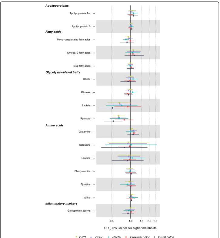
cancer. Fatty acids were unassociated with CRC risk
ex-cept for higher monounsaturated fatty acid levels which
were associated with a lower risk of rectal cancer (IVW
OR = 0.85, 95% CI = 0.75, 0.95; Fig.
4
). Lactate and
pyru-vate were inversely associated with CRC at 0.66 (95%
CI = 0.42, 1.03) times lower odds and 0.64 (95% CI =
0.52, 0.80) times lower odds, respectively. However,
these metabolites were positively associated with BMI,
and so directions were inconsistent with the mediation
of the adiposity-CRC relationship. Amino acids and
glycoprotein acetyls were unassociated with CRC risk.
Associations of BMI and WHR with CRC risk independent
of metabolites
The association of BMI with overall CRC was not
atten-uated following adjustment for various metabolite
clas-ses (Fig.
5
; Additional file
1
: Table S7). The univariable
IVW OR for BMI (per 4.77 kg/m
2higher, based on 67
SNPs) in relation to CRC was 1.12 (95% CI = 1.00, 1.26),
whereas this IVW OR was 1.14 (95% CI = 1.01, 1.29)
adjusting for VLDL lipids and 1.11 (95% CI = 0.99, 1.26)
adjusting for IDL and LDL lipids. Attenuation was
greater when adjusting the BMI-CRC association for
WHR (positive control), at IVW OR = 0.93 (95% CI =
0.78, 1.11). Results for WHR in relation to CRC were
directionally consistent as seen for BMI, with a lack of
attenuation upon adjustment for metabolite classes.
Discussion
We aimed to better estimate sex-specific effects of
adi-posity on CRC risk and to identify potential metabolic
mediators of the effects of adiposity on CRC, using
two-sample MR methods and expanded two-sample sizes. Our
re-sults, based on genetic instruments for adiposity that
were sex-specific and were not mutually conditioned,
suggest that higher BMI more greatly raises CRC risk
among men, whereas higher WHR more greatly raises
CRC risk among women. In sex-combined mediation
analyses, adiposity was associated with numerous
meta-bolic alterations, but none of these alterations explained
the associations between adiposity and CRC. More
de-tailed metabolomic measures are likely needed to clarify
the mechanistic pathways.
Observational [
3
,
47
] and MR [
7
–
9
] studies have
sug-gested adverse effects of adiposity on CRC risk, but
causal evidence has been lacking regarding sex
specifi-city. Previous MR studies suggested stronger effects of
BMI on CRC risk among women [
7
–
9
], which
contra-dicts observational suggestions of stronger effects among
men [
12
]. The genetic regulation of BMI and WHR
shows strong sexual dimorphism, thought attributable to
the influence of sex hormones, namely estrogen, and it
is important to capture these differences in MR
esti-mates [
48
,
49
]. Our new results are based on
instru-ments for BMI and WHR that were sex-specific and a
sample size for CRC that was six times larger than used
previously which enabled higher power relative to two
previous MR studies of BMI, WHR, and CRC risk [
7
,
8
]
(Additional file
2
: Figure S48). These new results suggest
that BMI more greatly raises CRC risk among men—a
reversal of previous MR estimates. This new pattern for
BMI and CRC (22% higher risk among men per 4.2 kg/
m
2and 9% higher risk among women per 5.2 kg/m
2) is
highly consistent with observational estimates reviewed
by IARC (22% higher risk in men and 9% higher risk in
women per 5 kg/m
2[
4
]). Our results also support a
re-versal of previous MR estimates for WHR, with risk now
appearing higher among women than among men. This
is unexpected since BMI and abdominal fat measures
correlate highly [
50
,
51
]; however, given that fat storage
is more peripheral in women [
18
,
19
], WHR (unadjusted
for BMI) may be a better proxy for the extremeness of
Fig. 3 Associations of BMI- or WHR-related lipid metabolites with CRC risk based on two-sample MR (IVW method). Estimates reflect the OR (95% CI) for CRC per SD higher metabolite that is associated (as an outcome) with BMI or WHR. +/− symbols indicate the direction of association of BMI or WHR with that metabolite
fat volume among women since fat may be stored more
abdominally only when peripheral fat stores are
over-whelmed. As a post hoc comparison, we repeated
ana-lyses of the main effects of adiposity on CRC using the
sex-combined adiposity instruments in relation to split
samples of men and women (Additional file
1
: Table S8,
A) to examine the potential for biased results. These
suggest that use of sex-combined instruments for BMI
and WHR would lead to the conclusion that both are
as-sociated with higher CRC risk in males as well as
fe-males, but with still higher risk with BMI among males
and with WHR among females, in contrast to previous
Fig. 4 Associations of BMI- or WHR-related non-lipid metabolites with CRC risk based on two-sample MR (IVW method). Estimates reflect the OR (95% CI) for CRC per SD higher metabolite that is associated (as an outcome) with BMI or WHR. +/− symbols indicate the direction of association of BMI or WHR with that metabolite
MR studies [
7
,
8
]. This suggests that discrepancies in the
result patterns are most likely due to the differences in
the power of the main adiposity-CRC relationship
(Add-itional file
2
: Figure S48).
SNP heterogeneity was high for BMI and WHR with
CRC, although this was similar between sexes and
direc-tions of effect from sensitivity models were consistent,
suggesting balanced SNP heterogeneity. One cause of
heterogeneity may be pleiotropy in violation of the
ex-clusion restriction criteria (assumption 3, Fig.
1
). This is
not unexpected due to the large number of SNPs
in-cluded in the adipose trait instruments and the many
underlying biological pathways that explain variation in
adiposity. A future approach to minimizing
heterogen-eity in instrument selection could be to analyze the
asso-ciation between subsets of genetic variants related to
specific pathways of BMI and WHR in relation to CRC;
this requires more biological knowledge of these genetic
variants than currently exists.
Given the difficulty of weight loss [
11
] and the ongoing
obesity epidemic, it is increasingly important to identify
the biological pathways which explain the effect of
adi-posity on the risk of chronic diseases including CRC
[
10
]. Adipose tissue is highly metabolically active and
Fig. 5 Associations of BMI and WHR with CRC risk independent of various metabolite classes based on multivariable MR. Metabolite classes are based on a single representative metabolite from a previous network analysis [43], as follows: VLDL (triglycerides in small VLDL); IDL and LDL (total cholesterol in medium LDL), HDL (triglycerides in very large HDL), 3 and PUFA (other polyunsaturated fatty acids than 18:2), Omega-6 (18:2, linoleic acid), MUFA and other fatty acids (Omega-9 and saturated fatty acids), glycemia (glucose), substrates (citrate), branched-chain amino acids (leucine), and other amino acids (glutamine). Adipose adjustments include the alternative adiposity trait (WHR or BMI) as a positive control
secretes pro-inflammatory cytokines such as interleukin
(IL)-6 and tumor necrosis factor (TNF)-alpha which may
promote tumor initiation [
52
]. Adipose tissue-derived
inflammation also promotes insulin resistance in glucose
storage tissues that can lead to hyperinsulinemia [
53
],
and insulin and insulin-like growth factors (IGF) such as
IGF-1 have pro-mitogenic and anti-apoptotic effects that
are cancer promotive [
47
,
54
–
58
]. Our current results
suggest effects of BMI or WHR on numerous lipids and
pre-glycemic traits; however, few of these traits had any
strong association with CRC risk, and the few that did
were in a direction that was inconsistent with a
mediat-ing role in the adiposity-CRC relationship. Results of a
series of multivariable MR models, which adjusted for
various metabolites considered representative of broader
metabolite classes [
42
], suggested that associations of
BMI and WHR with CRC risk were highly independent
of these metabolites. However, this analysis may be
lim-ited by weak instrument bias [
59
] given that
F-statistics
for metabolite instruments included in each
multivari-able MR model were relatively low. Nevertheless, the
re-sults of two complementary approaches to mediation
(two-step MR and multivariable MR) provide little
evi-dence that the effects of adiposity on CRC risk are
medi-ated by adiposity-relmedi-ated metabolites that are detectable
by NMR metabolomics. Future studies could examine
metabolites, proteins, hormones, and inflammatory
fac-tors that are detectable by other metabolomic and
prote-omic platforms.
The few traits that did show consistent directions of
effect included total lipids in IDL, LDL, and VLDL
parti-cles which were raised by BMI and which in turn raised
the risk of distal colon cancer specifically (not proximal
colon or rectal cancer). If robust, this pattern may reflect
differential sensitivity of colon regions to lipid exposure
owing to divergent functions (the distal colon functions
primarily in the storage of resultant fecal matter whereas
the proximal colon functions primarily in water
absorp-tion and fecal solidificaabsorp-tion [
60
]), or it may reflect
differ-ential detectability through screening (proximal colon
tumors tend to be detected in older ages and at more
advanced stages [
60
]). Colorectal anatomical regions
may also have distinct molecular features [
61
], e.g., the
distal colon may be more susceptible to p53 mutations
and chromosomal instability [
62
], whereas the proximal
colon may be more mucinous and susceptible to
micro-satellite instability and B-Raf proto-oncogene expression
[
63
,
64
]. Several meta-analyses of long-term follow-ups
of randomized controlled trials of LDL
cholesterol-lowering statin use suggested no strong evidence of a
protective effect of statin used on CRC risk [
65
–
67
];
CRC sub-sites were largely unexamined. One previous
MR study suggested an adverse effect of higher LDL
cholesterol, and a protective effect of genetically proxied
statin use, on overall CRC risk [
21
]; again, CRC sub-sites
were not examined. Prospective observational evidence
for LDL cholesterol and CRC risk is less consistent than
for total cholesterol or triglycerides; heterogeneity in
meta-analyzed effect estimates is much higher for LDL
cholesterol (82.7% based on an
I
2statistic) compared
with total cholesterol and triglycerides (46.7% and 47.8%,
respectively) [
68
]. Prospective estimates of lipoprotein
subclass measures from metabolomic platforms are
lack-ing as these are only recently available at scale.
The limitations of this study include the non-specificity
of genetic variants used as instruments for some
metabo-lites which stems from their expectedly correlated nature
(e.g., rs1260326, a SNP in
GCKR, was included in genetic
instruments for 54 metabolites). A total of 321 unique
SNPs was used to instrument 100 metabolites, but the
number of instruments available for a given metabolite was
typically small. This limits causal inference for individual
traits but should not prevent the identification of relevant
classes of traits (e.g., lipid, amino acid). It should also be
stressed that genetic variants used for metabolites may
alter the enzyme expression and so serve as instruments
for the metabolizing enzyme itself, not factors influenced
downstream of that enzyme. Since inference in MR applies
to the most proximal trait that the genetic variant relates
to [
15
], directing inference to specific glycolytic traits as
distinct from their downstream consequences like insulin
resistance [
69
] (a key result of higher fatness and trigger of
tumorigenesis [
61
]) is difficult and requires stronger
gen-etic instruments alongside mechanistic insights from
pre-clinical studies [
70
]. Adiposity was measured indirectly
using BMI and WHR because these correlate highly with
more objectively measured fat indexes [
50
,
51
] and allow
much larger GWAS sample sizes than otherwise possible
(comparably strong GWAS were unavailable for waist
cir-cumference). UK Biobank data are included within GWAS
for both the exposure and outcome used for MR estimates
of adiposity for CRC risk. Sample overlap in a two-sample
MR setting is reported to contribute to weak instrument
bias and inflated type one error rates, resulting in MR
esti-mates that are biased towards confounding-prone
observa-tional estimates [
71
]. However, given that the proportion
of sample overlap is presently low (< 5%) and estimated
F-statistics are relatively high (each > 70 for adiposity traits),
we do not expect considerable bias here. As a post hoc
comparison, we obtained CRC summary GWAS statistics
with UK Biobank excluded and repeated MR analyses of
adiposity for CRC risk. Estimates were largely consistent
with or without the inclusion of UK Biobank data
(Add-itional file
1
: Table S8, B). Our sex-specific MR
investiga-tions were confined to effects of adiposity on overall CRC
because sex-specific GWAS were unavailable for
site-specific CRC and metabolite outcomes. Sex-stratified
GWAS of such outcomes would enable these in the future.
Conclusions
Our results based on sex-specific MR instruments and
expanded sample sizes suggest that higher BMI more
greatly raises CRC risk among men, whereas higher
WHR more greatly raises CRC risk among women. In
sex-combined mediation analyses, adiposity was
associ-ated with numerous metabolic alterations, but none of
these alterations explained the associations between
adi-posity and CRC. More detailed metabolomic measures
are likely needed to clarify the mechanistic pathways.
Supplementary Information
The online version contains supplementary material available athttps://doi. org/10.1186/s12916-020-01855-9.
Additional file 1: Table S1. Genetic variants used to instrument BMI, WHR and metabolites. Table S2. Assesment of instrument strength. Table S3. Colorectal cancer case distributions by study, sex and site. Table S4. LogOR colorectal cancer per SD higher BMI or WHR. Table S5. Beta change in NMR-detected metabolite per SD higher BMI or WHR. Table S6. LogOR colorectal cancer per SD higher BMI or WHR-driven NMR-detected metabolite. Table S7. Risk of overall colorectal cancer per SD higher adipose or metabolite trait, estimated using multivariable Men-delian randomization. Table S8. Posthoc investigations.
Additional file 2: Figure S1. Scatter plot of SNP-BMI and SNP-CRC asso-ciations. Figure S2. Scatter plot of SNP-BMI and SNP-CRC associations (female specific). Figure S3. Scatter plot of SNP-BMI and SNP-CRC associ-ations (male specific). Figure S4. Scatter plot of SNP-BMI and SNP-colon cancer associations. Figure S5. Scatter plot of SNP-BMI and SNP-proximal colon cancer associations. Figure S6. Scatter plot of BMI and SNP-distal colon cancer associations. Figure S7. Scatter plot of SNP-BMI and SNP-rectal cancer associations. Figure S8. Forest plot showing individual SNP (black) and combined MR estimates (red; Egger and IVW) for the ef-fect of BMI on CRC. Figure S9. Forest plot showing individual SNP (black) and combined MR estimates (red; Egger and IVW) for the effect of BMI on CRC (female specific). Figure S10. Forest plot showing individual SNP (black) and combined MR estimates (red; Egger and IVW) for the effect of BMI on CRC (male specific). Figure S11. Forest plot showing individual SNP (black) and combined MR estimates (red; Egger and IVW) for the ef-fect of BMI on colon cancer. Figure S12. Forest plot showing individual SNP (black) and combined MR estimates (red; Egger and IVW) for the ef-fect of BMI on proximal colon cancer. Figure S13. Forest plot showing individual SNP (black) and combined MR estimates (red; Egger and IVW) for the effect of BMI on distal colon cancer. Figure S14. Forest plot showing individual SNP (black) and combined MR estimates (red; Egger and IVW) for the effect of BMI on rectal cancer. Figure S15. Leave-one-out plot showing the association between BMI and CRC, following SNP-by-SNP removal from the model. Figure S16. Leave-one-out plot show-ing the association between BMI and CRC (femalespecific), followshow-ing SNP-by-SNP removal from the model. Figure S17. Leave-one-out plot showing the association between BMI and CRC (malespecific), following SNP-by-SNP removal from the model. Figure S18. Leave-one-out plot showing the association between BMI and colon cancer, following SNP-by-SNP removal from the model. Figure S19. Leave-one-out plot show-ing the association between BMI and proximal colon cancer, followshow-ing SNP-by-SNP removal from the model. Figure S20. Leave-one-out plot showing the association between BMI and distal colon cancer, following SNP-by-SNP removal from the model. Figure S21. Leave-one-out plot showing the association between BMI and rectal cancer, following SNP-by-SNP removal from the model. Figure S22. Scatter plot of SNP-WHR and SNP-CRC associations. Figure S23. Scatter plot of SNP-WHR and CRC associations (female specific). Figure S24. Scatter plot of SNP-WHR and SNP-CRC associations (male specific). Figure S25. Scatter plot of SNP-WHR and SNP-colon cancer associations. Figure S26. Scatter plot of SNP-WHR and SNP-proximal colon cancer associations. Figure S27. Scatter plot of SNP-WHR and SNP-distal colon cancer associations. Figure
S28. Scatter plot of SNP-WHR and SNP-rectal cancer associations. Figure S29. Forest plot showing individual SNP (black) and combined MR esti-mates (red; Egger and IVW) for the effect of WHR on CRC. Figure S30. Forest plot showing individual SNP (black) and combined MR estimates (red; Egger and IVW) for the effect of WHR on CRC (female specific). Fig-ure S31. Forest plot showing individual SNP (black) and combined MR estimates (red; Egger and IVW) for the effect of WHR on CRC (male spe-cific). Figure S32. Forest plot showing individual SNP (black) and com-bined MR estimates (red; Egger and IVW) for the effect of WHR on colon cancer. Figure S33. Forest plot showing individual SNP (black) and com-bined MR estimates (red; Egger and IVW) for the effect of WHR on prox-imal colon cancer. Figure S34. Forest plot showing individual SNP (black) and combined MR estimates (red; Egger and IVW) for the effect of WHR on distal colon cancer. Figure S35. Forest plot showing individual SNP (black) and combined MR estimates (red; Egger and IVW) for the ef-fect of WHR on rectal cancer. Figure S36. Leave-one-out plot showing the association between WHR and CRC, following SNP-by-SNP removal from the model. Figure S37. Leave-one-out plot showing the associ-ation between WHR and CRC, following SNP-by-SNP removal from the model (female specific). Figure S38. Leave-one-out plot showing the as-sociation between WHR and CRC, following SNP-by-SNP removal from the model (male specific). Figure S39. Leave-one-out plot showing the association between WHR and colon cancer, following SNP-by-SNP re-moval from the model. Figure S40. Leave-one-out plot showing the as-sociation between WHR and proximal colon cancer, following SNP-by-SNP removal from the model. Figure S41. Leave-one-out plot showing the association between WHR and distal colon cancer, following SNP-by-SNP removal from the model. Figure S42. Leave-one-out plot showing the association between WHR and rectal cancer, following SNP-by-SNP removal from the model. Figure S43. Effects of BMI and WHR on circu-lating metabolite levels (NMR-detected metabolites, 1 of 5), based on two-sample MR (IVW models) in summary GWAS consortia data. Figure S44. Effects of BMI and WHR on circulating metabolite levels (NMR-de-tected metabolites, 2 of 5), based on two-sample MR (IVW models) in summary GWAS consortia data. Figure S45. Effects of BMI and WHR on circulating metabolite levels (NMR-detected metabolites, 3 of 5), based on two-sample MR (IVW models) in summary GWAS consortia data. Fig-ure S46. Effects of BMI and WHR on circulating metabolite levels (NMR-detected metabolites, 4 of 5), based on two-sample MR (IVW models) in summary GWAS consortia data. Figure S47. Effects of BMI and WHR on circulating metabolite levels (NMR-detected metabolites, 5 of 5), based on two-sample MR (IVW models) in summary GWAS consortia data. Fig-ure S48. Power curves for MR analyses, based on samples sizes for colo-rectal cancer in the present study (black), Thrift et al., 2015 (blue) and Jarvis et al., 2016 (purple). Upper and lower power curves describe gen-etic instruments explaining 5% and 0.3% of variance respectively for each study.
Acknowledgements
ASTERISK: We are very grateful to Dr. Bruno Buecher without whom this project would not have existed. We also thank all those who agreed to participate in this study, including the patients and the healthy control persons, as well as all the physicians, technicians, and students. COLON and NQplus: The authors would like to thank the COLON and NQplus investigators at Wageningen University & Research and the involved clinicians in the participating hospitals.
CCFR: The Colon CFR graciously thanks the generous contributions of their 42,505 study participants, dedication of study staff, and the financial support from the US National Cancer Institute, without which this important registry would not exist.
CORSA: We kindly thank all those who contributed to the screening project Burgenland against CRC. Furthermore, we are grateful to Doris Mejri and Monika Hunjadi for the laboratory assistance.
CPS-II: The authors thank the CPS-II participants and Study Management Group for their invaluable contributions to this research. The authors would also like to acknowledge the contribution to this study from central cancer registries supported through the Centers for Disease Control and Prevention National Program of Cancer Registries, and cancer registries supported by the National Cancer Institute Surveillance Epidemiology and End Results program.
Czech Republic CCS: We are thankful to all clinicians in major hospitals in the Czech Republic, without whom the study would not be practicable. We are also sincerely grateful to all patients participating in this study.
DACHS: We thank all participants and cooperating clinicians, and Ute Handte-Daub, Utz Benscheid, Muhabbet Celik, and Ursula Eilber for the excel-lent technical assistance.
EDRN: We acknowledge all the following contributors to the development of the resource: University of Pittsburgh School of Medicine, Department of Gastroenterology, Hepatology and Nutrition: Lynda Dzubinski; University of Pittsburgh School of Medicine, Department of Pathology: Michelle Bisceglia; and University of Pittsburgh School of Medicine, Department of Biomedical Informatics.
Harvard cohorts (HPFS, NHS, PHS): The study protocol was approved by the institutional review boards of the Brigham and Women’s Hospital and Harvard T.H. Chan School of Public Health, and those of participating registries as required. We would like to thank the participants and staff of the HPFS, NHS, and PHS for their valuable contributions as well as the following state cancer registries for their help: AL, AZ, AR, CA, CO, CT, DE, FL, GA, ID, IL, IN, IA, KY, LA, ME, MD, MA, MI, NE, NH, NJ, NY, NC, ND, OH, OK, OR, PA, RI, SC, TN, TX, VA, WA, and WY. The authors assume full responsibility for analyses and interpretation of these data.
Kentucky: We would like to acknowledge the staff at the Kentucky Cancer Registry.
LCCS: We acknowledge the contributions of Jennifer Barrett, Robin Waxman, Gillian Smith, and Emma Northwood in conducting this study.
NCCCS I & II: We would like to thank the study participants and the NC Colorectal Cancer Study staff.
PLCO: The authors thank the PLCO Cancer Screening Trial screening center investigators and the staff from Information Management Services Inc. and Westat Inc. Most importantly, we thank the study participants for their contributions that made this study possible.
The SCCFR and the PMH study graciously thanks the generous contributions of their study participants, dedication of study staff, and the financial support from the US National Cancer Institute, without which this important research was not possible. The content of this manuscript does not necessarily reflect the views or policies of the NIH or any of the collaborating centers in the CCFR, nor does mention of trade names, commercial products, or organizations imply endorsement by the US government, any cancer registry, or the CCFR.
SEARCH: We thank the SEARCH team.
SELECT: We thank the research and clinical staff at the sites that participated in the SELECT study, without whom the trial would not have been successful. We are also grateful to the 35,533 dedicated men who participated in SELECT.
Women’s Health Initiative: The authors thank the WHI investigators and staff for their dedication and the study participants for making the program possible. A full listing of WHI investigators can be found athttp://www.whi. org/researchers/Documents%20%20Write%20a%20Paper/WHI%2
0Investigator%20Short%20List.pdf.
Authors’ contributions
CJB, JAB, NM, ES, GDS, EEV, and MJG planned the study. CJB had access to the data and conducted the analyses. JAB, CJB, and EEV wrote the first draft. All authors read and approved the final manuscript.
Funding Author funding:
This publication is the work of the authors who are guarantors for its contents. CJB is supported by Diabetes UK (17/0005587), the Wellcome Trust (202802/Z/16/Z), and the World Cancer Research Fund (WCRF UK), as part of the World Cancer Research Fund International grant programme
(IIG_2019_2009). JAB is supported by the Cancer Research UK (C18281/ A19169) and the Elizabeth Blackwell Institute for Health Research, University of Bristol and the Wellcome Trust Institutional Strategic Support Fund (204813/Z/16/Z). NJT is a Wellcome Trust Investigator (202802/Z/16/Z); is the PI of the Avon Longitudinal Study of Parents and Children (MRC & WT 217065/Z/19/Z); is supported by the University of Bristol NIHR Biomedical Research Centre (BRC-1215-20011) and the MRC Integrative Epidemiology Unit (MC_UU_12013/3); and works within the CRUK Integrative Cancer Epidemiology Programme (C18281/A19169). EEV is supported by Diabetes UK (17/0005587) and the World Cancer Research Fund (WCRF UK), as part of
the World Cancer Research Fund International grant programme (IIG_2019_2009). GDS works in a unit funded by the UK Medical Research Council (MC_UU_00011/1) and the University of Bristol. The funders had no role in study design, data collection and analysis, decision to publish, or preparation of the manuscript. Where authors are identified as personnel of the International Agency for Research on Cancer/World Health Organization, the authors alone are responsible for the views expressed in this article and they do not necessarily represent the decisions, policy, or views of the International Agency for Research on Cancer/World Health Organization. Consortia funding:
Genetics and Epidemiology of Colorectal Cancer Consortium (GECCO): National Cancer Institute, National Institutes of Health, US Department of Health and Human Services (U01 CA164930, U01 CA137088, R01 CA059045, R21 CA191312).
ASTERISK: A Hospital Clinical Research Program (PHRC-BRD09/C) from the University Hospital Center of Nantes (CHU de Nantes) and supported by the Regional Council of Pays de la Loire, the Groupement des Entreprises Françaises dans la Lutte contre le Cancer (GEFLUC), and the Association Anne de Bretagne Génétique and the Ligue Régionale Contre le Cancer (LRCC).
The ATBC Study is supported by the Intramural Research Program of the US National Cancer Institute, National Institutes of Health, and by US Public Health Service contract HHSN261201500005C from the National Cancer Institute, Department of Health and Human Services.
CLUE II: This research was funded by the American Institute for Cancer Research and the Maryland Cigarette Restitution Fund at Johns Hopkins, the NCI (U01 CA86308, P30 CA006973 to W.G. Nelson), and the National Institute on Aging (U01 AG18033).
COLO2&3: National Institutes of Health (R01 CA60987).
ColoCare: This work was supported by the National Institutes of Health (grant numbers R01 CA189184 (Li/Ulrich), U01 CA206110 (Ulrich/Li/Siegel/ Figueireido/Colditz, 2P30CA015704-40 (Gilliland), R01 CA207371 (Ulrich/Li)), the Matthias Lackas Foundation, the German Consortium for Translational Cancer Research, and the EU TRANSCAN initiative.
The Colon Cancer Family Registry (CCFR,www.coloncfr.org) was supported in part by funding from the National Cancer Institute (NCI), National Institutes of Health (NIH) (award U01 CA167551), and through U01/U24 cooperative agreements from NCI with the following CCFR centers: Australasian (CA074778 and CA097735), Ontario (OFCCR) (CA074783), Seattle (SFCCR) (CA074794 (and R01 CA076366 to PAN)), USC Consortium (CA074799), Mayo Clinic (CA074800), and Hawaii (CA074806). Support for case ascertainment was provided in part from the Surveillance,
Epidemiology, and End Results (SEER) Program and the following US state cancer registries: AZ, CO, MN, NC, and NH; and by the Victoria Cancer Registry (Australia) and Ontario Cancer Registry (Canada). Additional funding for the OFCCR/ARCTIC was through award GL201-043 from the Ontario Re-search Fund (to BWZ), award 112746 from the Canadian Institutes of Health Research (to TJH), through a Cancer Risk Evaluation (CaRE) Program grant from the Canadian Cancer Society (to SG), and through generous support from the Ontario Ministry of Research and Innovation. The SCCFR Illumina HumanCytoSNP array was supported through NCI award R01 CA076366 (to PAN). The CCFR Set-1 (Illumina 1 M/1 M-Duo) and Set-2 (Illumina Omni1-Quad) scans were supported by NIH awards U01 CA122839 and R01 CA143247 (to GC). The CCFR Set-3 (Affymetrix Axiom CORECT Set array) was supported by NIH award U19 CA148107 and R01 CA81488 (to SBG). The CCFR Set-4 (Illumina OncoArray 600 K SNP array) was supported by NIH award U19 CA148107 (to SBG) and by the Center for Inherited Disease Re-search (CIDR), which is funded by the NIH to the Johns Hopkins University, contract number HHSN268201200008I. Colon Cancer Family Registry (CCFR): The content of this manuscript does not necessarily reflect the views or pol-icies of the NIH or any of the collaborating centers in the CCFR, nor does mention of trade names, commercial products, or organizations imply en-dorsement by the US government, any cancer registry, or the CCFR. COLON: The COLON study is sponsored by Wereld Kanker Onderzoek Fonds, including funds from grant 2014/1179 as part of the World Cancer Research Fund International Regular Grant Programme, by Alpe d’Huzes and the Dutch Cancer Society (UM 2012–5653, UW 2013-5927, UW2015-7946), and by TRANSCAN (JTC2012-MetaboCCC, JTC2013-FOCUS). The Nqplus study is sponsored by a ZonMW investment grant (98-10030); by PREVIEW, the pro-ject PREVention of diabetes through lifestyle intervention and population studies in Europe and around the World (PREVIEW) project which received
funding from the European Union Seventh Framework Programme (FP7/ 2007–2013) under grant no. 312057; by funds from TI Food and Nutrition (cardiovascular health theme), a public-private partnership on precompetitive research in food and nutrition; and by FOODBALL, the Food Biomarker Alli-ance, a project from JPI Healthy Diet for a Healthy Life.
Colorectal Cancer Transdisciplinary (CORECT) Study: The CORECT Study was supported by the National Cancer Institute, National Institutes of Health (NCI/ NIH), US Department of Health and Human Services (grant numbers U19 CA148107, R01 CA81488, P30 CA014089, R01 CA197350, P01 CA196569, R01 CA201407), and National Institutes of Environmental Health Sciences, National Institutes of Health (grant number T32 ES013678).
CORSA:“Österreichische Nationalbank Jubiläumsfondsprojekt” (12511) and Austrian Research Funding Agency (FFG) grant 829675.
CPS-II: The American Cancer Society funds the creation, maintenance, and updating of the Cancer Prevention Study-II (CPS-II) cohort. This study was conducted with Institutional Review Board approval.
CRCGEN: Colorectal Cancer Genetics & Genomics, Spanish study was supported by Instituto de Salud Carlos III, co-funded by FEDER funds—a way to build Europe—(grants PI14-613 and PI09-1286), Agency for Management of University and Research Grants (AGAUR) of the Catalan Government (grant 2017SGR723), and Junta de Castilla y León (grant LE22A10-2). The sample collection of this work was supported by the Xarxa de Bancs de Tumors de Catalunya sponsored by Pla Director d’Oncología de Catalunya (XBTC), Plata-forma Biobancos PT13/0010/0013, and ICOBIOBANC, sponsored by the Cata-lan Institute of Oncology.
Czech Republic CCS: This work was supported by the Grant Agency of the Czech Republic (grants CZ GA CR: GAP304/10/1286 and 1585) and by the Grant Agency of the Ministry of Health of the Czech Republic (grants AZV 15-27580A, AZV 17-30920A, and NV18/03/00199).
DACHS: This work was supported by the German Research Council (BR 1704/ 6-1, BR 1704/6-3, BR 1704/6-4, CH 117/1-1, HO 5117/2-1, HE 5998/2-1, KL 2354/3-1, RO 2270/8-1, and BR 1704/17-1); the Interdisciplinary Research Program of the National Center for Tumor Diseases (NCT), Germany; and the German Federal Ministry of Education and Research (01KH0404, 01ER0814, 01ER0815, 01ER1505A, and 01ER1505B).
DALS: National Institutes of Health (R01 CA48998 to M. L. Slattery). EDRN: This work is funded and supported by the NCI, EDRN Grant (U01 CA 84968-06).
EPIC: The coordination of EPIC is financially supported by the European Commission (DGSANCO) and the International Agency for Research on Cancer. The national cohorts are supported by Danish Cancer Society (Denmark); Ligue Contre le Cancer, Institut Gustave Roussy, Mutuelle Générale de l’Education Nationale, Institut National de la Santé et de la Recherche Médicale (INSERM) (France); German Cancer Aid, German Cancer Research Center (DKFZ), Federal Ministry of Education and Research (BMBF), Deutsche Krebshilfe, Deutsches Krebsforschungszentrum and Federal Ministry of Education and Research (Germany); the Hellenic Health Foundation (Greece); Associazione Italiana per la Ricerca sul Cancro-AIRCItaly and Na-tional Research Council (Italy); Dutch Ministry of Public Health, Welfare and Sports (VWS), Netherlands Cancer Registry (NKR), LK Research Funds, Dutch Prevention Funds, Dutch ZON (Zorg Onderzoek Nederland), World Cancer Research Fund (WCRF), Statistics Netherlands (The Netherlands); ERC-2009-AdG 232997 and Nordforsk, Nordic Centre of Excellence programme on Food, Nutrition and Health (Norway); Health Research Fund (FIS), PI13/00061 to Granada, PI13/01162 to EPIC-Murcia, Regional Governments of Andalucía, Asturias, Basque Country, Murcia and Navarra, ISCIII RETIC (RD06/0020) (Spain); Swedish Cancer Society, Swedish Research Council and County Councils of Skåne and Västerbotten (Sweden); Cancer Research UK (14136 to EPIC-Norfolk; C570/A16491 and C8221/A19170 to EPIC-Oxford), Medical Re-search Council (1000143 to EPIC-Norfolk, MR/M012190/1 to EPICOxford) (UK). EPICOLON: This work was supported by grants from Fondo de Investigación Sanitaria/FEDER (PI08/0024, PI08/1276, PS09/02368, P111/00219, PI11/00681, PI14/00173, PI14/00230, PI17/00509, 17/00878, Acción Transversal de Cáncer), Xunta de Galicia (PGIDIT07PXIB9101209PR), Ministerio de Economia y Competitividad (SAF07-64873, SAF 2010-19273, SAF2014-54453R), Fundación Científica de la Asociación Española contra el Cáncer (GCB13131592CAST), Beca Grupo de Trabajo“Oncología” AEG (Asociación Española de Gastroen-terología), Fundación Privada Olga Torres, FP7 CHIBCHA Consortium, Agència de Gestió d’Ajuts Universitaris i de Recerca (AGAUR, Generalitat de Catalunya, 2014SGR135, 2014SGR255, 2017SGR21, 2017SGR653), Catalan Tumour Bank
Network (Pla Director d’Oncologia, Generalitat de Catalunya), PERIS (SLT002/ 16/00398, Generalitat de Catalunya), CERCA Programme (Generalitat de Cata-lunya), and COST Action BM1206 and CA17118. CIBERehd is funded by the Instituto de Salud Carlos III.
ESTHER/VERDI. This work was supported by grants from the Baden-Württemberg Ministry of Science, Research and Arts and the German Cancer Aid.
Harvard cohorts (HPFS, NHS, PHS): HPFS is supported by the National Institutes of Health (P01 CA055075, UM1 CA167552, U01 CA167552, R01 CA137178, R01 CA151993, R35 CA197735, K07 CA190673, and P50 CA127003), NHS by the National Institutes of Health (R01 CA137178, P01 CA087969, UM1 CA186107, R01 CA151993, R35 CA197735, K07CA190673, and P50 CA127003), and PHS by the National Institutes of Health (R01 CA042182). Hawaii Adenoma Study: NCI grants R01 CA72520.
HCES-CRC: The Hwasun Cancer Epidemiology Study–Colon and Rectum Cancer (HCES-CRC; grants from Chonnam National University Hwasun Hospital, HCRI15011-1).
Kentucky: This work was supported by the following grant support: Clinical Investigator Award from Damon Runyon Cancer Research Foundation (CI-8); NCI R01CA136726.
LCCS: The Leeds Colorectal Cancer Study was funded by the Food Standards Agency and Cancer Research UK Programme Award (C588/A19167). MCCS cohort recruitment was funded by VicHealth and Cancer Council Victoria. The MCCS was further supported by Australian NHMRC grants 509348, 209057, 251553, and 504711 and by infrastructure provided by Cancer Council Victoria. Cases and their vital status were ascertained through the Victorian Cancer Registry (VCR) and the Australian Institute of Health and Welfare (AIHW), including the National Death Index and the Australian Cancer Database.
MEC: National Institutes of Health (R37 CA54281, P01 CA033619, and R01 CA063464).
MECC: This work was supported by the National Institutes of Health, US Department of Health and Human Services (R01 CA81488 to SBG and GR). MSKCC: The work at Sloan Kettering in New York was supported by the Robert and Kate Niehaus Center for Inherited Cancer Genomics and the Romeo Milio Foundation. Moffitt: This work was supported by funding from the National Institutes of Health (grant numbers R01 CA189184, P30 CA076292), Florida Department of Health Bankhead-Coley Grant 09BN-13, and the University of South Florida Oehler Foundation. Moffitt contributions were supported in part by the Total Cancer Care Initiative, Collaborative Data Services Core, and Tissue Core at the H. Lee Moffitt Cancer Center & Research Institute, a National Cancer Institute-designated Comprehensive Cancer Cen-ter (grant number P30 CA076292).
The Multiethnic Cohort Study is supported by the National Cancer Institute grant (CA164973).
NCCCS I & II: We acknowledge the funding support for this project from the National Institutes of Health, R01 CA66635 and P30 DK034987.
NFCCR: This work was supported by an Interdisciplinary Health Research Team award from the Canadian Institutes of Health Research (CRT 43821), the National Institutes of Health, US Department of Health and Human Services (U01 CA74783), and National Cancer Institute of Canada grants (18223 and 18226). The authors wish to acknowledge the contribution of Alexandre Belisle and the genotyping team of the McGill University and Génome Québec Innovation Centre, Montréal, Canada, for genotyping the Sequenom panel in the NFCCR samples. Funding was provided to Michael O. Woods by the Canadian Cancer Society Research Institute.
NSHDS: Swedish Cancer Society; Cancer Research Foundation in Northern Sweden; Swedish Research Council; J C Kempe Memorial Fund; Faculty of Medicine, Umeå University, Umeå, Sweden; and Cutting-Edge Research Grant from the County Council of Västerbotten, Sweden.
OFCCR: The Ontario Familial Colorectal Cancer Registry was supported in part by the National Cancer Institute (NCI) of the National Institutes of Health (NIH) under award U01 CA167551 and award U01/U24 CA074783 (to SG). Additional funding for the OFCCR and ARCTIC testing and genetic analysis was through a Canadian Cancer Society CaRE (Cancer Risk Evaluation) program grant and Ontario Research Fund award GL201-043 (to BWZ), through the Canadian Institutes of Health Research award 112746 (to TJH), and through generous support from the Ontario Ministry of Research and Innovation.
OSUMC: OCCPI funding was provided by Pelotonia, and HNPCC funding was provided by the NCI (CA16058 and CA67941).