.
Accuracy and the role of
experience in dynamic
computer guided dental
implant surgery: An in-vitro
study.
Treball Final de Grau
Adrià Jorba García
Universitat de Barcelona - Facultat de Medicina i Ciències de la Salut
Grau en Odontologia
Convocatòria Juny 2018
I
ndex
1. Abstract ... 5
2. Abstract (Catalan version) ...6
3. Introduction ...7
4. Aim... 9
5. Hypothesis ... 10
6. Materials and methods ... 10
7. Results... 24
8. Discussion... 26
9. Conclusions ... 31
1. A
bstract
Background: The development of new imaging technologies like Cone-Beam Computer Tomography (CBCT) has allowed a great advance in the pre-surgical implant planning. Computer Assisted Surgery (CAS) in implantology has been described aiming to minimize the differences between the preoperative planning and the final treatment outcome. The dynamic CAS, also known as surgical navigation system, allows to determine the real position of the surgical drill on the reconstructed 3D image of the CBCT. It guides the surgeon, while performing the surgical procedure, to the preoperative planned position. Aim: To assess the accuracy and the role of the surgeon’s experience comparing the implant placement using both freehand and a dynamic navigation system.
Materials and methods: A randomized in-vitro study was made. Six resin mandible models and 36 implants were used. Two investigators with different degrees of clinical experience placed implants using either the CAS Navident® system (Navident group) or the conventional freehand method (freehand group). Accuracy assessment was measured by overlapping the virtual presurgical placement of the implant in a CBCT and the real position in the postoperative CBCT. Descriptive and bivariate analysis of the data was made.
Results: The Navident group had a significantly higher accuracy for all studied variables, except for the 3D entry and depth deviation. This system significantly enhanced the accuracy of the unexperienced professional in several outcome variables in comparison with the freehand implant placement method. On the other hand, when the implants were placed by the experienced clinician, the Navident® system only allowed to improve the angulation deviation. If both degrees of experiences are compared, significant differences were only found when the freehand method was employed. The implants placed with the Navident® system had similar deviations.
Conclusion: The dynamic computer assisted surgery system Navident® allows a more accurate implant placement in comparison with the conventional freehand method, regardless of the surgeon’s experience. However, this system seems to offer important advantages to unexperienced professionals, since they can significantly reduce their deviations, achieving the same results of experienced clinicians.
2. A
bstract (Catalan version)
Introducció: La introducció de noves tecnologies en el camp del diagnòstic per la imatge, com és la Tomografia Computeritzada de Feix Cònic (TCFC), ha suposat un gran avenç en la planificació prequirúrgica d’implants. La Cirurgia Guiada per Ordinador (CGO) en odontologia s’ha descrit amb l’objectiu de minimitzar les diferències entre la planificació preoperatòria i la posició final de l’implant. La CGO dinàmica, també coneguda com sistemes de navegació quirúrgics, permet determinar la posició real de la fresa quirúrgica sobre la reconstrucció en 3D d’una TCFC. El sistema guia al cirurgià durant l’acte quirúrgic per assolir la posició planificada preoperatoriament.
Objectiu: Avaluar l’exactitud i la influència de l’experiència del cirurgià a l’hora de col·locar implants utilitzant el mètode convencional a mà alçada i un sistema de navegació dinàmic.
Material i mètode: S’ha dissenyat un estudi in vitro randomitzat. S’han utilitzat sis models de mandíbula de resina i 36 implants. Dos investigadors amb diferents graus d’experiència clínica en implantologia han col·locat els implants utilitzant el sistema de CGO Navident® (grup Navident) i el mètode convencional a mà alçada (grup mà alçada). L’avaluació de l’exactitud s’ha mesurat superposant la TCFC amb la planificació preoperatòria dels implants i una TCFC amb la posició real dels implants. S’ha realitzat un estudi descriptiu i bivariat de les dades.
Resultats: El grup Navident ha obtingut una exactitud significativament superior per a totes les variables excepte per a la desviació 3D a la plataforma i la profunditat. El sistema potencia significativament l’exactitud en diverses variables en comparació amb la col·locació d’implants a mà alçada. Per altra banda, quan els implants han sigut col·locats pel cirurgià experimentat, el sistema Navident® només ha permès millorar la desviació en l’angulació. Al comparar els dos graus d’experiència, només s’han trobat diferències significatives en usar el mètode convencional a mà alçada. Els implants col·locats amb el sistema Navident® han presentat desviacions similars.
Conclusions: El sistema dinàmic de navegació Navident® presenta una major exactitud quan es col·loquen implants en comparació amb el mètode convencional a mà alçada independentment de l’experiència del cirurgià. No obstant, aquest sistema sembla oferir importants avantatges al cirurgià inexperimentat en implantologia, ja que redueix significativament les desviacions, aconseguint així els mateixos resultats respecte a
3. I
ntroduction
Since Brånemark defined the concept of osseointegration in the mid-60s (1), oral rehabilitations have changed significantly due to the introduction of dental implants. Nowadays, dental implants have high success rates and are considered to be a reliable treatment option to rehabilitate both partially and totally edentulous patients. (2-3) An adequate surgical diagnosis is paramount to avoid complications and damage to important anatomical structures, to facilitate the prosthetic treatment and to evaluate the quality and quantity of available bone. (4-8)
The development of new imaging technologies like Cone-Beam Computer Tomography (CBCT) in 1998 by Mozzo et al. (9) has allowed a great advance in the pre-surgical planning in comparison with panoramic radiographies since it provides three-dimensional (3D) data about the patient’s anatomy. (10,11) In addition, it is now possible to virtually place the dental implants in its ideal position through different software programs using the data provided by CBCT scans. (4,12)
The reproduction of the position predefined on the CBCT is a sensible process since it consists on transferring the implant’s position of the CBCT to the patient’s mouth. Thus, a correct and accurate control of the drilling and insertion of the implant in the three dimensions of the space (mesio-distal; bucco-lingual; depth and angulation) must be assured. Other limitations must also be taken into account during the surgical procedure like the reduced field of vision or a possible movement of the patient during the surgical procedure. (4,6,11)
Several methods based on “Computer Assisted Surgery (CAS)” have been described aiming to minimize the differences between the preoperative planning and the final treatment outcome. CAS methods can be considered static when stereolithographic templates are employed during the drilling and the insertion of the dental implant; or dynamic when an intraoperative real-time tracking device of the drills and implants according to the planned path of insertion is used. (4,7,8,11,12)
The dynamic computer assisted surgery, also known as surgical navigation system, allows to determine the real position of the surgical drill on the reconstructed 3D image of the CBCT. It guides the surgeon, while performing the surgical procedure, to the preoperative planned position. (7,11)
Navident® (Navident®, ClaroNav Technology Inc.®, Toronto, Canada) is described as a cart-based, computerized, image-guided dental navigational system, operating as a combined package for performing both pre-operative planning and guided implant insertion. Using pre-acquired CT scans of the jaws, Navident® provides the dentist with implantation planning and real-time guidance during dental implants insertion. (13) The guidance function of Navident® is primarily provided using a visualization of the drill pose (tip location and shaft axis direction) relative to the desired pose of the implant, as planned on a pre-acquired CT image of the jaw. This visualization assists the dentist in implementing their implantation plan. (13)
SCIENTIFIC BACKGROUND
There are several published studies about dynamic computer assisted surgery systems and its high accuracy has been proven and assessed. It has been shown that sinus perforations or inferior alveolar nerve injuries during the drilling can be reduced by using these guided systems. (6, 14)
The results show very little deviation if a comparison is made between the preoperative planning and the final position of the implant. In the study published by Emery et al. (15) the accuracy of one dynamic navigation system was assessed, and the results showed an angular deviation of 1.09 ° + 0.55; the global deviation of the platform was 0.46 + 0.2mm and the global apex deviation was 0.48 + 0.21mm. These deviations are generally higher in the maxilla than in the mandible; and are also higher in totally edentulous patients than in partially edentulous patients. The figures of angular deviation of dental implants placed without any guides are much higher and can reach up to 10.4º + 5.41º. (15)
CLINICAL IMPLICATIONS
The need for an accurate system that guides the surgeon during an implantology procedure has led companies to develop new dynamic navigation systems to achieve the greatest possible accuracy. There are already some papers published showing a high accuracy of these systems, but new studies comparing different systems or methods must be done to show the benefits of the dynamic guided surgery in comparison with the freehand implant placement. (12,15)
The use of the computer guided surgery is usually indicated for complex cases in which anatomic situations, such as, the proximity of the inferior alveolar nerve, makes necessary a very accurate surgery to avoid injures. Hence, the knowledge of the maximum possible deviation of these systems is very relevant for the daily clinical practice. (11) Moreover, to the best of our knowledge, no data has been published on the role of the surgeon’s experience in the use of such technology.
This study is therefore important to shed light into the accuracy of this navigation system for dental implant placement and to determine if the surgeon’s experience influences the degree of deviations.
4. A
im
MAIN AIMS:- To compare the accuracy of a dynamic navigation system (Navident®, ClaroNav Technology Inc.®, Toronto, Canada) with the conventional freehand implant placement method.
- To compare the implant placement accuracy between experienced and unexperienced professionals using both freehand and a dynamic navigation system (Navident®, ClaroNav Technology Inc.®, Toronto, Canada).
5. H
ypothesis
MAIN HYPOTHESIS:- The dynamic navigation system (Navident®) is significantly more accurate than the conventional freehand method to place dental implants.
- Although both, experienced and unexperienced surgeons, significantly increase their accuracy when placing dental implants using a dynamic navigation system, this experimental method seems to be more beneficial for less experienced professionals.
6. M
aterials and
M
ethods
STUDY DESIGNA randomized in-vitro study comparing implant placement with the dynamic navigation system Navident® (Navident®, ClaroNav Technology Inc.®, Toronto, Canada) and with the conventional freehand method was made. The CONSORT guidelines were followed throughout the study. (16)
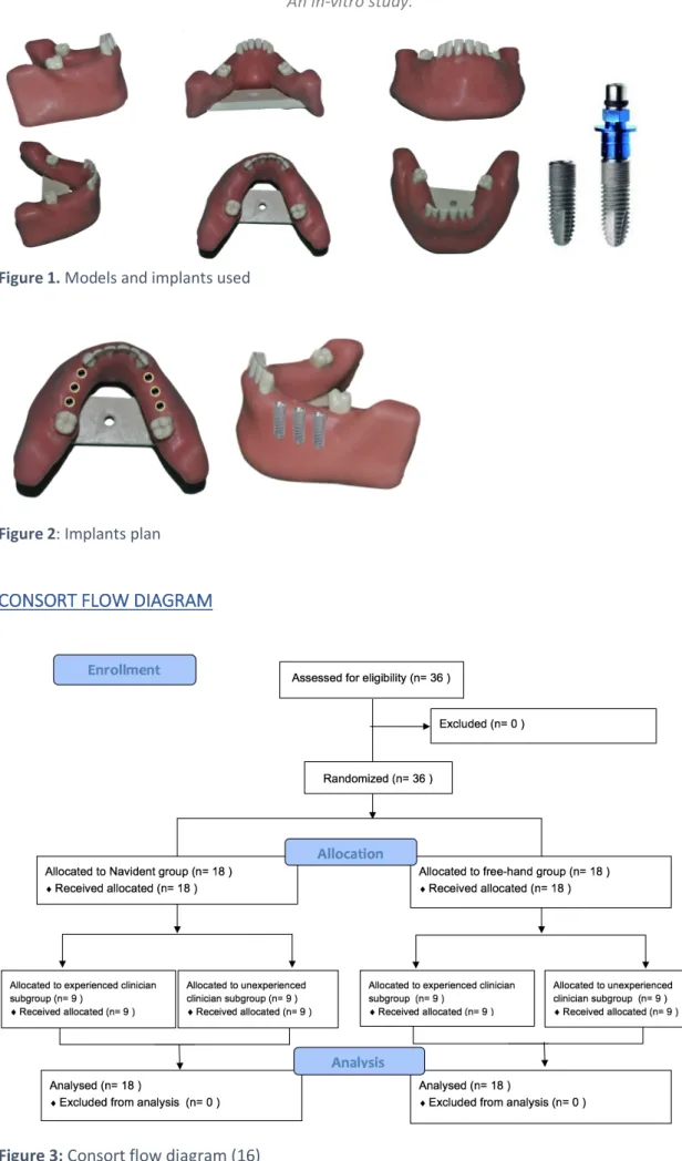
Two investigators placed 36 dental implants (Ticare InHex standard of 3.75mm x 10mm; MG Mozo-Grau®, Valladolid, España) in 6 partially edentulous mandibles models (BoneModels®, Castellón de la Plana, Spain) fabricated for this study (Figure 1). The models were made using the exact same template and were missing 3 adjacent teeth on both sides (first and second premolar and the first molar) (Figure 2). A radio-opacifier was employed in relevant structures like the adjacent teeth and the inferior alveolar canal. The 2 investigators had a different degree of experience: One was a last-year undergraduate student of the dental degree of the University of Barcelona while the other was an experienced surgeon (over 15 years of clinical experience in implant dentistry) with postgraduate training in Oral Surgery and Implantology. (Figure 3)
Figure 1. Models and implants used
Figure 2: Implants plan
CONSORT FLOW DIAGRAM
NAVIDENT®
Navident® (Navident®, ClaroNav Technology Inc.®, Toronto, Canada) is a computer guided surgery system that consists of a real-time computer dynamic navigation system, that allows the surgeon to detect the position of the drills and implants on a computer screen, taking into consideration the initial planification. The aim of Navident® is to guide drilling and implant insertion in a patient jaw during surgery using pre-acquired CBCT scans of the jaws.
The system provides a real time location of the hand-piece tip during the drilling and the implant placement. The software shows a dartboard on the screen which guides the surgeon to the preoperative planning. To improve the guidance, the software provides also images from the 3 cuts of the CBCT in which you can see the planification and the real-time drill position.
The Navident® system consists basically of 5 components (Figure 4):
1. A laptop with the Navident® software which provides integrated planning and navigation functionalities.
2. A handpiece attachment with an optically marked plastic part (“DrillTag”) which latches onto the adapter.
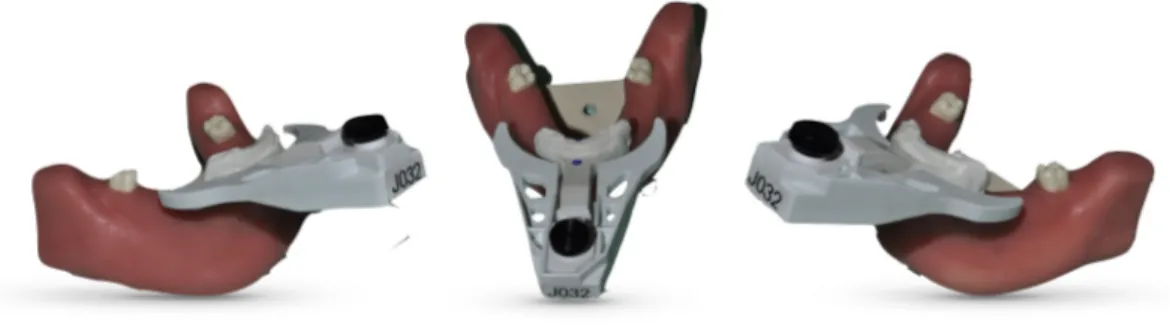
3. A patient jaw attachment consisting, for a partially edentulous jaw, of a custom splint (“NaviStent”), or, for a fully edentulous jaw, of a mini-implant and a matching bracket. In either case, an arm extending from the attachment is designed to connect to a “CT-Marker” part during the CT scan and to an optically marked plastic tag (“JawTag”) during surgery.
4. An optical position sensor (“MicronTracker”) which detects the special patterns printed on the DrillTag and JawTag and constantly reports their relative positions, to a small fraction of a millimeter, to the Navident® software. 5. A compact mobile cart which provides a foldable arm that, when extended,
.
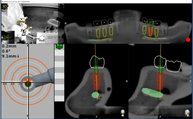
Navigation during the surgery is driven by a dynamic interface (Figure 5). In the top left part of the laptop screen, a video of what is recording the camera of the MicronTracker can be observed. Then in the right part of the screen, a panoramic reconstruction of the patient mandible where all the planned implants are visible in yellow can be seen. To aid the navigation, apart from the panoramic view, Navident® interface also show
Figure 4. Navident® parts. (1) Laptop, (2) DrillTag, (3) JawTag, (4) MicronTracker, (5) Mobile
different CBCT slices: frontal and sagittal; in these slices, implant planning appears in red and the position of the bur or dental implant in real-time in green.
In the lower left part of the screen, a dartboard with a handpiece is shown. Finally, next to the dart board, a ruler that shows the desired depth. To improve surgical navigation, a color legend is used to describe different degrees of deviation. Concerning the angulation, a visual color scale is used, red-yellow-green, where red is more than 6º deviation, yellow between 3-6º and green less than 3º. In the depth ruler a color scale is also used; Green means that drilling is more than 1mm away from the desired depth, yellow indicates that the last millimeter has been reached and red when the desired depth has been achieved (Figure 6). Furthermore, to provide a plus of safety, acoustic sounds are present when working in the proximity of the inferior alveolar nerve or when overdrilling of the implant site occurs.
Figure 6 Dartboard guidance aid. (1) Incorrect 2D positioning and deficient angulation). (2) Good
positioning and incorrect angulation. (3) Adequate positioning and angulation. (4) Correct positioning and angulation and yellow color in the depth ruler means that less than 1mm is necessary to reach the final drilling position (0,3mm in this case). (5) Red means the final position has been reached.
PRE-SURGICAL PLANNING
Before placing the dental implants, a splint (NaviStent) was firmly attached to the remaining anterior teeth of the mandible. Fiducials markers were attached to the splint and then CBCT scans (Planmeca ProMax® 3D Mid (Planmeca, Helsinki, Finland) with the following set up: 90Kv, 10mA, 13.9 seconds, 1245 DAP (mGy*cm2), 0,4mm Voxel) were
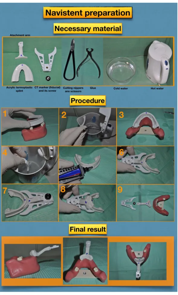
made to all models (Figure 7). Fabrication of the NaviStent splint is summarized in figure 8.
Figure 7. Model with the acrylic thermoplastic splint (NaviStent) and the fiducial marker
remaining teeth. Afterwards, remove the splint and place it on cold water. (4) Insert the splint arm in hot water for 1 minute. (5) Place glue to the anterior zone of the splint where the arm should be attached. (6) Attach the arm to the splint. (7) Before the resin is cooled, CT fiducial marker must be attached to the splint in order to do some small holes in the splint where the CT marker will rest. (8) Remove CT fiducial marker and ensure that small indents in splint are done and that the CT marker has only one possible position. (9) Finally, using nippers and scissors, cut the splint until it fits perfectly to the mandible.
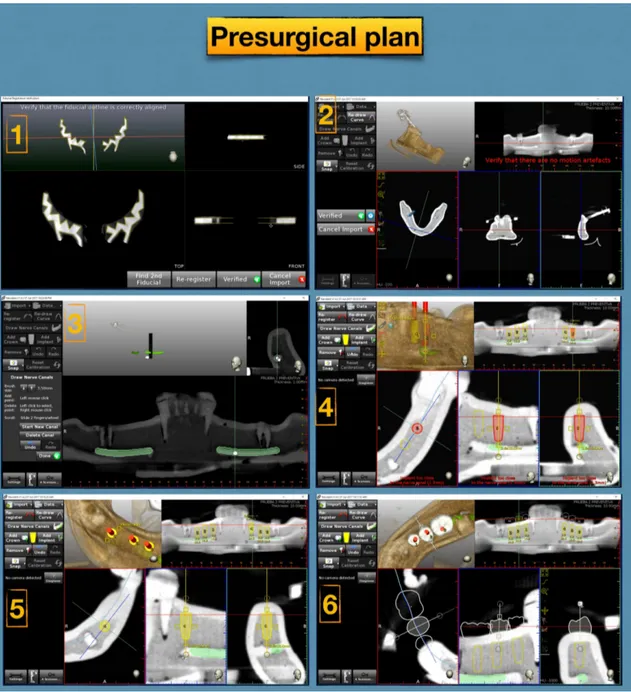
DICOM data of the CBCT was upload to the navigation system software (Navident®) and using its planning utilities, dental arch, inferior alveolar nerve path and the position of each implant were defined on the CBCT images. Implants were virtually placed in the first and second premolars and first molar on each side taking into account the most suitable position for the final restoration. Prosthetic crowns were also drawn on the CBCT image. This procedure is summarized in figure 9.
Aiming to minimize bias, during this phase, surgeons were blinded regarding the assigned group for each implant.
Figure 9. Presurgical plan using the Navident® planning tool. (1) Registration of the fiducials
markers is performed by the software when a new CBCT with the marker is imported. (2) Arch curve is drawn in the coronal slice of the CBCT. (3) The inferior alveolar nerve canal is drawn using the panoramic reconstruction and the frontal slice view. (4) Implants are added to the CBCT images, implant size is also determined. (5) Implants can be moved or angulated in all the dimensions of the space until optimal position is achieved (6) Prosthetic crowns can be also added to the CBCT image to ensure implant is place in an optimal prosthetic position.
DENTAL IMPLANT PLACEMENT
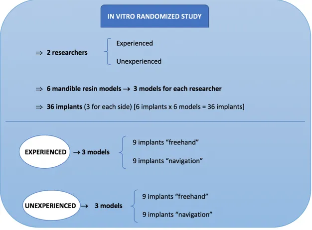
The models were randomly allocated to each investigator. Each participant placed 18 implants in three models in teeth positions 3.4, 3.5, 3.6 and 4.4, 4.5, 4.6. For each implant site, the use of navigation system and freehand system was assigned using a website generated random sequence (www.randomization.com). In order to guarantee the allocation concealment, the researcher was informed of the assigned group for each implant just before starting the drilling sequence (after the raising of the flap). Figure 10 shows the study planning.
Figure 10. Study design
Each model was placed in a preclinical learning dental simulator with limited mouth opening, and a latex face to limited visibility and to mimic the pressure due to facial soft tissues. The setting used for the study was very similar to a real clinical scenario in an ergonomic position (Figure 11).
Figure 11. Real clinical scenario reproduction
A crestal incision was made with a type 15C scalpel blade. Soft tissue was detached with a Freer elevator. Afterwards, drilling was performed while separating soft tissues with a Minnesota separator.
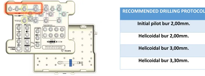
Surgeons used the recommended drilling protocol for Ticare InHex standard 3,75mm (MG Mozo Grau SA, Valladolid, Spain) dental implants (Figure 12). Drill axis and tip calibration were performed before starting drilling with a new bur in the Navident group.
Implants were placed with specific burs and implant carrier at 15 rpm and with a maximum torque of 50 N.cm. Before implant insertion, a new axis and tip calibration were performed.
Figure 12. Ticare surgical box and the recommended drilling protocol for the Ticare InHex
RECOMMENDED DRILLING PROTOCOL Initial pilot bur 2,00mm.
Helicoidal bur 2,00mm. Helicoidal bur 3,00mm. Helicoidal bur 3,30mm.
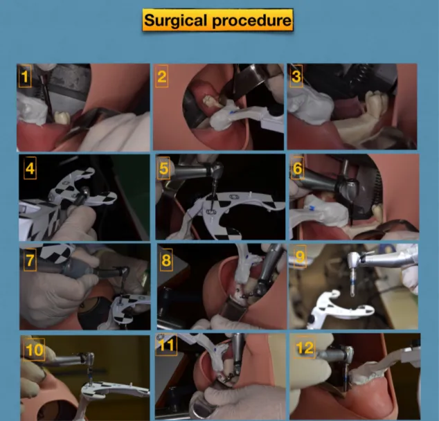
The surgical procedure using Navident® is summarized in Figure 13
Figure 13. Surgical procedure. (1) Crestal incision with a type 15C scalpel blade. (2) Flap
elevation. (3) Exposure of the bone crest. (4) Drill axis calibration. (5) Drill tip calibration. (6) Drill following real-time navigation. (7,8) Each new bur required a drill tip calibration. (9) Implant driver attached to the contra-angle. (10) Calibration of the implant’s tip. (11) Implant placement following real-time navigation images. (12) Insert the implant until the final position is achieved.
ACCURACY ASSESSMENT AND OUTCOME MEASUREMENTS
A second CBCT was performed after implant placement for each model (Planmeca ProMax® 3D Mid (Planmeca, Helsinki, Finland) with the following set up: 90Kv, 10mA, 13.9 seconds, 1245 DAP (mGy*cm2), 0,4mm Voxel). The accuracy of implant positioning
was assessed by a third independent and blinded investigator by overlapping the preoperative and postoperative CBCT and comparing the planned position with the final position of the dental implant. The overlapping of the two CBCT was performed using the software EvaluNav® (ClaroNav Technology Inc. ®, Toronto, Canada).
For each implant placed, the following variables were measured (Figure 14): • Entry 3D deviation
• Entry 2D deviation • Apex 3D deviation • Apex depth deviation • Angle deviation
ALLOCATION AND BLINDING
The models were randomly allocated to each researcher (experienced and unexperienced). As mentioned previously, implant sites were divided into two groups using a webpage random generated sequence (www.randomization.com). The control group consisted on freehand implant placement; and the test group (Navigation group) employed the navigation system Navident® to place the dental implants. The allocation ratio was 1:1. Surgeons were unaware of the assigned group until the they had to start drilling in order to assure allocation concealment. The researcher that analyzed the data was also blinded since the variables operator and method were codified.
Figure 14. Description of the main
SAMPLE SIZE CALCULATION
The sample size was calculated with G*Power v.3.1.3 (Heinrich-Heine Universität, Düsseldorf, Germany) taking into account that the primary outcome variable was angular deviation. Mean angulation deviation data were extracted from a previously published study (17). An alpha value of 0.05 and a statistical power of 90% were established.
The sample size calculation yielded that 36 implants were considered necessary for this study (18 implants for each group). Since the simulated surgeries were made by 2 different professionals (experienced and unexperienced), each surgeon will place 9 implants using a freehand procedure and 9 implants using the dynamic computed guided system.
STATISTICAL ANALYSIS
A third blind researcher performed the statistical analysis using the software Statistical Package for the Social Sciences (SPSS version 22.0; Armonk, NY, USA: IBM Corp.). The level of significance for all statistical tests was set at 5% (p <0.05).
Normality of scale variables (Entry 3D, Entry 2D, Apex 3D, Apex vertical, Angulation and Surgical time) was explored using the Shapiro-Wilks test and the visual analysis of normal P-P graphics and box diagrams. Descriptive analysis using the median and interquartile range (IQR) were calculated when normality was rejected. Where distribution was compatible with normality, the mean and standard deviation (SD) were used. Descriptive analysis for bivariable categoric variables was performed through absolute and relative frequency tables.
Once descriptive statistics were done, the possible relationship between variables was analyzed by using a bivariate analysis. To analyze the effect of the discrepancy regarding the method (freehand versus dynamic guided surgery), operator (experienced or unexperienced) and the interaction between the two variables, a two independent factors analysis of variance (Two-way ANOVA) was performed. The fulfillment of the conditions of application of the test has been carried out through the normality and homogeneity of the variances test. This analysis was completed using graphs of the
estimated averages. For statistically significant variables, the comparison between the corresponding groups was performed and an average estimation was given for each group.
7. R
esults
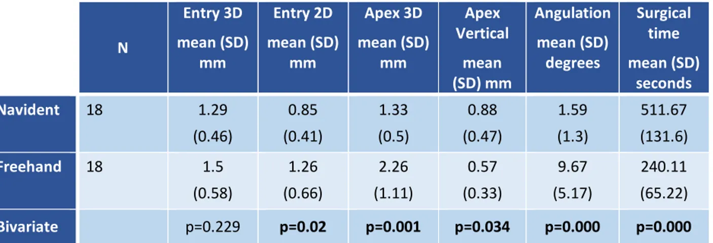
A total of 36 implants were analyzed. Descriptive and bivariate results of the main outcome variables comparing the 2 employed implant placement systems (freehand and Navident®) can be observed in Table 1. The Navident group had a significantly higher accuracy for all studied variables, except entry 3D and apex depth variable. On the other hand, this system significantly increased the surgical procedure time.
N Entry 3D mean (SD) mm Entry 2D mean (SD) mm Apex 3D mean (SD) mm Apex Vertical mean (SD) mm Angulation mean (SD) degrees Surgical time mean (SD) seconds Navident 18 1.29 (0.46) 0.85 (0.41) 1.33 (0.5) 0.88 (0.47) 1.59 (1.3) 511.67 (131.6) Freehand 18 1.5 (0.58) 1.26 (0.66) 2.26 (1.11) 0.57 (0.33) 9.67 (5.17) 240.11 (65.22) Bivariate p=0.229 p=0.02 p=0.001 p=0.034 p=0.000 p=0.000
Table 1. Descriptive and bivariate results for both groups of the main outcome variables.
The experienced clinician achieved a more adequate angulation with the Navident® system. However, dynamic guided surgery did not significantly improve the other studied parameters and increased the surgery time. On the other hand, the Navident® system enhanced the accuracy of the unexperienced professional regarding Entry 2D, Apex 3D and angulation. All the above-mentioned data can be observed in table 2.
Table 2. Differences
between the
experienced and
unexperienced clinician
divided by groups.
When analyzing the results regarding the role of the surgeon’s experience, the unexperienced professional had a much more pronounced improvement in most studied parameters (Figure 15). Indeed, when a freehand placement method was employed, the experienced clinician had significant better results in the 2D entry variables (p=0.014), apex 3D (p=0.008) and angulation deviations (p<0.001). However, when the Navident® system was used, these differences were neglectable (p>0.05).
EXPERIENCED UNEXPERIENCED
Navident Freehand Navident Freehand
Entry 3D mean (SD) mm 1.19 (0.45) (0.39) 1.28 (0.48) 1.39 (0.67) 1.71 Bivariate p=0.713 p= 0.182 Entry 2D mean (SD) mm 0.68 (0.42) 0.95 (0.52) 1.02 (0.33) 1.56 (0.67) Bivariate p=0.260 p=0.027 Apex 3D mean (SD) mm 1.24 (0.46) 1.73 (0.51) 1.43 (0.54) 2.78 (1.32) Bivariate p=0.191 p=0.001 Apex vertical mean (SD) mm 0.93 (0.35) (0.4) 0.63 (0.58) 0.83 (0.25) 0.52 Bivariate p=0.118 p= 0.134 Angulation mean (SD) degrees 2.15 (1.53) (3.17) 6.69 (0.75) 1.03 (5.16) 12.66 Bivariate p=0,004 p=0.000 Surgical time mean (SD) seconds 487.8 (82.2) (21.8) 200.2 (169.6) 535.6 (70.6) 280 Bivariate p=0.000 p=0.000
Figure 15: Differences between the experienced and unexperienced clinicians for both implant
placement systems.
Figure 15 shows that Navident group had a higher accuracy in all parameters except for the apical vertical depth deviation (Fig. 15–4). Also, the surgical time was 2-fold higher when the dynamic guided system was used (Table 1; Figure 15-6).
8. D
iscussion
The purpose of dynamic computer guided surgery systems in implantology is to minimize implant position deviation from the preoperative planning employing a real-time tracking of the drilling and implant insertion. Ewers et al. (18) after 12 years of clinical experience in this field and 395 implants placed with a dental navigation system, consider that this option provides excellent benefits specially in compromised situations.
The usefulness of navigation relies on its high accuracy, particularly necessary in some specific surgical situations: (I) when anatomic structures must be taken into account and depth control is important , (II) when clinicians wish to use a flapless approach, (III) when placement requires high accuracy on angulation and spacing between implants and adjacent tooth, especially in the esthetic zone or in the planification of implant-supported bridges, (IV) when implants must be placed in a tight interdental space and static guide tubes will interfere with the ideal implant position due to its size, (V) when direct visualization is expected to be difficult, such as in difficult-to-access locations or in limited mouth opening patients. (7, 19)
A meta-analysis about computer technologies applications in implantology (11) revealed that the entry point and apex accuracies are significant higher when dynamic navigation systems are used in comparison with the traditional static surgical guides. Mean values from this meta-analysis are in accordance with the outcomes obtained in the present
in-vitro study. However, these results should be interpreted with caution, since most data
concerning the accuracy of dynamic systems is extracted from in-vitro studies made with artificial models, which can lead to better results in comparison to real clinical scenarios. (11) Still, several authors show good results in clinical studies, and conclude that navigation systems are as good as static guides (14), and significantly better than freehand implant placement. (14,20)
Although the results obtained in our sample were excellent in terms of horizontal direction (entry and apex of the implant) and angulation, the outcomes related with the depth accuracy were not as good as expected. The overall mean error at depth was 0.88 mm (range from 0mm to 1.6mm), which shows an important discrepancy that may be considered unacceptable in anatomically compromised situations where the inferior alveolar nerve is at risk. Thus, in our opinion, a 2mm security margin should be implemented to all important anatomical structures in the pre-surgical planning. This is an extremely important issue since neuropathic pain and sensory alterations have been described after dental implant placement. (21,22)
Somogyi-Ganss et al. (8) published in 2015 an in-vitro study to assess the accuracy of the Navident® system and reported similar findings. Again, the results regarding depth deviation were disappointing (deviation ranged from 0 to 3,3mm). The improvement observed in the present sample can be related with the software updates provided by the company and small developments made to the system in the past 3 years. It is also interesting to stress that a systematic review on this subject was unable to perform a statistical analysis with the variable height deviation due to a large amount of studies that did not report this data in their results. (11) Several variables can explain vertical deviations. CBCT scan quality, registration error, planning error, precision of the tracking system, movement of the acrylic splint, imprecision of the operator while following the onscreen navigated path of drilling, small movement between the carrier, driver and implant, and finally during the overlapping of the two CBCTs scans are factors that can produce inaccuracies. (8,11,19, 23) In our opinion, the misfit of the splint is the most likely cause for deviations, since small movements might occur between presurgical CBCT scan and the surgical procedure. New methods of fiducial points markers and registration should be analyzed in future studies because an acrylic splint can be easily deformed.
Some authors report that a high accuracy can only be achieved by using bone fixed fiducials because dental or mucosal supported splints can originate deviations. However, this option might increase surgical morbidity of patients due to screw fixation. (12, 23)
The main drawbacks of dynamic computer guided surgery are a longer surgical time, the need for previous specific training with a preclinical model, the mandatory use of a splint that might be considered uncomfortable to patients, and the high costs associated with the device. On the other hand, these systems increase the accuracy in implant placement, offer especially good results to unexperienced professionals, and are not time-consuming when compared to static guidance options. Indeed, with dynamic navigation systems, it is possible to do the CBCT scan, the presurgical planning and the surgical procedure in just a few hours without the need to take impressions or fabricate any laboratory manufactured specific splints. (7,11,15) Advantages and disadvantages
Table 3. Dynamic guided surgery advantages and disadvantages
Experience is a very important factor when assessing the accuracy of implant placement. The dynamic guidance seems reduce the influence of surgeon’s experience. The present study has shown that the deviations were very small even for unexperienced clinicians. Block et al. (14) analyzed the role of prior experience with dental navigation systems in the accuracy of implant placement. As expected, surgeons with prior experience in dynamic assisted surgery, had better accuracy outcomes and a flat learning curve compared with expert professionals in implant dentistry but with no experience in navigation systems. However, the learning curve is quite fast since after 20 cases, these authors only found minimal accuracy differences between surgeons.
Although implant surgery is a common procedure in Dentistry, students consider that the dental degree offers insufficient information about implant-based treatments. (24) The fact that these procedures are usually complex, have high costs and depend on the experience of the professional are probably related with the few amount of dental
Dynamic computer guided surgery
Advantages Disadvantages
High accuracy Longer surgical time
All procedures the same day. Simple work flow
More complicated intraoperative procedures
Reduce risk for patients Learning curve Ability to change planification at any
time Splint might be uncomfortable to patients
Freehand flexibility Possible errors during registration No need to work with dental
technicians
Possible position deviation when replacing splint on the patient
Does not require special drill kits Single use splint and software license for each patient.
implant treatments made by dental students. Taking into account the results obtained in this in-vitro study, including preclinical and clinical training with dental implant dynamic guided surgery systems in dental degrees could be a good option to improve implantology skills and reduce complications. According to Casap et al. 2011 (25), last year dental students that use navigation systems improve their performance and are likely to use it in the future. This same paper showed that the learning curve was much higher with dynamic guidance in comparison with the conventional freehand method. (25)
STUDY LIMITATIONS
The major limitation of this study is it’s in-vitro design. Therefore, the generalization of our results should be taken with caution, especially those related with accuracy since they can be affected by clinical variables. On the other, the present study has a high internal validity and allows to control several confounding variables that cannot be manipulated in a real clinical scenario. Indeed, the fact that all anatomical (models, preclinical simulated patient, light conditions), surgical (drilling unit, implant system, implant length and diameter) and planification (CBCT, software used in the pre-surgical planning) variables were identical, allowed to analyze the effect of experience in the accuracy without interferences.
The limited sample size and the randomization system did not allow to evaluate the relation between accuracy and implant position (maxilla versus mandible; anterior versus posterior). This relevant issue also needs further research.
9. C
onclusions
1. The dynamic computer assisted surgery system Navident® allows a more accurate but slower implant placement in comparison with the conventional freehand method.
2. Both experienced and unexperienced clinicians improve the accuracy outcomes when the Navident® system is employed.
3. This dynamic computer assisted surgery system seems to be specially indicated in less experienced professionals, since they can significantly reduce their deviations, achieving the same results of experienced clinicians.
4. The most relevant deviations found were related with the depth of the implant. Therefore, a security margin of at least 2mm should be implemented when important anatomic structures are in the proximity.
5. Further research in a real clinical scenario is needed to confirm the present findings.
10. C
onclusions (Catalan version)
1. El sistema dinàmic de cirurgia guiada per ordinador Navident® permet una major exactitud però suposa un augment del temps de col·locació dels implants en comparació amb el mètode convencional a mà alçada.
2. Els dos cirurgians, l’experimentat i l’inexperimentat, han millorat els resultats respecte l’exactitud quan s’ha utilitzat el sistema Navident®.
3. Aquest sistema de cirurgia guiada dinàmica per ordinador sembla estar especialment indicat en professionals menys experimentats, ja que redueix significativament les desviacions respecte la planificació preoperatòria, obtenint així els mateixos resultats que un cirurgià experimentat en implantologia. 4. La desviació més rellevant observada ha sigut respecte la profunditat. Per tant,
s’hauria de tenir present un marge de seguretat d’almenys 2mm a l’hora de planificar un implant quan hi hagi estructures anatòmiques importants pròximes.
5. Futures investigacions en pacients són necessàries per confirmar les presents troballes.
R
eferences
1. Branemark PI, Adell R, Breine U, Hansson BO, Lindstrom J, Ohlsson A. Intra-osseous anchorage of dental prostheses. I. Experimental studies. Scand J Plast Reconstr Surg. 1969;3:81–100.
2. Pjetursson BE, Tan K, Lang NP, Brägger U, Egger M, Zwahlen M. A systematic review of the survival and complication rates of fixed partial dentures (FPDs) after an observation period of at least 5 years. Clin Oral Implants Res 2004;15:625–42.
3. Jung RE, Pjetursson BE, Glauser R, Zembic A, Zwahlen M, Lang NP. A systematic review of the 5-year survival and complication rates of implant-supported single crowns. Clin Oral Implants Res. 2008;19:119–30.
4. Vercruyssen M, Fortin T, Widmann G, Jacobs R, Quirynen M. Different techniques of static/dynamic guided implant surgery: Modalities and indications. Periodontol 2000. 2014;66:214-27.
5. Miller RJ, Bier J. Surgical navigation in oral implantology. Implant Dent. 2006;15:41-7.
6. D'haese J, Ackhurst J, Wismeijer D, De Bruyn H, Tahmaseb A. Current state of the art of computer-guided implant surgery. Periodontol 2000. 2017;73:121-33. 7. Block MS, Emery RW. Static or dynamic navigation for implant placement
choosing the method of guidance. J Oral Maxillofac Surg. 2016;74:269-77. 8. Somogyi-Ganss E, Holmes HI, Jokstad A. Accuracy of a novel prototype dynamic
computer-assisted surgery system. Clin Oral Impl Res. 2015;26:882–90
9. Mozzo P, Procacci C, Tacconi A, Martini PT, Andreis IA. A new volumetric CT machine for dental imaging based on the cone-beam technique: Preliminary results. Eur Radiol. 1998;8:1558–64.
10. Bornstein MM, Scarfe WC, Vaughn VM, Jacobs R. Cone beam computed tomography in implant dentistry: A systematic review focusing on guidelines, indications, and radiation dose risks. Int J Oral MaxIllofac Implants. 2014;29:55– 77.
11. Jung RE, Schneider D, Ganeles J, Wismeijer D, Zwahlen M, Hämmerle CHF, Tahmaseb A. Computer technology application in surgical implant dentistry: A systematic review. Int J Oral Maxillofac Implants. 2009;24:92–109
12. Gaggl A, Schultes G, Kärcher H, Navigational precision of drilling tools preventing damage to the mandibular canal. J Craniomaxillofac Surg. 2001;29:271-5.
13. US Food and Drug Administration [Internet]. US: Medical devices databases, 510(k) premarket notification, no. K161406. Navident®, ClaroNav Technology Inc.®
Available at: https://www.accessdata.fda.gov/cdrh_docs/pdf16/k161406.pdf.
Accessed on June 9, 2018.
14. Block MS, Emery RW, Lank K, Ryan J. Implant placement accuracy using dynamic navigation. Int J Oral Maxillofac Implants. 2017;32:92-9.
15. Emery RW, Merritt SA, Lank K, Gibbs JD. Accuracy of dynamic navigation for dental implant placement-model-based evaluation. J Oral Implantol. 2016;42:399-405.
16. Schulz KF, Altman DG, Moher D. CONSORT 2010 Statement: updated guidelines for reporting parallel group randomised trials. BMJ. 2010;340:332.
17. Hoffmann J, Westendorff C, Gomez-Roman G, Reinert S. Accuracy of navigation-guided socket drilling before implant installation compared to the conventional freehand method in a synthetic edentulous lower jaw model. Clin Oral Impl Res. 2005; 16:609–14.
18. Ewers R, Schicho K, Undt G, Wanschitz F, Truppe M, Seemann R, Wagner A. Basic research and 12 years of clinical experience in computer-assisted navigation
19. Elian N, Jalbout ZN, Classi AJ, Wexler A, Sarment D, Tarnow DP. Precision of flapless implant placement using real-time surgical navigation: A case series. Int J Oral Maxillofac Implants. 2008;23:1123–27.
20. Block MS, Emery RW, Cullum DR, Sheikh A. Implant placement is more accurate using dynamic navigation. J Oral Maxillofac Surg. 2017;75:1377-86.
21. Vázquez-Delgado E, Viaplana- Gutiérrez M, Figueiredo R, Renton T, Gay-Escoda C, Valmaseda-Castellón E. Prevalence of neuropathic pain and sensory alterations after dental implant placement in a university-based oral surgery department: A retrospective cohort study. Gerodontology. 2018;35:117-22. 22. Juodzbalys G, Wang HL, Sabalys G, Sidlauskas A, Galindo-Moreno P. Inferior
alveolar nerve injury associated with implant surgery. Clin Oral Implants Res. 2013;24:183-90.
23. Gaggl A, Schultes G. Assessment of accuracy of navigated implant placement in the maxilla. Int J Oral Maxillofac Implants. 2002;17:263–70.
24. Sánchez-Garcés MA, Berástegui-Jimeno E, Gay-Escoda C. Knowledge, aptitudes, and preferences in implant dentistry teaching/training among undergraduate dental students at the University of Barcelona. Med Oral Patol Oral Cir Bucal. 2017; 22:484-90.
25. Casap N, Nadel S, Tarazy E, Weiss EI. Evaluation of a navigation system for dental implantation as a tool to train novice dental practitioners. J Oral Maxillofac Surg. 2011;69:2548-56.