International Journal of
Immunopathology and Pharmacology 2016, Vol. 29(1) 3 –8
© The Author(s) 2015 Reprints and permissions:
sagepub.co.uk/journalsPermissions.nav DOI: 10.1177/0394632015617951 iji.sagepub.com
Introduction
The development of biomaterials for tissue engi-neering applications is continuously improving, accordingly to the needs to generate an ideal cell– extracellular matrix interaction.1 Generally, stem
cell fate, in vitro or in vivo, has been mainly associ-ated to molecular intracellular mediators and to growth factors (GFs)1 and many researchers
dem-onstrated that environmental factors contribute to the regulation of stem cell behavior and fate.
Stem cells seem to remember past physical signals: Yang et al. found that the human mesenchymal
Mechanical influence of tissue culture
plates and extracellular matrix on
mesenchymal stem cell behavior:
A topical review
Marco Tatullo,1,2 Massimo Marrelli,1,2 Giovanni Falisi,3 Claudio Rastelli,3 Francesca Palmieri,2 Marco Gargari,4
Barbara Zavan,5,* Francesco Paduano2,* and Vincenzo Benagiano6,*
Abstract
Tissue engineering applications need a continuous development of new biomaterials able to generate an ideal cell– extracellular matrix interaction. The stem cell fate is regulated by several factors, such as growth factors or transcription factors. The most recent literature has reported several publications able to demonstrate that environmental factors also contribute to the regulation of stem cell behavior, leading to the opinion that the environment plays the major role in the cell differentiation.
The interaction between mesenchymal stem cells (MSCs) and extracellular environment has been widely described, and it has a crucial role in regulating the cell phenotype. In our laboratory (Tecnologica Research Institute, Crotone, Italy), we have recently studied how several physical factors influence the distribution and the morphology of MSCs isolated from dental pulp, and how they are able to regulate stem cell differentiation. Mechanical and geometrical factors are only a small part of the environmental factors able to influence stem cell behavior, however, this influence should be properly known: in fact, this assumption must be clearly considered during those studies involving MSCs; furthermore, these interactions should be considered as an important bias that involves an high number of studies on the MSCs, since in worldwide laboratories the scientists mostly use tissue culture plates for their experiments.
Keywords
environmental factors, extracellular matrix (ECM), mesenchymal stem cells (MSCs), mesenchymal stem cell fate, mesenchymal stem cell behavior
Date received: 24 August 2015; accepted: 23 October 2015
1Unit of Maxillofacial Surgery, Calabrodental, Crotone, Italy 2Tecnologica Research Institute, Biomedical Section, Crotone, Italy 3 Department of Life, Health and Environmental Sciences, School of
Dentistry, University of L’Aquila, L’Aquila, Italy
4 Department of Clinical Sciences and Translational Medicine, University of Rome “Tor Vergata”, Rome, Italy
5Biomedical Department, University of Padova, Padova, Italy 6 Basic Medical Sciences, Neurosciences and Sense Organs, University
of Bari, Bari, Italy *Equal contributors.
Corresponding author:
Marco Tatullo, Scientific Director, Tecnologica Research Institute, St. E. Fermi - Crotone, Italy.
Email: [email protected] Editorial
stem cells (hMSCs) cultured on soft poly-ethylene glycol (PEG) hydrogel were stimulated in the activation of Yes-associated protein (YAP), a tran-scriptional co-activator with PDZ-binding domain (TAZ) and pre-osteogenic transcription factor (runt-related transcription factor 2 [RUNX2]), continuing to follow the fate, a kind of “memory”, depending on the previous culture on the tissue culture poly-styrene plates (TCPS).2 With these data, Yang et al.
concluded that stem cells possess a mechanical memory that can control the cells’ fate, on the base of past physical environments.2
Also Gilbert et al. achieved similar results, working on stem cells from muscle: the authors confirmed that cells remember the past mechanical signals derived from the in vitro culture; moreover, even after the in vivo application of these MSCs, their “memory” influences the long-term MSC fate.3
The recent literature has reported many studies focused on the influence of the extracellular matrix (ECM) on stem cell fate, with particular regard to the ECM geometry/topography, to the ECM mechanical properties, and to the transmission of mechanical or other biophysical factors to the cells.4 Nevertheless, many questions still remain to
be addressed; in fact, another set of studies reported a critical link between the ECM mechanical influ-ence and intracellular signaling. Dupont et al. and Halder et al. analyzed the transduction of mechani-cal cues by YAP and TAZ, two transcriptional co-activators that regulate RNA expression, and they revealed a critical interaction between the extracel-lular environment and the intracelextracel-lular signaling.5,6
In particular, they demonstrated that hMSCs seeded on substrata with stiff moduli of 40 kPa showed the activation of YAP/TAZ at the nucleus level, instead YAP/TAZ were deactivated (YAP/TAZ were located in the cytoplasm) when hMSCs were seeded on substrata with stiff moduli of 1 kPa. Moreover, they demonstrated that the role of YAP/ TAZ was related to the osteogenic differentiation.
On the bases of these data, Yang et al. began to investigate the effects of standard methods of culturing and expanding the MSCs into TCPs, by analyzing the implications of this environment on stem cell plasticity and by investigating whether or not stem cell fate is affected by all the physical sig-nals they have previously interacted. They assayed the hMSC behavior when they were cultured on substrates of different stiffness, ranging between 3 GPa (tissue culture plates [TCPS]) and 2 kPa (soft
hydrogel). In this way, they tested whether the past physical environment of TCPS, able to activate YAP and RUNX2, could override new mechanical signals coming from the soft hydrogel where hMSCs were plated. They observed that YAP remained nuclear (activated), even after the trans-fer from the TCP to the hydrogel. Furthermore, the authors used a photodegradable hydrogel, able to change its rigidity during the cell culturing: this new experiment confirmed that hMSCs can remember such important mechanical information. YAP/TAZ act as an intracellular mechanical rheo-stat, modulating the cell plasticity by a persistent presence in the nucleus: this work has shown that the mechanical influence effected by TCPS biases the hMSC behavior, even if stem cells are cultured on soft hydrogel, and makes these cells basically committed towards the osteogenic lineage.
Recently, it was shown that the stiffness of a flat surface regulates the stem cell differentiation, inde-pendently from the protein tethering and porosity. Wen et al. showed that by modulating the porosity of a substrate made of polyacrylamide gel without altering its stiffness, it did not significantly change the protein tethering, the substrate deformation, or the osteogenic and adipogenic differentiation of adipose-derived and marrow-derived hMSCs seeded on this substrate.7 Furthermore, they showed
that a different protein–substrate density changed tethering, but it did not influence the osteogenesis or the adipogenesis. Cell differentiation was also unaffected by the absence of protein tethering.
Banks et al. highlighted the need to selectively manipulate the biomaterial microenvironment, thus to identify the right synergies between biochemical and mechanical cues, for regenerative medicine applications.8 They reported an approach based
on carbodiimide cross-linking and benzophenone photo-immobilization chemistries. They orthogo-nally modified the stiffness in a way to immobilize the GFs content of a collagen-GAG (CG) biomate-rial. Moreover, they observed the single and combined effect of bone morphogenetic protein (BMP-2), a platelet-derived GF (PDGF-BB), together with the CG membrane on the bioactivity and osteogenic/adipogenic lineage-specific gene expression of adipose derived stem cells. They discovered that the stiffest substrates induced the osteogenic commitment of adipose-derived stem cells (ASCs), regardless of the presence of osteogenic growth factors, while a softer substrate needed a biochemical cues to modify the cell fate.
Yim et al. examined the cell signaling in stem cell differentiation, with a focus on stem cell inter-actions with biochemical and biophysical signals present in their extracellular environment.9 The
biophysical signals are transferred to the stem cells, by both the ECM and the externally applied forces. The authors investigated the mechanism of the differentiation induced by different ways of adhesion, different cytoskeletal contractility, and different Rho-guanosine triphosphatase (Rho-GTPase) signaling and nuclear regulation related to these biophysically induced differentiations.
Human embryonic stem cells (hESC) are able to sense the mechanical properties of their microenvi-ronment. Eroshenko et al. tested the hypothesis that hESCs accept mechanical cues for differentiation from the substrate by culturing them on flexible polydimethylsiloxane (PDMS) of varying stiffness, prepared using available commercial formulations and characterized for stiffness, surface properties, and efficiency of cell attachment and proliferation.10
They evaluated the utility of PDMS substrates for stem cell development and if those substrates medi-ated the cell differentiation. They concluded that PDMS substrates could be used to direct hESC fate towards early mesodermal lineages.
All these studies have differently enhanced the hypothesis that artificial substrate and ECM play a key role in regulating MSC fate during regenera-tive events.
Discussion
Stem cells regulate their fate by binding to the extracellular environment, where they may be exposed to various chemicals, and physical and mechanical signals.4 Previous studies showed that
these signals can be transduced and they can deeply influence the stem cell growth and differentiation, in vivo and in vitro. In this context, a growing lit-erature has shown the importance of intracellular mechano-transduction in stem cell differentiation.11,12
Recent reports demonstrated that the biophysical cues, such as substrate stiffness and topography, can direct stem cell differentiation and determine the cell fate; moreover, the same reports high-lighted how the cells integrate mechanical signals from the ECM and how they transduce them in a directed gene expression. Thus, ECM and the cell– ECM interactions are important in determining stem cell fate.13,14 The mechanism of the
biophysi-cally induced differentiation is not well understood, however numerous key-signaling components
showed to be involved in the environment-mediated differentiation.
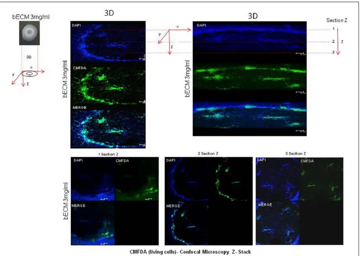
Interaction between MSCs and ECM is also recognized to have a crucial role in regulating stem cell phenotype. In our laboratory (Tecnologica Research Institute, Crotone, Italy), we recently observed that physical factors of the cell–culture environment were able to influence the distribution and shape of mesenchymal stem cells isolated from dental pulp (DPSC), and were able also to regulate the stem cell differentiation. By using hydrogel scaffolds derived from bovine bone extracellular matrix (bECM), we observed that cell distribution and cell morphology were influenced by the matrix stiffness, and this feature also promoted the odon-togenic and osteogenic differentiation (data not yet published). Cells seeded on bECM tended to grow in clusters, creating a circular structure that caused the hydrogel contraction. Moreover, DPSCs cul-tures in ECM hydrogels exhibited an increased level of osteogenic specific genes and odontogenic specific genes, if compared to polystyrene tissue culture plates. Moreover, we found that DPSCs seeded on bECM, when exposed to growth factors such as epidermal growth factor (EGF) and fibro-blast growth factor (FGF), yielded the most signifi-cant contractility (Figure 1) and showed a higher expression of the osteo/odontogenic specific genes (data not shown). Furthermore, DPSCs exhibited elongated spindle-shape morphology and no dead cells were observed all over the experiment. Confocal microscopy images showed that DPSCs were initially randomly distributed on the hydrogel surface, instead after the proliferation, cells were connected in order to stabilize the hydrogel con-traction (Figure 2). Our studies have been mainly focused on the use of MSCs in bone regeneration, taking into account that osteogenic differentiation of hMSCs is guided by various physical and bio-chemical factors. Jha et al. first highlighted the physical osteoinductive signals of the ECM niche, able to contribute to endochondral ossification of a cartilaginous skeleton template.15 In particular,
they evaluated the osteogenic differentiation of hMSCs cultured on low stiffness moduli (stiffness: 102, 390, or 970 Pa) made of poly-N-isopropy-lacrylamide (p(NIPAAm)) based on a semi- interpenetrating network (sIPN), modified with the integrin that engaging the bsp-RGD peptide (0, 105, or 210 µM).15 Cell adhesion and proliferation
and osteogenic differentiation of hMSCs, meas-ured by alkaline phosphatase (ALP), runt-related
transcription factor 2 (RUNX2), bone sialoprotein-2 (iBSP), and osteocalcin (OCN) protein expression, were the highest on those substrates with the high-est modulus and peptides concentration.
Sharma et al. investigated a substrate-dependent paracrine signaling, between the sub-populations of bone marrow stromal cells (BMSCs), able to alterate the neoformation of a new tendon at the bone enthesis.16 They used fibronectin (Fn) and
type-I collagen (Col) to functionalize the poly-acrylamide substrates and to approximate the elas-tic modulus of tendon granulation tissue and healing bone (10–90 kPa). BMSCs were cultured in growth media alone or media supplemented with soluble Col or Fn. More rigid substrates (70–90 kPa) induced osteogenic cell differentiation when functionalized with either Col or Fn. On broader mechanical gradient substrates (10–90 kPa), cell differentiation was markedly osteogenic on sub-regions of Fn functionalized substrates above 20 kPa, but osteogenic activity was inhibited on all
sub-regions of Col substrates. Osteogenic differen-tiation was not observed when cells were cultured on Fn substrates, if Col was present in the media or on the substrate (Fn/Col). Tenogenic differentia-tion markers were observed only on Col substrates with a moderate rigidity (30–50 kPa). They also analyzed the mediation of bone morphogenetic protein-2 (BMP-2); in particular the level of gene-expression of BMP-2 and of the transcription factor Smad8: they verified that BMP-2 average levels were similar to those levels observed in the cell population showing an arrested osteogenic differentiation after 14-day culture. Thus, they concluded that cell instructive biomaterials with mechanical and biochemical properties represent powerful tools for directing BMSC differentiation to tendon and bone; however, paracrine signals from tenogenic cells may delay osteogenesis in the healing enthesis.
Tilghman et al. analyzed how the cancer cells respond to changes in the mechanical properties
(rigidity/stiffness) of the microenvironment and how this response varies among cancer cell lines.17
In this study, they used a 96-well plate system with the wells filled with ECM-conjugated poly-acrylamide gels, to increase the stiffness to at least 50-fold across the plate. They determined how the changes in the rigidity of the extracellular matrix modulated the biological properties of tumor cells. They demonstrated the in vitro ability of these cells to grow on soft gel, just similar to their in vivo ability to grow in a soft tissue environment. Their observations suggested that the mechanical pro-perties of the matrix environment play a significant role in regulating the proliferation and the mor-phology of cancer cells.
All these studies could lead to different consid-erations about the role of the environment on the cell fate. The cell niche is a concept relatively new18 and many authors have demonstrated that
a scaffold can be multipotent19 and can induce
different phenotypes. On the other hand, cells can follow their own basal commitment20 or they can
be induced to differentiate into a phenotype defi-nitely different from the biological niche where they were harvested.21,22
Conclusions
The literature has reported several high-quality studies that clearly demonstrated the correlation between the change in cell shape and lineage com-mitment, between the cell–matrix interaction and the cell adhesion, particularly during the osteo-genic and odontoosteo-genic differentiation. Furthermore, our results reported in this paper also indicate that this correlation could be explained by the physical cues of matrix stiffness, as well as biochemical signals.
Often the behavior of a cell line is determined by numerous co-factors: the ECM, the cellular memory, the substrates, the presence of different temperatures and pressures, and recently, also acoustic waves have been investigated as potential factors capable of inducing a cellular response. Of
course, these growth conditions must be carefully standardized; when performing proper studies it is first important to know how these co-factors modify the growth and behavior of cells.
Recently, the concept of “cell niche” was widely studied: it defines the behavior of a cell line, based on environment where cells are grown. This con-cept is supported by recent papers describing how different parameters can lead to differentiation into different cell lineages.
Mechanical and geometrical factors are surely able to influence MSC behavior and fate, a fact to be carefully considered during further investigations into MSCs. Finally, these interactions should be considered as a very important bias that involves a lot of studies on MSCs, since in worldwide labora-tories scientists routinely use TCPS for cell culture.
Declaration of conflicting interests
The authors declared no potential conflicts of interest with respect to the research, authorship, and/or publication of this article.
Funding
This research received no specific grant from any funding agency in the public, commercial, or not-for-profit sectors.
References
1. Guilak F, Cohen DM, Estes BT, et al. (2009) Control of stem cell fate by physical interactions with the extracellular matrix. Cell Stem Cell 5: 17–26.
2. Yang C, Tibbitt MW, Basta L, et al. (2014) Mechanical memory and dosing influence stem cell fate. Nature
Materials 13: 645–652.
3. Gilbert PM, Havenstrite KL, Magnusson KE, et al. (2010) Substrate elasticity regulates skeletal muscle stem cell self-renewal in culture. Science 329: 1078–1081.
4. Daley WP, Peters SB and Larsen M (2008). Extracellular matrix dynamics in development and regenerative medicine. Journal of Cell Science 121: 255–264. 5. Dupont S, Morsut L, Aragona M, et al. (2011) Role
of YAP/TAZ in mechanotransduction. Nature 474: 179–183.
6. Halder G, Dupont S and Piccolo S (2012) Transduction of mechanical and cytoskeletal cues by YAP and TAZ. Nature Reviews Molecular Cellular Biology 13: 591–600.
7. Wen JH, Vincent LG, Fuhrmann A, et al. (2014) Inter-play of matrix stiffness and protein tethering in stem cell differentiation. Nature Materials 13: 979–987. 8. Banks JM, Mozdzen LC, Harley BA, et al. (2014)
The combined effects of matrix stiffness and growth factor immobilization on the bioactivity and
differentiation capabilities of adipose-derived stem cells. Biomaterials 35: 8951–8959.
9. Yim EK and Sheetz MP (2012) Force-dependent cell signaling in stem cell differentiation. Stem Cell
Research Therapy 3: 41.
10. Eroshenko N, Ramachandran R, Yadavalli VK, et al. (2013) Effect of substrate stiffness on early human embryonic stem cell differentiation. Journal of
Biological Engineering 7: 7.
11. Chen CS, Mrksich M, Huang S, et al. (1998) Micropatterned surfaces for control of cell shape, posi-tion, and function. Biotechnology Progress 14: 356–363. 12. Ghosh K and Ingber DE (2007) Micromechanical con-trol of cell and tissue development: Implications for tissue engineering. Advanced Drug Delivery Reviews 59: 1306–1018.
13. Trappmann B, Gautrot JE, Connelly JT, et al. (2012) Extracellular-matrix tethering regulates stem-cell fate. Nature Materials 11: 642–649.
14. Khetan S, Guvendiren M, Legant WR, et al. (2013) Degradation-mediated cellular traction directs stem cell fate in covalently crosslinked three-dimensional hydrogels. Nature Materials 12: 458–465.
15. Jha AK, Jackson WM and Healy KE (2014) Controlling osteogenic stem cell differentiation via soft bioinspired hydrogels. PLoS One 9: e98640. 16. Sharma RI and Snedeker JG (2012) Paracrine
interactions between mesenchymal stem cells affect substrate driven differentiation toward tendon and bone phenotypes. PLoS One 7: e31504.
17. Tilghman RW, Cowan CR, Mih JD, et al. (2010) Matrix rigidity regulates cancer cell growth and cellular phenotype. PLoS One 5: e12905.
18. Tatullo M, Marrelli M and Paduano F (2015) The regenerative medicine in oral and maxillofacial surgery: The most important innovations in the clinical application of mesenchymal stem cells. International
Journal of Medical Science 12(1): 72–77.
19. Aulino P, Costa A, Chiaravalloti E, et al. (2015) Muscle extracellular matrix scaffold is a multi-potent environment. International Journal of Medical
Science 12(4): 336–340.
20. Tatullo M, Falisi G, Amantea M, et al. (2015) Dental pulp stem cells and human periapical cyst mesenchy-mal stem cells in bone tissue regeneration: Comparison of basal and osteogenic differentiated gene expression of a newly discovered mesenchymal stem cell lineage.
Journal of Biological Regulators and Homeostatic Agents 29(3): 713–718.
21. Marrelli M, Paduano F and Tatullo M (2015) Human periapical cyst-mesenchymal stem cells differentiate into neuronal cells. Journal of Dental Research 94(6): 843–852.
22. Marrelli M, Paduano F and Tatullo M (2013) Cells isolated from human periapical cysts express mes-enchymal stem cell-like properties. International