UNIVERSITÀ DEGLI STUDI DI CATANIA DOTTORATODIRICERCAINBIOMEDICINATRASLAZIONALE
CURRICULUMINBIOMEDICINAMOLECOLAREGENOMICAEDEISISTEMICOMPLESSI:BASI CELLULARIEMOLECOLARIDELFENOTIPO-XIXCICLO
DIPARTIMENTODISCIENZEBIOMEDICHEEBIOTECNOLOGICHE
SEZIONE DI BIOLOGIA E GENETICA “GIOVANNI SICHEL”
Cristina Barbagallo
_______________LncRNA and circRNA expression profiles in tissues and serum exosomes of
colorectal cancer patients and cell lines
_______________ TESI DI DOTTORATO
_______________
Coordinatore del Dottorato: Prof. Lorenzo Malatino Coordinatore del Curriculum: Prof. Michele Purrello Tutor: Prof. Marco Ragusa
Table of contents
1. Abstract ... 1
2. Introduction ... 3
2.1 Long non-coding RNAs ... 8
2.1.1 LncRNA biogenesis ... 9
2.1.2 LncRNA functions ... 10
2.2 Circular RNAs ... 16
2.2.1 CircRNA biogenesis ... 17
2.2.2 CircRNA functions ... 22
2.3 Small non-coding RNAs ... 25
2.3.1 MiRNA biogenesis ... 25
2.3.2 MiRNA functions ... 28
2.4 Exosomes ... 30
2.4.1 Exosomes as mediators of intercellular communication ... 36
2.5 Colorectal cancer ... 39
2.5.1 CRC genetic alterations ... 40
2.5.2 CRC diagnosis and therapy ... 47
2.6 NcRNAs as mediators of tumor progression... 52
2.6.1 LncRNA role in CRC pathogenesis ... 52
2.6.3 MiRNAs involved in CRC carcinogenesis ... 57
2.7 Exosomes in tumor progression ... 58
2.8 NcRNAs in biological fluids ... 59
3. Materials and methods ... 61
3.1 Cell lines ... 61
3.2 Isolation of exosomes secreted by CRC cell lines ... 61
3.3 RNA isolation from cell lines and exosomes ... 62
3.4 RNA isolation from CRC formalin-fixed, paraffin-embedded samples ... 62
3.5 Exosome and RNA isolation from CRC patient serum ... 62
3.6 PCR primer design ... 63
3.7 LncRNA and circRNA expression analysis by Real-Time PCR ... 65
3.8 Computational analysis ... 65
3.9 Biological network construction and Gene ontology analysis ... 69
3.10 ROC curve analysis ... 69
3.11 CRC cell lines treatment with MAPK inhibitors ... 70
3.12 Protein extraction and Western Blot analysis ... 70
3.13 In vitro silencing of UCA1 ... 71
4. Results ... 72
4.1 NcRNA expression in CRC cell lines and exosomes ... 72
4.2 NcRNA expression in FFPE CRC biopsies ... 73
4.4 Analysis of GEO DataSets and identification of mRNA and miRNA targets of DE
ncRNAs ... 78
4.5 Analysis of gene ontologies ... 82
4.6 Evaluation of the diagnostic accuracy of DE ncRNAs in serum exosomes... 84
4.7 In vitro inhibition of MAPKs ... 86
4.7.1 Cell viability assay ... 86
4.7.2 Confirmation of MAPK inhibition through Western Blot analysis ... 86
4.7.3 MAPK inhibition affects ncRNA expression ... 87
4.7.4 Evaluation of correlation between ncRNA expression and ERK activation ... 88
4.8 UCA1 target expression after in vitro silencing ... 89
5. Discussion ... 91
5.1 I NcRNAs contribute to CRC progression ... 91
5.2 NcRNAs in serum exosomes of CRC patients: new diagnostic biomarkers? ... 96
5.3 Identification of miRNA and mRNA targets of DE ncRNAs: future perspectives for the understanding of miRNA-ncRNA and mRNA-ncRNA interaction effects ... 98
5.4 Altered expression of ncRNAs after treatment with MAPK inhibitors ... 100
5.5 In vitro silencing of UCA1 affects the expression of its targets ... 102
6. Conclusions and future perspectives ... 105
1. Abstract
In the last few years several studies demonstrated the fundamental role of non-coding RNAs (ncRNAs) in tumor onset and progression. While the involvement and the mechanism of action of microRNAs (miRNAs) have been widely investigated, little is known about long non-coding RNAs (lncRNAs) and circular RNAs (circRNAs), which seem to act through a plethora of molecular mechanisms regulating essential biological processes, such as cell cycle, splicing, chromatin remodeling, apoptosis, adhesion and migration. LncRNAs are non-coding RNAs longer than 200 nucleotides, with or without 5’-cap and poly(A) tail; circRNAs are circular molecules lacking free ends and thus resistant to exonucleasic degradation, transcribed from protein-coding genes. Several evidences showed that aberrant expression of lncRNAs and circRNAs is associated with various tumors, including CRC. In our study we analysed through Real-Time PCR the expression of a set of 17 lncRNAs and 31 circRNAs selected from literature in 20 CRC tissues compared to normal adjacent tissues (NATs), and in serum exosomes of 20 CRC patients compared to 20 healthy individuals. We identified 8 ncRNAs (CCAT1, CCAT2, CDR1AS, HOTAIR, MALAT1, TUG1, UCA1 and ZEB2AS1) differentially expressed (DE) in tissues and 3 ncRNAs (circ16, TUG1 and UCA1) DE in serum exosomes. Through gene ontology analysis we verified the involvement of DE ncRNAs in pathways associated with tumor progression; we also evaluated the diagnostic accuracy of ncRNAs deregulated in serum exosomes through ROC curves, suggesting their possible application in CRC non-invasive diagnosis. We observed that inhibition of MAPK pathway, associated with CRC, altered the expression of HOTAIR, MALAT1, TUG1 and UCA1 in HCT-116 colon cancer cells treated with U0126 (MEK1/2 specific inhibitor) compared to untreated cells, suggesting a possible connection between ncRNAs and MAPKs. Also, we performed a computational analysis to identify mRNAs and miRNAs involved in CRC and characterized by
Finally, we performed in vitro functional assays by silencing UCA1 expression through ASOs in HCT-116 cells and analysed the expression of its mRNA and miRNA targets, aiming to investigate the eventual degradation triggered by the interaction between ncRNAs and their miRNA/mRNA targets. Our study provide new data about aberrant expression of ncRNAs in CRC and their involvement in carcinogenesis; further analyses will be necessary to fully understand ncRNA molecular mechanisms and to evaluate their possible application in CRC diagnosis and therapy.
2. Introduction
A few years after the discovery of DNA structure, Francis Crick stated the central dogma of molecular biology: “Once information has got into a protein it can't get out again" (1); with this simple sentence he summarized the flow of genetic information through DNA, RNA and proteins (Figure 2.1).
Figure 2.1: the original concept diagram published by Francis Crick in 1956.
Shortly after, François Jacob and Jacques Monod confirmed the central role of RNA in genetic information transfer from DNA to proteins (2). At that time, with few evidences, it was assumed that all biological processes in cells were regulated by proteins, characterized by more
variety of new technologies and skills have been improved and a huge amount of sequencing data has been produced, leading researchers all over the world to go beyond these models and reconsider RNA role in cellular biology.
In 1990 an international consortium (including USA, United Kingdom, France, Australia, and China) launched the Human Genome Project, aiming to determine the entire sequence and to detect all genes of human genome (3). In 2001 a first draft of the human genome was published (4), followed by the final version in 2004 (5). Until then, scientists tried to indirectly estimate the total number of human protein-coding genes using many approaches, such as the typical vertebrate tissue mRNA complexity, the approximate ratio of typical gene size to the genome size, the number of CpG islands and the frequency of their association with known genes; finally it was supposed a total of 30,000-35,000 protein-coding genes in human genome (3). The final results of Human Genome Project revealed that our genome includes approximately 20,000-25,000 protein-coding genes (5), but this number has been recently corrected in about 19,000 genes (6). Surprisingly, this number is lower than expected, and very close to total protein-coding gene number of less complex organisms (Caenorhabditis elegans, 19,735 genes, Gallus gallus, 16,736 genes, Drosophila melanogaster, 14,889 genes, Mus musculus (21,839 genes) (7) (Figure 2.2).
Figure 2.2: total number of protein-coding genes comparison between Homo sapiens and inferior organisms (from https://www.sciencenews.org/article/more-chicken-fewer-grape).
This discovery surprised scientists, which believed that Homo sapiens higher complexity was due to higher gene number compared to inferior organisms. Also, it was observed that all genes identified during the Human Genome Project represent less than 2% of the entire genome, while in other organisms, eukaryotic or prokaryotic, the coding fraction of genome is considerably higher (8) (Figure 2.3).
This illusory paradox was partially explained by alternative splicing events and post-translational modifications, which allow cells to produce more proteins with different functions from a single gene. However, scientists concluded that a large fraction of the human genome, even 98%, was represented by “junk DNA”, that is DNA with no function.
In order to understand the molecular bases of Homo sapiens higher complexity and the “junk DNA” function, the ENCODE (Encyclopedia of DNA Elements) Project was launched with the purpose to identify all functional elements in the human genome (9). Final results, confirmed by following studies, showed that the 85-90% of human genome is pervasively transcribed: numerous new transcription start sites were identified, as well as many new transcripts, originated from genes located at non-coding regions or overlapping protein-coding loci (10, 11, 12). Since these new transcripts were not translated into proteins, they were defined non-coding RNAs (ncRNAs). It was recently estimated that non-coding gene number tends to exponentially increase ascending the evolutionary scale, while protein-coding gene number reaches a plateau: therefore, Homo sapiens higher complexity in comparison to inferior organisms with similar protein-coding gene number may be due to this new class of RNAs (13) (Figure 2.4).
Figure 2.4: correlation between organism complexity and
Vanhercke, The power of RNA: discover next generation biomarkers and therapeutic targets
Next Generation Sequencing (NGS) new technologies sequences, stressing their great structural heterogeneity observed in the structure of ncRNA gene
nucleotides to several hundreds of kilobases; ii) they lack
evolutionary conservation is often low or absent; iv) there is no preferential localization genome. Also, a relative tolerance to point mutations
make ncRNA genes difficult to identify,
however, it has been estimated that the human genome includes from 10,000 to 20,000 non coding genes (16). Classifying these molecules is also
currently used, based on ncRNA length or function.
i) long non-coding RNAs (lncRNAs), which are longer than 200 nucleotides, and ii) small non coding RNAs, whose length is equal or
molecules deeply analysed, such as microRNAs (miRNAs), small interfering RNAs (siRNAs), and
correlation between organism complexity and coding/non-coding genome fraction (from
Vanhercke, The power of RNA: discover next generation biomarkers and therapeutic targets
Next Generation Sequencing (NGS) new technologies led to the identif
eat structural heterogeneity. The same heterogeneity can
ncRNA genes: i) their length is extremely variable, from few tens of nucleotides to several hundreds of kilobases; ii) they lack ORFs (Open Reading Frames); iii) evolutionary conservation is often low or absent; iv) there is no preferential localization
a relative tolerance to point mutations has been observed (14
ult to identify, which is why their exact number is to date unknown; however, it has been estimated that the human genome includes from 10,000 to 20,000 non
). Classifying these molecules is also difficult: two different classificat
currently used, based on ncRNA length or function. According to length, ncRNAs are divided into coding RNAs (lncRNAs), which are longer than 200 nucleotides, and ii) small non coding RNAs, whose length is equal or inferior to 200 nucleotides; the latter includes ncRNA
ed, such as microRNAs (miRNAs), small interfering RNAs (siRNAs), and
coding genome fraction (from Kristiaan
Vanhercke, The power of RNA: discover next generation biomarkers and therapeutic targets).
identification of ncRNA The same heterogeneity can also be : i) their length is extremely variable, from few tens of ORFs (Open Reading Frames); iii) evolutionary conservation is often low or absent; iv) there is no preferential localization within the 14, 15). These features exact number is to date unknown; however, it has been estimated that the human genome includes from 10,000 to 20,000 non-protein : two different classification are According to length, ncRNAs are divided into coding RNAs (lncRNAs), which are longer than 200 nucleotides, and ii) small
non-the latter includes ncRNA ed, such as microRNAs (miRNAs), small interfering RNAs (siRNAs), and
housekeeping ncRNAs, constitutively expressed and responsible of fundamental physiological processes; ii) regulatory ncRNAs, whose expression can be induced in different cell histotypes, in defined development and differentiation stages or following external stimuli. Housekeeping ncRNAs include transfer RNAs (tRNAs), ribosomal RNAs (rRNAs), small nuclear RNAs (snRNAs), small nucleolar RNAs (snoRNAs), and telomerase RNAs, while regulatory ncRNAs comprise miRNAs, siRNAs, piRNAs and lncRNAs (14, 15).
2.1 Long non-coding RNAs
LncRNAs represent a highly heterogeneous class of ncRNAs, characterized by length greater than 200 nucleotides (cut-off value derived from biochemical techniques that allow to exclude all known classes of small RNAs) frequently reaching 100 kb (14). These transcripts are considered non-protein coding because they lack long ORF (more than 100 codons) and/or codon conservation; however, recent studies showed that some lncRNAs can code for small proteins (17). LncRNA genes are difficult to identify because of their variable features; indeed, they can:
i. Be located within intergenic regions, generating long intergenic non-coding RNAs (lincRNAs) (18);
ii. Be located inside introns of protein-coding genes (19);
iii. Partially overlap untranslated regions (UTRs) or protein-coding gene promoters (20, 21);
iv. Be pseudogenes whose transcripts (sense or antisense) control the expression of their protein-coding paralog genes (22, 23);
v. Lie within transcribed ultra-conserved regions (tUCRs), characterized by high evolutionary conservation and located at intra- or intergenic regions (24);
vi. Be located within the mitochondrial genome and regulated by proteins coded by nuclear genes (25).
The discovery of ncRNAs amazed researchers and started off a debate about the effective functions of these molecules, initially considered “transcriptional noise” (26, 27). Nevertheless, numerous studies have highlighted that ncRNA expression is tissue-specific, and it varies in different development or differentiation stages or in response to external stimuli (28, 29, 30, 31). These data allowed to go beyond the “transcriptional noise” hypothesis and sparked scientist interest in lncRNAs: recent studies showed that these molecules play an important role in biological processes, both physiological, such as cell cycle (32, 33), apoptosis (34, 35), differentiation (36), stemness (37), aging (38), and pathological (39, 40, 41). In particular, many researchers focused their attention on the possible involvement of lncRNAs in neoplastic processes, which by now seems to be demonstrated (42, 43, 44, 45, 46).
2.1.1 LncRNA biogenesis
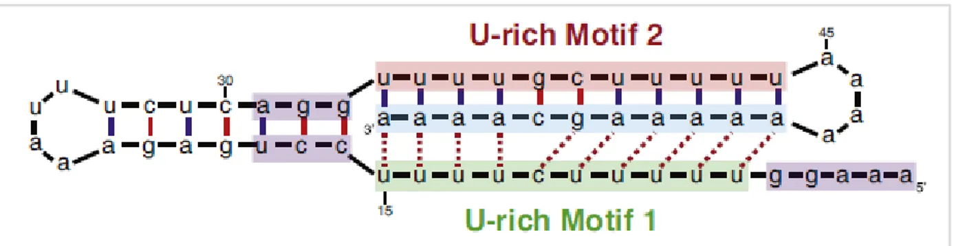
Most of lncRNAs share with mRNAs the transcription by RNA polymerase II, the epigenetic regulation (histone modifications) and splicing signals (47); only a small part of them is transcribed by RNA polymerase I and III (48, 49). About half of lncRNAs are post-transcriptionally processed undergoing the same steps of mRNAs (5’-cap addition, polyadenylation, splicing), although some of them are monoexonic and non-polyadenylated (47, 50). Poly(A) tail protects 3’-ends from enzymatic degradation; in its absence, the presence of alternative structures with similar function, such as triple helix, has been shown. This kind of structure has been identified at 3’-end of MALAT1 (metastasis-associated lung adenocarcinoma transcript 1), a 8.7-kb lncRNA transcribed from a gene located at chromosome 11. During MALAT1 processing, the primary transcript loses from its 3’-end a 61-nucleotide fragment, called mascRNA (MALAT1-associated small cytoplasmic RNA); hence, the new 3’-end of mature transcript presents a stem-loop structure and three conserved motifs, composed of two U-rich and one A-rich tracts; this A-rich tract is similar to
interact with each other and form a triple helix structure that stabilizes MALAT1 3’-end and protects it from enzymatic degradation (51) (Figure 2.5).
Figure 2.5: triple helix structure stabilizing MALAT1 3’-end in absence of poly(A) tail. Dashed lines show base triplets
(from Wilusz, Biochim Biophys Acta. 2016).
2.1.2 LncRNA functions
LncRNAs play a crucial role in cellular physiology through a plethora of functions and molecular mechanisms. It is now clear that they can regulate gene expression at different levels (transcriptional, post-transcriptional, translational, post-translational, epigenetic) (52) (Figure 2.6); however, the exact molecular mechanism has been elucidated only for few lncRNAs.
Figure 2.6: lncRNAs regulate cell physiology performing several functions (from Morris and Mattick, Nat. Rev. Genet. 2014).
Xist (X inactive specific transcript) is one of the first lncRNAs identified (53, 54). Xist is a 19-kb lncRNA transcribed by the X chromosome; it is involved in the X-chromosome inactivation process, the epigenetic mechanism leading to the random inactivation of one of the two X
chromosomes in female individual cells. X inactivation depends on histone modifications (in particular lysine methylation) performed by PRC1 and PRC2 (Polycomb-repressing complex 1 and 2), which in turn are recruited on the inactive X chromosome by Xist (55). Similarly, other lncRNAs can modulate gene expression by recruiting chromatin remodeling complexes: one of the best known is HOTAIR (Hox transcript antisense intergenic RNA), a 2.2-kb lncRNA transcribed from the antisense strand of HOXC (Homeobox C) cluster on chromosome 12 (56, 57). HOTAIR also interacts with PRC2, inducing the trimethylation of histone H3 lysine 27, and the consequent transcription inhibition, of HOXD (Homeobox D) cluster on chromosome 2 (58). Moreover, HOTAIR interacts not only with PRC2 through a 5’-domain, but also with LSD1 (lysine demethylase 1A) through a 3’-domain, thus modulating demethylation and transcriptional activation of the same cluster (57, 59). TUG1 (taurine up-regulated 1) is a 7.5-kb lncRNA located at chromosome 22; its expression is p53-mediated thanks to many highly conserved p53-binding sites included in its promoter. After DNA damage, p53 binds TUG1 promoter and activates TUG1 expression; the lncRNA in turn recruits PRC2 and inhibits expression of cell cycle regulator genes (60).
LncRNAs can also bind transcriptional factors, recruiting them on DNA or sequestering them, therefore regulating the expression of specific genes. PANDAR (promoter of CDKN1A antisense DNA damage activated RNA) is a 1.5-kb polyadenylated and capped lncRNA, antisense transcript of CDKN1A (cyclin-dependent kinase inhibitor 1A) on chromosome 6. Its expression is regulated by p53 after DNA damage; PANDAR binds the transcription factor NFYA (nuclear transcription factor Y subunit alpha), impeding it to bind the promoters of pro-apoptotic genes (61).
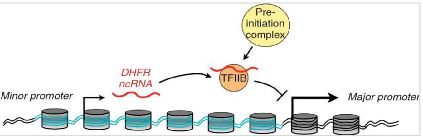
Some lncRNAs can interact with DNA inducing loop formation, thus moving closer two otherwise very distant regions; these lncRNAs act as “bridges” between promoters and enhancers or silencers. Their mechanism of action is not known yet, although it has been proposed the formation of a RNA-DNA-DNA triplex or a RNA-protein-DNA complex (62). The DHFR (dihydrofolate
reductase) locus on chromosome 5 contains two promoters: the major promoter codes for the DHFR mRNA, while the upstream minor promoter codes for a ncRNA which negatively regulates mRNA levels in cis and in trans: the DHFR ncRNA interacts with the major promoter, forming a stable complex, and with TFIIB (transcription factor IIB), causing PIC (pre-initiation complex) dissociation and transcription inhibition (63) (Figure 2.7).
Figure 2.7: DHFR ncRNA inhibits the transcription of DHFR mRNA by interacting with the major promoter and
TFIIB (from Martianov et al., Nature 2007).
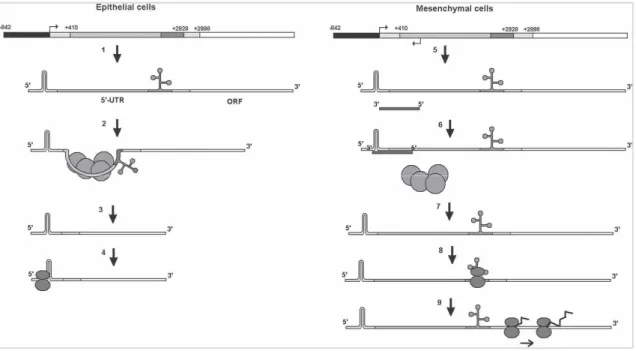
LncRNAs can act at post-transcriptional level modulating mRNA stability, translation and splicing. BACE1AS (BACE1 antisense RNA) is a 825-nucleotide lncRNA transcribed from a gene located at chromosome 11, apparently involved in Alzheimer’s disease pathogenesis. BACE1AS is the natural antisense of BACE1 (beta-secretase 1), a transmembrane protease that catalyzes the first step in amyloid beta peptide synthesis; amyloid beta peptide accumulates in Alzheimer’s patient cerebral neurons, generating the typical amyloid plaques. Thanks to sequence complementarity, BACE1AS binds BACE1 mRNA, thus stabilizing it and inducing increased amyloid beta production (64). ZEB2AS1 (ZEB2 antisense RNA 1), antisense transcript of ZEB2 (zinc finger E-box binding homeoE-box 2), is a 430-nucleotide lncRNA transcribed from chromosome 2. ZEB2 plays a key role in neoplastic processes by repressing E-cadherin expression, thus promoting epithelial-mesenchymal transition (EMT). ZEB2AS1 is abundantly expressed in mesenchymal cells through the activation of an alternative promoter; the lncRNA binds the 5’-UTR of ZEB2 mRNA,
masking splicing sites and preventing the removal of a long intron (about 2.5 kb) including an internal ribosome entry site (IRES). So, in presence of its antisense, ZEB2 mRNA is recognised by ribosomes and translated, producing a transcription factor that induces E-cadherin and EMT repression (20) (Figure 2.8).
Figure 2.8: ZEB2 mRNA alternative splicing mediate by its antisense ZEB2AS1 in mesenchymal cells; ZEB2 main
promoter is shown in black, IRES-containing 3-kb 5’-UTR in gray, ZEB2 ORF in white (from Beltran et al., Genes Dev.
2008).
Another function of lncRNAs is to regulate cellular localization of proteins, thus modulating their activity. NRON (non-protein coding RNA, repressor of NFAT), a 2.7-kb transcript of chromosome 9, controls the trafficking of NFAT (nuclear factor of activated T cells) transcription factor: NFAT is normally contained in the cytoplasm, but it is imported to the nucleus in response to calcium-dependent signals. NRON interacts with several proteins, including members of the importin-beta superfamily, thus inhibiting NFAT nuclear accumulation (52, 65).
Some lncRNAs play structural roles, allowing correct formation of specific cellular structures. NEAT1 (nuclear paraspeckle assembly transcript 1) is a 4-kb monoexonic polyadenilated lncRNA transcribed from chromosome 11; it is contained in the nucleus overlapping SC35 splicing
factor enriched regions; NEAT1 presence in the nucleus is essential for the formation of paraspeckles, nuclear domains involved in nuclear retention of specific mRNAs (66).
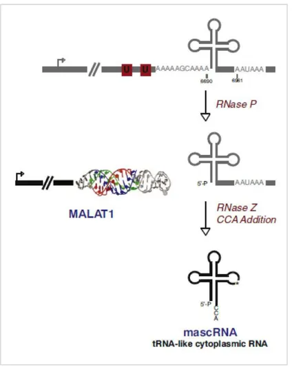
Recent studies revealed a connection between lncRNAs and small non-coding RNAs, such as miRNAs: some lncRNAs can be miRNA precursors or can interact with them, sequestering them and inhibiting their transcriptional repression activity; this function has been defined “miRNA sponge”. H19, the first lncRNA identified (67), is transcribed from a gene localized on chromosome 11, near IGF2 (insulin-like growth factor 2) locus; both genes are epigenetically silenced: only the maternal allele of H19 and the paternal allele of IGF2 are expressed, while the other ones are silenced by methylation (68). H19 gene produces a 2.3-kb capped, polyadenilated and spliced lncRNA which is expressed from the earliest stages of embryonic development in several organs and quite completely silenced after birth. H19 is the precursor of miR-675, which targets, among others, RB1 (retinoblastoma 1), a key cell cycle regulator (69). Plus, H19 contains many binding sites for let-7 miRNA family, well known tumor suppressor miRNAs; H19 can bind and sequester let-7 miRNAs, acting as “miRNA sponge” (70). TUSC7 (tumor suppressor candidate 7), a 2.1-kb lncRNA transcribed from chromosome 3, also acts as “sponge” for miR-211 thanks to two binding sites lying within its fourth exon (71). MALAT1 is highly expressed and retained inside the nucleus, where it acts as “riboregulator” modulating the expression of its target genes, involved in cell cycle regulation (51, 72). MALAT1 lacks poly(A) tail, and its 3’-end folds to form a tRNA-like secondary structure, recognised and cleaved by RNAse P; this cleavage creates a short fragment, subsequently processed by enzymes involved in tRNA biogenesis; at the end of the process, a 61-nucleotide mature transcript called mascRNA is produced (Figure 2.9). MascRNA is exported to the cytoplasm but, despites its structure, it doesn’t contribute to protein synthesis, and its function is still unknown (51). A recent study suggested a mascRNA involvement in immune system regulation: high levels of mascRNA (but not of MALAT1) have been observed in human immune system circulating cells; furthermore, mascRNA downregulation in monocytes-macrophages
strongly affects the expression of fundamental genes, including FASLG (Fas ligand), FAS (Fas cell surface death receptor), TNF-α (tumor necrosis factor alpha), and IL6 (interleukin 6) (73).
Figure 2.9: mascRNA biogenesis mediated by RNAse P cleavage of 3’-end of MALAT1 (from Wilusz, Genes Dev. 2012).
Therefore, it is clear that lncRNAs perform multiple functions through very heterogeneous molecular mechanisms, escaping any attempt at classification.
2.2 Circular RNAs
Next Generation Sequencing techniques led to the identification of a unique class of lncRNAs represented by circular non-coding RNA molecules, called circular RNAs (circRNAs). The first single-stranded circular RNA molecule was identified in 1976 within viroids, infectious
observed in other organisms, including animals, but they have been considered by-products of mRNA processing derived from splicing errors (75). Recently, several new circRNAs previously unknown have been identified, characterized by high expression levels in mammalian cells and, sometimes, by high evolutionary and expression conservation, suggesting a specific function in cellular physiology (76, 77).
2.2.1 CircRNA biogenesis
As it is known, one of the characteristics distinguishing prokaryotes and eukaryotes is the alternation of introns (non-coding segments) and exons (coding segments) in eukaryotic gene structure; after transcription, pre-mRNA undergoes a processing which includes splicing, that is intron removal by a ribonucleoprotein complex called spliceosome. Splicing has been widely studied; it consists of two phases, both regulated by the spliceosome: i) the 2’-OH of an intronic base, called branchpoint (usually an adenosine), performs a nucleophilic attack on the 5’-splicing site, localized at exon-intron boundary and identified by consensus dinucleotide GU, thus forming a phosphodiester 2’,5’-bond and releasing the 3’-OH end of the exon; ii) this 3’-OH in turn attacks on the 3’-splicing site, localized at intron-exon boundary and identified by consensus dinucleotide AG: hence, the two exons are bound through a phosphodiester 3’,5’-bond, while the intron is removed as a lariat, because of a 2’,5’-bond (Figure 2.10). This is an essential event finely regulated but also flexible, that allows cells to produce several protein variants modulating splicing sites choice (alternative splicing) (78).
Figure 2.10: splicing mechanism regulated by the spliceosome (from Matera et al,. Nat Rev Mol Cell Biol. 2014).
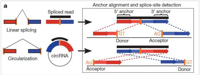
Many circRNAs are transcribed from protein-coding gene exons, so they are called exonic circRNAs (ecircRNAs); it has been proposed that ecircRNA biogenesis ecircRNA biogenesis is catalyzed by the spliceosome following the same splicing mechanism. This hypothesis is supported by several evidences: both splicing inhibitors and mutagenesis of splicing sites (GU and AG consensus sequences) significantly reduce circular RNA levels (79). The exact mechanism of ecircRNA biogenesis is still unknown: however, it seems that spliceosome regulates backsplicing (or inverse splicing) events, where a downstream 5’ splicing site (donor) is bound to an upstream 3’ splicing site (acceptor); this mechanism produces a head-to-tail junction, typical of backsplicing events (80) (Figure 2.11).
Figure 2.11: head-to-tail junction of two exons typical of backsplicing events (from Memczak et al., Nature 2013).
Three models for ecircRNA biogenesis have been proposed:
i. Lariat-driven circularization (Figure 2.12a): during splicing events, some exons can be skipped and removed together with introns (exon skipping); ecircRNAs can be produced by a second splicing on exon-containing lariats, before they are unraveled by debranching enzymes (77);
ii. Intron pairing-driven circularization (Figure 2.12b): intronic complementary motifs flanking circularized exons can anneal and promote circularization (77);
iii. Resplicing-driven circularization (Figure 2.12c): sometimes, a second splicing event can happen on a mature mRNA in presence of specific splicing signals. The first event is regulated by canonical splicing sites and removes introns from pre-mRNA; the second one requires cryptic splicing sites, which are recognised only in specific conditions, and induces exon circularization. Re-splicing is likely an aberrant phenomenon, often observed in carcinogenesis (81).
Figure 2.12: three models proposed to explain ecircRNA biogenesis a) Lariat-driven circularization: an
exon-containing lariat undergoes a second splicing; b) intron pairing-driven circularization: the annealing of intronic complementary motifs flanking circularized exons promotes circularization; c) resplicing-driven circularization: a second splicing event on a mature mRNA induces exon circularization (from Matera et al,. Nat Rev Mol Cell Biol.
2014).
As suggested by the intron pairing-driven circularization model, the presence of reverse complementary sequences (RCSs) in flanking introns can move exons closer and promote their circularization; indeed, several studies reported a positive correlation between flanking intron RCSs (often represented by Alu repeats in primates) and exon circularization (77, 82, 83). However, RCSs are not essential, and sometimes longer RCSs can even inhibit circularization through increased base-pairing stability; moreover, a gene can include many RCS copies competing with each other, so alternative circularization events can occur, thus raising ecircRNA number produced by a single gene (79, 84). Another factor promoting ecircRNA biogenesis is flanking intron length, that is
three- to fivefold longer than in randomly selected introns both in human genome, (77, 82), and in other species (79). Again, long flanking introns are not essential for ecircRNA biogenesis, but longer introns are likely to include more cis-acting regulatory element (77, 85).
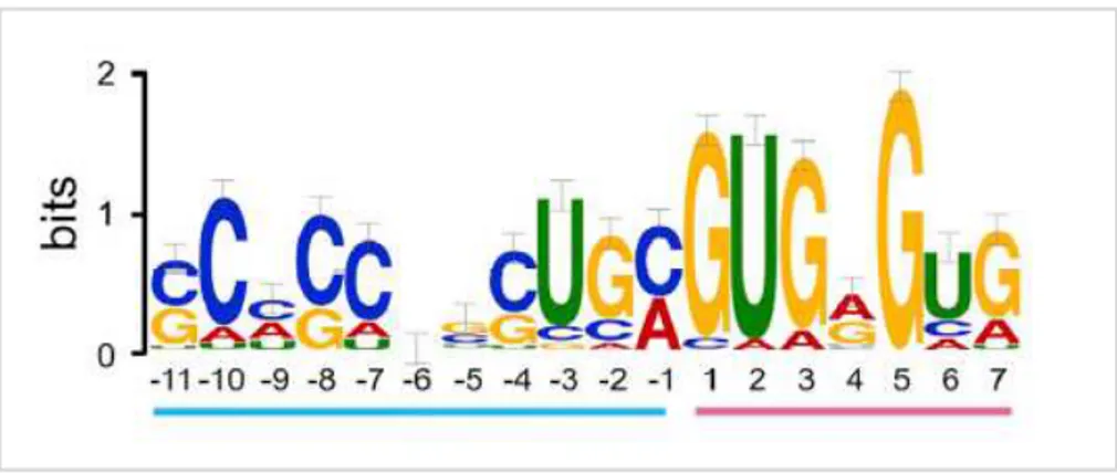
In addition to ecircRNAs, circular transcripts originated from spliced introns, called circular intronic RNAs (ciRNAs), have been identified. They are originated from the lariat, which is not debranched but circularized thanks to opportune signals, that are: i) a 7-nucleotide GU-rich sequence sited near to 5’ splicing site; ii) an 11-nucleotide C-rich sequence near to branchpoint; molecules participating in this process are still unknown (86) (Figure 2.13).
Figure 2.13: consensus sequences needed for ciRNA biogenesis: a 7-nucleotide GU-rich sequence sited near to 5’
splicing site (pink) and an 11-nucleotide C-rich sequence near to branchpoint (blue) (from Zhang et al., Mol Cell. 2013).
It has been hypothesized that the same factors regulating canonical (or linear) splicing also modulate backsplicing events; therefore, the two phenomena compete with each other to bind these molecules. A recent study showed that Quaking (QKI), an RNA-binding protein regulating alternative splicing, controls the expression of several circRNAs during EMT. QKI binds RCSs, moving circularized exons closer; the addition of QKI-binding sites in flanking introns highly increases circRNA production (87). ADAR (adenosine deaminase acting on RNA) protein family includes enzymes involved in RNA editing, a process modifying some nucleotides during pre-mRNA maturation. In particular, ADAR enzymes bind the adenosines of double-stranded RNA molecules, converting them into inosines through deamination. Recently, it has been observed that
the downregulation of ADAR enzymes induces increased circRNA expression, suggesting that these enzymes could negatively regulate circRNA biogenesis (83).
2.2.2 CircRNA functions
CircRNAs have been initially considered as splicing errors. Successively, some circRNAs showed evolutionary conservation between human and mouse and among species, probably because of the conservation of splicing sites; this observation led to the hypothesis of a molecular function performed by this class of ncRNAs (79). Another evidence supporting this hypothesis is the high number of different circRNAs produced by cells: according to the database circBase (http://www.circbase.org/), approximately 100.000 circRNAs have been identified in human cells (89). It has been estimated that circRNAs represent 1-10% of the entire cell transcriptome (76, 85, 90); most of them have very low expression levels (0.1-1%) compared to co-linear mRNAs, although sometimes the circular transcript exceeds the linear one (77); moreover, circRNAs are often tissue-specific, so they are expressed only in some cell histotypes, even if they tend to be more abundant in brain tissues (79).
Several studies reported the presence of ecircRNAs in the cytoplasm, while their biogenesis occurs within the nucleus: nucleus export systems likely regulate the transport of these molecules (76, 77, 90). One of ecircRNA functions, also performed by lncRNAs, is to bind miRNAs and modulate their action. Some ecircRNAs are strongly associated with Argonaute (Ago) proteins, key factors of induced gene silencing (RNA interference, RNAi), and include many miRNA-binding sites. The most known example is represented by a circRNA called CDR1AS/ciRs-7 (CDR1 antisense) and its 80 binding sites for miR-7. CircRNA-miRNA interaction causes a reduced miRNA availability within cell cytoplasm, and consequently a reduced translational repression of specific mRNA targets: increased CDR1AS expression induces an incremented
downregulation provokes reduced protein levels of miR-7 targets, because miR-7 is not sequestered by the circRNA (80, 91) (Figure 2.14). A bioinformatic study showed that circRNAs present miRNA-binding sites lacking polymorphisms, strengthening their miRNA sponge or miRNA reservoir function (92); nevertheless, most of circRNAs include a number of miRNA-binding sites comparable to co-linear mRNAs, suggesting that only some circRNAs act as miRNA sponge (90, 93).
Figure 2.14: CDR1AS/ciRS-7 acts as miR-7 “sponge” (from Hansen et al., Cancer Res. 2013).
Moreover, ecircRNAs can modulate parental gene expression through transcription regulation. Recent studies showed that some ecircRNAs are retained within the nucleus and interact with RNA polymerase II, suggesting an involvement in gene expression regulation; these ecircRNAs contain, in addition to exons, non-spliced introns, and therefore they have been called exon-intron circRNAs (EIcircRNAs) (94, 95, 96). EIcircRNAs are prevalently expressed in the nucleus; they promote parental gene transcription by binding small nuclear ribonucleoprotein U1 (snRNPU1), a splicing regulator also involved in the initial stages of transcription; the EIcircRNA-snRNPU1 complex interacts with RNA polymerase II on the promoter of the parental gene, promoting its expression (94) (Figure 2.15).
Figure 2.15: positive regulation of parental gene transcription mediated by EIcircRNAs: they bind snRNPU1 forming a
complex which interacts with RNA polymerase II, promoting transcription (from Li et al., Nat Struct Mol Biol. 2015).
Another study suggested that circRNA biogenesis itself reduces co-linear mRNA expression. MBNL1 (muscleblind like splicing regulator 1) gene codes for a splicing regulating protein, but also for a circRNA called circMbl, produced co-transcriptionally. circMbl flanking introns include several binding sites for MBNL protein: when highly expressed, MBNL protein binds circMbl, promoting its circularization to the detriment of splicing. Thus, the cell reacts to MBNL accumulation in two ways: i) splicing is inhibited, reducing mature mRNA production; ii) protein availability is decreased through its circMbl-mediated sequestering (97).
Despite many ecircRNAs include ORFs, to date contrasting evidences suggesting peptide production has been reported; therefore, the possibility that ecircRNAs act as mRNAs needs further investigations (98).
CiRNA functions are not fully understood. A recent study showed that ciRNAs present reduced evolutionary conservation between human and mouse, accumulate in the nucleus and contain few miRNA-binding sites; these observations suggest different functions and mechanisms compared to ecircRNAs. CiRNAs interact with phosphorylated RNA polymerase II, suggesting that they act as positive regulator of gene expression, consistently with their nuclear localization. Interestingly, ciRNA expression is positively correlated with parental gene levels: it has been
in the sites of synthesis. Low evolutionary conservation can be due to ciRNA origin: since introns don’t code for functional proteins, they undergo no or very low evolutive pressure and accumulate more mutations compared to exons (86).
2.3 Small non-coding RNAs
Small non-coding RNAs, characterized by length equal or inferior to 200 nucleotides, have been widely studied for many years. By now, it is well known that they regulate many biological processes: i) RNA synthesis, processing and translation and transcriptional initiation [piRNAs, PASRs (promoter-associated small RNAs)]; ii) RNA maturation (snoRNAs); iii) RNA degradation or translation inhibition (miRNA, siRNA) (15). Among all small non-coding RNAs, the most known and studied are definitely miRNAs, whose biogenesis and function as post-transcriptional repressors have been well characterized.
2.3.1 MiRNA biogenesis
MicroRNAs are a class of endogenous single-stranded RNAs consisting of approximately 18-25 nucleotides and expressed both in animals and in plants. MiRNA-coding genes are located at all chromosomes in humans, except for Y chromosome; frequently, genes coding for different miRNAs are located at adjacent loci on the same chromosome, forming clusters (e.g. let-7 cluster on chromosome 9, including hsa-let-7a-1, hsa-let-7f-1 e hsa-let-7d). Transcription of a cluster is simultaneous and generates a polycistronic primary transcript, successively processed into single miRNAs. MiRNAs belonging to the same cluster are often related to each other, suggesting that clusters derive from gene duplication; consequently, miRNAs from the same cluster are often functionally correlated, since function depends on sequence. Genomic localization of miRNA-coding genes is highly heterogeneous; indeed, they can be located at: i) intergenic regions, ii)
some miRNA-coding genes have an independent promoter (99). According to miRBase database (http://www.mirbase.org/), to date 1881 precursors and 2588 mature miRNAs have been identified, even if a recent study hypothesized a higher number of miRNA-coding genes in human genome (100).
MiRNA biogenesis starts in the nucleus; transcription is prevalently performed by RNA polymerase II, rarely by RNA polymerase III; transcription produces a long (many kb) primary transcript called pri-miRNA, which folds into a double-stranded hairpin structure; the hairpin includes a loop and a double-stranded stem, made up of 33 complementary base pairs (bp), and ends with two long single-stranded traits (101, 102, 103). Successively, pri-miRNA undergoes two sequential cleavages: the first cleavage occurs in the nucleus by Drosha, the second one in the cytoplasm by Dicer, two endonucleases including conserved RNAse III catalytic domains (RIIIDa, RRIIDb); these enzymes act in association with proteins containing double-stranded RNA-binding domains (dsRBDs) (99). The first cleavage performed by a complex including Drosha and Pasha (DGCR8) converts the pri-miRNA into a 700-nucleotide long molecule with a hairpin structure called pre-miRNA, which is exported to the cytoplasm by exportin-5 (Exp5), a GTP-dependent nuclear/cytoplasmic transporter (104). In the cytoplasm, pre-miRNA interacts with a big multiprotein complex called RISC loading complex (RLC), consisting of the endonuclease Dicer, TRBP (Tar RNA Binding Protein), which contains three dsRBDs, PACT (protein activator of PKR), and Argonaute protein Ago-2, an RNAse with catalytic function (105, 106, 107, 108). Argonaute (Ago) proteins are expressed in all eukaryotes and contain specific domains: PAZ and MID domains to bind RNA targets at 3’-end, and PIWI domain al 5’-end to catalyze RNA cleavage (109). Dicer catalyzes the cleavage of the hairpin loop, producing a 22-nucleotide long double-stranded miRNA characterized by mismatches and two protrusive nucleotides at both 3’-ends. This RNA duplex is released from RLC and splits into two strands, defined: i) guide strand, complementary to the targets, and therefore functional; ii) passenger strand (miRNA*), initially
considered non-functional and degraded (110) (Figure 2.16). Recent studies showed that passenger strand also is functional and accumulates in the cytoplasm (111, 112, 113): by now, it is known that every miRNA-coding gene originates two mature molecules, defined -5p and -3p depending on the duplex strand from which they derive.
Figure 2.16: miRNA biogenesis starts in the nucleus with the transcription of a long pri-miRNA performed by RNA
polymerase II or III; the pri-miRNA folds into a hairpin structure and undergoes an enzymatic cleavage performed by Drosha, generating the pre-miRNA, characterized by a stem-loop structure. The pre-miRNA is exported to the cytoplasm; the hairpin structure undergoes a cleavage performed by Dicer, generating a 22-nucleotide long
double-stranded miRNA (from Winter et al., Nat Cell Biol. 2009).
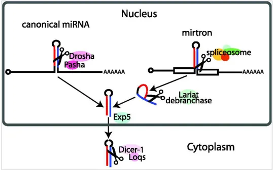
A particular miRNA subfamily includes genes located within protein-coding gene introns, which code for miRNA called mirtrons. Mirtron biogenesis is Drosha-independent: after splicing, they are processed into a hairpin RNA, which is exported to the cytoplasm by exportin-5 and directly cleaved by Dicer (114) (Figure 2.17).
Figure 2.17: mirtron biogenesis is Drosha-independent: after removal, the intron lariat is exported to the cytoplasm by
exportin-5 and cleaved by Dicer (from Ruby et al., Nature. 2007).
2.3.2 MiRNA functions
All miRNAs perform the same function of post-transcriptional repressors of gene expression through the same molecular mechanism.
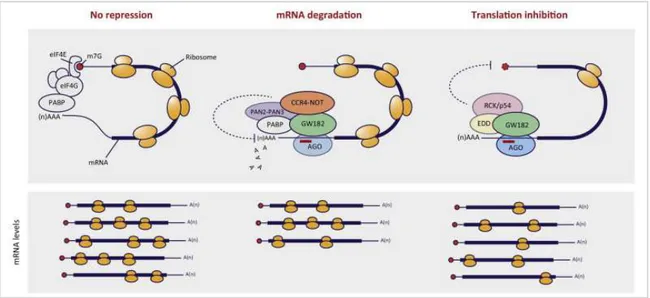
Mature miRNAs bind Ago-2 protein and are led to the RNA-induced silencing complex (RISC), the true effector of miRNA-mediated silencing; RISC consists of several proteins, including Ago. Once associated to RISC, mature miRNA becomes active and recognizes its mRNA target: in animals, interaction occurs between the 3’-UTR of mRNAs, where many miRNA-binding sites can be located, and the seed region of miRNAs, included between the second and the eighth nucleotide of the 5’-end of mature miRNAs. MiRNA-mRNA complementarity determines mRNA destiny: i) a perfect match causes the degradation of the mRNA, which is first deprived of its poly(A) tail; ii) a partial complementarity induces the inhibition of mRNA translation (115) (Figure 2.18).
Figure 2.18: miRNA-mediated post-transcriptional silencing occurs through mRNA degradation or through inhibition
of mRNA translation (from Vidigal and Ventura, Trends Cell Biol. 2015).
MiRNA-mediated mRNA degradation mechanism is still not clear; it seems that RISC recruits on mRNA target the effectors of deadenylation and degradation (exonucleases). This process starts in the cytoplasm and continues in specialized organelles called P-bodies (processing bodies), subcellular structures representing transcript storage and decay areas (109, 116). Translation inhibition seems to be due to RISC-included Ago proteins, which compete with eukaryotic translation initiation factors (eIFs) to bind mRNA 5’-cap: during the first stages of protein synthesis, eIF factors bind the 5’-cap, while PABPC1 [poly(A) binding protein cytoplasmic 1] binds poly(A) tail; eIFs-PABPC1 interaction induces the approach of the two ends of mRNA, promoting its recognition by ribosomes. When mRNA is recognized by miRNA loaded on RISC, Ago proteins bind its 5’-cap and prevent translation start (109).
It has been demonstrated that every miRNA can bind several mRNA targets, and, similarly, a single mRNA can be targeted by many miRNAs (99); therefore, miRNAs can be considered small effectors of wider regulatory pathways, controlling fundamental processes, such as cell cycle, differentiation, apoptosis, and also more complex processes regarding the entire organism, such as embryonic development, immune response, and many others.
2.4 Exosomes
In the last decade, the scientific community welcomed with great interest the discovery of a new mechanism of intercellular communication mediated by membranous vesicles, structures delimited by a phospholipid bilayer with transmembrane proteins; these vesicles carry the same molecules included within cell cytoplasm, such as hydrophilic solutes and biological macromolecules (proteins, lipids, RNAs), so they have been initially considered miniature cells. Further studies showed that cells secrete several types of vesicles, identified with different names (exosomes, exosome-like vesicles, microparticles, microvesicles, membrane-bound particles, apoptotic bodies, apoptotic microparticles), conferred in relation to vesicle dimension, density, origin and biogenesis. It is possible to classify vesicles in: i) exosomes, vesicles of endocytic origin secreted after multivesicular bodies and plasma membrane fusion; ii) shedding microvesicles, which directly origin from plasma membrane; iii) apoptotic bodies, derived from membrane blebbing during apoptosis (117, 118) (Figure 2.19).
Figure 2.19: different biogenesis of shedding microvesicles, exosomes and apoptotic bodies (from Akers et al., J Neurooncol. 2013).
Exosomes are small membrane vesicles characteristically cup-shaped, with a diameter of 30-100 nm (119, 120) (Figure 2.20). Similarly to cells, exosomes present lipid rafts on their membrane and contain proteins and RNAs; typically, it is believed that exosomes lack DNA, even if some studies recently showed the contrary (121, 122).
Figure 2.20: exosomes observed by transmission electron microscopy. Several stages of biogenesis are shown: i)
intraluminal vesicles within multivesicular bodies; ii) intraluminal vesicles formation through inward invagination of multivesicular body membrane; iii) multivesicular bodies and plasma membrane fusion; iv) exosome secretion (from
Sahoo et al., Circ Res. 2011).
Exosomes have been identified for the first time in 1987 as vesicles secreted during maturation of reticulocytes, red blood cells precursors (123); successively, it has been observed that similar structures are secreted by several cell histotypes, such as immune system cells (124, 125, 126), platelets (127), intestinal epithelial cells (128), cancer cells (129, 130). By now it is clear that many cell types, if not all, can secrete exosomes (131, 132, 133), which are released in extracellular space and biological fluids: indeed, exosomes have been identified in all body fluids analysed to
date, such as urine (134), seminal fluid (135), amniotic fluid (136), saliva (137), breast milk and plasma (138).
Exosome biogenesis begins on plasma membrane with an endocytosis event that generates a vesicle called early endosome, typically characterized by tube-like shape and cortical localization within the cytoplasm; early endosome undergoes a maturation process during which internal pH decreases because of the activation of membrane proton pumps, converting into a late endosome, spherical and closer to the nucleus. Late endosome evolves into a multivesicular body (MVB) when its membrane, through inward invaginations, originates intraluminal vesicles (ILVs). MVB fate is variable: indeed, MVBs can: i) fuse together with lysosomes, causing the degradation of their cargo by lysosome hydrolytic enzymes; ii) move toward plasma membrane, fuse with it and release ILVs in the extracellular space; secreted ILVs are called exosomes (139) (Figure 2.21)
Figure 2.21: exosome biogenesis starts with an endocytosis event and the formation of an early endosome; after a
maturation process, it becomes a late endosome, which converts into a multivesicular body after inward invaginations of its membrane, leading to the formation of intraluminal vesicles. Once secreted through MVBs and plasma membrane
fusion, ILVs are called exosomes (from Bellingham et al., Front. Physiol. 2012).
Exosome biogenesis regulation is still under investigation. A calcium-mediated mechanism modulating vesicles secretion has been proposed; calcium ions control similar events, such as neurotransmitter release through synaptic vesicle secretion, or lysosome fusion with the plasma
membrane; in particular, exosome secretion is induced by increased cytoplasmic levels of calcium ions in haematopoietic cells and oligodendrocytes (140, 141, 142). Similarly, plasma membrane depolarization due to the increase of potassium ions promotes exosome release in cortical neurons (143). Moreover, exosome biogenesis is impaired by inhibition of sphingomyelinases, ubiquitous enzymes controlling ceramide production through hydrolysis of sphingomyelin molecules included in plasma membranes: this evidence suggests that ceramide, of which exosome membranes are enriched, can act as exosome biogenesis regulator (144). Rab protein family includes small GTPases involved in trafficking and fusion of intracellular membranes; many members of this family (in particular Rab27a and Rab27b) regulate exosome biogenesis, even if it’s not clear if different members act in different cell types or at different stages of the process (145, 146, 147, 148, 149). Other proteins involved in exosome production are SNAREs (soluble N-ethylmaleimide-sensitive factor attachment protein receptors), a family of membrane fusion regulators; SNAREs modulate MVBs fusion with the plasma membrane. Vesicular SNAREs (v-SNAREs), included within MVB membrane, interact with target SNAREs (t-SNARE), located on the cytoplasmic side of the plasma membrane; this interaction causes the formation of a membrane-bridging SNARE complex, which controls the fusion of the membranes (150, 151) (Figure 2.22).
Figure 2.22: membrane fusion mediated by SNARE proteins: vesicular SNAREs interact with target SNAREs, moving
the two membranes closer and triggering their fusion (from David Tareste, Biophysics of membrane fusion).
secreted by several cell histotypes. However, several studies underline a specific cargo including lipids, proteins and RNAs:
i. Exosomal membranes are particularly enriched in cholesterol, ceramide, sphingomyelin, gangliosides and polyunsaturated fatty acids, while phosphatidylcholine and diacylglycerol levels are lower compared to donor cell membranes (152, 153); moreover, compared to cell membranes, exosomal membranes contain more phosphatidylserine on outer side, which could promote their internalization by recipient cells (154). In its entirety, exosomal membrane is more rigid than cell membrane, and this rigidity could be pH-dependent. This hypothesis isn’t surprising considering exosome biogenesis, since MVB pH is lower than cytosolic one; plus, it has been observed that exosome uptake increases in acid microenvironment, such as the cancer niche, where decreased pH makes exosome membrane more fluid, promoting its fusion with recipient cells (153, 155). In addition, exosome membrane rigidity confers on them greater resistance to degradation, making exosomes excellent vehicles for several different molecules (156, 157). Exosomes also contain lipids acting as signal mediators, such as prostaglandins, and enzymes essential for their production, among which phospholipases A and D (158). It has been demonstrated that, overall, exosomes present an 8.4-fold enrichment of lipids per mg of protein (152).
ii. Proteins identified within exosomes always come from donor cell cytoplasm, while organelle characteristic proteins have never been found within exosomes (159, 160); in particular, exosomes transport heat shock proteins, such as Hsc73 and Hsc90 chaperons, proteins constituting ESCRT (endosomal sorting complex required for transport) complex, such as Tsg101 (tumor susceptibility gene 101) and Alix; tetraspanins, among which CD63 and CD81; cell cycle regulators, such as
phosphatidylinositol-3-kinase (PI3K), mitogen-activated protein kinases (MAPKs) and ErbB family members; integrins; cytoskeleton proteins, such as actin, myosin and tubulins; metabolism enzymes, among which glyceraldehyde 3-phosphate dehydrogenase (GAPDH); proteins involved in signal transduction, such as beta-catenin (120, 161, 162, 163). Among all these proteins, tetraspanins are particularly abundant in exosomes, so they are considered exosome specific markers.
iii. Exosomes are enriched in RNA molecules long less than 200 nucleotides, typically mRNAs and miRNAs, while they lack ribosomal RNAs (164); recently, lncRNAs and circRNAs have been identified within exosomes (165, 166, 167, 168).
It has been hypothesized that exosome cargo is casually determined during ILV formation, because late endosome membrane invaginations could lead to random incorporation of surrounding portions of the cytoplasm (139); recently, a second theory is emerging, according to which cells strictly control molecular trafficking toward exosomes. This theory is supported by several studies demonstrating that exosome cargo depends on donor cell histotype and physiological state (169, 170, 171, 172, 173). Nevertheless, it is not clear yet how cells recognize those molecules that have to be directed toward exosomes: protein sequence or structure motifs (e.g., acylation sites or phospholipid-binding domains) and membrane myristoylation and palmitoylation sites which may mediate protein recognition have been identified (174); also RNA molecules seem to be identified through specific sequence motifs discriminating cellular and exosomal miRNAs; these consensus sequences seem to be recognized by ribonucleoproteins, such as hnRNPA2B1 (heterogeneous nuclear ribonucleoprotein A2B1) (173). ESCRT machinery involvement in exosomal cargo sorting seems to be certain. ESCRT consists of four multiprotein complexes, called ESCRT-0, ESCRT-I, ESCRT-II and ESCRT-III, usually recruited on the cytoplasmic side of endosome membrane to sort specific proteins within ILVs. This process requires ubiquitination of target proteins, recognized by Tsg101, included in ESCRT-I; the interaction between Tsg101 and target proteins activates
ESCRT-II: ESCRT-I and ESCRT-II together regulate the inward invaginations of MVB membrane, allowing cytoplasmic mRNAs, miRNAs and proteins to enter into the nascent ILVs. ESCRT-III is recruited by ESCRT-II to cleave the nascent ILVs into free vesicles within the MVB. All ESCRT subunits and free ubiquitin molecules are released within the cytoplasm to be recycled (Figure 2.23). However, ESCRT-independent mechanisms regulating exosomal cargo sorting have been proposed (175).
Figure 2.23: ESCRT complex involvement in exosomal cargo sorting: the ESCRT-0 complex recognizes ubiquitinated
target proteins in the cytoplasm and binds the ESCRT-I complex, which in turn recruits ESCRT-II subunits. ESCRT-I and ESCRT-II regulates the inward invaginations of MVB membrane, allowing cytoplasmic mRNAs, miRNAs and proteins to enter into the nascent ILVs. ESCRT-II recruits ESCRT-III subunits, which cleave the nascent ILVs into free
vesicles. All ESCRT subunits and ubiquitin molecules are released in the cytoplasm to be recycled (from Robbins and
Morelli, Nat Rev Immunol. 2014).
2.4.1 Exosomes as mediators of intercellular communication
Despite exosomes were initially considered cell “waste bins” secreted to remove dangerous or useless compounds, it is by now clear that their main function is to mediate intercellular communication. Once secreted, exosomes interact with recipient cells influencing their physiology.
Exosomes can act on the same donor cell in autocrine manner, on recipient cells near to donor cell in paracrine manner, or they can reach distant target cells through bloodstream and act in endocrine manner (176, 177, 178). The
investigation, even if four models have
with a surface receptor of the recipient cell, acting as a
membrane fuses with target cell membrane, transferring in its cytoplasm their entire cargo
2.24c); iii) a protein of exosome membrane undergoes a proteolytic cleavage in the extracellular space, generating a soluble ligand that interacts wi
(Figure 2.24d); iv) a recipient cell phagocytosis (Figure 2.24e) (155
Figure 2.24: proposed models explaining the molecular mechanism of interactions between exosomes and
cells. A) exosomes are released by donor cells; B)
recipient cell membrane; C) exosome membrane fuses with the extracellular space, a protein of exosome membrane
cell surface; E) a phagocytic cell absorbs t
act on the same donor cell in autocrine manner, on recipient cells near to donor cell in paracrine manner, or they can reach distant target cells through bloodstream and act in endocrine exact molecular mechanism of this interaction is still under investigation, even if four models have been proposed: i) a protein of exosome membrane
with a surface receptor of the recipient cell, acting as a ligand (Figure 2.24b); ii) membrane fuses with target cell membrane, transferring in its cytoplasm their entire cargo
a protein of exosome membrane undergoes a proteolytic cleavage in the extracellular space, generating a soluble ligand that interacts with its receptor on the surface of the target cell recipient cell with phagocytic activity absorbs the entire vesicle through
155, 179, 180, 181).
explaining the molecular mechanism of interactions between exosomes and
. A) exosomes are released by donor cells; B) a protein of exosome membrane interacts with a surface receptor on exosome membrane fuses with target cell membrane; D) through
exosome membrane generates a soluble ligand, which binds its receptor on recipient absorbs the entire exosome through phagocytosis (from Mathivanan, J Biotechnol
Biomater. 2012).
act on the same donor cell in autocrine manner, on recipient cells near to donor cell in paracrine manner, or they can reach distant target cells through bloodstream and act in endocrine xact molecular mechanism of this interaction is still under exosome membrane interact ligand (Figure 2.24b); ii) exosome membrane fuses with target cell membrane, transferring in its cytoplasm their entire cargo (Figure a protein of exosome membrane undergoes a proteolytic cleavage in the extracellular th its receptor on the surface of the target cell he entire vesicle through
explaining the molecular mechanism of interactions between exosomes and recipient exosome membrane interacts with a surface receptor on
; D) through a proteolytic cleavage in generates a soluble ligand, which binds its receptor on recipient
Whatever the mechanism, the interaction between exosomes and recipient cells induces a response modulating cell phenotype, because of a signal transduction event or the entrance of specific molecules carried by vesicles. Once entered into target cells, exosome mRNAs are actively translated into functional proteins (164); in particular, exosomes secreted by cells cultured in oxidative stress conditions, induced by hydrogen peroxide administration, confer on recipient cells higher resistance to the same treatment through RNA molecules transfer (169).
Among the first exosome-secreting cells identified there are several immune system cells; indeed, exosomes contribute to immune response modulation both mediating intercellular communication and acting as antigen-presenting vesicles thanks to their enrichment in class II MHC (major histocompatibility complex) molecules (124). Particularly, MHC-antigen complexes on exosome surface can directly interact with T-cell receptor; otherwise, the whole microvesicle can be captured by antigen-presenting cells (APCs) and indirectly activate T lymphocytes (182, 183, 184, 185); in addition, exosomes secreted by mature dendritic cells are more efficient in the activation of T lymphocytes than exosomes secreted by immature dendritic cells, suggesting that donor cell type influences exosome function (184, 186, 187).
As suggested by the presence of several molecules involved in signal transduction pathways within exosomes, these microvesicles mediate the transfer of pro-proliferative stimuli. First proteomic studies showed that beta-catenin, the main cytosolic regulator of Wnt pathway, is contained into exosomes; in addition, beta-catenin packaging into exosomes increases in presence of CD9 and CD82, two tetraspanins inhibiting Wnt pathway, and cytoplasmic protein levels decrease through its expulsion via exosome secretion (188).
Recently, the role played by exosomes in nervous system physiology has attracted increasing attention, since exosome secretion has been observed in neurons, Schwann cells, oligodendrocytes, astrocytes and microglia (133, 142, 143, 189). Exosomes produced by oligodendrocytes transfer enzymes, such as catalase and superoxide dismutase I, to neurons, helping
them fighting oxidative stress (190). Moreover, exosomes regulate synaptic plasticity by transporting AMPA (α-amino-3-hydroxy-5-methyl-4-isoxazolepropionic acid) glutamatergic receptor, thus modulating their number on post-synaptic membrane (189).
2.5 Colorectal cancer
Colorectal cancer (CRC) is one of the most common diseases in industrial society. About 875,000 new cases are reported every year, among which about 500,000 are fatal. In Western society, CRC represents the second leading cause of cancer death and the third most frequent cancer type (191).
CRC has the highest incidence rate among tumors in the Italian population: among men it represents 14% of all new tumor cases, being preceded only by prostate and lung cancer, while among women it represents 13% of new cases, being preceded only by breast cancer. The causes of CRC increased incidence observed in the last decades are population aging and higher spread of risk factors, such as the consumption of red meat, sausages, refined flours and sugars, obesity and reduced physical exercise, smoking and alcohol excess; however, prevention campaigns led in recent years to reduced incidence and mortality (192). Average onset age is 62 years, with an increased risk after 40 for both men and women (191). Prognosis is generally favorable and tends to get better, being 5-years survival rate increased from 50% in early 90s to 64% in men in 2006-2007, and from 51% to 63% in women (192).
The disease begins as a benign adenomatous polyp, which successively evolves into advanced adenoma with high-grade dysplasia and finally in invasive cancer. TNM (tumor-node-metastasis) classification allows to classify tumor stage according on three parameters: i) tumor extension (T), ii) lymph node involvement (N), iii) distant metastasis (M). Invasive cancers confined within colon wall (TNM stage I and II) are curable, but if not treated they spread to
IV). Stage I and II cancers are curable through surgical excision, and up to 73% of stage III tumors are curable by surgical excision associated with chemotherapy. Even if recent progresses in chemotherapy led to increased survival, stage IV cancers are usually incurable (191, 193).
Clearly, CRC prognosis depends largely on pathological stage at diagnosis and is influenced by several factors, among which the most important is tumor stage; bowel wall invasion degree, lymph node and distant metastases negatively influence prognosis (191).
2.5.1 CRC genetic alterations
CRC can be classified into two different forms: i) sporadic CRC is more common, representing about 75-80% of all cases; ii) familial (hereditary) CRC is rarer, even if it has been observed that a high percentage of CRC patients has relatives affected, with a familial degree comprised between the first and the third; this finding suggests that even in sporadic cancers familiarity leads to a higher risk of disease onset. According to genetic alterations involved in carcinogenesis, sporadic CRC can be further classified into two subtypes, associated with two main pathways involved in CRC: i) the “suppressor” or “canonical” pathway, which causes chromosomal instability (CIN); ii) the “mutator” pathway, associated with microsatellite instability (MSI) (194).
It is believed that the canonical pathway (about 80-85% of sporadic CRC cases) follows the sequence of molecular events described in Fearon and Vogelstein model (195), according to which specific genetic events correlate with tissue morphology during CRC carcinogenesis (Figure 2.25): the first genetic alteration is represented by tumor suppressor APC gene mutation during the adenoma stage, followed by KRAS and p53 mutations and chromosome 18q deletions. This model, although considered linear and simplistic, shows that mutations of tumor suppressor genes, such as APC and p53, and of oncogenes, such as KRAS, are fundamental events in the canonical pathway. Cancers characterized by CIN also present allelic imbalance, with frequent allelic loss on
Figure 2.25: molecular alterations during CRC carcinogenesis; the upper part shows mutations associated with the
“suppressor” pathway, the bottom those associated with the “mutator” pathway (from Walther et al., Nat Rev Cancer.
2009).
On the contrary, the “mutator” pathway is rarer, representing 15-20% of sporadic CRC cases. MSI cancers are characterized by high microsatellite instability (MSI-H), that is the accumulation of mutations in microsatellites, short non-coding DNA sequences repeated in tandem throughout the genome. This accumulation of mutations is due to other mutations occurring in genes coding for the mismatch repair (MMR) system, which normally corrects mistakes made by DNA polymerase during DNA replication; mutation-induced MMR system inactivation causes a further accumulation of mutations in microsatellite and in the entire genome. MSI-H sporadic cancers generally don’t show cytogenetic abnormalities or aneuploidy; plus, they present rare or no mutations typical of “suppressor” pathway, while other mutations have been described in microsatellite sequences of genes involved in carcinogenesis, such as TGFβRII, BAX, MSH3, MSH6, caspase 5, APC, beta-catenin. In addition, most of MSI-H sporadic cancers presents the CpG island methylator phenotype (CIMP), characterized by DNA aberrant methylation which causes silencing of functional genes (194).