SCUOLE DOTTORALE IN BIOLOGIA
SEZIONE: Biologia Applicata alla Salute dell’Uomo (BASU)
CICLO DEL CORSO DI DOTTORATO XXIV
New insights into Quorum Sensing and other
infection-related processes in Pseudomonas
aeruginosa and Burkholderia cenocepacia.
“Nuovi approfondimenti riguardo il Quorum Sensing e
altri processi legati all’infezione in Pseudomonas
aeruginosa e Burkholderia cenocepacia.”
Dottoranda
Francesca Longo
Docenti guida/Tutors:
Prof.ssa E. Zennaro
Dott.ssa
L.
Leoni
CONTENTS
RIASSUNTO ESTESO IN ITALIANO...………... I INTRODUZIONE……… I OBIETTIVI………... II RISULTATI……….. III CONCLUSIONI……… V ABSTRACT……….. A 1. INTRODUCTION………. 1 1.1 Cystic fibrosis……….. 1
1.1.1 Chronic infections in cystic fibrosis patients……….. 1
1.2 Pseudomonas aeruginosa……… 3
1.2.1 P. aeruginosa virulence………... 3
1.2.2 Quorum sensing…….……….. 6
1.2.3 The quorum sensing system in P. aeruginosa………. 7
• RsaL………... 9
• QscR………... 10
1.2.4 Quorum sensing and virulence……….... 11
1.2.5 Quorum sensing regulation in P. aeruginosa………...…... 12
• Vfr……….. 13
• MvaT……….. 14
1.3 Burkholderia cepacia complex……… 14
1.3.1 B. cepacia complex virulence………. 15
1.3.2 The RND efflux transporters in B. cepacia complex…………. 16
1.3.3 Quorum sensing of B. cepacia complex………...…….. 17
2. GENERAL RATIONALE AND AIMS………... 18
3. CHAPTER I Picking up novel P. aeruginosa QS regulators……… 20
3.1 Background and rationale……….. 20
3.2 Specific aim……….. 20
3.3 Results and discussion………. 21
3.3.1 Retrieving regulators by DNA-affinity chromatography……... 21
3.3.2 In vivo characterization of the putative QS regulators………... 27
• vfr mutant……….. 28
• mvaT and mvaU mutants……….. 31
• Mutants in the other putative regulators……… 34
3.3.4 PA3699 represses lasR expression………. 37
3.3.5 Purified PA3699 binds the PlasR promoter region……… 40
3.3.6 PA3699 represses pyocyanin production………... 42
3.4 Conclusion……… 45
4. CHAPTER II Screening for new P. aeruginosa QS inhibitors……….. 47
4.1 Background and rationale……….. 47
4.2 Specific aim……….. 48
4.3 Results and discussion………. 48
4.3.1 Screening of a library of FDA-approved compounds………… 48
4.4 Conclusion……… 50
5. CHAPTER III Deciphering the role of RND efflux transporters in B. cenocepacia. 51 5.1 Background, rationale and aims..……….. 51
5.2 Results and discussion………. 53
5.2.1 rnd mutation affects B. cenocepacia motility and biofilm……. 53
5.3 Conclusion……… 57
LIST OF ABBREVIATIONS………... 59
ACKNOWLEDGMENTS……… 61
I
RIASSUNTO ESTESO IN ITALIANO
INTRODUZIONE
Pseudomonas aeruginosa e Burkholderia cenocepacia, oltre a essere importanti patogeni nosocomiali, causano infezioni polmonari croniche e letali nella maggior parte dei malati di Fibrosi Cistica (FC). L’infezione polmonare cronica nei malati di FC è impossibile da eradicare mediante le tradizionali terapie antibiotiche (Gibson et al., 2003).
Questo progetto è focalizzato principalmente su P. aeruginosa, responsabile della grande maggioranza delle morti nei pazienti affetti da FC (Lyczac et al., 2002). In questo microrganismo la produzione di fattori di virulenza è regolata da un processo di comunicazione tra cellule chiamato Quorum Sensing (QS). Il QS è un fenomeno molto diffuso nei batteri e si basa sulla produzione di specifiche molecole segnale. Durante la crescita i batteri secernono molecole segnale che si accumulano nel mezzo circostante in modo proporzionale alla densità cellulare. Quando viene raggiunta una concentrazione soglia, un recettore viene attivato dal legame con la molecola segnale e regola l’espressione di specifici geni (Whitehead et al., 2001).
P. aeruginosa possiede due sistemi di QS che utilizzano acil-omoserina-lattoni (acil-HSL) come molecole segnale: il sistema las ed il sistema rhl.
La sintasi LasI, codificata dal gene lasI, produce N-3-ossi-dodecanoil-omoserina lattone (3-oxo-C12-HSL), in grado di legare il recettore LasR, codificato dal gene lasR. I geni rsaL e qscR codificano per due repressori di lasI (RsaL e QscR), che rispondono al 3-oxo-C12-HSL e che quindi costituiscono parte integrante del sistema las.
Il sistema rhl è organizzato in maniera simile al sistema las: i geni rhlI e rhlR codificano rispettivamente la sintasi (RhlI) e il recettore (RhlR) della molecola segnale N-butiril-omoserina lattone (C4-HSL).
I recettori LasR e RhlR, attivati dalle rispettive molecole segnale, regolano centinaia di geni, inclusi quelli di un terzo sistema di QS, basato su un diverso tipo di molecola segnale, e la maggioranza dei geni coinvolti nella virulenza (Schuster & Greemberg, 2006; Smith & Iglewski, 2003).
In P. aeruginosa molti regolatori influenzano l’espressione dei geni las e rhl, tuttavia il loro meccanismo d’azione è stato caratterizzato solo nel caso di Vfr, un attivatore che agisce in modo diretto sulla trascrizione del gene lasR (Albus et al., 1997). Esperimenti in modelli murini di infezione hanno dimostrato che, oltre ai mutanti di P. aeruginosa inattivati nei geni las e rhl, anche un mutante inattivato nel gene vfr presenta una virulenza fortemente ridotta rispetto al “wild type”, (Smith et al., 2004). Ciò suggerisce che anche i regolatori trascrizionali dei geni las e rhl potrebbero costituire bersagli per lo sviluppo di nuovi farmaci anti-Pseudomonas.
II
B. cenocepacia appartiene al gruppo “B. cepacia complex” (Bcc). Tale gruppo è composto da 17 specie di batteri strettamente correlate. I membri del gruppo Bcc infettano solo il 5% dei malati di FC, tuttavia, rispetto a P. aeruginosa, causano infezioni con decorso molto più grave, riducendo significativamente le aspettative di vita (Coenye et al., 2001; Isles et al., 1984).
Fra i meccanismi responsabili della elevata resistenza di B. cenocepacia alle tradizionali terapie antibiotiche un ruolo rilevante è svolto dalle pompe di efflusso appartenenti alla famiglia RND (Resistance-Nodulation-cell Division; Nikaido & Takatsuka, 2009). Di recente sta emergendo l’idea che le pompe di efflusso RND siano coinvolte, oltre che nella secrezione di composti tossici esogeni, anche nella secrezione di prodotti batterici importanti nella virulenza, inclusi fattori coinvolti nella formazione di biofilm (Piddock, 2006). Il biofilm consiste in una comunità batterica immersa in una matrice extracellulare auto-prodotta che conferisce al batterio resistenza sia agli antibiotici, sia nei confronti del sistema immunitario dell’ospite e gioca un ruolo chiave nelle infezioni croniche (Davies & Bilton, 2009). Le pompe di efflusso RND sono state studiate principalmente per il loro effetto sulla resistenza agli antibiotici, mentre si sa poco riguardo il loro impatto sulla fisiologia della cellula batterica e sull’espressione di fenotipi legati alla virulenza e alla formazione di biofilm.
OBIETTIVI
Dal momento che P. aeruginosa e B. cenocepacia sono altamente resistenti alle tradizionali terapie antibiotiche, studiarne i principali processi legati all’infezione potrebbe contribuire allo sviluppo di terapie alternative volte a inibire la virulenza anziché la crescita batterica.
Sia il QS che i regolatori dei geni del QS sono considerati buoni bersagli per lo sviluppo di nuove terapie anti-Pseudomonas. Per questo, il principale obiettivo di questo progetto è stato l’identificazione e la caratterizzazione funzionale di regolatori trascrizionali che regolano direttamente i geni dei sistemi di QS las e rhl in P. aeruginosa.
Un obiettivo secondario di questa parte del progetto, focalizzata su P. aeruginosa, è stato lo screening di una collezione di farmaci già approvati per l’uso nell’uomo, allo scopo di identificare quelli che avessero come attività secondaria l’inibizione del QS in P. aeruginosa.
Nella seconda parte di questo progetto è stato studiato il ruolo svolto da due pompe di efflusso RND nell’espressione di fenotipi collegati alla virulenza in B. cenocepacia.
III
RISULTATI
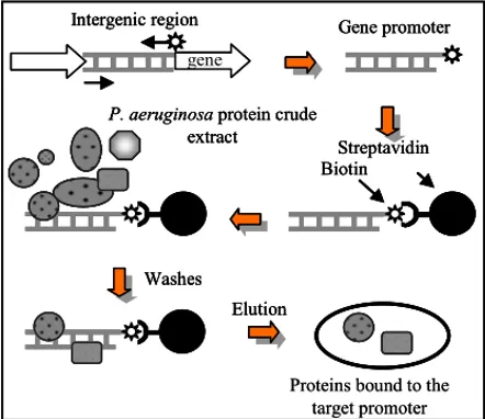
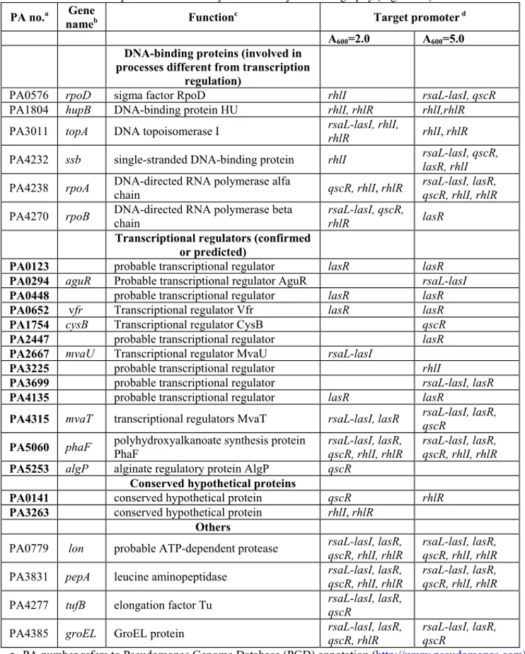
Allo scopo di individuare i regolatori trascrizionali dei geni dei geni las e rhl di P. aeruginosa, è stato usato il metodo della cromatografia per affinità al DNA (Fig. 8). Sei frammenti di DNA, corrispondenti ai promotori dei geni lasR, lasI, rsaL, qscR, rhlR e rhlI, sono stati indipendentemente coniugati a una resina cromatografica e incubati con estratti proteici crudi preparati a partire da colture di P. aeruginosa in fase di crescita esponenziale (A600= 2.0) e stazionaria (A600= 5.0). Le proteine in grado di legare in maniera specifica ogni regione promotore sono state eluite, separate su SDS-PAGE e identificate mediante spettrometria di massa MALDI-TOF (Fig. 9A-E).
In tutto sono state identificate 25 proteine, appartenenti a diverse classi funzionali (Tab. 1). Ben diciannove di queste sono classificate come fattori in grado di legare il DNA, di cui tredici sono regolatori trascrizionali (confermati o putativi) e sei sono proteine coinvolte in funzioni generiche collegate al processamento del DNA (come ad esempio le subunità della RNA polimerasi). Mentre queste ultime erano presenti su tutti i promotori usati come ligando, la maggior parte dei tredici regolatori è stata trovata essere associata ad un solo promotore, indicando che l’interazione tra proteina e promotore era specifica. È anche degno di nota il fatto che l’attivatore Vfr, già noto per legare il promotore di lasR, è stato identificato usando proprio questo promotore come ligando.
Oltre ai fattori in grado di legare il DNA, sono anche state identificate due proteine classificate come ipotetiche (a funzione sconosciuta) e quattro con funzione diversa dal legame al DNA. Si può supporre, in base alla loro funzione, che quest’ultime abbiano co-purificato con altre proteine (Tab. 1).
Le quindici proteine classificate come regolatori trascrizionali e come ipotetiche proteine sono state selezionate come possibili regolatori del QS e sono state quindi oggetto di ulteriori analisi.
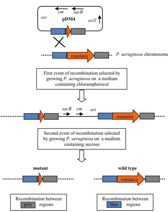
Per verificare in vivo l’effetto dei putativi regolatori identificati, sono stati creati quindici mutanti di P. aeruginosa, nei quali i geni codificanti i corrispondenti regolatori sono stati indipendentemente deleti (Fig. 10). Mediante l’uso di fusioni trascrizionali, l’attività dei promotori bersaglio è stata misurata durante l’intera curva di crescita nel “wild type” e nei corrispondenti mutanti.
Grazie a questa analisi, è stato dimostrato per la prima volta che le due proteine “histon-like” MvaT e MvaU regolano in modo diretto la trascrizione dei geni del sistema las. In particolare MvaT reprime la trascrizione di lasI e qscR e attiva quella di lasR (Fig. 14), mentre MvaU è un repressore sia di rsaL che di lasI (Fig. 16). MvaT e MvaU sono regolatori globali dell’espressione genica e, in alcuni casi, è stato riportato che possono anche interagire l’una con l’altra (Vallet et al., 2004; Castang et al., 2008; Li et al., 2009). L’interazione tra MvaT e MvaU nella regolazione dei geni del QS è quindi molto complessa e difficile da studiare anche perchè nel ceppo di P. aeruginosa preso in esame in questo studio il doppio
IV
mutante mvaT-mvaU è letale (Castang et al., 2008). Questo tema sarà oggetto di studi futuri nel nostro laboratorio.
Sorprendentemente, le mutazioni negli altri geni presi in esame in questo studio non hanno causato effetti evidenti sull’attività dei corrispondenti promotori bersaglio, almeno nelle condizioni di crescita standard nelle quali è stato eseguito il saggio di attività promotore. Una spiegazione possibile per questo risultato è che alcune di queste proteine non siano sufficientemente espresse e/o attive nelle condizioni usate, dato che il ruolo di questi regolatori è quella di modulare il QS in risposta a specifiche condizioni ambientali e metaboliche. Per verificare tale ipotesi l’attività dei promotori bersaglio è stata valutata in ceppi ricombinanti di P. aeruginosa che sovra-esprimono i putativi regolatori. Come sistema modello, sono stati presi in esame i sei fattori identificati usando come ligando il promotore del gene lasR.
I geni codificanti i sei putativi regolatori di lasR sono stati clonati in un plasmide di espressione arabinosio-inducibile, i plasmidi risultanti sono stati quindi indipendentemente introdotti in P. aeruginosa “wild type”, contenente la fusione trascrizionale per la misurazione dell’attività promotore di lasR. L’attività promotore di lasR è stata quindi misurata durante tutta la curva di crescita in presenza di arabinosio.
I risultati ottenuti mostrano che la sovra-espressione della proteina PA3699 reprime fortemente l’attività promotore di lasR, senza inibire la crescita (Fig. 17). La capacità di PA3699 di reprimere l’espressione di lasR è stata verificata e confermata anche nell’ospite eterologo Escherichia coli (Fig. 20).
La proteina PA3699 è stata purificata e la sua capacità di legare in modo specifico la regione di DNA contenente il promotore di lasR è stata dimostrata mediante un saggio di ritardo della motilità elettroforetica del DNA (Fig. 22).
Nel complesso, i risultati ottenuti dimostrano che PA3699 è un repressore diretto dell’espressione di lasR. Un aspetto rilevante di questo studio è che, sebbene la struttura cristallografica di PA3699 sia disponibile nelle banche dati dal 2009, questa è la prima volta che gli viene attribuita una funzione specifica.
Poichè la delezione del gene PA3699 non ha effetto sull’espressione di lasR nelle condizioni standard di laboratorio, è probabile che tale regolatore venga attivato da specifici stimoli ambientali e/o metabolici. Questo potrebbe essere il caso anche delle altre proteine identificate in questo studio come probabili regolatori del QS. Sono attualmente in corso studi volti all’identificazione degli stimoli ambientali e metabolici in grado di aumentare l’espressione/attività sia di PA3699 che degli altri putativi regolatori identificati.
Un obiettivo secondario di questa parte del lavoro è stato l’identificazione di inibitori del QS. Lo screening è stato effettuato usando un biosensore in grado di misurare i livelli di 3-oxo-C12-HSL. Tra i 1120 composti testati, 9 erano in grado di inibire la produzione di 3-oxo-C12-HSL in P. aeruginosa (Tab. 2). Il composto più promettente tra questi è al momento in corso di studio, per verificarne
V l’efficacia anti-Pseudomonas sia in vitro che in modelli murini d’infezione polmonare cronica.
Per quanto riguarda la parte del lavoro che ha riguardato B. cenocepacia, studi preliminari di trascrittomica avevano mostrato che i mutanti D4 e D9, inattivati rispettivamente nelle due pompe di efflusso RND-4 e RND-9, e il doppio mutante D4-D9 avevano un’espressione alterata di molti geni correlati al flagello. Dal momento che la motilità flagellare (un fenotipo chiamato “swimming”) è strettamente connessa alla virulenza e alla formazione di biofilm, è stato testato l’effetto di queste mutazioni sui fenotipi “swimming” e biofilm. I risultati ottenuti hanno mostrato che, in accordo con quanto ottenuto dai dati di trascrittomica, i mutanti D4 e D9 avevano, rispettivamente, un’aumentata e una diminuita capacità di motilità “swimming” rispetto al “wild type”. Il doppio mutante D4-D9 aveva un fenotipo simile al mutante D4 (Fig. 27). Ciò indica che l’inattivazione di diversi sistemi RND può avere effetti drammaticamente diversi su uno specifico fenotipo di virulenza. È interessante notare che tutti i mutanti mostravano una maggiore capacità di formare biofilm, rispetto al “wild type” (Fig. 28). Dal momento che la formazione di biofilm è un fenotipo complesso e pleiotropico non è possibile correlare questo risultato solo con l’espressione dei geni per il flagello.
CONCLUSIONI
I risultati ottenuti nella parte di progetto focalizzata su P. aeruginosa hanno permesso di identificare tre nuovi fattori trascrizionali responsabili della regolazione del QS, un processo fondamentale per l’espressione della virulenza in questo batterio. Inoltre, è stata identificata una molecola con spiccata attività anti-QS, quindi potenzialmente in grado di inibire la virulenza di P. aeruginosa.
Per quanto riguarda la parte di progetto che ha avuto come oggetto lo studio delle pompe d’efflusso RND, è rilevante notare che molte compagnie farmaceutiche e gruppi di ricerca stanno studiando inibitori delle pompe di efflusso (“efflux pump inhibitors”, EPIs) come adiuvanti nelle terapie antibiotiche. In questo studio è stato dimostrato, usando B. cenocepacia come sistema modello, che l’inattivazione di pompe di efflusso RND potrebbe aumentare la formazione di biofilm e, in alcuni casi, la motilità, facendo insorgere seri dubbi circa l’opportunità di usare gli EPIs nella terapia. Infatti, l’uso degli EPIs potrebbe da un lato potenziare l’effetto degli antibiotici, ma dall’altro potrebbe promuovere la formazione di biofilm e quindi l’instaurarsi dell’infezione cronica.
A
ABSTRACT
Pseudomonas aeruginosa and Burkholderia cenocepacia, beside their relevance as a nosocomial pathogens, cause lethal chronic lung infections in the vast majority of cystic fibrosis (CF) patients. Once established, the CF lung infection is impossible to eradicate with traditional antimicrobial therapies.
The main body of this project has been focused on P. aeruginosa. In this bacterium the Quorum Sensing (QS) system coordinates the production of virulence factors in a cell density-dependent manner via the secretion of specific signal molecules. During growth the bacteria secrete the signal molecules, which accumulate in the surrounding environment as the population density increases. When a threshold concentration is reached, the receptor is activated by the binding with the cognate signal molecule and triggers the expression of several genes, including virulence genes.
P. aeruginosa has two QS systems relying on the production of acylated- homoserine lactone (acyl-HSL) as signal molecules: the las and rhl systems. The lasI gene encodes the LasI synthase producing the N-3-oxo-dodecanoyl-homoserine lactone (3-oxo-C12-HSL) signal molecule, that binds to the LasR receptor encoded by lasR. The rsaL and qscR genes encode repressors of lasI and constitute an integral part of the las QS system, since they respond to 3-oxo-C12-HSL. The rhl system is organized similarly to the las one: the rhlI and rhlR genes encode, respectively, for the RhlI synthase and the RhlR receptor of the N-butyryl-homoserin lactone (C4-HSL) signal molecule. The signal-activated LasR and RhlR receptors regulate transcription of hundreds of genes, including the genes encoding a third QS system, based on the production of a different signal molecule, and the vast majority of virulence genes.
In P. aeruginosa many regulators have been found to affect the expression of the las and rhl genes, although their mechanism of action remains largely unknown. In particular, only the Vfr protein has been shown to directly activate lasR transcription. Besides las and rhl mutants, also a vfr mutant shows reduced virulence in murine models, suggesting that also the QS regulators could be feasible drug-targets.
The main objective of this project has been the identification and functional characterization of transcriptional factors that directly regulate the las and rhl genes. To achieve this objective, six DNA fragments corresponding to the promoters of lasR, lasI, rsaL, qscR, rhlR and rhlI were independently conjugated to a chromatography resin and incubated with protein crude extracts prepared from P. aeruginosa cultures. The proteins able to specifically bind each DNA bait were recovered, separated by SDS-PAGE and identified by MALDI-TOF mass spectrometry.
Overall, 25 proteins bound on the promoter regions of the QS genes were identified. Out of these, fifteen factors were selected for further analysis as possible
B
QS regulators. Noteworthy, the activator Vfr, already known to directly bind the lasR promoter, was recovered using this promoter as a bait.
A set of fifteen P. aeruginosa mutants, each one deleted in one of the genes encoding the selected proteins, was generated. In each mutant the activity of correspondent target promoter was compared with that of the wild type, by means of transcriptional fusions between the promoter region of the target genes and the reporter system lux. In this system the production of bioluminescence is proportional to the promoter activity. Among the newly identified factors, we showed for the first time that the histon-like proteins MvaT and MvaU directly control the transcription of the las genes. In particular, MvaU is a repressor of both rsaL and lasI transcription, while MvaT represses lasI and qscR transcription and activates lasR transcription. MvaT and MvaU are global regulators of gene expression and on some promoters can also interact each other. The interplay between these two proteins in regulating QS genes is a complex issue that will be the object of future studies.
Unexpectedly, mutations in the other genes here investigated did not cause evident effects on the activity of the corresponding target promoter under the tested laboratory conditions. A possible explanation for this result is that some of these factors are actually QS regulators, but they are not sufficiently expressed and/or active in the standard growth conditions we have used. To verify this hypothesis, we decided to test the activity of target promoters in recombinant P. aeruginosa strains over-expressing the putative QS regulators.
We focused on the six factors retrieved on the lasR promoter. The results showed that PA3699 over-expression strongly repressed lasR promoter activity during the whole growth curve, without affecting cell growth. The ability of PA3699 to repress lasR expression was also confirmed in the heterologous host Escherichia coli. Moreover, the PA3699 protein was purified and its capability to bind in vitro the lasR promoter region was demonstrated by electrophoretic mobility shift assay. Overall, these results show that PA3699 is a repressor of lasR expression. Since PA3699 mutation has no effect on lasR expression under standard cultural conditions, it is likely that this regulator is activated by specific environmental/metabolic stimuli. This could be the case also for the other proteins identified in this study. Environmental and metabolic stimuli increasing the expression/activity of PA3699 and of the other putative regulators identified in this project are currently under investigation.
A minor objective of this part of the project has been the screening of the Prestwick Chemical Library of FDA-approved drugs for the identification of possible secondary activities against P. aeruginosa QS. The screening was performed by using a biosensor able to detect 3-oxo-C12-HSL levels. Among the 1120 compounds tested, 9 displayed a QS-inhibitory effect. The anti-Pseudomonas activity of the most promising compound is currently under investigation in murine models of chronic lung infection.
C The second part of this project has been focused on B. cenocepacia, a member of the B. cepacia complex (Bcc). This group of strictly related bacteria is the second most important Gram negative pathogen, infecting the CF patients. Like many pathogens, including P. aeruginosa, B. cenocepacia is highly resistant to a wide range of antibiotics due to the production of drug-efflux pumps belonging to the Resistance-Nodulation-cell Division (RND) family.
It is emerging the notion that, beside exogenous toxic compounds, RND efflux pumps are involved in the secretion of bacterial products important for virulence, including factors involved in biofilm formation. The biofilm is a bacterial community encased in a self-produced extracellular matrix and confers resistance to antibiotics and to the host immune systems, playing a major role in CF lung infection. The RND efflux pumps have been mainly studied for their effect on antibiotic resistance, while little is known about their impact on cell-physiology and virulence-related phenotypes.
A preliminary transcriptomic study showed that mutants inactivated in the D4 and D9 RND- drug efflux systems of B. cenocepacia displayed altered expression of flagella-related genes. Since flagellar motility (a phenotype named swimming) is strictly connected to virulence and to biofilm formation, we tested the effect of these mutation on swimming and biofilm. Results showed that, in accordance with transcriptomic data, the D4 and D9 mutants showed increased and decreased swimming motility with respect to the wild type, respectively. Therefore the inactivation of distinct RND systems can have a dramatically different effect on a specific virulence-related phenotype.
Interestingly, both the mutants showed increased biofilm formation with respect to the wild type. Since biofilm formation is a pleiotropic and complex phenotype it is not easy to correlate this result only to flagella genes expression.
Many companies are developing Efflux Pumps Inhibitors (EPIs) as antibiotic therapy adjuvants. In this view, a relevant result of this research is that inactivation of efflux pumps can enhance biofilm formation and, sometimes, motility, raising serious concerns about the use of EPIs in therapy. Indeed, the use of EPIs could be, on one side positive for helping the antibiotic therapy, on the other side, it could promote biofilm formation and chronic infection. More detailed study on the effect of RND efflux pumps in virulence-related phenotypes and chronic infection are therefore strongly desirable.
1
1. INTRODUCTION
1.1 Cystic fibrosis
Cystic fibrosis (CF) is a recessive genetic disease caused by a mutation in a gene on the long arm of chromosome 7 that encodes for the cystic fibrosis transmembrane conductance regulator (CFTR; Welsh et al. 2001). CFTR is an ABC transporter-class ion channel that transports chloride and thiocyanate ions across epithelial cell membranes. Mutations of the CFTR gene affect functioning of the chloride ion channels in these cell membranes. CFTR is required to regulate the components of sweat, digestive juices, and mucus. The inability to regulate sodium and chloride transport due to an aberrant CFTR increases airway secretion viscosity (Welsh et al. 2001).
CF is considered one of the most common genetic diseases affecting roughly 70,000 persons worldwide. One child in approximately 2,500 of European carries two defective copies of
CFTR
(Ratjen & Doring, 2003).CF primarily affects the airways and submucosal glands with sparing of the interstitium and alveolar spaces until late in the disease (Tomashefski et al. 1989). The role of ion transport in the maintenance of lung health is complex. The recent development of a porcine model of CF has provided a further step forward our understanding of the basic mechanisms whereby the loss of CFTR function leads to parenchymal lung damage (Rogers et al. 2008). These animals developed lung disease and demonstrated abnormal bacterial clearance in the absence of significant inflammation (Stoltz et al. 2010), suggesting that infection of the airways may be the primary event that initiates the progression of CF lung disease.
1.1.1 Chronic infections in cystic fibrosis patients
In almost all patients with CF, bacteria can be isolated from airway secretions at some point in their lives. For the majority, such infections begin in childhood and become chronic by early adulthood.
In the healthy host, bacteria that enter the lungs are cleared rapidly, without the initiation of an inflammatory response. This process involves a variety of innate host defence strategies, both mechanical (mucociliary clearance) and immunological (resident macrophages and antimicrobial peptides). In certain clinical situations this first line of defence fails, and the normally harmless bacteria survive, multiply, and lead to organ damage. Such conditions are encountered in mechanically ventilated patients in intensive care, in those with defective immunity or severe burns, and in patients with CF. The mechanisms underlying the early acquisition of infection in CF are complex and incompletely understood. Briefly, hypotheses include impaired mucociliary clearance related to low airway surface liquid volume, increased availability of cell surface receptors and impaired ingestion of bacteria by epithelial cells. Once within the airway, conditions such as
2
hypoxia within mucus plugs may attract motile organisms capable of anaerobic survival and encourage biofilm formation (Davies & Bilton 2009).
CF has a unique set of bacterial pathogens that are frequently acquired in an age-dependent sequence and, among them, only Staphylococcus aureus may be pathogenic in immunocompetent individuals. Pseudomonas aeruginosa, Burkholderia cepacia, nontypeable Haemophilus influenzae, Stenotrophomonas maltophilia, and Achromobacter xylosoxidans are all considered opportunistic pathogens. Other organisms seen in CF that are also generally nonpathogenic in the healthy host include Aspergillus and nontuberculous mycobacteria.
Early infections in CF airways are most frequently caused by S. aureus and H. influenzae, organisms that may be seen in other young children with chronic illnesses and in adults with non–CF bronchiectasis. S. aureus is often the first organism cultured from the respiratory tract of young children with CF. However, there still is a continue debate about the significance of S. aureus in the pathogenesis of CF lung infection. Historically, significant improvements in patient longevity have been associated with the advent of antistaphylococcal therapy. H. influenzae is also isolated from the respiratory tract early in the course of CF. The role of H. influenzae in progressive airway infection and inflammation in patients with CF has not been clearly demonstrated, although this organism is known to be pathogenic in patients with non–CF bronchiectasis (Gibson et al. 2003).
In the CF patients P. aeruginosa plays a particularly important role: the inability to regulate sodium and chloride transport due to an aberrant CFTR increases airway secretion viscosity, and within the resulting thick mucus in the lung P. aeruginosa find a favourable niche (Lyczak et al., 2000). 80 % to 95 % of CF patients succumb because of respiratory failure brought on by chronic P. aeruginosa infection and concomitant airway inflammation. Up to 97 % of CF patients are infected with P. aeruginosa by the age of 3 years (Lyczac et al., 2002; Murray et al., 2007).
Among the organisms involved in CF airways B. cepacia is particularly relevant, besides P. aeruginosa. Although this bacterium infects a small percentage of CF patients (around 5%), the so-called “B. cepacia syndrome” leads to high fevers, bacteremia, rapid progression to severe necrotizing pneumonia and death. The majority of B. cepacia infected patients have more severe course with decline in lung function and increased mortality with respect to P. aeruginosa infection. However, B. cepacia is not a single species but rather a group of closely related species, thus the organism should be called B. cepacia complex. Seventeen distinctive species of B. cepacia have been identified. The vast majority of CF airway infections with B. cepacia complex are caused by B. cenocepacia, B. multivorans and B. vietnamiensis (Gibson et al. 2003).
3
1.2 Pseudomonas aeruginosa
P. aeruginosa is a highly adaptable bacterium that can colonize various environmental niches, including soil and marine habitats, plants, animals and humans. The completion of the sequencing project of the genome of P. aeruginosa PAO1 demonstrated that the ecological versatility is indeed reflected in its gene content, by its relatively large genome (6.3 Mbp) and genetic complexity (5570 open reading frames, ORFs), comparable to that of the simple eukaryote Saccharomyces cerevisae (Stover et al., 2000). Compared to the majority of known sequenced bacterial genomes, the genome of P. aeruginosa possesses an overall larger number of genes encoding outer membrane proteins, efflux systems and multiple chemotaxis systems, which may contribute to its pathogenesis. Moreover, up to 10 % of the assigned ORFs are classified as transcriptional regulators, reflecting the ability of P. aeruginosa to respond and adapt to environmental fluctuations (Stover et al., 2000; Goodman & Lory, 2004).
Besides being the major cause of death in CF patients (see above), P. aeruginosa is an opportunistic human pathogen that may cause acute infections in hospitalized patients and immuno-compromised individuals, for example those with AIDS, neutropenic patients undergoing chemotherapy, and burn victims (Driscoll et al., 2007).
Infections caused by P. aeruginosa are not only very frequent, but they are also associated with high morbidity and mortality rates when compared with infections caused by other bacterial pathogens (Osmon et al., 2004). This is mainly due to the fact that P. aeruginosa infections are hard to eradicate because this microorganism is intrinsically resistant to many antibacterials, including β-lactams, macrolides, tetracyclines, co-trimoxazole and most fluoroquinolones and it is particularly prone to acquire new resistances in the hospital environment by horizontal gene transfer (Latifi et al., 1995). Also the P. aeruginosa tendency to colonize surfaces in organized communities, termed biofilms, makes P. aeruginosa less susceptible to antibiotics and disinfectants, anchors the cells to the surface they colonize (for instance to the medical devices) and protects the bacteria from the host defences, such as lymphocytes, phagocytes, antibodies and the ciliary action of the respiratory tract (Greenberg, 2003; Hentzer et al., 2004). These characteristics make P. aeruginosa a particularly dangerous pathogen.
1.2.1 P. aeruginosa virulence
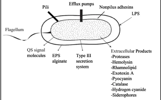
The capacity of P. aeruginosa to produce such diverse infections, is due to an arsenal of virulence factors. These factors are collectively capable of causing extensive tissue damages, bloodstream invasion and dissemination in humans and other mammals (Fig. 1; reviewed by Smith & Iglewski, 2003).
4
Flagellum
QS signal molecules EPS
alginate Type III secretion system
LPS Pili Efflux pumps Nonpilus adhesins
Extracellular Products -Proteases -Hemolysin -Rhamnolipid -Exotoxin A -Pyocyanin -Catalase -Hydrogen cyanide -Siderophores Flagellum QS signal molecules EPS
alginate Type III secretion system
LPS Pili Efflux pumps Nonpilus adhesins
Extracellular Products -Proteases -Hemolysin -Rhamnolipid -Exotoxin A -Pyocyanin -Catalase -Hydrogen cyanide -Siderophores
Figure 1. Virulence and antibacterial resistance factors in P. aeruginosa (modified from Van Delden &
Iglewski, 1999).
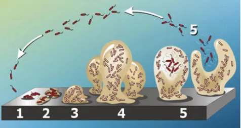
Some of the virulence factors that confer to the pathogen the ability to colonize the host are cell surface virulence factors Actually the cell surface itself is a virulence factor because it contains very immunogenic compounds, like the lipopolysaccharide (LPS). Flagella and pili involved in different kind of motility and in chemotaxis, display a critical role in pathogenesis, by adhering to epithelial cells and stimulating an inflammatory response (DiMango et al., 1995; Adamo et al., 2004). When P. aeruginosa chronically infects patients, it adapts to the biofilm mode of growth. The generally accepted definition of a biofilm is “a community of cells attached to a surface or to each other, imbedded in a self-made, protective matrix of extracellular polymeric substances (EPS)” (Fig. 1; Kirisits & Parsek, 2006).
The clinical implications of bacterial biofilms are particularly pronounced (Davies, 2002). Biofilms may form on any foreign object inserted into the human body, and also in the lungs of CF patients P. aeruginosa forms biofilm during chronic infection. In the biofilm mode the bacteria are highly tolerant to the action of several antimicrobial agents including antibiotics, disinfectants, and to the action of the immune system (Donlan & Costerton, 2002; Drenkard, 2003).
5
Figure 2. Schematic representation of different stages of biofilm formation. In the colonization stage
planktonic cells adhere to the substrate (1) and start to proliferate forming microcolonies (2). During the maturation stage the microcolonies grow (3) forming three-dimensional structures known as “mushrooms” (4). Finally, bacterial cells leave the mushrooms leading to biofilm dispersion (5; modified from Kirisits & Parsek, 2006).
P. aeruginosa also produces several extracellular products that, after the initial step of colonization, cause extensive tissue damage, bloodstream invasion and dissemination. Some of these extracellular products, besides having the role of favoring pathogen dissemination, also provide nutrients to P. aeruginosa by causing host tissues damage. These secreted factors are elastases, alkaline proteases, exotoxins and hemolysins that also contribute to the infection in lung disease by destroying the protective glicocalix of the respiratory epithelium (Kipnis et al., 2006). Other secreted factors are toxic compounds such as hydrogen cyanide and pyocyanin: hydrogen cyanide is a potent poison that blocks cytochrome oxidase, leading to the inhibition of mitochondrial respiration (Gallagher & Manoil, 2001). Pyocyanin is a blue pigment metabolite of P. aeruginosa that has been shown to have numerous pathogenic effects such as increasing IL-8, depressing host-response, and inducing apoptosis in neutrophils (Denning et al., 1998; Leidal et al., 2001; Allen et al., 2005). In animal models of acute and chronic lung infection, pyocyanin was shown to be essential for P. aeruginosa virulence (Lau et al., 2004). Additionally, due to its known oxidoreductive properties, pyocyanin oxidizes glutathione and inactivates catalase in respiratory epithelial cells thus participating in oxidative-stress related damage (O’Malley et al., 2003; O’Malley et al., 2004).
The request of iron in P. aeruginosa is supported by the production of the two siderophores, pyoverdine and pyochelin, that are small molecules chelating iron from the iron-poor environment encountered in the host, allowing its utilization in P. aeruginosa metabolism (Buckling et al., 2007).
6
1.2.2 Quorum sensing
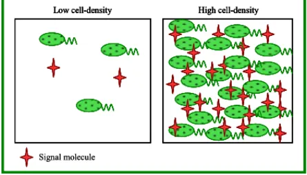
For many years, researchers thought of bacteria as individual cells designed to proliferate under various conditions but unable to interact with each other and to collectively respond to environmental stimuli, as it is typical for multicellular organisms. This view began to change few decades ago with the discovery of the cooperative regulation of luminescence in the Gram-negative marine bacterium Vibrio fischeri (Nealson et al., 1970) and regulation of the genetic competence in the Gram-positive bacterium Streptococcus pneumoniae (Tomasz, 1965). These bacteria were show to coordinate their behavior via the secretion of specific signaling molecules in a population density-dependent manner. During growth the bacteria secrete these signal molecules that accumulate in the surrounding environment as the population density increases until a critical threshold concentration is reached, which then triggers expression of certain sets of genes. This type of cell-to-cell communication was termed “quorum sensing” (QS) in order to emphasize the fact that a sufficient number of bacteria, the bacterial “quorum”, is needed to induce or repress expression of target genes (Fig. 3; Fuqua et al., 1994; Whitehead et al., 2001).
QS has been proven to play an important role in the physiology of various bacterial species. Beside bioluminescence and genetic competence, QS is also involved in the regulation of a wide variety of different physiological processes including antibiotic biosynthesis, motility, plasmid conjugal transfer, biofilm formation, and the production of bacterial virulence factors in plant, animal and human pathogens (Miller & Bassler, 2001; Whitehead et al., 2001; Camara et al., 2002; Fuqua & Greenberg, 2002; Lazdunski et al., 2004).
Furthermore, evidence has accumulated that some bacterial signal molecules are used not only as population density sensors within the same species but also for communication between bacteria of different species or genera occupying the same ecological niche, and even to interact with their eukaryotic hosts.
In Gram-negative bacteria the most common signal molecule used is an acylated homoserine lactone (acyl-HSL) molecule and, in some cases, a single bacterium possesses multiple acyl-HSL signal molecules.
The first acyl-HSL-based QS system that has been studied is the Lux system of V. fisheri. It consists of a synthase, LuxI (encoded by luxI gene), that produces the 3-oxohexanoyl-homoserine lactone (3-oxo-C6-HSL) signal molecule and a signal-receptor protein LuxR (encoded by luxR gene) which acts as transcriptional regulator (Nealson et al., 1970).
Since then, a wide number of LuxI-like and LuxR-like proteins have been shown to be involved in acyl-HSL mediated QS systems in a number of Gram-negative bacteria (Whitehead et al., 2001).
7
Low cell-density High cell-density
Signal molecule
Low cell-density High cell-density
Low cell-density High cell-density
Low cell-density High cell-density
Signal molecule
Figure 3. A simple representation of QS. The signal molecule is constitutively produced at a basal level
by the bacterial cells. At low-cell densities very little signal molecule is present. As cell density increases, the signal molecule accumulates until a threshold level is reached. This signal molecule concentration is responsible for a coordinate transcriptome reprogramming in the whole bacterial population.
1.2.3 The quorum sensing system of P. aeruginosa PAO1
One of the most extensively studied acyl-HSL-dependent cell-to-cell communication systems is the one of P. aeruginosa.
P. aeruginosa shows three QS systems, two of which are acyl-HSL-dependent systems.
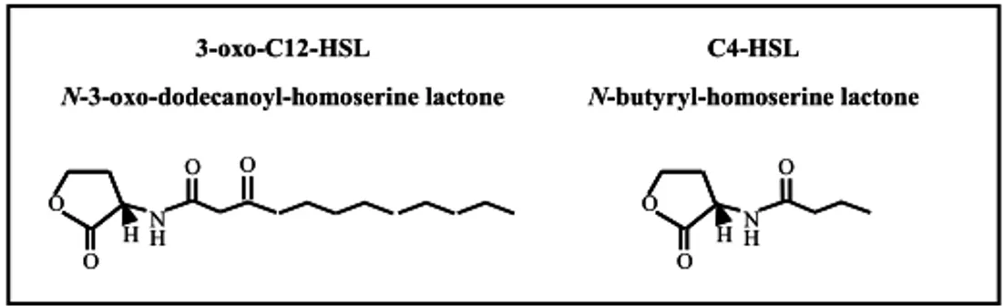
The first is the las system and consists of the LuxR-like transcriptional regulator LasR (codified by lasR gene) and of the LuxI-like acyl-HSL synthase LasI (codified by lasI gene), which directs the synthesis of N-3-oxododecanoyl-homoserine lactone (3-oxo-C12-HSL; Fig.4; Schuster & Greenberg, 2006 ).
At low-cell density the 3-oxo-C12-HSL molecule is synthesized by LasI at basal levels and is secreted into the surrounding media where it become diluted. In this phase, no QS-response occurs (Pearson et al., 1999). With the increasing of cell density, the signal molecule accumulates, until its concentration reaches a threshold level. At this critical concentration, 3-oxo-C12-HSL binds the cognate QS-activator LasR (Fuqua et al., 1996). The LasR/3-oxo-C12-HSL complex increases lasI transcription, triggering a positive feedback loop that leads to the amplification of 3-oxo-C12-HSL production (Seed et al., 1995). Moreover, activated LasR exerts its role of global transcriptional regulator drastically reprogramming the P. aeruginosa PAO1 transcriptome.
Among the genes regulated by the LasR/3-oxo-C12-HSL complex there is the second acyl-HSL system: the rhl system, that is organized similarly to the las one. The rhl QS-system is based on the production of N-butyryl-homoserine lactone (C4-HSL; Fig.4) by the LuxI-like synthase RhlI, codified by the rhlI gene. Also in this case the synthase gene has a cognate LuxR-like transcriptional regulator, RhlR, codified by the rhlR gene, that is activated by the binding with the cognate
8
signal molecule C4-HSL (Latifi et al., 1996; Pesci et al., 1997). As schematized in figure 5, the rhl system, being dependent on LasR/3-oxo-C12-HSL complex for its activation, is hierarchically below the las system.
N-3-oxo-dodecanoyl-homoserine lactone 3-oxo-C12-HSL C4-HSL N-butyryl-homoserine lactone O O H O H N O O H O H N O N-3-oxo-dodecanoyl-homoserine lactone 3-oxo-C12-HSL C4-HSL N-butyryl-homoserine lactone O O H O H N O O H O H N O
Figure 4. Structure of acyl-HSL molecules exploited by P. aeruginosa for cell-to-cell communication
(modified from Juhas et al., 2005).
LasR and RhlR transcription regulators bind to specific lux-box-like sequences, called las-boxes in P. aeruginosa (Whiteley et al., 1999; Schuster et al., 2004). LasR forms a dimer and binds las-boxes only in the presence of 3-oxo-C12-HSL (Kiratisin et al., 2002), whereas RhlR dimerizes and binds DNA both in the presence and absence of C4-HSL, but it is active only as RhlR/C4-HSL complex (Medina et al., 2003; Ventre et al., 2003).
The third QS-response involves another chemical signal, distinct from the above mentioned acyl-HSLs. This signal, 2-heptyl-3-hydroxy-4-quinolone (PQS), adds a further level of complexity to the QS network, providing another link between the las and rhl QS-systems (Diggle et al., 2003).
Briefly, the phnAB operon is required for the conversion of 2-amino-2-deoxy-isochorismic acid to anthranilic acid, which is subsequently converted to 2-heptyl-4-quinolone (HHQ) by the enzymes encoded by the pqsABCD genes, belonging to the pqsABCDE operon. The role of the last gene of this operon, pqsE, is not jet known. (Deziel et al., 2004; Lepine et al., 2004). HHQ is then converted to PQS by the product of the pqsH gene. Recent experiments showed that the transcription of pqsABCDE operon is negatively regulated by the rhl system and positively regulated by the las system (Diggle et al., 2003). Pesci and co-workers demonstrated that the effects of LasR and RhlR on the pqsABCDE operon occur indirectly through the control of the transcriptional regulator PqsR (Pesci et al., 1999). It was demonstrated that pqsR transcription is under the positive control of LasR and under the negative control of RhlR. PqsR in turn has a positive effect on the transcription of pqsABCDE, directly binding to the promoter of this operon (Wade et al., 2005). Thus the production of the PQS signal molecule is dependent on the ratio 3-oxo-C12-HSL/C4-HSL (McGrath et al., 2004). This study also demonstrated that PqsR binding to the pqsABCDE promoter is augmented by the presence of PQS, implying that PQS acts as a coinducer of PqsR (Wade et al.,
9 2005). Moreover PqsR is a global regulator controlling the expression of several genes of P. aeruginosa (Fig. 5).
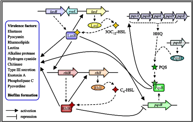
RhlI RhlR 3OC12-HSL C4-HSL LasI activation repression lasI lasR LasR rhlI rhlR Virulence factors: Elastases Pyocyanin Rhamnolipids Lectins Alkaline protease Hydrogen cyanide Chitinase Type III secretion Exotoxin A Phospholipase C Pyoverdine Biofilm formation pqsH pqsA pqsB pqsC pqsD pqsE HHQ PqsH PQS pqsR PqsR rsaL RhlI RhlR 3OC12-HSL C4-HSL LasI activation repression lasI lasR LasR rhlI rhlR Virulence factors: Elastases Pyocyanin Rhamnolipids Lectins Alkaline protease Hydrogen cyanide Chitinase Type III secretion Exotoxin A Phospholipase C Pyoverdine Biofilm formation pqsH pqsA pqsB pqsC pqsD pqsE HHQ PqsH PQS pqsR PqsR rsaL RhlI RhlR 3OC12-HSL C4-HSL LasI activation repression lasI lasR LasR rhlI rhlR Virulence factors: Elastases Pyocyanin Rhamnolipids Lectins Alkaline protease Hydrogen cyanide Chitinase Type III secretion Exotoxin A Phospholipase C Pyoverdine Biofilm formation pqsH pqsA pqsB pqsC pqsD pqsE HHQ PqsH PQS pqsR PqsR rsaL RhlI RhlR 3OC12-HSL C4-HSL LasI activation repression lasI lasR LasR rhlI rhlR Virulence factors: Elastases Pyocyanin Rhamnolipids Lectins Alkaline protease Hydrogen cyanide Chitinase Type III secretion Exotoxin A Phospholipase C Pyoverdine Biofilm formation pqsH pqsA pqsB pqsC pqsD pqsE HHQ PqsH PQS pqsR PqsR rsaL
Figure 5. Schematic representation of the central core of the QS network in P. aeruginosa, with las, rhl,
and pqs systems. Relevant QS-regulated virulence factors are listed (modified from Camara et al., 2002 and Diggle et al., 2003).
The two regulators RsaL and QscR are considered an integral part of the las QS system, since they respond to 3-oxo-C12-HSL signal molecule.
• RsaL
In P. aeruginosa lasR and lasI genes are separated by a 367 bp intergenic region, and are transcribed from independent promoters in the same direction (Seed et al., 1995). In this intergenic region, Iglewski and co-workers in late 90’s, identified a gene, rsaL, located on the opposite strand to lasR and lasI (Fig. 5). This gene encodes a protein, RsaL, of 80 aminoacids and 9.4 kDa (de Kievit et al., 1999).
RsaL represses lasI transcription by direct binding to the lasI promoter (Rampioni et al., 2006) and it is a member of the tetrahelical superclass of helix-turn-helix proteins (Rampioni et al., 2007a).
Most recent studies highlighted the major role played by RsaL in QS regulation and P. aeruginosa physiology. When P. aeruginosa is cultured in complex media, levels of 3-oxo-C12-HSL attain a steady state level (homeostasis) in the range of a few μM when the cells are still in the exponential phase, despite the fact that expression of the 3-oxo-C12-HSL synthase gene (lasI) is regulated by a positive
10
feedback through the LasR/3-oxo-C12-HSL complex. This implies the existence of a negative regulatory element balancing the positive feedback exerted by LasR/3-oxo-C12-HSL. The level of 3-oxo-C12-HSL in a rsaL mutant culture continues to rise in late logarithmic and stationary phase to a level about 10-fold the level reached in a wild type culture. This result demonstrated that RsaL is the regulatory element responsible of 3-oxo-C12-HSL homeostasis (Rampioni et al., 2007b).
When the quorum concentration of 3-oxo-C12-HSL is reached, the binding of the LasR/3-oxo-C12-HSL complex at a unique site located in the rsaL-lasI intergenic region triggers the divergent transcription of both genes. Consequently, the levels of 3-oxo-C12-HSL and of RsaL increase. The binding of RsaL to a unique site on the intergenic region simultaneously represses the transcription of rsaL and lasI. This generates a negative feedback loop, which counteracts the positive feedback loop mediated by LasR/3-oxo-C12-HSL (Rampioni et al., 2007b).
Homeostasis would allow a population of P. aeruginosa cells to maintain 3-oxo-C12-HSL at an appropriate level in a given environment and to change steady-state levels of 3-oxo-C12-HSL as environmental conditions change. However, other mechanisms can play roles in controlling 3-oxo-C12-HSL homeostasis, including for instance, signal-molecule-degrading enzymes and import-export pumps (Köhler et al., 2001; Aendekerk et al., 2002; Huang et al., 2006; Sio et al., 2006).
Furthermore, transcription profiling revealed that RsaL regulates 130 genes independent of its effect on QS signal molecule production, including genes involved in virulence. RsaL can repress pyocyanin and hydrogen cyanide virulence genes in two ways: directly, by binding to their promoters, and indirectly, by decreasing levels of the signals for their QS-signal-dependent transcription. These findings highlight the importance of RsaL as a global regulator of P. aeruginosa physiology that provides a counterbalance to 3-oxo-C12-HSL-dependent gene activation via multiple mechanisms (Rampioni et al., 2007b).
• QscR
The genome of P. aeruginosa revealed the presence of a LuxR-like factor, QscR, codified by the qscR gene. Differently from the homologous lasR and rhlR genes, this member of the LuxR family is not genetically linked to a gene encoding for a cognate acyl-HSL synthase. Therefore QscR is classified as an “orphan” QS signal receptor. In a P. aeruginosa qscR mutant the lasI and rhlI genes are prematurely transcribed, producing an increment in the expression of some QS controlled virulence genes, such as those involved in the production of pyocyanin, elastase and hydrogen cyanide. Indeed, the P. aeruginosa qscR mutant is hypervirulent (Chugani et al., 2001). Several possible mechanisms had been proposed to explain the role of QscR in QS regulation.
At very low concentrations of acyl-HSLs, inactive heterodimers can be formed between QscR and LasR or QscR and RhlR; while increased acyl-HSL concentrations result in QscR-acyl-HSL interaction inducing dissociation of QscR
11 and relasing LasR and/or RhlR (Ledgham et al., 2003). The current model is therefore that QscR inhibits LasR and RhlR at low concentrations of acyl-HSL through formation of inactive heterodimers with LasR and/or RhlR, thus inhibiting the expression of QS-regulated genes (reviewed by Venturi, 2006).
In accordance with the fact that QscR protein was shown to contain both putative acyl-HSL and DNA-binding domains (Chugani et al., 2001), it was recently demonstrated that the 3-oxo-C12-HSL-QscR complex can bind the promoter region of PA1897 gene, indicating that it can directly regulate gene transcription . To exert this function, QscR borrows the signal produced by LasI (Lee et al., 2006b).
Comparison of the transcriptional profiles of a qscR null mutant and the wild type strain at different stages of culture growth, revealed that more than 400 genes were differentially expressed, many of which were repressed, while a smaller set was activated by QscR (Lequette et al., 2006). In the same work, it was also demonstrated that a QscR protein lacking the DNA-binding domain is still able to regulate the majority of these genes, but not all of them. These data on one hand confirm that main role of QscR is that of inhibiting the activity of LasR and/or RhlR, on the other hand provide evidence that QscR can also directly modulate P. aeruginosa transcriptome, and suggest that QscR controls its own regulon, which only in part overlaps with the LasR and RhlR ones.
Another significant finding regarding QscR is that this regulator was demonstrated to have broader signal specificity than LasR does. Indeed, QscR can respond to other acyl-HSL molecules. This finding suggests that QscR might also respond to signals produced by other bacteria coexisting with P. aeruginosa (Lee et al., 2006b). This information is clinically relevant, since the lungs of the CF patients are often co-colonized by P. aeruginosa and by the C8-HSL-producing bacterium B. cenocepacia (Govan & Deretic, 1996).
1.2.4 Quorum sensing and virulence
In the whole, the QS-circuit regulate about 10 % of all the P. aeruginosa PAO1 genome, and has a key role in the infection process regulating the production of many virulence factors and the formation of biofilm (Fig. 7; reviewed in Kirisits & Parsek, 2006 and in Schuster & Greenberg, 2006).
P. aeruginosa strains carrying mutation in the QS genes were less virulent in several mouse models, including acute pneumonia and chronic lung infection models. Similarly, attenuation of virulence was also observed using alternative infection models like C. elegans, A. thaliana, D. melanogaster and Dictyostelium discoideum (reviewed in Smith & Iglewski, 2003, Juhas et al., 2005 and Diggle et al., 2006).
Functional QS systems have been found to be important for establishment of P. aeruginosa infection and for its reduced clearance in animal models of virulence. If the animals are infected with QS mutants, the immune response is faster, the polymorphonuclear leukocytes respond by developing stronger oxidative bursts
12
and antibodies accumulate faster in the infected lung (Rasmussen & Givskov, 2006). Moreover, 3-oxo-C12-HSL also interacts directly with the host organism; indeed it stimulates interferon-γ and IL-8 production, inhibits interleukin-12 and tumour necrosis factor α, promotes immunoglobulin-E production, and causes apoptosis in macrophages and neutrophils. In this respect, the QS signal molecules can be considered as virulence factors themselves (Wagner et al., 2006).
The above data strongly suggest that P. aeruginosa QS plays a primary role during infection. As a matter of fact, acyl-HSLs and PQS can be detected at biologically-significant concentrations in sputum, bronchoalveolar and mucopurulent fluids from CF patients and in other clinical samples (Singh et al., 2000; Collier et al., 2002; Middleton et al., 2002; Diggle et al., 2006).
The P. aeruginosa QS system regulates functions that strongly affect the structural and chemical-physical features of the biofilm, including the production of rhamnolipids, the secretion of DNA, swarming motility and the pyoverdine-mediated uptake of iron. Moreover, QS mutants have defects in biofilm structure and produce biofilms that are more susceptible to detergents and antibiotics (Kirisits & Parsek, 2006).
Therefore, the ultimate role of the P. aeruginosa QS system is to coordinate the expression of virulence factors and biofilm production at the level of the whole bacterial population.
In summary, the QS system is important in both acute and chronic infections. In the first case it is likely that QS enable P. aeruginosa to overcome host defence mechanisms, since isolated production of extracellular virulence factors by a small number of bacteria would probably lead to an efficient host response, neutralizing these compounds. In the chronic infection, the role of QS is strictly linked with the biofilm mode of growth. In both cases, the QS signal molecules participate to the cross-talk between host and parasite enhancing the fitness of the P. aeruginosa community in the host environment.
1.2.5 Quorum sensing regulation in P. aeruginosa
As previously mentioned, P. aeruginosa is an organism with complex genome. This genetic complexity is highlighted by the presence of a large proportion of regulatory genes, which make up almost 9 % of the total number of ORFs, further highlighting metabolic plasticity and capability of adaptation to several environments. Several recent studies have shown that QS in Pseudomonas is integrated with certain aspects of cell physiology and that it responds to other environmental signals, beside cell density. Production of QS signal molecules and virulence factors must be finely controlled and timed to achieve a productive and persistent infection. This is also stressed by the overlap of the acyl-HSL QS regulon with other regulons controlled by global regulators, providing a high degree of interconnectivity among different signalling networks. The integration of QS into additional regulatory circuits increases the range of environmental and
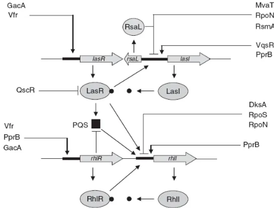
13 metabolic signals that affect QS gene expression beyond cell density as well as further tuning the timing of the QS response. At least 17 gene products that have an impact on the production of QS signal molecules have been identified. These regulators are important in modulating the production of signal molecules and virulence factors likely in response to stimuli deriving from the environment (e.g. from the host during infection; Fig. 6; reviewed in Venturi, 2006). Most of these QS regulators have been identified by genetic analysis and only in few cases their mechanism of action is known.
Figure 6. Regulation of the las and rhl quorum-sensing systems in P. aeruginosa. The las system is at
the top of the hierarchy, regulating the rhl system and PQS production. Arrows on the promoter regions of the indicated genes mean positive regulation, whereas a short parallel line indicates negative regulation (Venturi, 2006).
• Vfr
The QS and cyclic AMP (cAMP) signalling networks are linked via the transcriptional regulator Vfr, a member of the cAMP receptor protein (CRP) family. Vfr was first identified in P. aeruginosa as a virulence factor regulator, due to its positive effect on the production of several virulence factors, such as proteases and exotoxin A. This effect is probably mediated by LasR, since Vfr positively regulates transcription of lasR by directly binding to a CRP consensus sequence present in the lasR promoter region (Albus et al., 1997). Also the rhlR promoter has been shown to be positively regulated by Vfr, but it is not known
14
whether Vfr directly activates the rhlR promoter through a Vfr-binding site or if it acts indirectly via other regulators (Medina et al., 2003).
As mentioned above, Vfr is homologous to the cAMP receptor protein family and contains a cAMP binding domain. However, differently from Escherichia coli and other enterobacteriaceae, cAMP levels in pseudomonads are not related to glucose uptake and utilization (West et al., 1994). Therefore the role of cAMP in Pseudomonas, as well as the environmental stimuli to which Vfr respond, are still far to be understood.
• MvaT
The MvaT protein belongs to the H-NS family (histon-like nucleoid structuring). The proteins of this family are involved in processes of compacting chromosomal DNA, working as global gene expression regulators in Gram-negative bacteria (Dorman, 2004). The structure of these proteins includes two domains, with a flexible linker between them. The N-terminal domain is involved in the formation of oligomers, while the C-terminal domain is involved in the DNA-binding (Dorman et al., 1999; Tendeng & Bertin, 2003).
In P. aeruginosa MvaT regulates the expression of genes involved in virulence, in arginine metabolism, in antibiotic resistance and in biofilm formation, through direct binding to the promoters or through indirect regulation (Diggle et al., 2002; Vallet et al., 2004; Westfall et al., 2004 and 2006; Li et al., 2009).
Interestingly a mvaT mutant was shown to over-produce 3-oxo-C12-HSL and C4-HSL signal molecules and to regulate some QS-dependent virulence phenotypes (Diggle et al., 2002).
1.3 Burkholderia cepacia complex
Burkholderia cepacia was described in 1950 as Pseudomonas cepacia, the
phytopathogen responsible for the sour skin disease in onions (Burkholder, 1950). Molecular taxonomic analysis of this bacterium and closely related species, recovered from diverse environments, including pathogens of plants, animals, and humans, led to their inclusion into the new genus Burkholderia (Yabuuchi et al.,1992), now comprising over 50 species. Since then, impressive advances have been achieved in the taxonomy of B. cepacia and related species, now collectively known as the B. cepacia complex (Bcc), comprising of at least 17 different but closely related species, phenotypically similar: non-fermenting, aerobic, Gram-negative β-proteobacteria (Coenye et al., 2001). The genome of these strains consists of three chromosomes and many of them contain also plasmids. The progress in the taxonomy of these bacteria has been mainly due to their emergence as important opportunistic pathogens, capable of causing life-threatening infections in immunocompromised patients, in patients with chronic granulomatous disease, and especially in 2 to 8% of CF patients (Gibson et al., 2003).
15 Although strains from all the Bcc species are capable of causing infections to CF patients, their prevalence varies geographically. For example, while B. multivorans is predominant in Europe, B. cenocepacia predominates in North America (Govan
et al., 2007).
The B. cenocepacia epidemic ET12 lineage that originated in Canada and spread to Europe has been one of the most prevalent Bcc genotypes isolated from CF patients, with strain J2315 being studied in depth as model strain (Foweraker, 2009). The genome of B. cenocepacia J2315, a multidrug-resistant CF isolate was recently published (Holden et al., 2009).
The 8.06-Mb genome of this highly transmissible pathogen, consisting of three circular chromosomes and a plasmid, encodes a broad array of functions typical of metabolically versatile genus Burkholderia, as well as several virulence and drug resistance functions (Holden et al., 2009).
In CF patients, antibiotics are used to clear early infection, treat acute exacerbations of chronic infection and reduce their relapse frequency. Despite the heavy use of antibiotics in CF, over the last decades, B. cepacia complex has emerged as an important respiratory pathogen in the CF community. After colonization with a Bcc strain, few patients experience an asymptomatic carriage, while the majority experiences an increased decline of pulmonary function, associated with chronic infection and exacerbation episodes. Dramatically, a significant percentage of the Bcc-infected patients will develop a rapid and fatal necrotizing pneumonia known as the cepacia syndrome (Isles et al., 1984).
1.3.1 B. cepacia complex virulence
Compared to the advances achieved in the taxonomy, knowledge on the molecular mechanisms underlying Bcc pathogenicity and progress on the development of new therapeutic agents are still limited.
In order to successfully establish an infection, after entering into the respiratory tract of the CF patient, bacteria have to adhere to host mucosal or epithelial surfaces. In the case of the CF lung, the thickened mucus layer provides an ideal environment for microbial colonization, due to defective mucus clearance, reduced efficacy of antimicrobial peptides, and enhanced inflammatory response (Boucher 2007). The ability to cross the epithelial barrier and gain access to the blood stream seems to be restricted to Bcc strains, as other CF pathogens usually do not cause bacteremia.
During the interaction with the CF host, several virulence factors are thought to play critical roles for the success of the pathogen, although their precise contribution to the overall Bcc pathogenicity remains to be thoroughly elucidated.
Extracellular lipase, metalloproteases and serine proteases are thought to play roles directly related to the interaction with epithelial cells (McClean & Callaghan 2009).
Bacterial surface structures like the lipopolysaccharide (LPS), flagella and pili are also important in the interaction with the CF host. Flagella are required for
16
many biological processes, for example motility, production of biofilms, adherence and invasion into host cells (Mahenthiralingam et al., 2005; Moens & Vanderleyden, 1996; O’Toole & Kolter 1998). Indeed B. cepacia complex bacteria are motile and they possess one or longer polar flagella responsible for swimming motility. Flagella represent one of the virulence factors which contribute to the development of disease caused by these bacteria as shown by in vivo data (Urban et al., 2004). They have been described as a major factor contributing to host inflammatory responses to bacteria due to the interaction of bacterial flagellin with the Toll-like receptor 5 (TLR5) (Hayashi et al., 2001; Liaudet et al., 2003). The production and assembly of these multi-component structures involve more than 40 genes. In particular, members of the Bcc express one of the two types of flagellin that can be distinguished by size: 55 kDa for type I and 45 kDa for type II (Hales et al., 1998).
The production of siderophores such as pyochelin, salicylic acid, cepabactin, and ornibactin, also contribute to Bcc pathogenesis (Agnoli et al., 2006).
Protein secretion is also an important mechanism by which bacteria are able to deliver proteins to the environment and to host cells, being able to influence the host response and being crucial for virulence and survival. Several transport systems have been implicated in the secretion of many virulence factors by Bcc strains such as proteases, hemolysins, and adhesins, among others. Type I and type II secretion systems were shown to be responsible for the secretion of proteins with hemolytic activity in isolates of the B. cenocepacia J2315 (Whitby et al., 2006).
Another important feature of Bcc is their ability to form biofilms, communities within which bacteria live in a sessile lifestyle, protected from environmental insults and aggression from the immune system defences of the host. In addition, Bcc bacteria in biofilms have been demonstrated to be more resistant to antibiotics than planktonic cells, contributing to their persistence in the CF lung (Caraher et al., 2007). The characterization of the chemical structure and composition of the EPS showed that it is composed of glucose, mannose, rhamnose, galactose, and glucuronic acid (Cescutti et al., 2000).
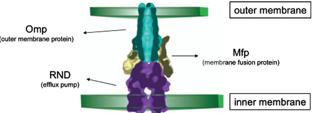
1.3.2 The RND efflux transporters in B. cepacia complex
B. cenocepacia and other members of the Bcc demonstrate high-levels of intrinsic resistance to most clinically relevant antibiotics, complicating the treatment of the infection (Waters & Ratjen, 2006). In CF isolates, multi-drug resistance (MDR) is defined as resistance to all of the agents belonging to at least two of three classes of antibiotics, such as quinolones, aminoglycosides, and β-lactam agents, including monobactams and carbapenems (Saiman & Siegel, 2003). Particularly interesting among mediators of MDR in Gram-negative bacteria are transporters belonging to the RND (Resistance-Nodulation-Cell Division) family, whose members catalyze the active efflux of many antibiotics and chemotherapeutic agents (Nikaido & Takatsuka, 2009).