Edited by:
Adrian Preda,
University of California, Irvine, USA
Reviewed by:
Anthony John Hannan,
University of Melbourne, Australia
Omar Cauli,
University of Valencia, Spain
Francis Bambico,
University of Toronto, Canada
*Correspondence:
Silvana Gaetani
[email protected]
Specialty section:
This article was submitted to
Neuropharmacology,
a section of the journal
Frontiers in Neuroscience
Received: 29 May 2015
Accepted: 14 December 2015
Published: 12 January 2016
Citation:
Romano A, Tempesta B, Micioni Di
Bonaventura MV and Gaetani S
(2016) From Autism to Eating
Disorders and More: The Role of
Oxytocin in Neuropsychiatric
Disorders. Front. Neurosci. 9:497.
doi: 10.3389/fnins.2015.00497
From Autism to Eating Disorders and
More: The Role of Oxytocin in
Neuropsychiatric Disorders
Adele Romano
1
, Bianca Tempesta
1
, Maria Vittoria Micioni Di Bonaventura
2
and
Silvana Gaetani
1
*
1Department of Physiology and Pharmacology “Vittorio Erspamer”, Sapienza University of Rome, Rome, Italy,
2Pharmacology Unit, School of Pharmacy, University of Camerino, Camerino, Italy
Oxytocin (oxy) is a pituitary neuropeptide hormone synthesized from the paraventricular
and supraoptic nuclei within the hypothalamus. Like other neuropeptides, oxy can
modulate a wide range of neurotransmitter and neuromodulator activities. Additionally,
through the neurohypophysis, oxy is secreted into the systemic circulation to act
as a hormone, thereby influencing several body functions. Oxy plays a pivotal role
in parturition, milk let-down and maternal behavior and has been demonstrated to
be important in the formation of pair bonding between mother and infants as well
as in mating pairs. Furthermore, oxy has been proven to play a key role in the
regulation of several behaviors associated with neuropsychiatric disorders, including
social interactions, social memory response to social stimuli, decision-making in the
context of social interactions, feeding behavior, emotional reactivity, etc. An increasing
body of evidence suggests that deregulations of the oxytocinergic system might be
involved in the pathophysiology of certain neuropsychiatric disorders such as autism,
eating disorders, schizophrenia, mood, and anxiety disorders. The potential use of
oxy in these mental health disorders is attracting growing interest since numerous
beneficial properties are ascribed to this neuropeptide. The present manuscript will
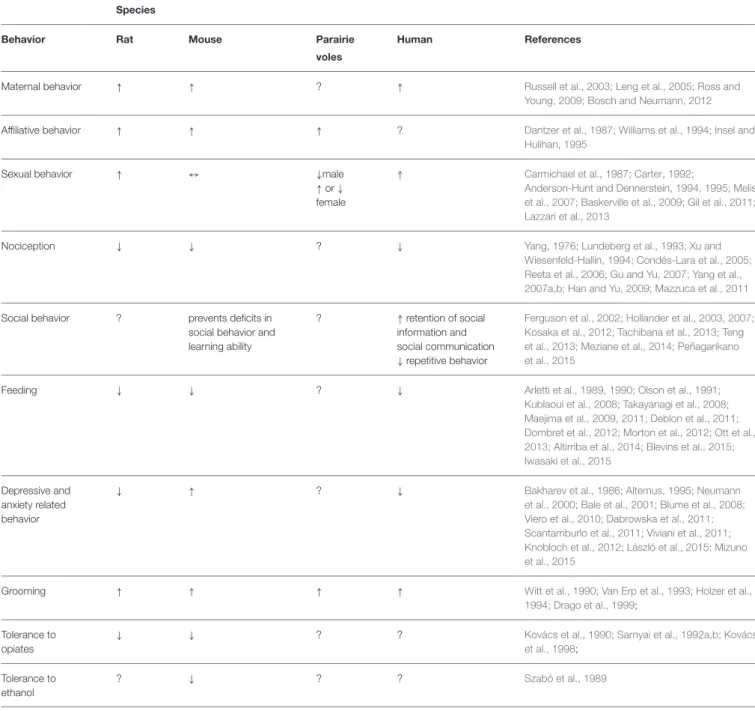
review the existing findings on the role played by oxy in a variety of distinct physiological
and behavioral functions (Figure 1) and on its role and impact in different psychiatric
disorders. The aim of this review is to highlight the need of further investigations on this
target that might contribute to the development of novel more efficacious therapies.
Keywords: oxytocinergic system, autism, eating disorders, anxiety, mood disorders
INTRODUCTION
History and Structure of Oxytocin
The neuropeptide oxytocin (oxy), was discovered in 1906 by Henry Dale, who observed that
extracts from human posterior pituitary gland were able to induce uterus contractions in a pregnant
cat; the name oxy, derives from the Greek words “ωκνξ τoκoxξ,” meaning “swift birth.”
In 1984, Ivell and Richter elucidated the structure of the oxy gene (
Ivell and Richter, 1984
),
and in 1992 the sequence of the oxy receptor (oxyr) was reported (
Kimura et al., 1992
). Oxy is a
nonapeptides with a disulfide bridge between Cys residues 1 and 6 that creates a six-amino acid
cyclic structure and a COOH-terminal α-amidated three-residue tail.
FIGURE 1 | Oxytocin regulatory control of different and complex
processes.
Synthesis and Localization
Oxy is synthesized as a preprohormone precursor protein that
includes the oxy carrier protein neurophysin I (
Brownstein
et al., 1980
). Although neurophysin is apparently devoid of any
biologically activity, different observations suggests that it might
exert a protective role against oxy enzymatic damage and it has
been extensively studied for its involvement in the regulation of
oxy neurosecretory pathways (
Legros and Geenen, 1996; de Bree,
2000
). The inactive preprohormone protein is hydrolyzed by a
variety of enzymes in small fragments and the last reaction that
generates oxy is catalyzed by a peptidylglycine alpha-amidating
monooxygenase (
Brownstein et al., 1980; Burbach et al., 2001;
von Eggelkraut-Gottanka and Beck-Sickinger, 2004
). The oxy
gene, located on chromosome two in mice, three in rats, and 20
in humans (
Dutil et al., 2001
), is composed of three exons, each
of them encoding for a particular portion of the peptide (
Gimpl
and Fahrenholz, 2001
).
The Oxytocinergic System
Hypothalamus-Hypophysis
The
hypothalamic-neurohypophysial
system
represents
the major oxy neurosecretory system and consists of the
paraventricular (PVN) and supraoptic (SON) nuclei (
Swanson
and Kuypers, 1980; Rhodes et al., 1981
) and their axons reaching
the neurohypophysis. However, the neurons of PVN and SON
project extensively also to other brain areas such as the arcuate
nucleus (Arc), the median eminence (ME), the lateral septum
(LS) and the medial amygdala nucleus (MeA;
Pittman et al.,
1981
). Within the PVN, two populations of oxy neurons have
been identified: “magnocellular” and “parvocellular” neurons.
Oxy is mainly synthesized in the magnocellular portions of the
PVN and SON (
Swaab et al., 1975
). Oxytocinergic magnocellular
neurons terminate in the posterior lobe of the pituitary gland and
also innervate the Arc, the LS, the MeA, and the ME (
Pittman
et al., 1981
). Once activated, magnocellular oxytocinergic
neurons, release oxy from the nerve terminals to the posterior
pituitary; from here oxy is secreted into the bloodstream,
so that it can produce its effect on organs expressing oxyrs
located in the rest of the body (
Gimpl and Fahrenholz, 2001
).
The release of oxy from the neurohypophysis into the blood
stream is not accompanied by an increase of the peptide at the
central level, thus indicating that oxy does not readily cross the
blood brain barrier (BBB). In accordance with this observation,
peripheral stimulation of oxytocinergic system such as during
milk suckling or vaginal dilatation may not change oxy levels in
the central nervous system (CSF;
Gimpl and Fahrenholz, 2001
).
Moreover, while stimulation of the PVN evokes oxy release at
both central and pheripheral level, electrical stimulation of the
rat neurohypophysis only evokes oxy release into the blood
(
Harris et al., 1981
); moreover oxy circulating levels are reduced
after hypophysectomy in rats while its concentration increases
in the CSF (
Dogterom et al., 1977
). The oxy half-life is 28 min in
the CSF, compared to 1–2 min into the blood; in the CSF, oxy is
normally present at concentrations of 10–50 pM, slightly higher
than those in plasma (
Jones and Robinson, 1982; Meyer et al.,
1987
). Moreover, oxy release has been shown to occur not only
at the axonal levels but also locally from dendrites in both the
PVN and SON (
Neumann et al., 1996
).
Furthermore, oxy can act as an intrinsic self neuromodulator;
oxy release within the SON is pivotal for the coordinated
depolarization of oxy neurons during lactation and for the
positive feedback mechanism mediated by oxyrs on its own
release during parturition (
Neumann et al., 1996
).
The
parvocellular
neurons,
that
are
smaller
than
magnocellular neurons, are located in the dorsal-caudal
portion of the PVN and terminate principally to the nucleus
of solitary tract (NST), the dorsal motor nucleus of the vagus
(DMNV), the rostral ventrolateral medulla, and the sympathetic
centers in the spinal cord (
Amico et al., 1990; Rinaman, 1998;
Tóth et al., 1999
).
Finally, it has been proposed that oxy may participate in
the physiological regulation of the adenohypophysial hormones
prolactin adrenocorticotropic hormone (
Page et al., 1990
), and
gonadotropins (
Robinson and Evans, 1990
). This hypothesis
is supported by evidence demonstrating that hypothalamic
oxytocinergic fibers reach also the anterior part of the pituitary
gland via the hypophyseal portal system (
Amar and Weiss, 2003
).
Oxytocin Receptor
The oxy receptor (oxyr) is a 389 aminoacid polypeptide
belonging to the G-protein coupled receptor family with seven
transmembrane domains. Specifically this receptor is coupled to
a Gq/11α protein that stimulates the activity of phospholipase
C. This leads to the generation of second messengers, release of
Ca
2+
from the intracellular storages, and activation of protein
kinase type C. These two conditions can trigger several cellular
events such as the contraction of smooth cells, cellular excitation,
and modification of gene expressions (
Gimpl and Fahrenholz,
2001
).
The oxyr has been identified not only in the brain but also in
several peripheral organs. In the rat CNS, oxyrs are present in
several regions, including the olfactory system, cortex, thalamus,
basal ganglia, ventromedial region of the hypothalamus, bed
nucleus of the stria terminalis, central amygdala, ventral