SCHOOL OF MEDICINE
Department of Translational Medicine
PhD Program in Sciences and Medical Biotechnology
XXXII CYCLE
BIOLOGICAL AND CLINICAL IMPLICATIONS
OF BIRC3 MUTATIONS
IN CHRONIC LYMPHOCYTIC LEUKEMIA
Tutor: Coordinator:
Chiar.mo Prof. Gianluca Gaidano Prof.ssa Marisa Gariglio
Candidate: Dr Chiara Favini
Matricula: 20022483
2
INDEX
SUMMARY ... 3
SOMMARIO ... 4
1. INTRODUCTION ... 5
2. AIM OF THE STUDY ... 7
3. MATERIALS AND METHODS ... 8
3.1. Patients ... 8
3.2. Cancer personalized profiling by deep sequencing (CAPP-seq) ... 8
3.3. Bioinformatic pipeline for variant calling after CAPP-seq ... 9
3.4. Statistical analysis ... 11
3.5. Cell studies ... 12
3.6. Western blot analysis ... 12
3.7. RNA extraction and gene expression profiling ... 13
3.8. Knockdown of MAP3K14 by RNA interference ... 13
3.9. Inhibitor studies ... 14
3.10. In vitro drug responses in primary CLL cells ... 14
3.11. Apoptosis assay ... 15
4. RESULTS ... 16
4.1 Patients harboring BIRC3 mutations are at risk of failing FCR ... 16
4.2 BIRC3 mutations associate with activation of non-canonical NF-κB signaling ... 17
4.3 BIRC3 mutations confer resistance to fludarabine in primary CLL cells ... 19
5. DISCUSSION ... 20 6. REFERENCES... 22 7. TABLES ... 28 8. FIGURES LEGENDS ... 30 8. FIGURES ... 33 9. SUPPLEMENTARY TABLES ... 39
3
SUMMARY
The current shift of therapy of chronic lymphocytic leukemia (CLL) towards novel targeted agents mandates the identification of molecular predictors to inform on who can still benefit from chemoimmunotherapy and who can be instead early considered for novel targeted agents. Fludarabine, cyclophosphamide, and rituximab (FCR) is the most effective chemoimmunotherapy regimen for the management of CLL and represents the current standard of care for young and fit patients devoid of TP53 disruption. A retrospective multicenter cohort of 287 untreated patients receiving first-line FCR was analyzed by targeted next generation sequencing of 24 recurrently mutated genes in CLL. By univariate analysis adjusted for multiple comparisons BIRC3 mutations identify a poor prognostic subgroup of patients failing FCR (median progression free survival: 2.2 years, p < 0.001) similar to cases harboring TP53 mutations (median progression free survival: 2.6 years, p < 0.0001). BIRC3 mutations maintained an independent association with an increased risk of progression with a hazard ratio of 2.8 (95% confidence interval 1.4-5.6, p = 0.004) in multivariate analysis adjusted for TP53 mutation, 17p deletion and IGHV mutation status. The functional implications of BIRC3 mutations are largely unexplored and little is known about the prognostic impact of BIRC3 mutations in CLL cohorts homogeneously treated with first line FCR. By immunoblotting analysis, we showed that the non-canonical NF-κB pathway is active in BIRC3 mutated cell lines and in primary CLL samples, as documented by the stabilization of MAP3K14 and by the nuclear localization of p52. In addition, BIRC3 mutated primary CLL cells are less sensitive to fludarabine. If validated, BIRC3 mutations may be used as a new molecular predictor to select high-risk patients for novel frontline therapeutic approaches.
4
SOMMARIO
Le terapie innovative per la leucemia linfatica cronica (CLL) includono nuovi agenti i quali richiedono l'identificazione di predittori molecolari per determinare i pazienti che possono ancora beneficiare della chemio-immunoterapia e coloro che, invece, necessitano di trattamento con nuovi farmaci. La terapia di prima linea per pazienti giovani, in buone condizioni cliniche e privi di aberrazioni del gene TP53, prevede l’utilizzo dello schema immunochemioterapico FCR, la combinazione dei farmaci Fludarabina, Ciclofosfamide e Rituximab. Il DNA genomico di 287 pazienti, trattati con terapia secondo lo schema FCR, è stato raccolto alla diagnosi e sottoposto al sequenziamento di 24 geni ricorrentemente mutati nella CLL, mediante tecnica di Next Generation Sequencing (NGS). In analisi univariata, le mutazioni di BIRC3 identificano un sottogruppo di pazienti con prognosi sfavorevole (sopravvivenza mediana libera da progressione: 2,2 anni, p <0,001) simile ai casi che presentano mutazioni di TP53 (sopravvivenza mediana libera da progressione: 2,6 anni, p <0,0001). Le mutazioni di BIRC3 rimangono associate ad un maggior rischio di progressione (HR) in analisi multivariata corretta per mutazione di TP53, delezione 17p e stato mutazionale di IGHV: HR 2,8 (95% I.C. 1,4-5,6, p = 0,004). Le implicazioni funzionali delle mutazioni di BIRC3 sono in gran parte inesplorate e poco si conosce circa il loro impatto prognostico in coorti di pazienti trattati omogeneamente con FCR. Mediante analisi di immunoblotting è stato dimostrato che la via non canonica di NF-κB è attiva sia nelle linee cellulari BIRC3 mutate che nelle cellule primarie, come documentato dalla stabilizzazione di MAP3K14 e dalla localizzazione nucleare di p52. Inoltre, le cellule primarie BIRC3 mutate sono meno sensibili al trattamento con Fludarabina. Se validate, le mutazioni di BIRC3 potrebbero essere utilizzate come nuovo predittore molecolare per selezionare pazienti ad alto rischio per nuovi approcci terapeutici in prima linea.
5
1. INTRODUCTION
Chronic lymphocytic leukemia (CLL) is the most common type of adult leukemia in the western world with marked genetic and clinical variability.1 The clinical course of CLL ranges from a very indolent condition,
with a nearly normal life expectancy, to rapidly progressive leading to early death.2 Fludarabine,
cyclophosphamide, and rituximab (FCR) is the most effective chemoimmunotherapy regimen for the management of CLL, and represents the current standard for untreated patients who are young and in good physical condition3,4 except for patients with TP53 alterations.5 Though the majority of CLL patients
receiving FCR as frontline therapy are destined to relapse, a subgroup of cases may experience a durable first remission. The current shift of therapy of CLL towards novel targeted agents mandates the recognition of molecular predictors to identify patients who can still benefit from chemoimmunotherapy and those who should instead be considered for novel targeted agents upfront. In the case of FCR, the immunoglobulin heavy chain genes (IGHV) mutational status and Fluorescence In Situ Hybridization (FISH) karyotype stratify: i) low-risk patients carrying mutated IGHV genes and devoid of both del11q and del17p who maximally benefit from such treatment; ii) intermediate-risk patients harboring unmutated IGHV genes and/or del11q in the absence of del17p who are a case mix of good and poor responders to FCR; iii) high-risk patients harboring del17p who are unsuitable for chemoimmunotherapy.3 Deletion of 17p and
TP53 mutations capture most, routinely analyzed in clinical practice, but not all patients who are refractory
to chemo-immunotherapy, which prompts the identification of additional biomarkers associated with early failure of FCR.3-5 B-cell neoplasia often pirates signaling pathways by molecular lesions to promote survival
and proliferation. Though according to bioinformatics criteria BIRC3 (also known as cIAP2) is one of the candidate driver genes of CLL, the functional implications of BIRC3 mutations are partially unexplored.7-9
6 Nuclear factor-κB (NF-κB) signaling is a key component CLL development and evolution.10 Two NF-κB
pathways exist namely, canonical and noncanonical.11 The former is triggered by the B-cell receptor (BCR)
signaling via the Bruton’s tyrosine kinase (BTK), while the latter is activated by members of the tumor necrosis factor (TNF) cytokine family.12 Upon receptor binding, the TRAF3/MAP3K14-TRAF2/BIRC3 negative
regulatory complex of non-canonical NF-κB signaling is disrupted, MAP3K14 (also known as NIK), the central activating kinase of the pathway, is released and activated to induce the phosphorylation and proteasomal processing of p100, thereby leading to the formation of p52-containing NF-κB dimers. The p52 protein dimerizes with RelB to translocate into the nucleus, where it regulates gene transcription. BIRC3 is a negative regulator of non-canonical NF-κB. Physiologically, BIRC3 catalyzes MAP3K14 protein ubiquitination in a manner that is dependent on the E3 ubiquitinine ligase activity of its C-terminal RING domain. MAP3K14 ubiquitination results into its proteasomal degradation.13 Also, little is known about the prognostic impact
7
2. AIM OF THE STUDY
We aimed at refining the genetic-based stratification of FCR-treated CLL patients. The aims of this study are:
• To identify molecular predictors in FCR treated patients;
8
3. MATERIALS AND METHODS
3.1. Patients
The study was designed as a retrospective observational analysis from a multicenter cohort of 287 (275 with complete clinical and molecular data) untreated CLL receiving first-line therapy with FCR in 17 different hematological centers. The following biological material was collected: i) 280 tumor genomic DNA (gDNA) and 7 tumor RNA isolated from peripheral blood (PB) before treatment start; and ii) paired germline gDNA from saliva from 14 cases. Normal gDNA from 22 healthy donors was also used to set the experimental background of the deep next generation sequencing (NGS) approach. Tumor and normal gDNA was extracted according to standard procedures.14 Tumor RNA was extracted according to the TRIzol Reagent
protocol (Life Technologies). The clinical database was updated in April 2018. Patients provided informed consent in accordance with local Institutional Review Board requirements and the Declaration of Helsinki. The study was approved by the Ethical Committee of the Ospedale Maggiore della Carità di Novara associated with the Amedeo Avogadro University of Eastern Piedmont (study number CE 67/14).
3.2. Cancer personalized profiling by deep sequencing (CAPP-seq)
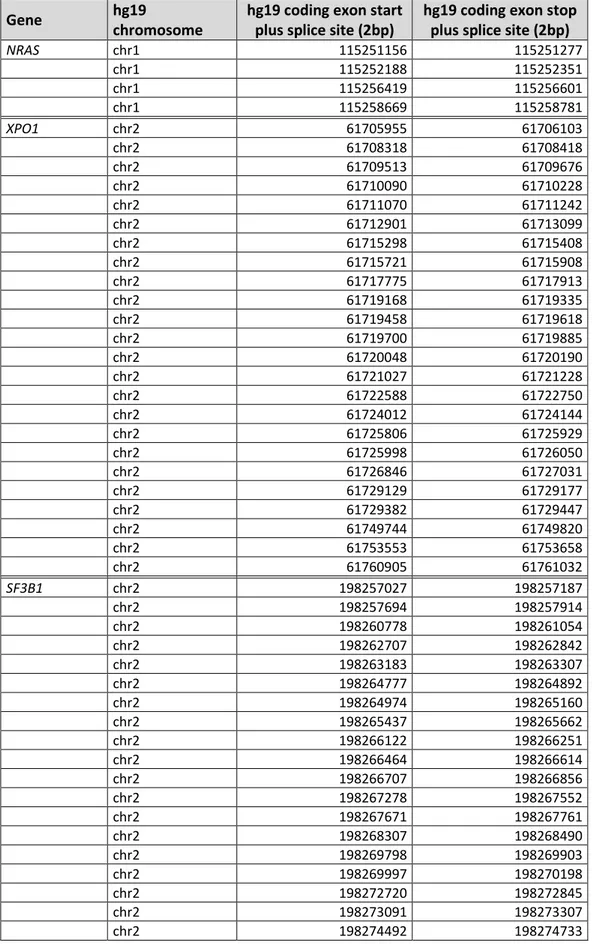
A targeted resequencing gene panel15 was designed to include: i) coding exons plus splice site of 24
CLL genes known to be implicated in CLL pathogenesis and/or prognosis; ii) 3’UTR of NOTCH1; and iii) enhancer and promoter region of PAX5 (size of the target region: 66627bp) (Table S1).8,9 Tumor and
germline gDNA were quantified using the Quant-iTTM PicoGreen dsDNA Assay kit (ThermoFisher Scientific)
and 400 ng were sheared through sonication (Covaris M220 focused-ultrasonicator) before library construction to obtain 200-bp fragments. The size of the DNA fragments was checked using the Bioanalyzer
9 (Agilent Technologies). The NGS libraries for gDNA were constructed using the KAPA Library Preparation Kit (Kapa Biosystems) and NGS libraries for RNA were constructed using RNA Hyper Kit (Roche) following the manufacturer’s instructions. Hybrid selection was performed with the custom SeqCap EZ Choice Library (Roche NimbleGen). Multiplexed libraries (n = 10 per run) were sequenced using 300-bp paired-end runs on a MiSeq sequencer (Illumina) to obtain a coverage of at least 2000x in >90% of the target region (66627bp) in 80% of cases (Table S2).
3.3. Bioinformatic pipeline for variant calling after CAPP-seq
Initially, FASTQ sequencing reads were deduped. We deduped FASTQ sequencing reads from gDNA by utilizing the FastUniq v1.1 software, that collapses as duplicate reads only those fragments (read pairs) with 100% sequence identity that also share genomic coordinates. The same approach was also used to dedupe germline gDNA and normal gDNA from 22 healthy donors, to avoid the introduction of biases in variant calling due to the application of different deduplication protocols. Then, the deduped FASTQ sequencing reads were locally aligned to the hg19 version of the human genome assembly using the BWA v.0.6.2 software with the default setting, and sorted, indexed and assembled into a mpileup file using SAMtools v.1. The aligned read families were processed with mpileup using the parameters -A -d 10000000. For cases provided with paired germline gDNA, single nucleotide variations and indels were called in tumor gDNA vs germline gDNA, respectively, with the somatic function of VarScan2 using the parameters min-coverage 1 --min-coverage-normal 1 --min-coverage-tumor 1 --min-var-freq 0--min-freq-for-hom 0.75 --somatic-p-value 0.05 --min-avg-qual 20 --strand-filter 1 --validation 1. For cases lacking paired germline gDNA, single nucleotide variations and indels were called in tumor gDNA using the CNS function of VarScan2 using the
10 parameters min-coverage 0 min-readge 2 min-avg-qual 20 min-var-freq 0 min-freq-for-hom 0.75 --p-value 0.05 --strand-filter 1 --output-vcf 1 --variants 0. The variants called by VarScan 2 were annotated using the SeattleSeq Annotation 138 tool by using the default setting. Variants annotated as SNPs according to dbSNP 138 (with the exception of TP53 variants that were manually curated and scored as SNPs according to the IARC TP53 database), intronic variants mapping > 2 bp before the start or after the end of coding exons, and synonymous variants were then filtered out. The following strict post-processing filters were then applied to the remaining variants to further improve variant call confidence. To filter out variants below the base-pair resolution background frequencies in gDNA across the selector, for cases provided with paired germline gDNA, the Fisher's exact test was used to test whether the frequency of the variant called by VarScan 2 was significantly higher from that called in the corresponding paired germline gDNA, after adjusting for multiple comparisons by Bonferroni test [multiple comparisons corrected p threshold = 0.00000018761163, corresponding to alpha of 0.05/(66627 x 4 alleles per position]. Accordingly, variants represented in > 10 reads of the paired germline and/or variants with a somatic p value from VarScan2 > 0.00000018761163 were no further considered. To filter out systemic sequencing errors, a database containing all germline and normal gDNA background allele frequencies was assembled. Based on the assumption that all background allele fractions follow a normal distribution, for both cases provided with paired germline gDNA and cases lacking paired gDNA, a Z-test was employed to test whether a given variant in the tumor gDNA differed significantly in its frequency from typical germline or normal gDNA background at the same position in all the other germline and normal gDNA samples, after adjusting for multiple comparisons by Bonferroni test [multiple comparisons corrected p threshold = 0.00000018761163, corresponding to alpha of 0.05/(66627 x 4 alleles per position]. Variants that did not pass this filter were no
11 further considered. Variant allele frequencies for the resulting candidate mutations and the background error rate were visualized using IGV.
3.4. Statistical analysis
Progression free survival (PFS) was the primary endpoint and was measured from date of treatment start to date of progression according to IWCLL-NCI guidelines (event), death (event) or last follow-up (censoring). Overall survival (OS) was measured from date of initial presentation to date of death from any cause (event) or last follow-up (censoring). Survival analysis was performed by Kaplan-Meier method and compared between strata using the Log-rank test. A false discovery rate approach was used to account for multiple testing, and adjusted p-values were calculated using the Bonferroni correction. A maximally selected rank statistic was used to determine the optimal cut-off for variant allele frequency (VAF) based on the Log-rank statistics. A cut-off of 3% of VAF was set for TP53 mutations and of 10% for all the other genes. The adjusted association between exposure variables and PFS was estimated by Cox regression. Internal validation of the multivariate analysis was performed using a bootstrap approach to estimate means and confidence intervals of hazard ratios (HR), and percentage of selection for each variable in the model. The number of bootstrap samples used was 1000. Statistical significance was defined as p value < 0.05. The analysis was performed with the Statistical Package for the Social Sciences (SPSS) software v.24.0 (Chicago, IL), with R statistical package 3.1.2 and with GraphPad version 7 (GraphPad Software Inc).
12
3.5. Cell studies
The human CLL cell line MEC1, the SMZL cell lines SSK41, VL51, and the MCL cell lines MAVER-1, Z-138 and JEKO-1 were cultured under standard conditions in RPMI-1640 medium with L-glutamine supplemented with 10% fetal calf serum (FCS), Penicillin (100 U/ml) and Streptomycin (100 U/ml) (Sigma Aldrich). Human HEK-293T cells were maintained in Iscove’s Modified Dulbecco Medium (IMDM) supplemented with 10% fetal calf serum, 100 U/ml penicillin, 100 U/ml streptomycin and 2mM L-glutamine (Sigma Aldrich) under identical conditions.
Three primary cells samples known to harbor heterozygous inactivating mutations of BIRC3 were included in the experiments. Two BIRC3 wild type cases were used as controls.
3.6. Western blot analysis
The entire non-canonical NF-κB pathway was assessed using the following specific primary antibodies: anti-BIRC3 (Cell Signaling, #3130), anti-TRAF2 (Cell Signaling, #4712), anti-TRAF3 (Cell Signaling, #4729), anti-MAP3K14 (Cell Signaling, #4994), anti-Phospho-NF-κB2 p100 (Cell Signaling, #4810), anti-NF-κB2 p100/p52 (Cell Signaling, #4882). Anti--actin (Sigma Aldrich, #A2066) was used as loading control. The
Qproteome Nuclear Protein Kit (Qiagen) was used according to the manufacturer’s instructions to isolate nuclear proteins from cells. Anti-β-tubulin (Sigma Aldrich, #T5201) and anti-BRG1 (G-7) (Santa Cruz Biotechnology, #17796) were used as controls for the purity of the cytoplasmic and nuclear fractions, respectively. Horseradish peroxidase-conjugated goat anti-mouse COR, #926-80010) or anti-rabbit (LI-COR, #926-80011) antibodies were used to highlight binding by enhanced chemiluminescence with the
13 Clarity Western ECL Substrate (Biorad). Image acquisition and densitometric analyses were performed using the Molecular Imager Gel Doc XR System and the Quantity One software (Biorad).
3.7. RNA extraction and gene expression profiling
Total RNA was extracted from exponentially growing cell lines by TRIzol reagent (Life Technologies), and retro-transcribed using the Reverse Transcription Kit (Applied Biosystems). Quantitative real-time PCR (qRT-PCR) was conducted with the Step One Plus apparatus (Step One software 2.0; Applied Biosystems) using commercially available TaqMan Gene expression assays (TNFAIP3: Hs00234713_m1; NFKB2: Hs00174517_m1; NFKBIA: Hs00153283_m1; NFKBIE: Hs00234431_m1; PLEK: Hs00950975_m1; WNT10: Hs00228741_m1; IL2RG: Hs00953624_m1; RELB: Hs00232389_m1; MALT1: Hs01120052_m1) (BIRC3: Hs00985031_g1) (Life Technologies). Reactions were done in triplicate from the same cDNA (technical replicates). The comparative CT method (ΔΔCT) was used to calculate relative expression levels of the gene under analysis, using GAPDH (Hs03929097_g1) as internal references.
3.8. Knockdown of MAP3K14 by RNA interference
Lentiviruses expressing 3 short hairpin RNAs (shRNAs) targeting MAP3K14, as well as the scrambled shRNA, were produced and cloned into the BamHI/HindIII cloning sites of the pGFP-C-shLenti vectors (OriGene Technologies). Within the 5’-LTR and 3’-LTR regions, each pGFP-C-shLenti vector contains an shRNA expression cassette driven by an U6 promoter, a puromycin resistance marker driven by a SV40 promoter and a GFP driven by a CMV promoter. The shRNA expression cassette consists of 29 bp target-gene-specific sequence, a 7 bp loop, and another 29 bp reverse complementary sequence, followed by a
14 TTTTTT termination sequence. The HEK293T cell line was co-transfected with expression (3 different pGFP-C-MAP3K14-shLenti or pGFP-C-non-effective-shLenti) vectors and adjuvant vectors (pMDL, REV and VSV-G). Fluorescence microscope was utilized to check the expression of the GFP in the transfected HEK293T cell line. After virus titration, the VL51 cell line was infected with lentiviruses harboring the shRNAs against MAP3K14 and the scrambled through a spinoculation protocol. After four days, infected cells were monitored by flow cytometry for the expression of the GFP and were selected by puromycin (1.5 µg/mL). Cell viability was monitored by Tripan blue counting.
3.9. Inhibitor studies
Cells were put under starvation in RPMI 0.1% Fetal Bovine Serum (FBS) 24h before treatment. Then they were seeded at 8000 cells per well in a 96-well U-bottom plate and treated with 1 µM, 5 µM and 10 µM of Ibrutinib (PCI-32765, Selleckchem) or vehicle (DMSO). Relative growth was determined by a Cell-Titer Glo (CTG) Luminescent Cell Viability Assay (Promega) 72h and 96h after treatment, according to the manufacturer’s instructions and luminescence was quantified using a Victor X (PerkinElmer) multilabel reader. Treatments were done in triplicate (biological replicates).
3.10. In vitro drug responses in primary CLL cells
Leukemic cells were purified using Ficoll-Hypaque (Sigma Aldrich) from PB of CLL patients. Staining with CD19 and CD5 confirmed that in all samples leukemic cells were >90%. Patients were then divided into BIRC3 mutated (MUT) or wild-type (WT). TP53 mutated samples (and BIRC3 WT) were selected as positive
15 reagents from Sigma) at a density of 5x10⁶/ml and both dose- and time-dependent responses were analyzed. Specifically, CLL cells were exposed to fludarabine) for 24-48 hours. Fludarabine was used at 1-5-10-25 µM and venetoclax at 5-10-50-100-500-2000 nM.
3.11. Apoptosis assay
Drug-induced apoptosis was measured using the eBioscience™ Annexin V Apoptosis Detection Kit APC (ThermoFisher) following the manufacturer’s instruction. Data were acquired using a FACSCanto II cytofluorimeter (BD Biosciences) and processed with DIVA v6.1.3 and FlowJo Version 9.01 (TreeStar). Apoptosis assays were analyzed using the two-way ANOVA test.
16
4. RESULTS
4.1 Patients harboring BIRC3 mutations are at risk of failing FCR
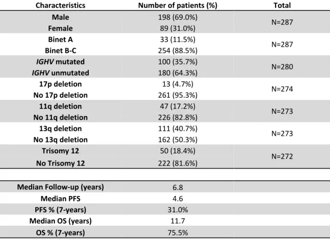
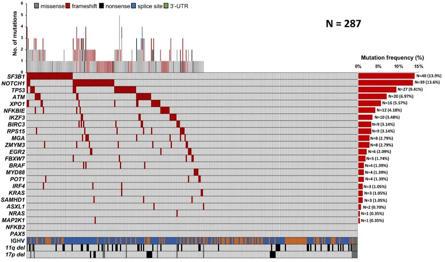
Mutational profiling was performed in 287 patients who received first line FCR. The baseline features of the study cohort were consistent with progressive, previously untreated CLL (Table 1). The median follow-up was 6.8 years, with a median PFS and OS of 4.6 and 11.7 years, respectively (Table 1) consistent with clinical trial cohorts.16 As expected, SF3B1 and NOTCH1 were the most frequently mutated genes
identified in 13.9% and in 13.6% of patients respectively, followed by TP53 in 9.4% and ATM in 6.9% of patients, reflecting the data reported in previous studies.8,9,17 Overall, 154/287 (53.6%) cases harbored at
least one non-synonymous somatic mutation in one of the 24 CLL genes included in our panel (range: 1-5 mutation per patient), which is consistent with the typical mutational spectrum of the coding genome of CLL requiring first line treatment. (Figure 1; Table S3).8,9,18 Outside of the coding genome, we identified one
single mutation in the 3’ region of NOTCH1 (c.*378A>G) already reported.8
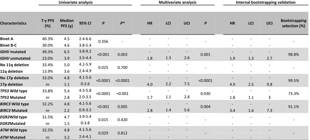
By univariate analysis adjusted for multiple comparisons, among the genes analyzed in our panel, only TP53 mutations (median PFS of 2.6 years; p < 0.0001) and BIRC3 mutations (median PFS of 2.2 years; p <
0.001) (Figure 2 A) associated with significantly shorter PFS (Table 2). The PFS after FCR of BIRC3 mutated patients was similar to that of cases harboring TP53 disruption (Figure 2 B). Consistently, BIRC3 mutated patients had a lower likelihood of achieving complete response (22.2%) at the end of FCR compared to BIRC3 wild type cases (76.7%; p=0.001). Well known molecular prognostic biomarkers of CLL, such as
unmutated IGHV gene status and 17p deletion also associated with a significantly shorter PFS, supporting the representativeness of the study cohort (Table 2). By multivariate analysis including variables showing a
17 multiplicity adjusted significant association with PFS, BIRC3 mutations maintained an independent association with PFS, with a HR of 2.8 (95% C.I. 1.4-5.6, p = 0.004) (Table 2).
4.2 BIRC3 mutations associate with activation of non-canonical NF-κB signaling
In order to comprehensively map unique BIRC3 mutations in CLL, we compiled somatically confirmed variants identified in the current CLL study cohort with those identified in previous studies17 or listed in
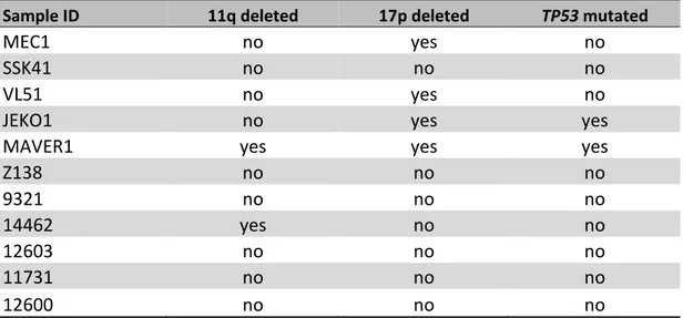
public CLL mutation catalogues (Figure 3 A). Virtually all BIRC3 mutations were represented by frameshift or stop codons clustering in two hotspot regions comprised between amino acid 367-438 and amino acid 537-564. BIRC3 variants were predicted to generate aberrant truncated transcripts causing the elimination or truncation of the C-terminal RING domain of the BIRC3 protein. The RING domain of BIRC3 harbors the E3 ubiquitin ligase activity that is essential for proteasomal degradation of MAP3K14, the central activating kinase of the noncanonical NF-κB signaling. This observation points to non-canonical NF-κB activation through MAP3K14 stabilization as the predicted functional consequence of BIRC3 mutations in CLL. The non-canonical NF-κB signaling was profiled by immunoblotting in B-cell tumor cell lines and primary CLL cells with different genetic make-up in the non-canonical NF-κB pathway to verify whether BIRC3 mutations lead to constitutive non-canonical NF-κB activation. Additional genetic features of the above mentioned cell lines and primary CLL cells are shown in Table S4. In the VL51 SMZL cell line and in the MEC1 CLL cell lines, both harboring endogenous truncating mutations of the BIRC3 gene, non-canonical NF-κB signaling was constitutively active, as documented by the stabilization of MAP3K14, phosphorylation of NF-κB2, its
processing from p100 to p52, as well as nuclear localization of p52 (Figure 3 B-D). Consistent with the biochemical clues of non-canonical NF-κB activation, the gene expression signature of the VL51 and MEC1
18 cell lines was significantly enriched of non-canonical κB target genes (Figure 3 E-F). Non-canonical NF-κB signaling in BIRC3 mutated cells was consistent with that of MCL cell lines known to harbor a disrupted TRAF3/MAP3K14-TRAF2/BIRC3 negative regulatory complex by loss of TRAF3 or TRAF2.19 As BIRC3 mutated
cell lines, also primary CLL samples harboring inactivating mutations of BIRC3 showed stabilization of MAP3K14 and NF-κB2 processing from p100 to p52 (Figure 3 C), thus confirming that non-canonical NF-κB activation is also a feature of primary cells harboring BIRC3 variants.
MAP3K14 was genetically targeted by shRNA to test whether BIRC3 mutated cells are addicted of its stabilization. Compared to non-targeting shRNA, the most efficient anti MAP3K14 shRNA-D resulted in a partial silencing of MAP3K14 and in a decreased NF-κB2 processing from p100 to p52. This translated into a
reduced cell viability of the BIRC3 mutated VL51 cell line transduced with shRNA-D. This observation indicates that MAP3K14 stabilization is a vulnerability of BIRC3 mutated cells (Figure 4).
In order to test the contribution of BTK to noncanonical NF-κB signaling when it is activated through BIRC3 mutations, BIRC3 mutated cell lines, as well as cell lines harboring a disrupted or competent
TRAF3/MAP3K14-TRAF2/BIRC3 negative regulatory complex were treated with ibrutinib at different dosage and non-canonical NF-κB signaling activation probed by immunoblotting of the NF-κB2 processing from p100 to p52. Processing from p100 to p52 was unaffected by ibrutinib treatment in cell lines harboring BIRC3 mutations (Figure 5) or a disrupted TRAF3/MAP3K14-TRAF2/BIRC3 negative regulatory complex
consistent with the notion that BIRC3 mutations activate non-canonical NF-κB by bypassing BTK blockade by ibrutinib19.
19
4.3 BIRC3 mutations confer resistance to fludarabine in primary CLL cells
We performed in vitro pharmacological studies on primary CLL cells to verify the vulnerabilities of BIRC3 mutated cells. CLL cells purified from patients carrying BIRC3 mutations were treated with increasing
doses of fludarabine. Drug-induced apoptosis was compared to samples harboring TP53 mutations, which represent a control for fludarabine resistance. CLL cells devoid of genetic lesions on either BIRC3 or TP53 were adopted as a control cohort for fludarabine sensitivity. Molecular characteristic of the ex-vivo CLL cells are listed in Table S5. BIRC3 mutated cells showed a delayed fludarabine-induced cell death, as no response was observed after 24-hour treatment, at variance with TP53 and BIRC3 wild type samples. At this time point, cell viability curves of BIRC3 mutated samples were almost completely overlapping with that of TP53 disrupted samples, which are known to be fludarabine resistant (Figure 6 A). At 48 hours, BIRC3 mutated cells had viability that was lower than that of TP53 mutated samples, but higher than that of TP53 and BIRC3 wild type samples (Figure 6 B).
In order to assess whether BIRC3 mutations interfere with apoptosis, primary CLL cells were treated with venetoclax. Venetoclax treatment resulted in similar reduction of cell viability in BIRC3 mutated cells, TP53 mutated cells and BIRC3/TP53 wild type cells (Figure 6 C, D). Such divergent sensitivity to fludarabine
and venetoclax of BIRC3 mutated CLL cells indirectly suggests that BIRC3 mutations likely affect the upstream DNA damage response pathway rather than the downstream apoptosis among mechanisms of cell death induction.
20
5. DISCUSSION
The results of this study provide the evidence that: i) BIRC3 mutated patients fail FCR chemoimmunotherapy analogous to cases harboring TP53 disruption; and that ii) BIRC3 mutations associate with activation of the non-canonical NF-κB pathway and with resistance to fludarabine in vitro.
The mere presence of somatic mutations is insufficient to implicate a gene in cancer. Cancer geneticists and bioinformaticians differentiate “passengers” events, likely being randomly acquired, to distinguish them from mutations targeting candidate “cancer driver” genes, likely implicated in the tumor biology, according to a statistical definition. Any given gene is labeled as candidate “cancer driver” if it harbors somatic point mutations at a statistically significant rate or pattern in cancer samples. In CLL, more than 40 genes fulfill the statistical definition of candidate “cancer driver”, including BIRC3, but few of them are biologically validated (i.e. SF3B1, NOTCH1, TP53, ATM, FBXW7).8,9,20-23 The BIRC3 (Baculoviral IAP Repeat
Containing 3) gene codes for a protein that ubiquitinates and negatively regulates the central activating kinase of the non-canonical NF-κB pathway, namely MAP3K14.24,25 In lymphoid malignancies, the NF-κB
pathway is a pivotal and positive mediator of cell proliferation and survival.7,26,27 In CLL, BIRC3 mutations
are absent in monoclonal B-cell lymphocytosis (MBL) patients, are rare at the time of diagnosis (3-4%), but are detectable in approximately 25% of fludarabine refractory patients.17 In this study, we verified the
biological consequences of BIRC3 mutations by showing that they associate with activation of the non-canonical NF-κB pathway, that BIRC3 mutated lymphoid cells are addicted of non-non-canonical NF-κB pathway, and that BIRC3 mutated CLL are resistant to fludarabine both in vitro and in patients. It still remains to be clarified whether NF-κB activation is the only molecular pathway that causes chemo-refractoriness in BIRC3 mutated CLL or whether other mechanisms are also involved.27-31
21 The introduction of FCR has represented a breakthrough in the management of young and fit CLL patients with an improvement in both PFS and OS compared to previous regiments. In both clinical trials and real life cohorts,3-5 IGHV mutation status and TP53 disruption sorted out as strong predictors of poor
response to FCR. However, these molecular biomarkers do not fully capture all high-risk patients destined to relapse. We propose BIRC3 mutations as a new biomarker for the identification of high-risk patients failing FCR similarly to cases harboring TP53 disruption. If validated in independent series, BIRC3 mutations may turn out as a new molecular predictor of FCR resistance to be use for selecting patients to be treated with novel targeted agents.
Non-canonical NF-κB activation by BIRC3 mutations by-pass the block of BTK by ibrutinib. Consistently, NF-κB activation and cell survival is unaffected by ibrutinib in both CLL cells (our study) and mantle cell lymphoma cells.19 If this pre-clinical evidence will be validated in ibrutinib-treated patients, BIRC3 mutations
22
6. REFERENCES
1. Zhang S, Kipps TJ. The pathogenesis of chronic lymphocytic leukemia. Annu Rev Pathol. 2014;9:103-18.
2. Rossi D, Gaidano G. The clinical implications of gene mutations in chronic lymphocytic leukemia. Br J Cancer. 2016;114(8):849-54.
3. Rossi D, Terzi-di-Bergamo L, De Paoli L, Cerri M, Ghilardi G, Chiarenza A, Bulian P, Visco C, Mauro FR, Morabito F, Cortelezzi A, Zaja F, Forconi F, Laurenti L, Del Giudice I, Gentile M, Vincelli I, Motta M, Coscia M, Rigolin GM, Tedeschi A, Neri A, Marasca R, Perbellini O, Moreno C, Del Poeta G, Massaia M, Zinzani PL, Montillo M, Cuneo A, Gattei V, Foà R, Gaidano G. Molecular prediction of durable remission after first-line fludarabine-cyclophosphamide-rituximab in chronic lymphocytic leukemia. Blood. 2015;126(16):1921-4.
4. Fischer K, Bahlo J, Fink AM, Goede V, Herling CD, Cramer P, Langerbeins P, von Tresckow J, Engelke A, Maurer C, Kovacs G, Herling M, Tausch E, Kreuzer KA, Eichhorst B, Böttcher S, Seymour JF, Ghia P, Marlton P, Kneba M, Wendtner CM, Döhner H, Stilgenbauer S, Hallek M. Long-term remissions after FCR chemoimmunotherapy in previously untreated patients with CLL: updated results of the CLL8 trial. Blood. 2016;127(2):208-15.
5. Thompson PA, Tam CS, O'Brien SM, Wierda WG, Stingo F, Plunkett W, Smith SC, Kantarjian HM, Freireich EJ, Keating MJ. Fludarabine, cyclophosphamide, and rituximab treatment achieves long-term disease-free survival in IGHV-mutated chronic lymphocytic leukemia. Blood. 2016;127(3):303-9.
23 6. Hallek M, Cheson BD, Catovsky D, Caligaris-Cappio F, Dighiero G, Döhner H, Hillmen P, Keating M, Montserrat E, Chiorazzi N, Stilgenbauer S, Rai KR, Byrd JC, Eichhorst B, O'Brien S, Robak T, Seymour JF, Kipps TJ. Guidelines for diagnosis, indications for treatment, response assessment and supportive management of chronic lymphocytic leukemia. Blood. 2018;131(25):2745-2760.
7. Asslaber D, Wacht N, Leisch M, Qi Y, Maeding N, Hufnagl C, Jansko B, Zaborsky N, Villunger A, Hartmann TN, Greil R, Egle A. BIRC3 expression predicts CLL progression and defines treatment sensitivity via enhanced NF-κB nuclear translocation. Clin Cancer Res. 2019;25(6):1901-1912.
8. Puente XS, Beà S, Valdés-Mas R, Villamor N, Gutiérrez-Abril J, Martín-Subero JI, Munar M, Rubio-Pérez C, Jares P, Aymerich M, Baumann T, Beekman R, Belver L, Carrio A, Castellano G, Clot G, Colado E, Colomer D, Costa D, Delgado J, Enjuanes A, Estivill X, Ferrando AA, Gelpí JL, González B, González S, González M, Gut M, Hernández-Rivas JM, López-Guerra M, Martín-García D, Navarro A, Nicolás P, Orozco M, Payer ÁR, Pinyol M, Pisano DG, Puente DA, Queirós AC, Quesada V, Romeo-Casabona CM, Royo C, Royo R, Rozman M, Russiñol N, Salaverría I, Stamatopoulos K, Stunnenberg HG, Tamborero D, Terol MJ, Valencia A, López-Bigas N, Torrents D, Gut I, López-Guillermo A, López-Otín C, Campo E. Non-coding recurrent mutations in chronic lymphocytic leukaemia. Nature. 2015;526(7574):519-524. 9. Landau DA, Tausch E, Taylor-Weiner AN, Stewart C, Reiter JG, Bahlo J, Kluth S, Bozic I, Lawrence M,
Böttcher S, Carter SL, Cibulskis K, Mertens D, Sougnez CL, Rosenberg M, Hess JM, Edelmann J, Kless S, Kneba M, Ritgen M, Fink A, Fischer K, Gabriel S, Lander ES, Nowak MA, Döhner H, Hallek M, Neuberg D, Getz G, Stilgenbauer S, Wu CJ. Mutations driving CLL and their evolution in progression and relapse. Nature. 2015;526(7574):525-530.
24 10. Mansouri L, Papakonstantinou N, Ntoufa S, Stamatopoulos K, Rosenquist R. NF-κB activation in chronic lymphocytic leukemia: A point of convergence of external triggers and intrinsic lesions. Semin Cancer Biol. 2016;39:40-48
11. Bonizzi G, Karin M. The two NF-kappaB activation pathways and their role in innate and adaptive immunity. Trends Immunol. 2004;25(6):280-288.
12. Oeckinghaus A, Hayden MS, Ghosh S. Crosstalk in NF-κB signaling pathways. Nat Immunol. 2011;12(8):695-708.
13. Sun SC. The noncanonical NF-κB pathway. Immunol Rev. 2012;246(1):125-140.
14. Miller SA, Dykes DD, Polesky HF. A simple salting out procedure for extracting DNA from human nucleated cells. Nucleic Acids Res. 1988;16(3):1215.
15. Newman AM, Bratman SV, To J, Wynne JF, Eclov NC, Modlin LA, Liu CL, Neal JW, Wakelee HA, Merritt RE, Shrager JB, Loo BW Jr, Alizadeh AA, Diehn M. An ultrasensitive method for quantitating circulating tumor DNA with broad patient coverage. Nat Med. 2014;20(5):548-554.
16. Hallek M, Fischer K, Fingerle-Rowson G, Fink AM, Busch R, Mayer J, Hensel M, Hopfinger G, Hess G, von Grünhagen U, Bergmann M, Catalano J, Zinzani PL, Caligaris-Cappio F, Seymour JF, Berrebi A, Jäger U, Cazin B, Trneny M, Westermann A, Wendtner CM, Eichhorst BF, Staib P, Bühler A, Winkler D, Zenz T, Böttcher S, Ritgen M, Mendila M, Kneba M, Döhner H, Stilgenbauer S; International Group of Investigators; German Chronic Lymphocytic Leukaemia Study Group. Addition of rituximab to fludarabine and cyclophosphamide in patients with chronic lymphocytic leukaemia: a randomised, open-label, phase 3 trial. Lancet. 2010;376(9747):1164-1174.
25 17. Rossi D, Fangazio M, Rasi S, Vaisitti T, Monti S, Cresta S, Chiaretti S, Del Giudice I, Fabbri G, Bruscaggin A, Spina V, Deambrogi C, Marinelli M, Famà R, Greco M, Daniele G, Forconi F, Gattei V, Bertoni F, Deaglio S, Pasqualucci L, Guarini A, Dalla-Favera R, Foà R, Gaidano G. Disruption of BIRC3 associates with fludarabine chemorefractoriness in TP53 wild-type chronic lymphocytic leukemia. Blood. 2012;119(12):2854-2862.
18. Stilgenbauer S, Schnaiter A, Paschka P, Zenz T, Rossi M, Döhner K, Bühler A, Böttcher S, Ritgen M, Kneba M, Winkler D, Tausch E, Hoth P, Edelmann J, Mertens D, Bullinger L, Bergmann M, Kless S, Mack S, Jäger U, Patten N, Wu L, Wenger MK, Fingerle-Rowson G, Lichter P, Cazzola M, Wendtner CM, Fink AM, Fischer K, Busch R, Hallek M, Döhner H. Gene mutations and treatment outcome in chronic lymphocytic leukemia: results from the CLL8 trial. Blood. 2014;123(21):3247-3254.
19. Rahal R, Frick M, Romero R, Korn JM, Kridel R, Chan FC, Meissner B, Bhang HE, Ruddy D, Kauffmann A, Farsidjani A, Derti A, Rakiec D, Naylor T, Pfister E, Kovats S, Kim S, Dietze K, Dörken B, Steidl C, Tzankov A, Hummel M, Monahan J, Morrissey MP, Fritsch C, Sellers WR, Cooke VG, Gascoyne RD, Lenz G, Stegmeier F. Pharmacological and genomic profiling identifies NF-κB targeted treatment strategies for mantle cell lymphoma. Nature Medicine. 2014;20(1):87-92.
20. Grossmann V, Kohlmann A, Schnittger A, Weissmann S, Jeromin S, Kienast J, Kern W, Haferlach T, Haferlach C. Recurrent ATM and BIRC3 mutations in patients with chronic lymphocytic leukemia (CLL) and deletion 11q22-q23. Blood. 2012;120(21):1771.
21. Rose-Zerilli MJ, Forster J, Parker H, Parker A, Rodríguez AE, Chaplin T, Gardiner A, Steele AJ, Collins A, Young BD, Skowronska A, Catovsky D, Stankovic T, Oscier DG, Strefford JC. ATM mutation rather than
26 BIRC3 deletion and/or mutation predicts reduced survival in 11q-deleted chronic lymphocytic leukemia: data from the UK LRF CLL4 trial. Haematologica. 2014;99(4):736-742.
22. Baliakas P, Hadzidimitriou A, Sutton LA, Rossi D, Minga E, Villamor N, Larrayoz M, Kminkova J, Agathangelidis A, Davis Z, Tausch E, Stalika E, Kantorova B, Mansouri L, Scarfò L, Cortese D, Navrkalova V, Rose-Zerilli MJ, Smedby KE, Juliusson G, Anagnostopoulos A, Makris AM, Navarro A, Delgado J, Oscier D, Belessi C, Stilgenbauer S, Ghia P, Pospisilova S, Gaidano G, Campo E, Strefford JC, Stamatopoulos K, Rosenquist R; European Research Initiative on CLL (ERIC). Recurrent mutations refine prognosis in chronic lymphocytic leukemia. Leukemia. 2015;29(2):329-336.
23. Nadeu F, Delgado J, Royo C, Baumann T, Stankovic T, Pinyol M, Jares P, Navarro A, Martín-García D, Beà S, Salaverria I, Oldreive C, Aymerich M, Suárez-Cisneros H, Rozman M, Villamor N, Colomer D, López-Guillermo A, González M, Alcoceba M, Terol MJ, Colado E, Puente XS, López-Otín C, Enjuanes A, Campo E. Clinical impact of clonal and subclonal TP53, SF3B1, BIRC3, NOTCH1, and ATM mutations in chronic lymphocytic leukemia. Blood. 2016;127(17):2122-2130.
24. Vince JE, Wong WW, Khan N, Feltham R, Chau D, Ahmed AU, Benetatos CA, Chunduru SK, Condon SM, McKinlay M, Brink R, Leverkus M, Tergaonkar V, Schneider P, Callus BA, Koentgen F, Vaux DL, Silke J. IAP antagonists target cIAP1 to induce TNFalphadependent apoptosis. Cell. 2007;131(4):682-693.ù 25. Jost PJ, Ruland J. Aberrant NF-kappaB signaling in lymphoma: mechanisms, consequences, and
therapeutic implications. Blood. 2007;109(7):2700-2707.
26. Raponi S, Del Giudice I, Ilari C, Cafforio L, Messina M, Cappelli LV, Bonina S, Piciocchi A, Marinelli M, Peragine N, Mariglia P, Mauro FR, Rigolin GM, Rossi F, Bomben R, Dal Bo M, Del Poeta G, Diop F, Favini C, Rossi D, Gaidano G, Cuneo A, Gattei V, Guarini A, Foá R. Biallelic BIRC3 inactivation in chronic
27 lymphocytic leukaemia patients with 11q deletion identifies a subgroup with very aggressive disease. Br J Haematol. 2019;185(1):156-159.
27. Hewamana S, Lin TT, Jenkins C, Burnett AK, Jordan CT, Fegan C, Brennan P, Rowntree C, Pepper C. The novel nuclear factor-kappaB inhibitor LC-1 is equipotent in poor prognostic subsets of chronic lymphocytic leukemia and shows strong ynergy with fludarabine. Clin Cancer Res. 2008;14(24):8102-8111.
28. Beg AA, Baltimore D. An essential role for NF-κB in preventing TNF-α-induced cell death. Science. 1996;274:782–784.
29. Wang CY, Mayo MW, Baldwin AS, Jr. TNF- and cancer therapy-induced apoptosis: potentiation by inhibition of NF-κB. Science. 1996;274:784–787.
30. Webster GA, Perkins ND. Transcriptional cross talk between NF-κB and p53. Mol Cell Biol. 1999;19:3485–3495.
31. Nakanishi C, Toi M. Nuclear factor-κB inhibitors as sensitizers to anticancer drugs. Nat Rev. 2005;5:297–309
28
7. TABLES
Table 1. Clinical data of FCR-treated CLL patients
Characteristics Number of patients (%) Total
Male 198 (69.0%) N=287 Female 89 (31.0%) Binet A 33 (11.5%) N=287 Binet B-C 254 (88.5%) IGHV mutated 100 (35.7%) N=280 IGHV unmutated 180 (64.3%) 17p deletion 13 (4.7%) N=274 No 17p deletion 261 (95.3%) 11q deletion 47 (17.2%) N=273 No 11q deletion 226 (82.8%) 13q deletion 111 (40.7%) N=273 No 13q deletion 162 (50.3%) Trisomy 12 50 (18.4%) N=272 No Trisomy 12 222 (81.6%)
Median Follow-up (years) 6.8
Median PFS 4.6
PFS % (7-years) 31.0%
Median OS (years) 11.7
OS % (7-years) 75.5%
29
Table 2. Univariate and multivariate analysis of PFS
Univariate analysis Multivariate analysis Internal bootstrapping validation
Characteristics 7-y PFS
(%)
Median
PFS (y) 95% CI P P* HR LCI UCI P HR LCI UCI
Bootstrapping selection (%) Binet A 40.3% 4.5 2.4-6.6 0.356 - - - - Binet B-C 30.0% 4.6 3.8-5.4 - - - - IGHV mutated 49.3% 6.5 3.8-9.2 <0.001 0.003 - - - 0.001 - - - 98.8% IGHV unmutated 23.0% 3.9 3.5-4.4 1.8 1.3 2.6 1.9 1.3 2.7 No 11q deletion 33.4% 5.0 4.2-5.9 0.025 0.700 - - - - 11q deletion 13.9% 3.6 2.4-4.9 - - - - No 17p deletion 33.0% 4.8 4.1-5.6 <0.0001 <0.0001 - - - <0.0001 - - - 99.5% 17p deletion nr 1.1 0-2.6 4.0 2.2 7.5 4.9 2.5 9.8 TP53 Wild type 33.8% 5.4 4.3-5.8 <0.0001 <0.001 - - - 0.030 - - - 73.3% TP53 Mutated nr 2.8 2.0-3.5 1.7 1.1 2.8 1.8 1.1 3
BIRC3 Wild type 32.2% 4.8 4.1-5.6
<0.001 0.005 - - - 0.004 - - - 91.1%
BIRC3 Mutated nr 2.2 0.9-3.5 2.8 1.4 5.6 3.4 1.6 7.3
EGR2Wild type 31.5% 4.7 3.9-5.4
0.015 0.420 - - - -
EGR2Mutated nr 1.5 0-3.8 - - - -
ATM Wild type 32.5% 4.8 4.1-5.6
0.029 0.812 - - - - - - - -
ATM Mutated nr 3.2 2.4-4.1 - - - - - -
P, P-value; P*, Bonferroni correction; PFS, progression free survival; CI, confidence interval; HR, hazard ratio; LCI, lower confidence interval; UCI, upper confidence interval; IGHV, immunoglobulin heavy variable gene; nr, not reached
30
8. FIGURES LEGENDS
Figure 1. Mutational profile of the treated cohort. Case-level mutational profiles of 287 patients
FCR-treated patients. Each column represents one tumor sample, each row represents one gene. The fraction of tumors with mutations in each gene is plotted on the right. The number and type of mutations in each patient is plotted above the heat map. Mutations are highlighted in red. IGHV mutational status, 17p deletion and 11q deletion are plotted in the bottom of the heatmap.
Figure 2. Kaplan-Meier estimates of progression free survival in BIRC3 mutated patients. (A) Cases
harboring BIRC3 mutations are represented by the red line. Cases wild type for this gene are represented by the blue line. (B) Cases harboring BIRC3 mutations are represented by the red line. Cases harboring TP53 disruption (including TP53 mutation and/or 17p deletion) are represented by the yellow line.
Patients devoid of BIRC3 mutation and TP53 disruption are represented by the blue line. The Log-rank statistics p values are indicated adjacent curves.
Figure 3: Non-canonical NF-κB pathway is active in BIRC3 mutated CLL cell lines and primary samples. (A) Disposition of BIRC3 mutations across the protein. The mutations identified by Landau et al.9, Puente
et al.8 and from public CLL mutation catalogue (COSMIC v85) are plotted in grey. Individual BIRC3
mutations identified in the current studied cohort and in our previous study17 are plotted in red. (B)
Western blot analysis of BIRC3 protein expression and NF-κB2 activation and processing in the SMZL cell
31 Z-138 cell lines were used as positive controls of non-canonical NF-κB activation, harboring genetic activation of non-canonical NF-kB signaling. The JEKO-1 and HEK 293T cell lines were used as negative controls for non-canonical NF-κB signalling. α-actin was used as a loading control. Color codes indicate the gene status in each cell lines. The aberrant BIRC3 band expressed in MEC1 and VL51 cell lines correspond in size to the predicted BIRC3-truncated protein, encoded by the mutant allele. (C) Western blot analysis showing BIRC3 expression and NF-kB2 processing in purified primary tumor cells from 5 CLL and SMZL patients carrying wild-type or disrupted BIRC3. Color codes indicate the gene status in each cell lines. The aberrant BIRC3 bands in patients 09321, 14462 and 12603 correspond in size to the predicted BIRC3-truncated protein encoded by the mutant allele. α-actin was used as a loading control. (D) Western blot of whole cell extract, cytoplasmic or nuclear fractions of the SMZL and CLL cell lines probed for the NF-κB2 subunits p100 and p52. The MAVER-1 and Z-138 cell lines served as positive controls while the
JEKO-1 and HEK 293T cell lines were used as negative controls. ß-tubulin and BRG1 served as controls for the purity of the cytoplasmic and nuclear fractionations, respectively. (E) GSEA enrichment score and distribution of non-canonical NF-κB target genes along the rank of transcripts differentially expressed in the SMZL cell lines SSK41, VL51 and in the CLL cell line MEC1. The JEKO-1 cell line was used as negative control. (F) Validation of non-canonical NF-κB target genes expression in the same SMZL and CLL cell lines as determined by quantitative real-time RT-PCR. Changes of genes expression were normalized to GAPDH expression; relative quantities were log2 normalized to control samples (MCL cell line JEKO-1).
Figure 4: Knockdown of MAP3K14 by RNA interference in VL51 cells. (A) Western blot analysis for
32 blue after transduction with lentiviral vectors expressing the shRNAD_MAP3K14 (in red), a scrambled shRNA (in blu), and in non transfected cells (in green).
Figure 5: Non-canonical NF-κB pathway is not switched off by ibrutinib in BIRC3 mutated cell lines.
Western blot showing p100/p52 expression in (A) MEC1 and (B) VL51 cell lines that harbors BIRC3 mutations. (C) MAVER-1 and (D) Z-138 cell lines, known to be affected by noncanonical NF-κB pathway gene mutations and resistant to ibrutinib were used as positive controls. (E) JEKO-1 cell line, known to be devoid of NF-κB pathway gene mutations and sensitive to ibrutinib was used as negative control. All cell lines were treated with different concentrations of ibrutinib for 72 and 96 hours.
Figure 6: Responses of primary cells lines to fludarabine and venetoclax. Viability of BIRC3 mutated (n =
6 patients, red line), TP53 mutated (n = 8 patients, black line) and wild type (n = 7 patients, blue line) primary CLL cells treated with different concentrations of fludarabine for (A) 24 hours and (B) 48 hours and of venetoclax for (C) 24 hours and (D) 48 hours The pairwise p values have been listed in the tables below the respective figures. M, mutated; WT, wild type; NT, not treated.
33
8. FIGURES
34
Figure 2. Kaplan-Meier estimates of progression free survival in BIRC3 mutated patients.
35
Figure 3: Non-canonical NF-κB pathway is active in BIRC3 mutated CLL cell lines and primary
samples.
36
Figure 4: Knockdown of MAP3K14 by RNA interference in VL51 cells.
37
Figure 5: Non-canonical NF-κB pathway is not switched off by ibrutinib in BIRC3 mutated cell lines.
38
Figure 6: Responses of primary cells lines to fludarabine and venetoclax.
39
9. SUPPLEMENTARY TABLES
Table S1. Target region
Gene hg19
chromosome
hg19 coding exon start plus splice site (2bp)
hg19 coding exon stop plus splice site (2bp)
NRAS chr1 115251156 115251277 chr1 115252188 115252351 chr1 115256419 115256601 chr1 115258669 115258781 XPO1 chr2 61705955 61706103 chr2 61708318 61708418 chr2 61709513 61709676 chr2 61710090 61710228 chr2 61711070 61711242 chr2 61712901 61713099 chr2 61715298 61715408 chr2 61715721 61715908 chr2 61717775 61717913 chr2 61719168 61719335 chr2 61719458 61719618 chr2 61719700 61719885 chr2 61720048 61720190 chr2 61721027 61721228 chr2 61722588 61722750 chr2 61724012 61724144 chr2 61725806 61725929 chr2 61725998 61726050 chr2 61726846 61727031 chr2 61729129 61729177 chr2 61729382 61729447 chr2 61749744 61749820 chr2 61753553 61753658 chr2 61760905 61761032 SF3B1 chr2 198257027 198257187 chr2 198257694 198257914 chr2 198260778 198261054 chr2 198262707 198262842 chr2 198263183 198263307 chr2 198264777 198264892 chr2 198264974 198265160 chr2 198265437 198265662 chr2 198266122 198266251 chr2 198266464 198266614 chr2 198266707 198266856 chr2 198267278 198267552 chr2 198267671 198267761 chr2 198268307 198268490 chr2 198269798 198269903 chr2 198269997 198270198 chr2 198272720 198272845 chr2 198273091 198273307 chr2 198274492 198274733
40 chr2 198281463 198281637 chr2 198283231 198283314 chr2 198285150 198285268 chr2 198285751 198285859 chr2 198288530 198288700 chr2 198299694 198299723 MYD88 chr3 38180153 38180521 chr3 38181353 38181491 chr3 38181877 38182061 chr3 38182246 38182341 chr3 38182621 38182777 FBXW7 chr4 153244035 153244303 chr4 153245334 153245548 chr4 153247156 153247385 chr4 153249358 153249543 chr4 153250822 153250939 chr4 153251882 153252022 chr4 153253746 153253873 chr4 153258952 153259090 chr4 153268080 153268225 chr4 153271192 153271278 chr4 153332453 153332955 IRF4 chr6 393150 393370 chr6 394818 395009 chr6 395844 395937 chr6 397105 397254 chr6 398825 398937 chr6 401421 401779 chr6 405015 405132 chr6 407452 407600 NFKBIE chr6 44226953 44227023 chr6 44227777 44228021 chr6 44228185 44228278 chr6 44229360 44229587 chr6 44230294 44230401 chr6 44232716 44233502 POT1 chr7 124464016 124464130 chr7 124465304 124465413 chr7 124467266 124467361 chr7 124469306 124469398 chr7 124475331 124475470 chr7 124481025 124481234 chr7 124482859 124483019 chr7 124486994 124487054 chr7 124491924 124492007 chr7 124493024 124493194 chr7 124499009 124499168 chr7 124503402 124503696 chr7 124510963 124511097 chr7 124532318 124532436 chr7 124537217 124537227 BRAF chr7 140434397 140434572 chr7 140439610 140439748 chr7 140449085 140449220 chr7 140453073 140453195
41 chr7 140453985 140454035 chr7 140476710 140476890 chr7 140477789 140477877 chr7 140481374 140481495 chr7 140482819 140482959 chr7 140487346 140487386 chr7 140494106 140494269 chr7 140500160 140500283 chr7 140501210 140501362 chr7 140507758 140507864 chr7 140508690 140508797 chr7 140534407 140534674 chr7 140549909 140550014 chr7 140624364 140624503 NOTCH1 chr9 139390512 139392020 NOTCH1_3'UTR chr9 139388893 139390524 PAX5 chr9 37370906 37371645 chr9 37033543 37034202 EGR2 chr10 64572967 64574230 chr10 64575619 64575789 NFKB2 chr10 104155676 104155777 chr10 104155999 104156101 chr10 104156155 104156296 chr10 104156471 104156590 chr10 104156650 104156822 chr10 104157048 104157175 chr10 104157273 104157452 chr10 104157727 104157852 chr10 104157958 104158064 chr10 104158131 104158290 chr10 104158485 104158631 chr10 104159034 104159264 chr10 104159323 104159485 chr10 104159826 104159961 chr10 104160024 104160258 chr10 104160401 104160591 chr10 104160693 104160816 chr10 104160926 104161098 chr10 104161225 104161325 chr10 104161491 104161684 chr10 104161794 104161926 chr10 104161998 104162143 ATM chr11 108098352 108098425 chr11 108098501 108098617 chr11 108099903 108100052 chr11 108106395 108106563 chr11 108114678 108114847 chr11 108115513 108115755 chr11 108117689 108117856 chr11 108119658 108119831 chr11 108121426 108121801 chr11 108122562 108122760 chr11 108123542 108123641 chr11 108124539 108124768 chr11 108126940 108127069
42 chr11 108128206 108128335 chr11 108129711 108129804 chr11 108137896 108138071 chr11 108139135 108139338 chr11 108141789 108141875 chr11 108141976 108142135 chr11 108143257 108143336 chr11 108143447 108143581 chr11 108150216 108150337 chr11 108151720 108151897 chr11 108153435 108153608 chr11 108154952 108155202 chr11 108158325 108158444 chr11 108159702 108159832 chr11 108160327 108160530 chr11 108163344 108163522 chr11 108164038 108164206 chr11 108165652 108165788 chr11 108168012 108168111 chr11 108170439 108170614 chr11 108172373 108172518 chr11 108173578 108173758 chr11 108175400 108175581 chr11 108178622 108178713 chr11 108180885 108181044 chr11 108183136 108183227 chr11 108186548 108186640 chr11 108186736 108186842 chr11 108188098 108188250 chr11 108190679 108190787 chr11 108192026 108192149 chr11 108196035 108196273 chr11 108196783 108196954 chr11 108198370 108198487 chr11 108199746 108199967 chr11 108200939 108201150 chr11 108202169 108202286 chr11 108202604 108202766 chr11 108203487 108203629 chr11 108204611 108204697 chr11 108205694 108205838 chr11 108206570 108206690 chr11 108213947 108214100 chr11 108216468 108216637 chr11 108218004 108218094 chr11 108224491 108224609 chr11 108225536 108225603 chr11 108235807 108235947 chr11 108236050 108236235 BIRC3 chr11 102195241 102196095 chr11 102196195 102196298 chr11 102198781 102198863 chr11 102199626 102199678 chr11 102201728 102201974 chr11 102206695 102206953
43 chr11 102207489 102207534 chr11 102207638 102207833 KRAS chr12 25368375 25368496 chr12 25378546 25378709 chr12 25380166 25380348 chr12 25398206 25398318 MAP2K1 chr15 66679683 66679767 chr15 66727362 66727577 chr15 66729081 66729232 chr15 66735615 66735697 chr15 66736991 66737047 chr15 66774090 66774219 chr15 66777325 66777531 chr15 66779563 66779632 chr15 66781550 66781616 chr15 66782053 66782103 chr15 66782837 66782955 MGA chr15 41961082 41962166 chr15 41988262 41989231 chr15 41991040 41991149 chr15 41991251 41991367 chr15 41999915 42000067 chr15 42000291 42000416 chr15 42002878 42003557 chr15 42005338 42005704 chr15 42019367 42019614 chr15 42021351 42021557 chr15 42026699 42026802 chr15 42028368 42028906 chr15 42032240 42032411 chr15 42034733 42035380 chr15 42040824 42041135 chr15 42041298 42042823 chr15 42046624 42046775 chr15 42049955 42050057 chr15 42052510 42052737 chr15 42053926 42054058 chr15 42054316 42054570 chr15 42057073 42057270 chr15 42058191 42059488 TP53 chr17 7572927 7573010 chr17 7573925 7574035 chr17 7576851 7576928 chr17 7577017 7577157 chr17 7577497 7577610 chr17 7578175 7578291 chr17 7578369 7578556 chr17 7579310 7579592 chr17 7579698 7579723 chr17 7579837 7579912 IKZF3 chr17 37922032 37922756 chr17 37933893 37934030 chr17 37944500 37944637 chr17 37947658 37947846 chr17 37948915 37949196
44 chr17 37985629 37985751 chr17 37988320 37988434 chr17 38020322 38020429 RPS15 chr19 1438374 1438477 chr19 1438785 1438911 chr19 1440007 1440262 chr19 1440337 1440471 SAMHD1 chr20 35521335 35521471 chr20 35526223 35526364 chr20 35526841 35526949 chr20 35532558 35532654 chr20 35533765 35533908 chr20 35539619 35539738 chr20 35540862 35540957 chr20 35545123 35545235 chr20 35545350 35545454 chr20 35547765 35547924 chr20 35555583 35555657 chr20 35559161 35559280 chr20 35563430 35563594 chr20 35569440 35569516 chr20 35575139 35575209 chr20 35579837 35580046 ASXL1 chr20 30946548 30946665 chr20 30954176 30954279 chr20 30955479 30955582 chr20 30956807 30956936 chr20 31015920 31016061 chr20 31016117 31016235 chr20 31017130 31017244 chr20 31017693 31017866 chr20 31019113 31019297 chr20 31019375 31019492 chr20 31020672 31020798 chr20 31021076 31021730 chr20 31022224 31025151 ZMYM3 chrX 70460766 70460960 chrX 70461075 70461196 chrX 70462018 70462276 chrX 70462818 70462936 chrX 70463677 70463832 chrX 70464150 70464322 chrX 70464638 70464745 chrX 70465187 70465337 chrX 70465516 70465694 chrX 70465834 70465950 chrX 70466201 70466364 chrX 70466443 70466544 chrX 70467193 70467362 chrX 70467582 70467758 chrX 70468010 70468164 chrX 70468286 70468376 chrX 70468534 70468653 chrX 70468867 70469021 chrX 70469309 70469531
45 chrX 70469874 70470055 chrX 70470280 70470578 chrX 70471026 70471096 chrX 70471406 70471453 chrX 70472437 70473105
46
Table S2. Target region with ≥1000X and ≥2000X coverage
Material Sample ID Target Region Coverage (%)
≥ 1000X ≥ 2000X RNA 3389 29.6 19.9 RNA 3390 49.8 28 RNA 3392 86.8 31.1 RNA 3393 55.2 33.6 RNA 3394 58.6 37.5 RNA 3395 91.7 38.8 RNA 3396 62 39.5 gDNA 4822 66.5 44.5 gDNA 4937 88.2 46.7 gDNA 4997 92.3 88.2 gDNA 5564 67.6 49 gDNA 5868 86 58 gDNA 6415 92.6 64 gDNA 7274 91 67.4 gDNA 7302 86.5 73.8 gDNA 8461 98.3 74.5 gDNA 9248 98.4 74.5 gDNA 9289 95.9 75.1 gDNA 9291 89.7 75.2 gDNA 10676 100 99.9 gDNA 10851 89.2 75.4 gDNA 12162 98.3 76.3 gDNA 12640 98.9 90.2 gDNA 12955 99.8 97.4 gDNA 13318 99.9 99.2 gDNA 13426 66.4 46.5 gDNA 13443 72.2 40.7 gDNA 14261 90.2 78 gDNA 14293 95.8 78.9 gDNA 14419 94.2 79.3 gDNA 14462 99.9 96.3 gDNA 14687 100 99.8 gDNA 14821 69.6 40.3 gDNA 14830 75.4 31.6 gDNA 15065 99.7 79.5 gDNA 15257 97.8 79.6 gDNA 15280 98.3 80 gDNA 15361 95.7 80.7 gDNA 15367 95.6 80.9 gDNA 15426 87.8 80.9 gDNA 15448 68.7 39.2 gDNA 15467 96.2 82.5 gDNA 15505 96.3 82.8 gDNA 15522 96.7 83.6 gDNA 15537 97.8 83.7 gDNA 15562 96.5 83.7 gDNA 15597 93.8 84.6 gDNA 15871 98.5 84.8 gDNA 15891 97.5 85 gDNA 15997 91.3 85.6
47 gDNA 16243 99.9 98.8 gDNA 17548 99.6 97.4 gDNA 17698 86.6 68 gDNA 18723 97.7 86 gDNA 18736 98.1 86.2 gDNA 19184 97.5 86.5 gDNA 19189 97 87 gDNA 19363 86.9 74.3 gDNA 19402 99.7 97.9 gDNA 19405 100 99.6 gDNA 19533 98.2 87 gDNA 20010 98.6 87.3 gDNA 20196 90.1 87.6 gDNA 20802 98.1 87.8 gDNA 20803 98.2 88 gDNA 20804 99.1 91.2 gDNA 20805 98.6 90.3 gDNA 20806 98.2 89.1 gDNA 20807 98.4 90.1 gDNA 20808 96.2 89.4 gDNA 20809 96.3 89.7 gDNA 20810 99.8 91.1 gDNA 20811 98.4 90.5 gDNA 20812 95.0 90.1 gDNA 20813 98.6 90.4 gDNA 20814 98.4 90.4 gDNA 20815 99.1 90.7 gDNA 20816 99.2 90.9 gDNA 20817 95.6 91.2 gDNA 20818 99.4 91.7 gDNA 20819 98.4 91.7 gDNA 20820 98.8 92.3 gDNA 20821 98.8 92.3 gDNA 20822 98.2 92.5 gDNA 20823 99.3 92.8 gDNA 20824 98.9 92.9 gDNA 20825 99.3 93.1 gDNA 20826 98.7 93.3 gDNA 20827 98.1 93.4 gDNA 20828 99.8 93.4 gDNA 20829 99 93.5 gDNA 20830 98.2 93.7 gDNA 20831 98.9 93.7 gDNA 20832 99.2 93.8 gDNA 20833 99 93.9 gDNA 20834 99.5 94.5 gDNA 20835 99.3 94.6 gDNA 20844 99.3 94.7 gDNA 20845 98.4 94.9 gDNA 20846 99.4 95.3 gDNA 20847 99.9 95.4 gDNA 20848 99.1 95.4 gDNA 20849 99.4 95.4 gDNA 20850 98.8 95.4 gDNA 20851 99.2 95.6
48 gDNA 20852 99.2 95.6 gDNA 20853 99.7 95.9 gDNA 20854 99.7 95.9 gDNA 20855 99 96.1 gDNA 20856 99.2 96.1 gDNA 20857 99.4 96.2 gDNA 20858 99.4 96.3 gDNA 20859 99.9 96.5 gDNA 20860 99.8 96.5 gDNA 20861 99.4 96.6 gDNA 20864 99.4 96.6 gDNA 20865 99.1 96.7 gDNA 20866 99.7 96.7 gDNA 20867 99.3 96.8 gDNA 20868 99.4 96.8 gDNA 20869 99.8 96.8 gDNA 20870 99.6 96.8 gDNA 20871 99.1 96.9 gDNA 20872 99.3 96.9 gDNA 20873 99.1 96.9 gDNA 20874 99.9 97 gDNA 20875 99.4 97.1 gDNA 20876 99.2 97.1 gDNA 20896 99.4 97.1 gDNA 20897 99.1 97.2 gDNA 21006 99.4 97.2 gDNA 21007 99.6 97.2 gDNA 21008 99.3 97.2 gDNA 21009 99.4 97.3 gDNA 21010 99.4 97.3 gDNA 21011 99.1 97.3 gDNA 21012 99.2 97.3 gDNA 21013 99.5 97.3 gDNA 21014 99.4 97.4 gDNA 21015 99.6 97.4 gDNA 21016 99.3 97.4 gDNA 21017 99.2 97.4 gDNA 21018 99.3 97.4 gDNA 21019 99.3 97.5 gDNA 21020 99.5 97.5 gDNA 21021 99.4 97.5 gDNA 21022 99.2 97.5 gDNA 21045 99.2 97.6 gDNA 21046 99.6 97.6 gDNA 21047 99.4 97.6 gDNA 21048 99.5 97.6 gDNA 21049 99.4 97.7 gDNA 21050 99.3 97.7 gDNA 21051 99.5 97.7 gDNA 21052 99.6 97.7 gDNA 21053 99.6 97.7 gDNA 21054 99.5 97.8 gDNA 21095 99.9 97.8 gDNA 21096 99.6 97.8 gDNA 21097 99.4 97.8
49 gDNA 21098 99.6 97.8 gDNA 21099 99.4 97.8 gDNA 21189 99.5 97.8 gDNA 21190 99.5 97.8 gDNA 21191 99.4 98 gDNA 21193 99.8 98 gDNA 21194 99.7 98 gDNA 21195 99.7 98 gDNA 21196 99.5 98 gDNA 21197 99.4 98.1 gDNA 21198 99.6 98.1 gDNA 21199 99.5 98.1 gDNA 21291 99.7 98.1 gDNA 21292 99.5 98.1 gDNA 21293 99.6 98.1 gDNA 21294 99.8 98.2 gDNA 21295 99.7 98.2 gDNA 21296 99.6 98.2 gDNA 21297 99.8 98.2 gDNA 21299 99.8 98.2 gDNA 21300 99.5 98.3 gDNA 21301 99.6 98.3 gDNA 21302 99.8 98.3 gDNA 21303 99.5 98.3 gDNA 21304 99.5 98.3 gDNA 21305 99.9 98.3 gDNA 21306 99.6 98.4 gDNA 21307 99.7 98.4 gDNA 21308 99.6 98.4 gDNA 21309 99.8 98.4 gDNA 23050 99.7 98.4 gDNA 23051 99.6 98.5 gDNA 23052 99.7 98.5 gDNA 23053 99.9 98.5 gDNA 23054 99.6 98.5 gDNA 23055 99.8 98.5 gDNA 23056 99.9 98.5 gDNA 23057 99.6 98.5 gDNA 23058 99.8 98.5 gDNA 23059 99.8 98.5 gDNA 23060 99.9 98.6 gDNA 23061 99.8 98.6 gDNA 23062 99.7 98.6 gDNA 23063 99.6 98.6 gDNA 23064 99.7 98.6 gDNA 23065 99.7 98.7 gDNA 23066 99.7 98.7 gDNA 23067 99.6 98.7 gDNA 23068 99.9 98.7 gDNA 23069 99.8 98.7 gDNA 23070 99.9 98.7 gDNA 23071 99.6 98.7 gDNA 23072 100 98.7 gDNA 23073 99.9 98.7 gDNA 23074 99.7 98.7
50 gDNA 23075 99.9 98.7 gDNA 23076 99.9 98.8 gDNA 23077 99.7 98.8 gDNA 23078 99.7 98.8 gDNA 23079 99.6 98.8 gDNA 23080 99.9 98.8 gDNA 23081 99.9 98.8 gDNA 23082 99.8 98.8 gDNA 23083 99.9 98.8 gDNA 23084 99.9 98.8 gDNA 23085 99.9 98.9 gDNA 23086 99.9 98.9 gDNA 23087 99.9 98.9 gDNA 23088 99.9 98.9 gDNA 23089 99.9 98.9 gDNA 23090 99.9 98.9 gDNA 23091 99.7 98.9 gDNA 23092 99.5 98.9 gDNA 23093 99.8 98.9 gDNA 23094 99.7 98.9 gDNA 23095 99.9 98.9 gDNA 23096 99.9 98.9 gDNA 23097 99.9 98.9 gDNA 23098 99.9 98.9 gDNA 23099 99 99 gDNA 23100 99.9 99 gDNA 23101 99.7 99 gDNA 23102 99.9 99 gDNA 23103 99.9 99 gDNA 23104 99.9 99 gDNA 23105 99.9 99 gDNA 23106 99.7 99.1 gDNA 23107 99.9 99.1 gDNA 23108 99.9 99.1 gDNA 23109 99.9 99.1 gDNA 23110 99.9 99.1 gDNA 23111 99.9 99.1 gDNA 23112 99.9 99.1 gDNA 23113 99.9 99.1 gDNA 23114 99.9 99.1 gDNA 23115 99.8 99.1 gDNA 23116 99.9 99.1 gDNA 23117 99.9 99.1 gDNA 23118 99.9 99.2 gDNA 23119 99.9 99.2 gDNA 23120 99 99.2 gDNA 23121 99.9 99.2 gDNA 23122 99.8 99.2 gDNA 23123 99.9 99.2 gDNA 23124 99.9 99.3 gDNA 23125 99.9 99.3 gDNA 23126 99.9 99.3 gDNA 23127 99.9 99.4 gDNA 23128 99.9 99.4 gDNA 23129 99.9 99.4
51 gDNA 23130 99.9 99.4 gDNA 23131 99.9 99.4 gDNA 23132 99.9 99.4 gDNA 23133 99.9 99.4 gDNA 23134 99.9 99.5 gDNA 23135 99.9 99.5 gDNA 23136 99.8 99.5 gDNA 23137 99.9 99.5 gDNA 23138 100 99.5 gDNA 23139 99.9 99.5 gDNA 23140 100 99.6 gDNA 23141 100 99.6 gDNA 23142 99.9 99.6 gDNA 23143 99.9 99.6 gDNA 23144 100 99.8 gDNA 23145 100 99.9 gDNA 23146 100 99.9