RIASSUNTO
La sindrome genetica rara da disabilità intellettiva e disturbo del ritmo cardiaco (acronimo: IDDCA, MIM # 617173) è una malattia multi-sistemica a trasmissione autosomica recessiva con esordio precoce, associata a varianti patogenetiche nel gene GNB5. Il quadro delle manifestazioni cliniche
varia da una forma più grave caratterizzata da ritardo nello sviluppo psicomotorio, disabilità
intellettiva, encefalopatia epilettica, anomalie visive e aritmie cardiache (IDDCA), a una forma più lieve associata al ritardo del linguaggio, disturbo da deficit di attenzione, deficit cognitivo, in
presenza o assenza di aritmia cardiaca (LADCI). Trattandosi di una sindrome rara, pochi casi sono
stati descritti in tutto il mondo, di cui l’ottanta per cento caratterizzati da aritmia cardiaca. Sebbene
sia stata stabilita la causa genetica della sindrome, non è ancora chiaro il meccanismo alla base della malattia, rendendo più difficile lo sviluppo di trattamenti mirati per una possibile terapia.
Il gene GNB5 codifica per Gβ5, un membro che diverge dalle altre sub-unità beta della famiglia delle proteine G eterotrimeriche. Caratteristica peculiare della proteina GNB5 è la sua capacità di formare complessi proteici con i membri del gruppo R7 dei regolatori della segnalazione cellulare mediata dalle proteine G (R7-RGS), con i quali GNB5 esercita un effetto di regolazione negativa sulla segnalazione cellulare mediata dall’attività dei recettori accoppiati alle proteine G (GPCR), inibendo il rilascio di diversi neurotrasmettitori implicati nell'apprendimento, nel controllo motorio oltre che nella vista.
Le peculiari manifestazioni cliniche riscontrate nei pazienti affetti dalla sindrome da IDDCA, riconducibili a convulsioni epilettiche, disabilità intellettiva e bradicardia, possono essere attribuite ad alterazioni dell'eccitabilità cellulare poiché GNB5, insieme al gruppo delle proteine R7-RGS, è un membro cruciale nella modulazione della cinetica di attivazione dei canali del potassio attivati dalle proteine G (GIRK) e di altri canali ionici, implicati nella regolazione della funzione neuronale e cardiaca. Questo è supportato dai dati riportati in letteratura, che identificano GNB5 come parte di un complesso multi-proteico composto da GPCR/GNB5/R7-RGS/GIRK. La nostra ipotesi progettuale è che le principali manifestazioni riscontrate nei pazienti siano causate principalmente dalla destabilizzazione di tali complessi proteici, con conseguente alterazione dell’eccitabilità cellulare.
Nel nostro studio dimostriamo come il segnale cellulare mediato dalla proteina GNB5 sia essenziale per il controllo del sistema parasimpatico sulla frequenza cardiaca. Nello specifico, abbiamo ingegnerizzato cellule staminali pluripotenti indotte (hiPSC), in cui abbiamo rimosso il gene GNB5 (hiPSC), sottoposte in seguito al differenziamento cellulare in senso cardiogenico. I nostri risultati preliminari ottenuti dai cardiomiociti derivati dalle linee cellulari di hiPSCKO (iCMKO), mostrano
una frequenza cardiaca alterata caratterizzata da una spiccata bradicardia se confrontata con quella dei cardiomiociti di controllo (iCMWT). È interessante notare che, in accordo con l'ipotesi di ipersensibilità muscarinica, l'applicazione di un antagonista dei recettori muscarinici cardiaci o di un bloccante dei canali GIRK, in parte favoriscano un recupero del fenotipo bradicardico associato all’assenza della proteina GNB5. Questi risultati indicano che il nostro modello cellulare "in vitro" è efficace per approfondire le conoscenze sui meccanismi molecolari alla base della sindrome da IDDCA e che pertanto potrebbe essere un buon modello per testare e identificare nuove molecole da utilizzare come punto di partenza per la sperimentazione di un programma finalizzato a una possibile terapia.
UNIVERSITÀ DEGLI STUDI DI FOGGIA
FACULTY OF MEDICINE AND SURGERY
PhD Course in Experimental and Regenerative Medicine
XXXI Cycle
Dissecting the IDDCA (Intellectual Developmental Disorder with
Cardiac Arrhythmia) syndrome pathogenic mechanisms
Tutor PhD Student
Prof. V.M Fazio Natascia Malerba
Supervisor
Dr. Giuseppe Merla
I INDEX
ABSTRACT ... 1
INTRODUCTION Human Induced Pluripotent Stem Cells (hiPSCs)... 2
G-Protein Coupled Receptors (GPCRs) ... 3
Regulator of G-protein signaling (RGS) ... 5
G-protein beta subunits ... 5
G protein subunit beta 5 (GNB5) ... 6
G protein-gated K+ channels (GIRK) ... 6
R7-RGS/GNB5 Complex ... 7
G-protein β subunits and Human Genetic Diseases ... 8
IDDCA/LADCI Syndromes ... 9
Gnb5 mouse models ... 10
AIM OF THE THESIS ... 12
MATHERIAL AND METHODS Self-replicate mRNA-based reprogramming of human induced pluripotent stem cell (hiPSC) lines ... 13
hiPSC-derived cardiomiocytes differentiation ... 14
Karyotyping ... 14
GNB5 sequence mutation ... 14
Functional evaluation of GNB5 variants ... 15
Reverse Transcription (RT-PCR) ... 16
Real-time polymerase chain reaction (qPCR) ... 16
Western Blotting analysis ... 18
Immunofluorescence staining ... 18
II
Electrophysiological analysis ... 19
Transcriptome profiling by RNA-seq ... 20
RESULTS Recruitment of IDDCA patients ... 21
GNB5 nonsense and frameshift variants ... 25
Generation of hiPSCs from patients and controls skin fibroblasts ... 26
Analysis of the GNB5 gene expression ... 29
Generation and selection of GNB5 hiPSC_KO ... 30
Generation and characterization of hiPSC-derived cardiomyocytes (iCM) ... 31
DISCUSSION ... 35
CONCLUSIONS ... 37
FUTURE PERSPECTIVES ... 38
REFERENCES ... 39
1
ABSTRACT
Homozygous and compound heterozygous pathogenic variants in GNB5 have been recently associated with a spectrum of clinical presentations varying from a severe multisystem form of the disorder including intellectual disability, early infantile developmental and epileptic encephalopathy, retinal abnormalities and cardiac arrhythmias (IDDCA) to a milder form with language delay, attention-deficit/hyperactivity disorder, cognitive impairment, with or without cardiac arrhythmia (LADCI). Very few affected patients have been described worldwide with ~80% of them having cardiac arrhythmia. Although genetic cause of IDDCA has been established, we have a poor understanding of the disease mechanism and are thus unable to work towards developing targeted treatments for this devastating syndrome. The GNB5 gene encodes for Gβ5, a divergent member of the beta subunits of heterotrimeric G proteins. A unique hallmark of GNB5 is its ability to form complexes with members of the R7 group of regulators of G-protein signalling (R7-RGS), that serve as negative regulators of GPCR signalling, inhibiting the release of several neurotransmitters implicated in learning, motor control, and vision, among others. Based on the IDDCA patient’s symptoms, epileptic encephalopathy/seizures, cognitive impairment, and bradycardia can be attributed to alterations in cell excitability and, indeed, GNB5, together with R7-RGS proteins, is a crucial player of the GPCR cascade, including neuronal and cardiac signalling mediated by GIRK and other ion channels. Supporting this, GNB5 has been recently identified as part of a protein complex composed of GPCR/GNB5/R7-RGS/GIRK. We hypothesize that the prominent cardiac manifestation of IDDCA is caused mainly by destabilization of such complexes resulting in impaired cell excitability.
Here we show that GNB5 signaling is essential for parasympathetic control of heart rate. Specifically, we engineered human induced pluripotent stem cells (hiPSCs) knocked-out for
GNB5 (hiPSCKO) that was subjected to cardiogenic differentiation. Our preliminary results
show that hiPSCKO-derived cardyomyocites (iCMKO) exhibit altered heart rate with marked bradycardia when compared to control cardyomyocytes (iCMWT). Interestingly, and in agreement with the hypothesis of muscarinic hypersensitivity, application of a muscarinic antagonist, or GIRK channels blocker, partially rescue the bradycadic phenotype associated with GNB5 KO.
These results indicate that our cellular models are “in vitro” effective models to deeper our knowledge about the molecular mechanisms of IDDCA syndrome, and could be used to test and identify some candidate molecules for further therapeutic approaches or as a starting point for drug optimization program
2
INTRODUCTION
Human Induced Pluripotent Stem Cells (hiPSCs)In 2006, Takahashi and Yamanaka demonstrated the possibility to reprogram the fate of both murine and human somatic cells bringing them back to a pluripotent state. They showed that four exogenous reprogramming factors, including Oct 3/4 (Octamer-binding transcription factor-3/4), Sox2 (Sex-determining region Y)-box 2, Klf4 (Kruppel Like Factor-4), and
c-Myc nicknamed the “OSKM factors”, all have key roles in human induced pluripotent stem
cells (hiPSC) generation. These factors are pluripotency-associated genes expressed early during embryonic development and are involved in the maintenance of pluripotency and self-renewal (Takahashi and Yamanaka, 2006; Takahashi et al., 2007). Prior to the discovery of hiPSCs, human embryonic stem cells (hESCs), derived from the inner cell mass (ICM) of a blastocyst of pre-implantation stage embryo, was the most well-known pluripotent stem cells. However, broad application on hESCs remains challenging due to the technical difficulties like immune rejection after transplantation of non-autologous cells and ethical concerns associated with the use of human embryos for research (Soldner and Jaenisch, 2018). The hiPSCs - defined as “embryonic stem cell-like” - have a self-renewal capability in culture, can differentiate into cell types from all three germ cell layers (ectoderm, mesoderm, and endoderm), and resolves many limitations associated with the use of hESCs.
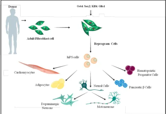
The strength of this technology resides in the possibility to obtain large quantities of cells from patients carrying a specific genetic background, including those alterations responsible for the pathology and is particularly crucial for those diseases for which we face with the impossibility to access the diseases-relevant human tissues. The hiPSCs can proliferate extensively in culture and differentiate into all types of cells of the human body, thus recapitulating the “human disease in a Petri dish” (Figure 1).
3
Figure 1. Generation of hiPSCs from skin fibroblast cultures and their ability to differentiate in different cell types.
Derivation of hiPSC from somatic cells (Takahashi et al., 2007) has generated significant enthusiasm for its potential application in basic and translational research, since they carry the same genome as the patient they were generated from, and offer the opportunity to generate disease-specific and patient specific hiPSCs for modeling human diseases, drug development and screening, and individualized regenerative cell therapy (Omole and Fakoya, 2018). Moreover, its current combination with genome editing by CRISPR/Cas9 has further enhanced the diagnostic and therapeutic power of the hiPSCs (Hotta and Yamanaka, 2015). Their application in regenerative medicine allows us to improve our knowledge of human genetic diseases, for which a therapy is not available yet.
G-Protein Coupled Receptors (GPCRs)
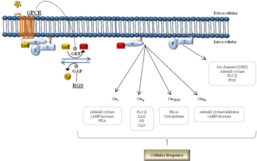
G-protein-coupled receptors (GPCRs) are the largest group of cell-surface seven-transmembrane proteins (TM) (Kamato et al., 2013) which has a ligand-binding pocket in the extracellular region, the TM region which contains seven TM α-helices, and a cytoplasmic interacting region (Pierce et al., 2002; Lagerstrom and Schioth, 2008) to mediate intracellular signalling crucial for neuronal communication, including regulation of the antagonistic effects of the parasympathetic and sympathetic branches of the autonomic nervous system throughout the body. GPCRs are encoded by nearly 800 different genes in the human genome and represent the largest TM receptors family found in humans (Bockaert and Pin, 1999). There are five subclasses of receptors on the bases of their sequences and structural
4
similarities, rhodopsin (class A), secretin (class B), glutamate (class C), adhesion and Frizzled/Taste2 (reviewed in (Zhao et al., 2016)). GPCRs transduce different extracellular stimuli by activating different isoform of G proteins (Gs, Gq/11, Gi, G12/13), which promote
different signalling cascades inside cells (Azzi et al., 2003; Fredriksson et al., 2003; Rosenbaum et al., 2009; Rajagopal et al., 2010).
G-proteins exist as heterotrimeric complex composed of three subunits, a guanine-nucleotide binding α-subunit (Gα) with GTPase activity and a tight dimer consisting of β and γ subunits (Gβγ) (Gilman, 1987; Morris and Malbon, 1999; McCudden et al., 2005). The combinatorial association of the different G protein subunits, composed of at least 20 different Gα, 6 different Gβ (including the two isoforms Gβ5 and Gβ5L), and 13 different Gγ subunits (Watson et al., 1996; Vincent et al., 2016) provides the level of selectivity that is needed to generate the wide range of signals activated by G proteins (Robishaw and Berlot, 2004; Dupre et al., 2009; Khan et al., 2013) in modulating many important cellular functions, including the release of hormones and growth factors, the regulation of cell contraction and migration, as well as cell growth and differentiation (Trivellin et al., 2014; Krishnan et al., 2015; Trivellin et al., 2016). Ligand binding of a GPCR acts as guanine-nucleotide exchange factors (GEFs) that promote a GPCR conformational change that then activates trimeric G-proteins by promoting the exchange of GDP to GTP on the Gα subunit, and its dissociation from Gβγ dimer. Both Gα-GTP and Gβγ subunits modulate downstream intracellular signalling by interacting with specific effector proteins (Figure 2) (Gilman, 1987; Neer, 1995; Clapham and Neer, 1997; Cabrera-Vera et al., 2003; Smrcka, 2008; Vilardaga et al., 2010).
5
Regulator of G-protein signaling (RGS)
The propagation of the GPCR cascades is controlled by the action of the regulators of G protein signalling (RGS). The regulator of RGS acts limiting the active Gα species lifetime by their GTPase activating (GAP) function, accelerating Gα GTP hydrolysis and thus resulting in a response deactivation of GPCRs-initiated signalling and its re-association with Gβγ dimer (Neer, 1995; Berman et al., 1996; Hunt et al., 1996; Watson et al., 1996; Hepler et al., 1997;
Kozasa et al., 1998; De Vries et al., 2000). The RGS proteins are encoded by more than 30
genes in mammals and have been classified into six subfamilies (Anderson et al., 2009; Aguado et al., 2016). Specifically, the R7 group of RGS (R7-RGS) include four highly homologous mammalian proteins namely RGS6, RGS7, RGS9, RGS11 (Sondek and Siderovski, 2001; Xie et al., 2010) which are highly expressed in neurons (Gold et al., 1997). Like other members of the larger RGS protein family, R7-RGS proteins contain i) a C-terminal core RGS homology domain that act as GTPase activating protein (GAP) (Ross and Wilkie, 2000); ii) the N-terminal Disheveled, EGL-10, Pleckstrin (DEP) and DEP helical extension (DHEX) protein domains, that are essential for protein-protein interactions critical for plasma membrane anchors (Tayou et al., 2016); and iii) the centrally located domain referred to as a Gγ-like (GGL) domain, which is responsible for the direct interaction with the divergent Gβ subunit, Gβ5 (Tayou et al., 2016).
G-protein beta subunits
G-proteins exist as heterotrimeric complex composed of three subunits, α, β, and γ (Gβγ) (Gilman, 1987; Morris and Malbon, 1999; McCudden et al., 2005). The G protein beta subunits (Gβ) belong to the beta-propeller family of proteins characterized by seven regular WD-40 repeats (WD-40) and a coiled coil domain at the far end N-terminus. The WD40 (also known as WD or beta-transducin repeats) domain is one of the most abundant and interacting domains in the eukaryotic genome; each of them is approximately 40 amino acids long and is characterized by a conserved tryptophan (Trp; W) -aspartic (Asp; D) acid pair, from which the WD40 domain is named (Watson et al., 1994; Sondek et al., 1996). In proteins the WD repeats folds into a β-propeller architecture, providing a platform for protein–protein or protein–DNA interaction, and to coordinate downstream cellular events including signal transduction, autophagy, and apoptosis (Neer et al., 1994; Jain and Pandey, 2018). The coiled-coil domain involving α-helical segments at the N termini of the β and γ subunits and contributes in βγ dimerization and effector signalling (Pellegrino et al., 1997).
In humans five different Gβ subunits, encoded by GNB1 to GNB5 genes have been identified. The first four Gβ1-4 subunits share between 80 and 90% sequence identity while Gβ5 shares
6
only 50% of sequence homology with the other four members, and is the only one that can have cellular other than cell membrane localization (Fletcher et al., 1998).
G protein subunit beta 5 (GNB5)
The GNB5 gene (MIM 604447), (NM_006578; located at 15q21.2 chromosome region) encodes for Gβ5 subunit of the heterotrimeric G-protein complex and encodes for a protein of 353 amino acid residues with 11 coding exons. GNB5 is broadly expressed in neural, neuroendocrine and other excitable tissue such as heart muscle (Slepak, 2009; Posokhova et al., 2010) with an additional longer Gβ5 isoform (Gβ5L) that is expressed in the vertebrate retina photoreceptors (Watson et al., 1994; Watson et al., 1996; Gautam et al., 1998; Robishaw and Berlot, 2004; McCudden et al., 2005).
The Gβ5 is a divergent member of the Gβ family which exhibits distinct biochemical properties respect to other members (Fletcher et al., 1998). Recent evidence suggests the existence of supramolecular complexes with members of the R7 group of regulators of G-protein signalling (R7-RGS). The obligatory interaction between Gβ5 and R7-RGS, by the GGL domain, is required for their mutual stability, indeed without their partner, both proteins are rapidly degraded in cells (Chen et al., 2000; Simonds and Zhang, 2000; Martemyanov et al., 2005). Consistently, in knockout mouse studies, the genetic ablation of the Gnb5 resulted in the instability of all R7-RGS proteins (Chen et al., 2003). GNB5 has an unusual selectivity for its effectors, as it potently regulates the activities of PLC β2, N-type calcium channels, and G protein-coupled inwardly-rectifying potassium (GIRK) channels (Watson et al., 1994; Yoshikawa et al., 2000).
G protein-gated K+ channels (GIRK)
G protein-coupled inwardly-rectifying potassium (GIRK) (Kir3) channels are important transducers of inhibitory neurotransmitter effects in heart and brain. They regulate heartbeat, neuronal excitability and plasticity, analgesia, alcohol and drug effects, and are implicated in a number of disorders such as epilepsy, Down syndrome, bipolar disorder, atrial fibrillation and primary aldosteronism (Yakubovich et al., 2015; Chen et al., 2017).
The GIRK family has four members: Kir3.1 (GIRK1), Kir3.2 (GIRK2), Kir3.3 (GIRK3) and Kir3.4 (GIRK4). Most of the GIRK channels are heterotetramers in native tissues, especially Kir3.1–Kir 3.2 in the brain and Kir3.1–Kir 3.4 in the heart, control the excitability of neurons and the heart rate, respectively (Noma and Trautwein, 1978; Kubo et al., 1993; Lesage et al.,
1994; Hibino et al., 2010). GIRK channels are directly activated by Gβγ subunits when Gi/o
7
dissociation of Gβγ from Gα. As a consequence of GIRK channels activation, the
corresponding outward potassium (K+) current moves the membrane potential to a more
hyperpolarized potential, far from the firing threshold therefore delaying the triggering of the next action potential (Yoshikawa et al., 2000; Yakubovich et al., 2015).
R7-RGS/GNB5 Complex
In hippocampal CA1 pyramidal neurons, R7-RGS/GNB5 forms macromolecular complexes
with the GABAB receptors (GABABR) and the G protein-coupled inwardly-rectifying
potassium (GIRK) channels (Fajardo-Serrano et al., 2013). For instance, co-expression of R7-RGS/GNB5 with the anchoring protein R7BP enhances the ability of RGS7 to increase muscarinic M2 receptor-dependent GIRK channel activation, presumably due to the stimulation of the catalytic activity of RGS7 (Yang et al., 2010; Zhou et al., 2012). The
activation of postsynaptic GABABR on pyramidal neurons produces slow inhibitory
postsynaptic currents (sIPSCs), which counteract the excitatory influence of ionotropic glutamate receptors to shape neuronal output (Ulrich and Bettler, 2007; Luscher and
Slesinger, 2010). As a result, GABABR signalling profoundly affects hippocampal synaptic
plasticity and has marked effects on memory formation (Davies et al., 1991; Schuler et al.,
2001; Ostrovskaya et al., 2014). Most of the postsynaptic inhibitory effect of GABABR
stimulation in the hippocampus is mediated by activation GIRK channels; which inhibit neuronal excitability via hyperpolarizing K+ efflux (Luscher and Slesinger, 2010). Blockade
of GABABR or GIRK channels by either pharmacological manipulations or genetic knockout
ablates the slow IPSC, and blunts a form of hippocampal synaptic plasticity known as
depotentiation (Chung et al., 2010). In hippocampal pyramidal neurons, GABABR-GIRK
signalling is negatively modulated by RGS7-Gβ5-R7BP complex. RGS7 is closely
co-localized with both GABABR and GIRK2-containing channels (Fajardo-Serrano et al., 2013).
Moreover the interaction between RGS7 with GIRK channels is mediated byGβ5 subunit and
is facilitated by R7BP, to limit the duration of GIRK activity evoked by GABABR in
hippocampal neurons (Zhou et al., 2012). Given the established role of GABAB receptors and
GIRK channels in many neurological diseases (Lujan and Ciruela, 2012; Lujan et al., 2014), the regulation of GNB5 is likely crucial in such pathologies..
As in neurons, the R7-RGS/GNB5 complex negatively regulates the GIRK channels also in cardiomyocytes (Posokhova et al., 2010). Cardiac rate is controlled by the sympathetic and parasympathetic branches of the autonomic nervous system. In the sinus node, the
8
parasympathetic modulation of cardiac output is mediated primarily by acetylcholine (ACh)
release from the vagus nerve which binds and activates the muscarinic receptor (M2R).
The activation of M2R triggers the release of inhibitory G proteins, activates the potassium
current IKACh by GIRK channels (M2R-IKACh); that hyperpolarize the membrane potential far
from the firing threshold thus slowing action potential rate. Activation of M2R associated G
proteins also inhibits the adenylyl cyclase (AC) activity. AC inhibition causes a decrease in the intracellular concentration of cAMP, a decrease in f-channels (HCN) activity and consequently a slower rate of the sinus node (DiFrancesco et al., 1989; DiFrancesco, 2010; Yang et al., 2010). All of these ionic mechanisms are thus potentially involved in the cholinergic regulation of heart rate (Mangoni and Nargeot, 2008).
In cardiac tissue, the M2R-IKACh signalling is negatively regulated by RGS6 which is
implicated in modulation of parasymphatetic stimulation of the heart. In particular, a specific
interaction of the Gβ5 and RGS6 - via the GGL domain - prominently accelerate IKACh
deactivation kinetics in sinoatrial nodal cells and cardiac myocytes (Kulkarni et al., 2018) functioning to prevent parasymphathetic override and severe bradycardia (Yang et al., 2010). The physical association between RGS6 and Gβ5 is critical for their expression
and/or stability (Posokhova et al., 2010). RGS6-GNB5 involvement in M2R-IKACh signalling
is mediated by a direct protein-protein interaction with the cardiac-specific GIRK subunit,
GIRK4 (Posokhova et al., 2010), suggesting that the association of RGS6-GNB5-IKACh also
plays critical role in controlling heart rate.
G-protein β subunits and Human Genetic Diseases
In human pathogenic variants in each of the five distinct G proteins β-subunits, encoded by the GNB1-GNB5 genes, cause genetic disorders with variable brain and heart involvement. Germline de novo GNB1 variants cause severe neurodevelopmental disability, hypotonia, and seizures (Petrovski et al., 2016); GNB3 bi-allelic loss-of-function (LoF) variants has been linked to congenital stationary night blindness (Vincent et al., 2016), recessive retinopathy in human (Arno et al., 2016) retinal degeneration in chicken (Tummala et al., 2006) and a reduced cone sensitivity with bradycardia in mice (Nikonov et al., 2013; Ye et al., 2014). A single nucleotide polymorphism (SNP) in GNB3 was associated with postural tachycardia syndrome (Nakao et al., 2012), incidence of cardiovascular disease and stroke (Zhang et al., 2005). GNB2 pathogenic variants cause sinus node and atrioventricular conduction dysfunction (Stallmeyer et al., 2017), while, GNB4 causes a dominant form of Charcot-Marie-Tooth (Soong et al., 2013).
9
IDDCA/LADCI Syndromes
In 2016 whole-exome sequencing successfully identified homozygous and compound heterozygous variants in the GNB5 (NM_006578; MIM #617173) gene as the cause of IDDCA/LADCI (IDDCA, MIM #617173; LADCI MIM#617182) (Lodder et al., 2016; Shamseldin et al., 2016) syndromes. The transmission pattern of them was consistent with an autosomal recessive inheritance. Homozygous and compound heterozygous loss of function (LoF) pathogenic variants in GNB5 gene were associated with a severe neurological phenotype including epileptic seizures, intellectual disability, delayed psychomotor development, hypotonia, and sick sinus syndrome (Figure 3).
Figure 3. IDDCA patient’s phenotype
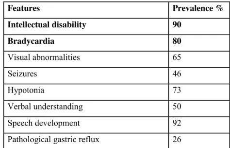
Additional finding includes visual abnormalities, with nystagmus and in some cases with reduction in cone and rode functions, and gastric reflux (Table 1) (Lodder et al., 2016). In contrast homozygous carriers of the most frequent missense variant p.(Ser81Leu) were characterized by the mildest form of the syndrome carrying mild intellectual disability in combination with language delay, attention-deficit/hyperactivity disorder, with or without cardiac arrhythmia (LADCI) (Shamseldin et al., 2016; Turkdogan et al., 2017; Malerba et al., 2018; Vernon et al., 2018). GNB5 mutations have been identified in various ethnic groups including Jordan, Puerto Rico, India, Morocco, Brazil, Saudi, Europe, Caucasus and Cambodia (Lodder et al., 2016; Shamseldin et al., 2016; Turkdogan et al., 2017; Malerba et al., 2018; Vernon et al., 2018). Since the first reports, additional affected individuals have been reported, delineating the spectrum of the phenotype with about 80% of them described with cardiac conduction dysfunction and 90% with severe intellectual disability. The disease prognosis is variable and in some instances lethal, particularly if the cardiac complications emerge as reported in (Turkdogan et al., 2017).
10
Table 1. Clinical features of IDDCA patients
Features Prevalence % Intellectual disability 90 Bradycardia 80 Visual abnormalities 65 Seizures 46 Hypotonia 73 Verbal understanding 50 Speech development 92
Pathological gastric reflux 26
References: (Lodder et al., 2016; Shamseldin et al., 2016; Turkdogan et al., 2017; Malerba et al., 2018; Vernon et al., 2018).
Consistent with manifestations present in affected human individuals we showed that treatment of gnb5 zebrafish mutant knock out larvae with carbachol (CCh, a parasympathomimetic compound that activates acetylcholine receptors of the heart and the GNB5-RGS-GIRK channel pathway) resulted in a strong decrease of the heart rate, whereas it had little effect on wild-type and sibling larvae consistent with loss of negative regulation of the cardiac GIRK channel by GNB5-RGS. In contrast, treatment with the sympathetic agonist isoproterenol resulted in an increased heart rate that was similar in wild-type, sibling, and gnb5 mutant larvae (Lodder et al., 2016). These results indicate that GNB5 is crucial for parasympathetic control of heart rate, but not for sympathetic control, suggesting that lack of
GNB5 is associated with extreme bradycardia at rest, highlighting the hypothesis that GIRK
channels or any component of the signalling mechanism that regulates GIRK channel activity could be a potential therapeutic target for IDDCA.
Gnb5 mouse models
Gnb5-null mouse models have been described (Krispel et al., 2003; Zhang et al., 2011).
Ablation of the mouse ortholog of Gnb5 resulted in phenotypes reminiscent of those of IDDCA patient, specifically marked neurobehavioral abnormalities including learning deficiencies, hyperactivity, impaired gross motor coordination, abnormal gait (Zhang et al., 2011), defective visual adaptation (Krispel et al., 2003), perturbed development and
11
functioning of retinal bipolar cells (Rao et al., 2007), smaller body size in the pre-weaning period and high pre-weaning mortality. Moreover, whereas IDDCA patients show sinus bradycardia, which result in a dysfunction of the sinoatrial node in generating or transmitting an action potential to the atrial tissue, the cardiac phenotype of Gnb5 null animals has never been studied. Interestingly, mice lacking Rgs6, the Gnb5-dependent RGS protein that is enriched in heart tissue, or Girk4 subunit, exhibited bradycardia and hypersensitivity to parasympathomimetics or diminished heart rate and heart rate variability responses to the cholinergic agonist (CCh), respectively (Posokhova et al., 2010; Kulkarni et al., 2018).
12
AIM OF THE THESIS
Genomic approaches have led to the evidence that variants in GNB1-5 genes cause a variety of human genetics diseases. However, the signalling responsible for these diseases is largely unknown, even though it likely involves dysfunction of ion channels. IDDCA/LADCI are a new autosomal recessive syndromes clinically well-defined, with no treatment or therapy available yet. The emerging involvement of GNB5 mutations in patients with such diseases, strengthened our interest in the study of the disease. The phenotypic consequence of GNB5 deficiency in affected individuals has its critical role in pathways specifically related to neuronal and cardiac functions. Specifically, since ~80% of affected patients showed cardiac arrhythmia, with ~20% of them were found death during sleep, probably due to sinus node dysfunction, we moved our interest to understand the effects of the cardiac phenotype shared in such patients driven by lack of functional GNB5 at the functional level and to link it to clinical phenotype, thereby leading to an explanation for some of the pathogenic mechanisms of IDDCA.
Although immediate benefits for patients are unpredictable, this basic science study will hopefully unravel molecular pathways that will open the way to new therapeutic targets. We propose to dissect the pathogenic mechanisms at the molecular/functional level of a novel intellectual developmental disorder with cardiac arrhythmia (IDDCA, MIM#617173) by generating and characterizing human induced pluripotent stem cells (hiPSCs), hiPSC-derived cardiomyocyte (iCM) and hiPSC-derived neurons, of the pathology. Small-scale drug screening will be performed to identify compounds that may rescue the GNB5-associated phenotype.
13
MATERIAL AND METHODS
Self-replicate mRNA-based reprogramming of human induced pluripotent stem cell (hiPSC) lines
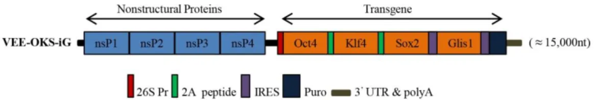
Cell reprogramming, a derivation of human induced pluripotent stem cells (hiPSC) from somatic cells (Takahashi et al., 2007) provides an unlimited source for the possibility to model in culture dish any desired specialized cell, thus enabling the characterization of the biological mechanisms underlying the human pathology. We established hiPSC lines through a mRNA-based technology, an efficient method to generate integration free and virus-free human iPSCs using a single transfection step. It entails the use of a positive, single-stranded RNA species derived from non-infectious, self-replicating Venezuelan equine encephalitis (VEE) virus. This technology consists of a polycistronic, self-replicative RNA system that consistently express the reprogramming factors Oct4, Klf4, Sox2, and Glis1 (OKS-iG) (Figure 4) (Yoshioka et al., 2013).
Figure 4. Structure of the Simplicon™ RNA replicon. The RNA replicon encodes four non-structural replication complex proteins (nsPs) as a single ORF in the 5’end of the RNA. At the 3’end, the viral structural proteins ORFs are replaced with the OKS-iG transgenes. Locations of 26S internal, 2A peptide, IRES and Puromycin (Puro)-resistance gene are indicated.
For reprogramming, 2x10⁵ fibroblasts were seeded on a 6-well plate (day 0) and maintained
in fibroblast growth medium. The following day (day 1), the cells at 60-80% confluent were
transfected using SimpliconTM RNA Reprogramming Kit (Merck-Millipore), according to
manufacturer’s instructions. During the next several days (day 2–4), 0.75µg/mL of puromycin was added in the Stage 1 Medium and was used to select for cells that have taken up the VEE-OKS-iG RNA. At day 10, RNA transfected cells puromycin-resistant at 70% confluent, were replated onto one 6-well plate containing inactivated mouse embryonic fibroblasts (MEFs) feeder cells, and cultured in Stage 2 Medium until colonies were manually isolated by picking. On day 28, individual hiPSC colonies were picked and re-seeded on 24-well plate
14
coated with Matrigel matrix (Corning) in feeder-free condition, cultured in Essential 8 Medium (Thermo Fisher Scientific) and propagated using ReLeSR (Stem Cell Technologies).
hiPSC-derived cardiomiocytes differentiation
Cardiomyogenic differentiation was performed by monolayer culture on hiPSC cultured in
feeder free condition, plating 20,000 cells/cm2 on 24-well plate coated with Matrigel, using
PSC Cardiomyocyte Differentiation Kit (Thermo Fisher Scientific) that guarantees high yield of cardiac differentiation. After splitting, hiPSCs were allowed to grow to almost confluence and then maintenance medium were changed to medium A of the kit, which is designed to favour mesodermal differentiation. After 48h, medium was changed to medium B that induces differentiation of cardiac precursors and after other 48h the medium was changed to medium C that favours cardiomyocytes survival and maintenance. Around day 10 of differentiation, the culture starts to show spontaneous beating. Cells were kept in culture for allowing cell maturation and analysis were carried out at day 30. Spontaneously beating regions were manually dissected and plated on gelatin-coated dishes for further analyses.
Karyotyping
It is well known, that long-term culture induces genetic aberrations in pluripotent cell lines (Liang and Zhang, 2013). To assess the genome integrity of the genetic material, hiPSC were treated with 0.1 µg/mL colcemid (Gibco) for 1.5 h at 37°C in an atmosphere containing 5%
CO2. The cells were then treated with Accutase, washed in DPBS and incubate in a hypotonic
solution (0,075M KCl pH7.0) for 25 min at 37°C. Thereafter the cells were fixed with methanol and glacial acetic acid (3:1 ratio) three times and stained with Giemsa stain (Gibco) on a glass slide (Fisher Scientific). The chromosomes were visualized with a 1000X objective (Zeiss, Germany) and analyzed by G-banding at GAG 300-400 band resolution in average, using Applied Imaging Cito-Vision (Version 7.5).
GNB5 sequence mutation
Genomic DNAs were extracted from fibroblasts and hiPSCs using Allprep DNA/RNA Mini kit (Qiagen Hilden, Germany), according to the manufacturer’s instructions.
15
GNB5 (MIM # 604447, NM_006578) mutations were confirmed by direct DNA sequencing
for all patients and their available family members. PCR amplifications were carried out in a final volume of 50 µl consisting of:
• 50 ng genomic DNA
• 2.5 mM deoxyribonucleotides
• 15 pmol/µl of sense and antisense primers
• 1X Reaction Buffer (200mM Tris-HCl/ pH 8.8 at 25°), 100 mM KCl, 100mM
(NH4)2SO4, 1.0% Triton X-100 and 21 mg/ml nuclease-free BSA) and
• 1 U of Taq DNA polymerase.
Denaturation was carried out at 95° for 30 s, annealing at 60°C for 30 s, extension at 72° for 30 s, for 35 cycles. Primers were designed using the Primer 3 Output program
(http://frodo.wi.mit.edu/primer3/), amplicons and primers were checked both by BLAST and
BLAT against the human genome to ensure specificity. The amplified products were subsequently purified (Exo Sap) and sequenced with a ready reaction kit (BigDyeTerminator v1.1 Cycle, Applied Biosystems). The fragments obtained were purified using DyeEx plates (Qiagen) and resolved on an automated sequencer (3130xl Genetyc analyzer DNA Analyzer, ABI Prism). Sequences were analyzed using the Sequencer software (Gene Codes, Ann Arbor, Michigan). All mutations were described following the recommendations of the
Human Genome Variation Society (http://www.hgvs.org/mutnomen).
Functional evaluation of GNB5 variants
Primary fibroblasts of family A affected individuals II.1 and II.2 (Lodder et al., 2016) were obtained from skin biopsies according to standard protocols. The cell cultures were maintained in DMEM/F12 supplemented with 10% FBS and 1% antibiotics mixture (penicillin and streptomycin 10.000 U/ml, Thermo Fisher Scientific) in a humidified
atmosphere with 5% CO2. Total RNA was obtained from primary fibroblasts using the
RNeasy Mini kit (Qiagen) according to the manufacturer's specifications, and followed by DNase (Qiagen) treatment for the genomic DNA elimination. Retrotranscribed cDNA was synthesized from 1 μg of total DNA-purified RNA using the Quantitect Reverse Transcription Kit (Qiagen), according to the manufacturer's instructions. Synthesized cDNAs were used as templates for RT-PCR amplifications involving the region carrying the splice-site variant. To discriminate and tally expressed alleles cDNAs were amplified by PCR from the members of family A, inserted into the plasmid vector pCR™4-TOPO® (Life Technologies) and sequenced. Non-sense Mediated Decay (NMD) was assayed by treating fibroblasts with
16
Puromycin and Cycloheximide at a concentration of 200 µg/ml for 8h and 4h, respectively. After incubation, the fibroblasts were harvested, total RNA was extracted and RT-PCR reactions were performed. ImageJ software (U. S. National Institutes of Health, Bethesda, Maryland, USA) was used to quantify the intensity of the gel bands by first calibrating images using control bands and then manually selecting the bands for the measurement.
Reverse Transcription (RT-PCR)
1 µg RNA was reverse transcribed using the Quantitect Transcription kit (Qiagen). RT-PCR produces DNA copies (complementary DNA, or cDNA) of a RNA template, by using the enzyme reverse transcriptase, and the resulting single-stranded cDNA can be amplified using traditional or real-time PCR. Reverse transcriptase enzyme, in general, has 3 distinct enzymatic acitivities: an RNA-dependent DNA polymerase, a hybrid-dependent exoribonuclease (Rnase H), and a DNA-dependent DNA polymerase. For reverse transcription in vitro, the first 2 activities are utilized to produce single-stranded cDNA: RNA-dependent DNA-polymerase activity (reverse transcription) transcribes cDNA from an RNA template, and RNase H activity specifically degrades only the RNA in RNA: DNA hybrids. The purified RNA samples were incubated in 1X gDNA Wipeout Buffer at 42°C for 2 minutes to remove contaminating genomic DNA. After genomic DNA elimination, the RNA samples were reverse transcribed using a master mix prepared from Reverse Transcriptase (1U), 1X RT Buffer, and RT Primer Mix, a optimized blend of oligo-dT and random primers. The entire reaction was performed at 42°C for 30 minutes and then inactivated at 95°.
Each cDNA sample was measured by using a Nanodrop spectrophotometer (NanoDrop Technologies, Willmington, Delaware, USA) and used in qPCR for i) NMD-assay in primary fibroblasts, ii) evaluation of mRNA levels for GNB5 expression, iii) hiPSCs characterization against pluripotency markers (OCT4, SOX2, NANOG, REX1 and LIN28), iv) iCMs characterization.
Real-time polymerase chain reaction (qPCR)
qPCR is a quantitative PCR method which enables both detection and quantification (as absolute number of copies or relative amount when normalized to DNA input or additional normalizing genes) of one or more specific sequences in a DNA sample.
Based on the molecule used for the detection, the real time PCR techniques can be categorically placed under two heads:
17
• non-specific fluorescent dyes that intercalate with double-stranded DNA, such as
SYBR Green, which binds to the minor groove of the DNA double helix, and is the most widely used double-strand DNA-specific dye reported for real time PCR
• sequence-specific DNA probes consisting of oligonucleotides that are labeled with a
fluorescent reporter, which permits detection only after hybridization of the probe with its complementary DNA target, such as Molecular Beacons, TaqMan Probes, FRET Hybridization Probes, Scorpion Primers.
Oligos for qPCR were designed using the Primer3 with default parameters. Amplicons and primer pairs were checked both by BLAST and BLAT against the human genome to ensure specificity.
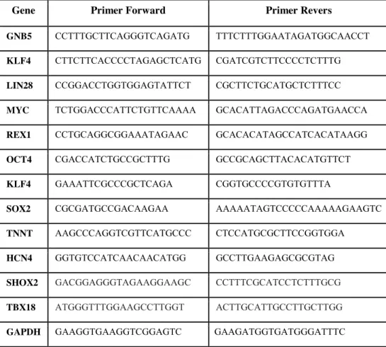
Target genes expression was examined by amplification with the primer sets described in the Table 2.
Table 2. Oligos used for qPCR analysis
Gene Primer Forward Primer Revers
GNB5 CCTTTGCTTCAGGGTCAGATG TTTCTTTGGAATAGATGGCAACCT
KLF4 CTTCTTCACCCCTAGAGCTCATG CGATCGTCTTCCCCTCTTTG
LIN28 CCGGACCTGGTGGAGTATTCT CGCTTCTGCATGCTCTTTCC
MYC TCTGGACCCATTCTGTTCAAAA GCACATTAGACCCAGATGAACCA REX1 CCTGCAGGCGGAAATAGAAC GCACACATAGCCATCACATAAGG OCT4 CGACCATCTGCCGCTTTG GCCGCAGCTTACACATGTTCT
KLF4 GAAATTCGCCCGCTCAGA CGGTGCCCCGTGTGTTTA
SOX2 CGCGATGCCGACAAGAA AAAAATAGTCCCCCAAAAAGAAGTC
TNNT AAGCCCAGGTCGTTCATGCCC CTCCATGCGCTTCCGGTGGA
HCN4 GGTGTCCATCAACAACATGG GCCTTGAAGAGCGCGTAG SHOX2 GACGGAGGGTAGAAGGAAGC CCTTTCGCATCCTCTTTGCG TBX18 ATGGGTTTGGAAGCCTTGGT ACTTGCATTGCCTTGCTTGG GAPDH GAAGGTGAAGGTCGGAGTC GAAGATGGTGATGGGATTTC
GAPDH was used as housekeeping genes. The reactions were run in triplicate in 10 μl of final volume with 10 ng of sample cDNA, 0.3 mM of each primer, and 1X Power SYBR Green PCR Master Mix (Applied Biosystems). Reactions were set up in a 384-well plate format with a Biomeck 2000 (Beckmann Coulter, Milan, Italy) and run in an ABI Prism7900HT (Applied
18
Biosystems) with default amplification conditions. Raw Ct values were obtained using SDS 2.3 (Applied Biosystems). Calculations were carried out by the comparative Ct method.
Western Blotting analysis
Protein extracts were resolved on home made 12% gels (for GNB5) and transferred to PVDF membranes (GE Healthcare) according to the manufacturer’s instructions. Antibodies used were: mouse polyclonal antibody to GNB5 (ptoteintech), and rabbit monoclonal anti-GNB5 (Abcam). Horseradish peroxidase conjugated anti-mouse (Santa Cruz), anti-rabbit (Santa Cruz) antibodies, and the ECL chemiluminescence system (GE Healthcare) was used for detection. Quantitation of band signal intensity was obtained by the ImageJ software (http://imagej.nih.gov/ij/). Values are expressed as fold differences relative to the wild-type protein sample, set at 1, after normalization for the loading control.
Immunofluorescence staining
The hiPSC cultures were fixed with 4% paraformaldehyde for 15 min at room temperature (RT). Fixed cells were washed twice in PBS, permeabilized and blocked for 30 min at RT with PBS containing 20% donkey serum and 0.1% TritonX-100. Cells were then incubated with primary antibodies for 2 h at RT in 20% donkey serum in PBS in oscillation, followed by three washing steps with PBS. The secondary antibodies were thereafter added for 1 h at RT in oscillation in the dark, followed by three washing with PBS, nuclei counterstain with DAPI (300mM) and image acquisition on Inverted Fluorescence Microscope (Axiovert 200M, Zeiss Carl; Software: AxioVision release 4.7.2 Dec 2008). Non-fixed hiPSC cultures were also incubated for StainAlive against TRA-1-60 antibody, to a final concentration of 2.5 µg/ml in fresh cell culture medium and incubate for 30 minutes at 37°C and 5% CO2. The cells were washed gently twice with cell culture medium and then were examined under an Inverted Fluorescence Microscope using the appropriate filters.
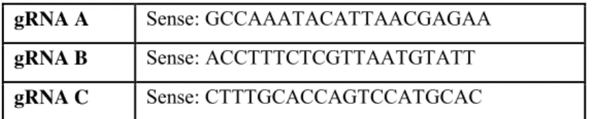
GNB5 endogenous tagging via CRISPR/Cas9
2,5x10⁵ hiPSC were plated in the matrigel-coated wells with 10µM of Rock inhibitor. After
24 hours, the cells were transiently transfected with GNB5_CRISPR/Cas9 KO Plasmid using the Lipofectamine 3000 reagent according to manufacturer’s instructions. The GNB5_CRISPR/Cas9 KO Plasmid system consists of a pool of 3 plasmids (Table 3), each
19
encoding the Cas9 nuclease and a different target-specific 20 nt guide RNA (gRNA) designed for maximum knockout efficiency.
Table 3. Gβ5 CRISPR/Cas9 KO Plasmid (h) gRNA plasmids gRNA A Sense: GCCAAATACATTAACGAGAA
gRNA B Sense: ACCTTTCTCGTTAATGTATT gRNA C Sense: CTTTGCACCAGTCCATGCAC
After 48 hours, we enriched the transfected cells by FACS analysis based on their EGFP fluorescent signal. The pool of FACS-isolated transfected hiPSCs were plated into matrigel-coated 10cm plate and cultivated sparsely. After approximately 20 days colonies were large enough for their manual picking and expansion. Each clone isolated were analyzed for genome modifications by Sanger Sequencing.
Electrophysiological analysis
The recording of action potentials was performed on small aggregates of dissociated cardiomyocytes between day 28 and day 33. Patch-Clamp experiments were conducted between 48 and 72 hours after isolation (between day 30 and 35) in Whole-Cell configuration and in Current Clamp mode.
During the experiment, the cardiomyocytes are kept at a constant temperature of 37 ° C and continuously perfused with a physiological solution (Tyrode) as follows: NaCl 137 mM, KCl 5 mM, CaCl2 2 mM, MgCl2 1 mM, HEPES-NaOH 10 mM , D-Glucose 10 mM, pH = 7.4. The borosilicate micropipettes had a resistance of 7-8MΩ used once filled with an intracellular solution thus composed: K-aspartate 130 mM, 10 mM NaCl, 5 mM EGTA-KOH, 10 mM Hepes, 2 mM CaCl2, 2 mM MgCl2, ATP (Na + -sel) 2 mM, Creatine Phosphate 5 mM, GTP (Na + -salt) 0.1 mM, pH = 7.2.
To evaluate the response to modulators of the frequency of action potentials, the cardiomyocytes were perfused with Tyrode added with 100 nM charbacol, (CCh) a stable
agonist of M2-muscarinic receptors or with Tertiapin-Q, a blocker of the GIRK channels and
compared with the frequency in only tyrode to evaluate the increase or the decrease. For the analysis of electrophysiological data, the Clampfit 10.6 and OriginPro 2016 software was used.
20
Transcriptome profiling by RNA-Seq
For transcriptome profiling we performed RNA-seq using paired-end sequences of poly-A RNA after rRNA removal and fragmentation steps. Libraries were prepared using Illumina TruSeq RNA Library Prep Kit v2. Paired-end 75 bp sequence reads were generated with the Illumina NextSeq500 platform, reaching high coverage (~40M reads per sample). We processed three biological replicates for each cell line, a number that will assure the removal of intra-sample variability and potential outliers.
We used the RAP pipeline (D'Antonio et al., 2015)– part of the CINECA’s bioinformatics resources – to process and analyse the RNA-seq data.
21
RESULTS
Recruitment of IDDCA patients
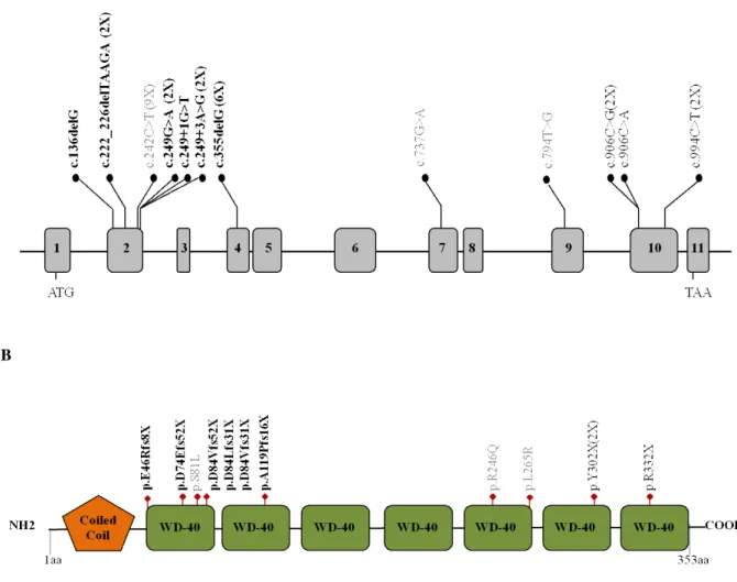
Since 2016, when the syndrome was identified, 26 patients with 12 different GNB5 variants (Figure 5) have been identified (Lodder et al., 2016; Shamseldin et al., 2016; Turkdogan et al., 2017; Malerba et al., 2018; Vernon et al., 2018) (Merla and Malerba, unpublished). The clinical evaluation included medical history interviews, a physical examination and review of medical records (Table 4). The following types of GNB5 mutations have been identified: 6 frameshift (6/12, 50%), 3 missense (3/12, 25%), and 3 nonsense (3/12 25%). All the GNB5 identified variants were inherited from asymptomatic parents.
A
B
Figure 5. (A) Distribution of the GNB5 variants (NM_006578) across the gene with exons indicated by grey boxes. The frameshift variants are designated in bold, missense variants are in gray, and nonsense variants are in black. The number of patients with each variant is indicated in parentheses. (B) Schematic representation of the GNB5 protein with Coiled coil and WD-40 repeats domains.
22
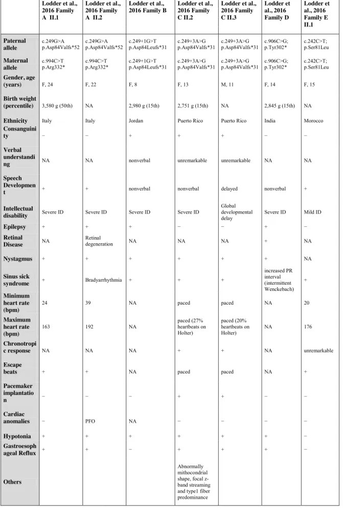
Table 4. Clinical features of GNB5-patients reported so far
Lodder et al., 2016 Family A II.1 Lodder et al., 2016 Family A II.2 Lodder et al., 2016 Family B Lodder et al., 2016 Family C II.2 Lodder et al., 2016 Family C II.3 Lodder et al., 2016 Family D Lodder et al., 2016 Family E II.1 Paternal allele c.249G>A p.Asp84Valfs*52 c.249G>A p.Asp84Valfs*52 c.249+1G>T p.Asp84Leufs*31 c.249+3A>G p.Asp84Valfs*31 c.249+3A>G p.Asp84Valfs*31 c.906C>G; p.Tyr302* c.242C>T; p.Ser81Leu Maternal allele c.994C>T
p.Arg332* c.994C>T p.Arg332* c.249+1G>T p.Asp84Leufs*31 c.249+3A>G p.Asp84Valfs*31 c.249+3A>G p.Asp84Valfs*31 c.906C>G; p.Tyr302* c.242C>T; p.Ser81Leu
Gender, age
(years) F, 24 F, 22 F, 8 F, 13 M, 11 F, 14 F, 15
Birth weight
(percentile) 3,580 g (50th) NA 2,980 g (15th) 2,751 g (15th) NA 2,845 g (15th) NA
Ethnicity Italy Italy Jordan Puerto Rico Puerto Rico India Morocco
Consanguini
ty − − + + + − −
Verbal understandi
ng NA NA nonverbal unremarkable unremarkable NA NA Speech
Developmen
t + + nonverbal nonverbal delayed nonverbal + Intellectual
disability Severe ID Severe ID Severe ID Severe ID
Global developmental
delay Severe ID Mild ID
Epilepsy + + + − − + − Retinal Disease NA Retinal degeneration NA NA NA + NA Nystagmus + + + + + + NA Sinus sick syndrome + Bradyarrhythmia + + + increased PR interval + (intermittent Wenckebach) Minimum heart rate (bpm) 24 39 NA paced paced NA 20 Maximum heart rate (bpm) 163 192 NA paced (27% heartbeats on Holter) paced (20% heartbeats on Holter) NA 176 Chronotropi c response NA NA NA + + NA unremarkable Escape
beats + + NA paced paced NA +
Pacemaker implantatio n − − − + + − − Cardiac anomalies − PFO NA − − − − Hypotonia + + + + + + − Gastroesoph ageal Reflux + + − + + + − Others Abnormally mithocondrial shape, focal z-band streaming and type1 fiber predominance
23 Lodder et al., 2016 Family E II.3 Lodder et al., 2016 Family F Shamseldin et al., 2016 V:1 Shamseldin et al., 2016 V:2 Shamseldin et al., 2016 V:3 Shamseldin et al., 2016 IV:1 Shamseldin et al., 2016 IV:6
Paternal allele c.242C>T; p.Ser81Leu c.242C>T, p.Ser81Leu c.242C>T, p.Ser81Leu c.242C>T, p.Ser81Leu c.242C>T, p.Ser81Leu c.242C>T, p.Ser81Leu c.242C>T, p.Ser81Leu
Maternal allele c.242C>T; p.Ser81Leu c.242C>T, p.Ser81Leu c.242C>T, p.Ser81Leu c.242C>T, p.Ser81Leu c.242C>T, p.Ser81Leu c.242C>T, p.Ser81Leu c.242C>T, p.Ser81Leu Gender, age
(years) M, 10 M, 25 F, 12 F, 11 F, 5 F, 7 F, 11
Birth weight
(percentile) NA NA 50th centile 50th centile NA 50th centile NA
Ethnicity Morocco Brazil Saudi Saudi Saudi Saudi Saudi
Consanguinity − + + + + - - Verbal under standing NA NA + + + + + Speech Development + NA Severe language
delay Severe language delay Severe language delay Severe language delay Severe language delay
Intellectual disability
Mild ID Mild ID normal IQ, but school performance issues normal cognitive development NA normal IQ NA Epilepsy − − NA NA NA NA NA Retinal Disease NA NA NA NA NA NA NA Nystagmus − NA NA NA NA NA NA Sinus sick syndrome + + NA NA NA NA NA NA NA NA NA NA Minimum heart rate (bpm) 16 NA NA NA NA NA Maximum heart rate (bpm) 180 NA NA NA NA NA NA Chronotropic response unremarkable NA NA NA NA NA NA Escape beats + NA NA NA NA NA NA Pacemaker implantation + NA NA NA NA NA NA Cardiac anomalies − NA NA NA NA NA NA
Hypotonia impaired fine motor skills − - - NA + NA
Gastroesophageal
Reflux − NA NA NA
Others ADHD, Hyperactivity Inattentive type ADHD motor delay motor delay ADHD, mild motor delay
24 Turkdogan et al., 2017 V.1 Turkdogan et al., 2017 IV.14 Vernon et al., 2018 Malerba et al., 2018 Poke and Sadleir 2017 (unpublished) Poke and Sadleir 2017 (unpublished) Kannu and Shao 2018 (unpublished) Kannu and Shao 2018 (unpublished) Paternal allele c.355delG; p.Ala119Profs *16 c.355delG; p.Ala119Profs *16 c.737G>A; p.Arg246Gln c.222_226delT AAGA; p.Asp74Glufs* 52 c.906C>G;
p.Tyr302* c.136delG; p.Glu46Argfs*8 c.794T>G; p.Leu265Arg c.906C>A; pTyr302*
Maternal allele c.355delG; p.Ala119Profs *16 c.355delG; p.Ala119Profs *16 c.222_226delTAA GA; p.Asp74Glufs*52 c.242C>T;
p.Ser81Leu c.906C>G; p.Tyr302* c.136delG; p.Glu46Argfs*8 c.794T>G; p.Leu265Arg c.906C>A; pTyr302*
Gender, age (years) M, 3 F,11 M, 2 F, 2.5 M,2.5 M, 10 F, 9 F, 3 Birth weight (percentile) NA 1800 g (<1st) 3311 g (50th) 1698 g (<1st) (Intrauterine growth restricted)
3300g (50th) 2740g (3rd) 2585 g (25-50%tile) No records but 7.49kg at 5 months
Ethnicity NA NA European/Caucasian European/Caucasian Pakistan Cambodian Cambodian South Asian
Consanguin
ity + + − −
First cousins
once removed Second cousins
+ (Maternal grandmother is Paternal grandmother sister) - Verbal understandi ng "no developmental milestones" "no developmental
milestones" nonverbal + noverbal nonverbal limited -
Speech Developme nt "no developmental milestones" "no developmental milestones" nonverbal expressive
speech delay nonverbal nonverbal fourteen months
Severe language delay
Intellectual
disability Severe ID Severe ID Severe ID Mild ID Severe DD Severe ID severe ID Severe ID Epilepsy + + − − + + - EEG showed slow wave,
no seizure
Retinal Disease
Retinal
degeneration Retinal degeneration
Severe reduction in cone and rode
function − NA No (normal examination 2010) Bilateral high myopia Cortical blindness, ERG anmoral wave forms Nystagmus + + + strabismus roving eye movements, vertical nystagmus roving eye movements - + Sinus sick syndrome Sinus arrhythmia/ Sinus bradycardia Sinus arrhythmia/ Sinus bradycardia Sinus arrhythmia/
Sinus bradycardia + + + Sinus bradycardia + Sinus bradycardia with skips Minimum heart rate (bpm) NA NA 71 36 NA NA 38 28 Maximum heart rate (bpm) NA NA 183 176 NA NA 171 151 Chronotrop ic response NA NA NA NA + NA NA Escape
beats NA NA + (prior to pacing) + + + - +
Pacemaker implantatio n
Refused by
parents Refused by parents + + - - - -
Cardiac anomalies − − − − - - - - Hypotonia + + + + + + - +++ Gastroesop hageal Reflux normal (abdominal US examination) normal (abdominal US examination) + − - + - - Others autistic (midline hand automatism, no eye contact) autistic (midline hand automatism, no eye contact) hypertonia Laryngomalacia
hypertonia, ear tubes
No dystonia. Non
25
GNB5 nonsense and frameshift variants
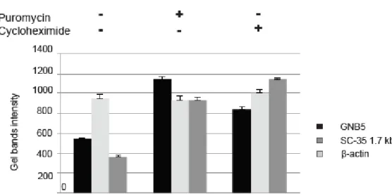
The prevalence of GNB5 variants found in our cohort is predicted to lead to truncated protein suggesting a loss of function event. In order to investigate the possibility that GNB5 mRNA carrying truncating variants may result in the partial transcripts degradation, we investigated the nonsense-mediated mRNA decay pathway. The r.[249G>A; 249-250ins25] allele is predicted to encode a truncated polypeptide containing a stretch of 52 incorrect amino acids starting with an Asp to Val substitution at position 84 (p.(Asp84Valfs52X)). Cloning and sequencing of amplicons from individual A.II.1 and A.II.2 (Lodder et al., 2016) further revealed that all transcripts containing the paternal allele were aberrantly spliced. Using the same approach, we examined the relative abundance of the c.249G>A and c.994C>T alleles. Both altered transcripts are less frequent than their unchanged counterpart in parents’ cells of (A.I.2) and father (A.I.1) (5 out of 14 amplicons (35.7%) for the paternal allele and 2 out of 15 (13.3%) for the maternal one). These results prompted us to hypothesize that both mutant alleles of family A might be targeted by nonsense-mediated mRNA-decay (NMD) pathway. We measured by RT-PCR the levels of GNB5 mRNA in proband’s fibroblasts after treatment with puromycin or cycloheximide, two known NMD inhibitors. The mRNA level of GNB5 was restored after treatment with both NMD inhibitors suggesting that the c.249G>A and c.994C>T variants trigger NMD of the corresponding transcripts and thus represent LoF alleles (Figure 6).
Figure 6. Molecular characterization of Family A variants. The physiological NMD substrate SC-35 1.7 Kb was included as positive control (dark grey bars). β-actin (light grey bars) was used as internal control.
26
Generation of hiPSCs from patients and controls skin fibroblasts
Starting from skin biopsy, we generated primary fibroblast cell lines from all members of the Family A described in (Lodder et al., 2016), that consists of two severely affected sibling carrying compound heterozygous p.Asp84Valfs52*/Arg332* (p.D84Vfs52X/p.R332X) GNB5 variants and their heterozygous healthy carrier parents, as well as an additional patient carrying compound heterozygous p.Asp74Glufs52*/Ser81Leu (p.D74Efs8X/p.S81L) GNB5 variants (Malerba et al., 2018). Thanks to already settled collaborations we got skin fibroblasts from three novel unpublished cases bearing the homozygous frameshift variants p.Glu46Argfs8*, the nonsense mutation p.Tyr302*, and the missense homozygous variant p.Leu265Arg respectively (Figure 7).
Figure 7. Sanger Sequence electropherogram of the GNB5 mutations in fibroblast cohort of IDDCA patients
Skin fibroblast derived-hiPSC lines have been established according to standard protocol (Figure 8) (See Material and Methods), and characterized from all the individuals reported in Table 5, along with those from two heterozygous healthy carrier parents and four unrelated healthy controls.
27
Figure 8. Generation of IDDCA patients-derived hiPSC lines by integration-free, virus-free reprogramming. (Above) Key steps and media requirements are indicated: i) plating of skin fibroblasts in fibroblast medium; ii) administration of synthetic mRNAs alongside the B18R inhibitor of interferon response; and iii) identification and picking of suitable hiPSC clones. (Below): Representative time course of human hiPS colonies generated using Human Simplicon RNA Reprogramming Kit. Colonies start to emerge from Day 25-28 and are ready to be picked at Day 32.
Table 5. Genotype of IDDCA patient-derived hiPSC lines. Of note, hiPSC from six healthy (GNB5 wild-type genotype) individuals have been also generated as control cohort. Gender and age is reported for each hiPSC cell line. F, female. M, male. GDB, Biobank identification code.
Cells that showed morphological evidence of reprogramming were selected manually. Two independent hiPSC clones per individual have been characterized (Figure 9-10) using standard methods by i) growth kinetics and maintenance of undifferentiated morphology upon culturing in feeder-free conditions; ii) expression of standard human pluripotency markers by immunofluorescence (against OCT4, NANOG, SOX2, and TRA-1-60); iii) quantitative RT- PCR evaluation of pluripotency markers (NANOG, OCT4, LIN28, and SOX2); and iv) Short Tandem Repeat (STR) and karyotyping analysis to confirm clones identity and genomic integrity, respectively. Mutation of GNB5 was confirmed by Sanger Sequencing.
Lodder et al., 2016 Family A II.1 GDB 813 Lodder et al., 2016 Family A II.2 GDB 813-6 Malerba et al., 2018 GDB 1337 Unpublished case II (Poke and Saldeir)
GDB 1307_Z2
Unpublished case III (Kannu and Shao)
GDB 1378 Paternal allele c.249G>A p.D84Vfs52X c.249G>A p.D84Vfs52X c.222_226delTAAGA p.D74Efs52X c.136delG p.E46Rfs8X c.794T>C p.L265R Maternal allele c.994C>T p.R332X c.994C>T, p.R332X c.242C>T, p.S81L c.136delG, p.E46Rfs8X c.794T>C p.L265R Gender, age (ys) F, 24 F, 22 F, 3 M 10 F, 9
28
Figure 9. Representative characterization of unrelated controls, healthy parents and IDDCA patients-derived hiPSC lines by Immunofluorescence detection for expressing pluripotency markers NANOG, OCT4 SOX2, and TRA-1-60. Nuclei were stained with DAPI (blue). Images acquired with fluorescence microscope 40X magnification.
29
Figure 10. Quantification of pluripotency markers by RT-qPCR. The expression of endogenous pluripotency-associated markers SOX2; NANOG; LIN28, and OCT4 were confirmed by RT-qPCR in all fibroblast-derived hiPSC lines. A primary fibroblast (FIBROBLAST CTRL) was used as negative control. The results of three independent experiments are expressed as mean ± SD.
Analysis of the GNB5 gene expression
GNB5 mRNA levels were measured by RT-qPCR in primary skin fibroblasts, 5 from IDDCA patients, 2 from healthy parents and 2 unrelated controls. We performed three RT-qPCR experiments (each in triplicate) to quantify the relative transcript concentrations of the corresponding gene, by using the comparative cycle threshold (Ct) method with the formula: 2^(ΔCt). Each input was normalized to GAPDH.
As reported in Figure 11 (upper panel), the endogenous GNB5 mRNA expression level was consistently decreased in healthy parents (blue) and in affected patients (pink) when compared to unrelated controls (green), suggesting the correlation between the genotype and the gene dosage.
This data was also confirmed in our cohort of fibroblasts and fibroblast-derived hiPSCs by RNA-Seq analysis as shown in Figure 11 (low panel).
30
Figure 11. GNB5expression levels on primary skin fibroblasts and hiPSCs included in our study. Upper panel; data from RT-qPCR analysis. Low panel; data from RNA-Seq assay. The results of three independent experiments are expressed as mean ± SD. Student’s t-test (*: p-value <0.01; **: p-value <0.001; ***: p-value <0.0001; ****: p-value <0.0001).
Generation and selection of GNB5 hiPSC_KO
The skin fibroblast-derived hiPSC lines selected in this study (4 from IDDCA patients, 2 from healthy parents and 4 from unrelated controls) represent our cohort of patient-specific. In order to gain deeper insight into GNB5 function in IDDCA patients, we selectively knocked-down (KO) its expression via CRISPR-Cas9 system. hiPSC_GNB5_KO (hiPSCKO) and hiPSC_WT (hiPSCWT) isogenic cell lines have been preferred to patient and control-derived hiPSC lines to keep the same genotypes through the whole study and to avoid the genotype-intrinsic influence of each IDDCA patient and control cell lines.
The PCR amplification and Sanger Sequencing have been performed on extracted DNA of the same hiPSCs, showed a deletion of five nucleotides (c.204_208delCATGG) in exon 2 of the gene, which gives rise a premature stop codon of the protein, p.C68fsX, confirmed by Sanger sequencing on RT-PCR amplicon (Figure 12A). We confirmed that the cell lines showed a normal karyotype (Figure 12B). Moreover, the absence of the GNB5 protein and the decrease of its expression level in hiPSCKO cells were evaluated by Western blot and RT-qPCR, respectively (Figure 12C-D).
31
Figure 12. Characterization of hiPSCKO cell line. (A) Sanger sequencing on RT-PCR amplicon; (B) hiPSC KO karyotype analysis, (C) absence of the GNB5 protein in hiPSCKO by Western blot and (D) decrease of its mRNA expression levels by RT-qPCR. Student’s t-test (*:p-value< 0.01; **p-value< 0.001; ***p-value< 0.0001).
We generated three independent clones each for KO, each carrying the same mutation c.204_208delCATGG. The presence of off-targets, predicted by target sites prediction tool (https://crispr.cos.uni-heidelberg.de/) was ruled out by PCR and Sanger Sequencing analysis of the in silico predicted off targets genes.
Table 6. Oligos used for off targets genes analysis
Gene Primer Forward Primer Revers
IGSF6 TGGACTACACTCATGAGGCC GTATGAAGGCCACGCACAC
MAST2 TGAGGCCTCCCATCATCATC GGGAGCTGCTCTGTGATGAA
Generation and characterization of hiPSC-derived cardiomyocytes (iCM)
The use of animal models have greatly contributed to our understanding of the etiology and mechanisms of diseases; however due to interspecies physiologic differences between human and animal cardiomyocytes in terms of cell biological, mechanical and electrophysiological properties animal model do not accurately represent the physiology of human cardyomyocites (CMs) (Denayer et al., 2014).