Depatment of Economics
P
HD
COURSE IN“H
EALTHY FOODS:
INNOVATION AND MANAGEMENT”
XXX
CYCLEGreen degradation of mycotoxins by biotechnological
application of enzymes from Pleurotus spp.
PhD candidate: Dr. Martina Loi Tutor: Dr. Giuseppina Mulè Co-tutor: Dr. Francesca Fanelli
List of abbreviations iii
List of tables v
List of figures v
Introduction
1. General introduction and background on mycotoxin 1
1.1. Overview of the main mycotoxins 6
1.1.1. Aflatoxins 6
1.1.2. Fumonisins 8
1.1.3. Zearalenone 10
1.1.4. Trichothecenes 11
1.1.5. Ochratoxins 13
1.2. Prevention and reduction strategies 14
1.2.1. Prevention in pre-harvest 14
1.2.2. Post-harvest reduction strategies 16
2. Laccases 19
2.1. Overview, occurrence and physiological role of laccase enzymes 19
2.2. Molecular structure and catalytic activity 20
2.3. Laccase mediator system 24
2.4. Laccase applications 31
2.5. Laccase as tool for mycotoxins bioremediation in feed, food and
bioenergy supply chains 33
Aim and outline of the thesis 45
Chapters
3. Mycotoxin Biotransformation by Native and Commercial Enzymes: Present
and Future Perspectives 47
4. Aflatoxin B1 and M1 Degradation by Lac2 from Pleurotus pulmonarius and
Redox Mediators 99
5. In vitro single and combined mycotoxins degradation by Ery4 laccase
from Pleurotus eryngii and redox mediators 126 6. Aflatoxin M1 removal and biotechnological application of a laccase
from Pleurotus eryngii for milk safety 144
8. Supplementary data 180 8.1 Zearalenone degradation in naturally contaminated maize flour by Ery4
laccase mediator system 180
8.2 Optimization of AFB1 degradation by LMS with the Design
Of Experiment 182
8.3 Aflatoxin M1 degradation by immobilized Ery4 and syringaldehyde 185
Mycotoxins are toxic secondary metabolites produced by filamentous fungi mainly belonging to Aspergillus, Penicillium, Fusarium and Alternaria genera. They can be found as common contaminants of cereals, fruits, seeds and spices as a result of fungal spoilage. Mycotoxin contamination is an significant health and economic concern worldwide. Some of them were recognized by the International Agency of Research on Cancer (IARC) as carcinogenic (aflatoxin of the B and G series), possible carcinogenic (aflatoxin M1, AFM1;
fumonisin B1, FB1; ochratoxin A, OTA) to humans. Moreover, they exert both acute and
chronic toxic effects towards humans and animals. Because of mycotoxin contamination, billions of dollars are lost every year due to unsold commodities, decrease of animal health and productivity or to sustain a complex and integrated mycotoxin management system. Prevention strategies are not completely effective and require the implementation of novel post-harvest methods, able to mitigate or remove mycotoxins from contaminated materials.
The aim of this thesis was to evaluate and study the capability of laccase enzymes to reduce mycotoxin contamination both in vitro and in contaminated materials through an environmental friendly and mild approach. In addition up to eight different redox mediators were used within the laccase mediator system (LMS) to maximize mycotoxin degradation.
Within this purpose, the activity of two different purified LCs, native Lac2 from Pleurotus pulmonarius and the recombinant Ery4 from P. eryngii, was tested towards the main classes of mycotoxins.
Lac2 was identified and evaluated for the in vitro degradation of aflatoxins, while Ery4 was tested towards AFB1, AFM1, FB1, OTA, deoxynivalenol (DON), zearalenone (ZEN)
and T-2 toxin. The preliminary screening revealed that the inclusion of a toxin - specific redox mediator is required to achieve high levels of degradation with both enzymes. However, the use of the LMS resulted ineffective for DON and not efficient for OTA.
also achieved for AFB1/ZEN and FB1/T-2 toxin pairs.
In addition, LMS treatment was successfully tested in real artificially and naturally contaminated matrixes (milk and maize flour) for AFM1 and ZEN degradation.
Despite the great potentialities showed by both enzymes in the field of bioremediation, LCs remain versatile biocatalysts which can be applied in various food processes. LMS application in milk was deeply investigated to evaluate the effects on milk proteins and the possibility of manufacturing a curd with improved technological and nutritional properties.
The results presented in this thesis lay the basis for the development of a biotransformation methods based on a LMS approach and open new perspective for the use of this versatile and green biocatalyst in the field of safety and quality of mycotoxin contaminated commodities.
ABTS: 2-azino-di-[3-ethylbenzo-thiazolin-sulphonate] AFB1: a atoxin B1
AFD1: a atoxin D1
AFL: a atoxicol AFM1: a atoxin M1
AFO: a atoxin oxidase enzyme AFQ1: a atoxin Q1
AFs: a atoxins ALA: -lactalbumin
ANOVA: analysis of variance AP1: aminopentol 1
-lactoferrin BLG: -lactoglobulin CGA: chlorogenic acid cLC: commercial laccase CN: casein
CPA: carboxypeptidase A CPY: carboxypeptidase Y CT: coated and tough DE: degrading enzyme DOE: Design of experiments DON: deoxynivalenol
DPs: degradation products
EDC: endocrine disrupting chemical EFSA: European Food Safety Authority ER: estrogen receptors
FB1: fumonisin B1
GAPs: Good Agricultural Practices HFB: hydrolyzed FB1
HR-ESI-MS: High Resolution electrospray ionization mass spectrometry HT-2: HT-2 toxin
IARC: International Agency of Research on Cancer LC: laccase
LCs: laccases
LMS: laccase mediator system MnPs: Manganese Peroxidases NIV: nivalenol
OTA: ochratoxin A OT : ochratoxin
Phe: phenylalanine molecule Prx: peroxiredoxin
SDS PAGE: sodium dodecyl sulphate poly acrylamide gel electrophoresis T-2: T-2 toxin
TEAC: trolox equivalent antioxidant capacity TGase: translutaminase
UHT: ultra high temperature VAN: vanillin WP: whey protein ZEN: zearalenone -ZAL: -zearalanol -ZEL: -zearalenol -ZAL: -zearalanol - -zearalenol
List of tables
Table 1-1. Mycotoxin classification and properties 5
Table 3-1. Aflatoxins degrading enzymes 14
Table 3-2. Fumonisin B1 degrading enzymes 56
Table 3-3. Trichothecenes degrading enzymes 65
Table 3-4. Zearalenone degrading enzymes 73
Table 4-1. Summary of Lac2 purification from P. pulmonarius culture filtrate 110 Table 4-2. Lac2 identification by LC MS/MS 112 Table 4-3. Aflatoxin concentrations in samples after Lac2 LMS treatment 115 Table 6-1. Summary of Ery4 purification from Saccharomyces cerevisiae
culture filtrate 150
Table 6-2. Absolute values and degradation percentages of the time
course degradation of AFM1 with Ery4 and SA 150
Table 7-1. Content of retained proteins from curd samples 171 Table 7-2. Antioxidant activity of supernatants from in vitro digested curds 173
Table 8-1. Zearalenone degradation 180
Table 8-2. Experimental runs of D-optimal design and response for AFB1 182
List of figures
Figure 1-1. Aflatoxins chemical structures 7
Figure 1-2. Fumonisins chemical structures 9
Figure 1-3. Structural analogies between zearalenone and 17- 10
Figure 1-4. Trichothecenes general structure 12
Figure 1-5. Chemical structure of type A and B trichothecenes 12 Figure 1-6. Chemical structure of ochratoxin A and derivatives 13
Figure 2-1. LC copper centers 21
Figure 2-2. Laccase catalytic cycle 23
Figure 2-4. Chemical structures of the major natural and artificial
redox mediators 27
Figure 2-5. ABTS oxidation by laccase and electron transfer mechanism 29
Figure 2-6. Hydrogen atom transfer mechanism 30 Figure 2-7. TEMPO oxoammonium cation catalysis by laccase 30 Figure 2-8. Ionic mechanism 31 Figure 3-1. Chemical structure and features of ciclopentenone and difurocoumarolactone aflatoxin series. 54
Figure 3-2. Chemical structures of ochratoxin A and its degradation products, ochratoxin- 61
Figure 3-3. Type B fumonisins chemical structure 63 Figure 3-4. Chemical structure of trichothecenes 67 Figure 3-5. Chemical and structural analogies between zearalenone -estradiol 71 Figure 4-1. Zymography using ABTS as the substrate and SDS PAGE of P. pulmonarius LC preparations 11
Figure 4-2. AFB1 and AFM1 degradation by Lac2 LMS 113
Figure 4-3. Examples of HPLC chromatograms of AFB1 and AFM1 114
Figure 5-1. Residual aflatoxin B1 in samples treated with different LMS 134
Figure 5-2. Residual fumonisin B1 in samples treated with different LMS 135
Figure 5-3. Residual ochratoxin A in samples treated with different LMS 137
Figure 5-4. Residual T-2 toxin in samples treated with different LMS 149
Figure 5-5. Residual AFB1 and ZEN in samples treated with different LMS 140
Figure 6-1. Time course degradation of AFM1 in buffer solution and in skimmed UHT milk by Ery4 and SA 151
Figure 6-2. SDS PAGE of milk samples 152
Figure 7-1. SDS-PAGE of UHT milk samples treated with LMS 166
Figure 7-2. SDS-PAGE of UHT milk samples treated with CGA-LMS 169
Figure 7-3. SDS-PAGE of full fat milk samples treated with Ery4 and commercial laccase CGA-LMS 172
Figure 7-4. SDS-PAGE of curds proteins manufactured with LCs 175 Figure 8-1. Scatter plot of aflatoxin B1 degradation by Ery4 and SA 184
Figure 8-2. Time course of in vitro degradation of aflatoxin M1
1. General introduction and background on mycotoxins
Mycotoxins are secondary metabolites produced by fungi mainly belonging to Fusarium, Aspergillus and Penicillium genera, which display toxic, carcinogenic, teratogenic and mutagenic activity towards humans and animals (Bennet and Klich, 2003).
Mycotoxins are low molecular weight metabolites, which exhibit very diverse chemical structure, toxicity and are produced by different organisms. Due to their huge heterogeneity, mycotoxins are not easy to classify. They can be grouped according to the chemical structure (e.g. coumarins, lactones etc.), target organ (e.g. nephrotoxic, hepatotoxic etc.) or producer fungus (Aspergillus toxins, Penicillium toxins). Cereals, seeds, spices, fresh and dried fruits are the major food commodities susceptible to mycotoxin contamination. The most important mycotoxins are shown in Table 1-1.
Mycotoxin contamination poses a severe risk for the health and economy of food and feed supply chains. The Food and Agriculture Organization (FAO) estimates that up to 25% of food commodities is contaminated with mycotoxins and from 30 to 100% of it is co-contaminated by multiple mycotoxins.
Cereals are the main source of mycotoxin exposure. At the same time, they represent the staple food of human diet worldwide, and have a considerable role in animal nutrition and biofuel production. Cereals by-products are precious ingredients in feed manufacture, and contribute to maintain the sustainability of food processing industries by converting wastes and by-products into valuable products (Pinotti et al., 2016).
Mycotoxigenic fungi are able to contaminate crops, both pre and post-harverst. Improper storage or prolonged drying can stimulate fungal growth and mycotoxin production (IARC, 2012).
Mycotoxins are stable to the most commonly used food processing methods, thus they can be found in processed products, including sterilized, extruded and
fermented ones. Carry over in animal products was also documented for AFB1 (as
AFM1 in milk and dairy) and OTA (Volkel et al., 2011).
Being secondary metabolites, mycotoxin production is not easy to predict, hence it depends on several environmental, as well as biological factors, such as the complex host-pathogen interactions (Logrieco et al., 2002). Among the environmental factors, the most important ones are temperature, nutrient availability, atmospheric gases, pH, water activity, photoperiod and light intensity (Fanelli et al., 2015; Fanelli et al., 2013). On the other hand, the size of the inoculum, presence- absence of competing microflora and strain-host variability are determining biological factors (Popovski et al., 2013).
Generally, a wide number of different mycotoxins may be found in the same product since a single fungal species can produce several toxic metabolites, or several species can be present simultaneously, producing different toxins (Pereira et al., 2014).
Aflatoxin B1 (AFB1) is the most toxic mycotoxin and was classified as
carcinogenic to humans (group 1) by the International Agency of Research on Cancer (IARC) in 2002 (IARC, 2002). Aflatoxin M1 (AFM1), ochratoxin A (OTA)
and fumonisin B1 (FB1), were recognized as possibly carcinogenic to humans
(group 2B) due to the limited availability of their toxicological data on humans (IARC, 2002).
Toxic outcomes of mycotoxin ingestion are classified into acute or chronic mycotoxicosis, depending on the type of mycotoxin assumed and its daily intake. Mycotoxicosis symptoms include gastro intestinal inflammation, anorexia, immune dysfunction, growth impairment and endocrine disorders. In addition, synergistic or additive effects have to be taken into account because of the co-occurrence of multiple toxins on the same product (Speijers et al., 2004).
c impact of mycotoxin contamination is also a topic of great interest. In addition to the money loss due to unsold commodities, which cannot enter the market
(Commission Regulation 1881/2006), additional costs have to be considered. Food and feed recalls or rejections at the borders, the reduction in animal productivity, sanitary treatments for mycotoxicosis, the safe disposal of contaminated biomasses, the development of detection and quantification methods as well as the development of strategies able to reduce mycotoxins occurrence have a great impact on the economic balance of mycotoxins contamination (Marroquín-Cardona et al., 2014). Economic losses are difficult to quantify, but range in the order of billions of dollars annually worldwide (Schmale and Munkvold, 2017).
Mycotoxin occurrence is regulated in many countries worldwide. The European Union has a solid legislation covering both food and feed commodities. Commission Regulation (EC) No. 1881/2006 sets mycotoxins maximum levels specifically established for food products (e.g. type pf food commodity, raw or processed) and consumer category (e.g., adults or infants). As regards trichothecenes T-2 and HT-2 toxins (T-2, HT-2), only indicative levels in cereals and cereals products have been established (Commission Recommendation 2006/576/EC; Commission Recommendation 2013/165/EU), while for the so called emerging mycotoxins (e.g., enniatins, beauvericin, fusaric acid and moniliformin) neither, maximum nor indicative levels have been yet established.
Different regulation limits were set in approximately 100 countries worldwide; however, they are still lacking in some African and Latin American regions. During the last 10 years, many efforts were made to harmonize regulations in several economic communities, such as EU (European Union), MERCOSUR (Mercado Cómun del Sur, which includes Argentina, Brazil, Paraguay, Uruguay and Venezuela), Australia and New Zealand (Egmond et al., 2007).
Regulation limits are formulated in accordance to toxicological and occurrence data, but can significantly vary. In US and China, the limit for AFM1 in milk, for
example, is 10 times higher than EU limits (0.5 vs 0.05 µg/Kg).
The differences in food safety standards and regulation on mycotoxin occurrence have an important impact on food and feed trade, with both economic
and health related implications. In developing countries, poor control measures, less strict regulations increase the overall mycotoxin exposure risk. High quality food is exported, while the most contaminated one is retained or imported for the local trade (Wu et al., 2012).
In tro du ctio n 5
Table 1-1. Mycotoxin classification and properties
Food commodity Maize, wheat, rice,
sorghum, nuts Cereals, grapes dried
fruits, wine, coffee
Cereals, cereal product Maize, maize products, sorghum Cereals, cereal product Chemical group Difuranocoumarin Dihydro-isocoumarin linked -phenylalanine Sesquiterpenoid Diesthers of acyls Resorcilyc acid lactones Toxicity Hepatotoxic, carcinogenic (liver) Nephrotoxic Cytotoxic Hepatotoxic and carcinogenic (esophagus) Estrogenic Main producing organisms Aspergillus flavus, A. parasiticus, A. niger Aspergillus ochraceus, Penicillium verrucosum Fusarium graminearum F. poae, F. sporotrichioides, F. equiseti F. verticillioides, F. proliferatum, F. fujikuroi F. culmorum, F. graminearum Mycotoxin Aflatoxin B1, B2,G1,G2 Ochratoxin A, B, C Deoxynivalenol Nivalenol T-2 toxin HT-2 toxin Fumonisin B1, B2, B3, B4 Zearalenone -Zearalenol -Zearalenol Mycotoxin class Aflatoxins Ochratoxins Trichothecenes Fumonisins Zearalenons
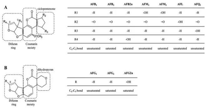
1.1. Overview of the main mycotoxins 1.1.1. Aflatoxins
derives from its principal fungal producer, Aspergillus flavus (Hesseltine, et al., 1966). AFs group includes more than 20 fungal secondary metabolites synthetized by Aspergillus species. AFs are primarily produced in the field, under elevated temperature and water stress conditions, but also during storage when relative humidity is maintained at 65% or during prolonged drying (Villers et al., 2014). Maize (Zea mays L.) is the major crop susceptible to Aspergillus spp. infection and subsequent AF contamination, together with nuts and dried fruits (Masood et al., 2015). Nuts, nut products and seeds were the most notified AF contaminated commodity by the Rapid Alert Alarm System for Food and Feed (RASFF) entering EU borders in 2016 (RASFF, 2016).
Afs are difuranocumarins
chemical structure (Fig. 1-1). The difurocoumarocyclopentenone group includes
AFB1 2 2a, AFM1 2 1 (AFQ1), and
1,
2 2a.
B and G designations refer to their characteristic blue and green fluorescence under UV-light, while the M refers to the first evidence of AFM1 occurrence in the
milk of lactating cows fed with AFB1 contaminated feed.
AFs of the B and G series co-occur in cereals and their derived products, fruits, oilseeds, nuts, tobacco and spices, while the M serie, AFL and AFQ1 can be found
in food as carry-over products of AFB1 contaminated feeds. In fact, in vivo AFB1 is
readily metabolized through hydroxylation (to AFM1 or, to a lesser extent, to
AFQ1) or reduction (to AFL). Among in vivo metabolites, AFM1 deserves particular
attention because it is excreted through the mammary glands of lactating humans and animals and can be found as natural contaminant in milk and breast milk.
AFs toxicity was discovered in 1960, when 100,000 turkeys died from AF induced liver necrosis (Blount, 1961). Their toxicity is both acute, at high doses, and chronic, if low amounts are assumed for a long period of time. Common symptoms of acute aflatoxicosis include vomiting, abdominal pain, pulmonary oedema, fatty infiltration and necrosis of the liver. Chronic assumption of AFs leads to liver cancer.
AF poisoning has been reported in developing countries such as India and Kenya, where poor agronomical practices, poor control measure and improper storage systems are in use. Additionally, environmental conditions are extremely favourable to fungal growth and mycotoxin production (Wild et al., 2015). One of the largest aflatoxicosis outbreak occurred in rural areas of Kenya leading to 314 cases and 125 deaths, due to the consumption of contaminated home-grown maize. AFs consumption is also a serious health threat for breast fed infants, since AFM1 has been recently detected also in breast milk (Ishikawa et al., 2016).
Figure 1-1. Aflatoxins chemical structure. Toxic determinants are highlighted in red (C-8 C-9 double bond) and blue (ester bond in the lactone moiety). Tables show substituent groups of different analogues.
Both B and G series were enlisted in group 1 by IARC (2002) and are carcinogenic to humans and animals. Upon ingestion, they are metabolized in the liver by cytochrome P450 (CYP450) microsomal enzymes to aflatoxin-8,9-epoxide. The epoxide ring is a crucial determinant for AFs carcinogenicity, since it is responsible for the binding to N7-guanine and the subsequent G to T transversions in the DNA molecule (Essigmann et al., 1977). Activated AFs are also able to form schiff bases with cellular and microsomal proteins (via methionine, histidine and lysine), thus leading to acute toxicity (Eaton et al., 1997). The lactone ring also plays a role in AFs toxicity and carcinogenicity, since
1 (AFD1), which still retains the 8,9-dihydrofuran double bond but not
the lacton ring, lacks the strong in vivo DNA binding activity of AFB1,
demonstrating that DNA alkylation depends upon both difuranocumarin and lactone moieties (Schroeder et al., 1985).
1.1.2. Fumonisins
Fumonisins are a wide group of mycotoxins produced by several Fusarium species, including F. verticillioides, F. proliferatum, F. oxysporum and F. globosum, commonly found as saprophytes on maize, wheat and other cereals. 28 different fumonisins have been described so far and divided into A, B, C and P-series. The B-serie includes the most common and toxic fumonisins, with FB1 being the most
abundant.
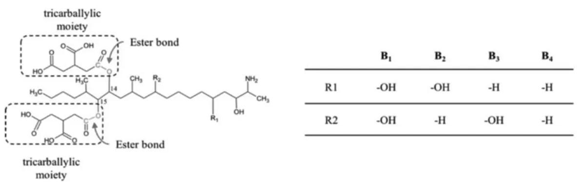
Chemically, fumonisins are composed by a long 19- or 20 carbon aminopolyol backbone substituted with two tricarballylic acid moieties (Fig. 1-2) which resembles the sphingoid bases sphinganine (SA) and sphingosine (SO).
The structural similarity to SA and SO is responsible for FB1 toxicity. FB1 acts
as a competitive substrate for ceramide synthase, which is technically a SA and SO N-acetyltransferase. As a consequence, ceramide biosynthesis is disrupted, SA and SO accumulate in cells and tissues leading to inhibition of cell growth, apoptosis, liver and kidney disfunction (Soriano et al., 2005). Other specific toxic effects on animals include porcine pulmonary edema and equine leukoencephalomalacia.
In 1993 and 2002 FB1 was enlisted in group 2B by the IARC, thus possible
carcinogenic to humans (IARC, 2002). Although carcinogenicity was not directly proven in humans, it has been positively correlated with the occurrence of oesophageal cancer in certain areas of South Africa and China (Marasas, 1996), while evidence of the carcinogenic effect on animals has already been proven in rats and mices (Stockmann-Juvala et al., 2008).
Both the tricarballylic moieties and the amino group are responsible for FB1
toxicity. Hydrolysed FB1 (HFB1) and N-acetyl FB1 are significantly less toxic or
unable to disrupt ceramide biosynthesis.
Maize is the main food commodity affected by FB1 contamination, although it
was also rarely found in sorghum and sporadically in wheat, asparagus, tea, and cowpea (Jackson, 2013).
Figure 1-2. Fumonisins (FBs) chemical structures. FB1ester bonds hydrolyzed by the main
degrading pathways, leading to the formation of HFB1 and the two tricarballylic acid
moieties, are indicated in red. Table shows substituent groups of different fumonisin analogues.
Available data on carry over exclude that FB1 transfer occurs from feed to
animal tissues or animal derivatives, such as milk and eggs (EFSA, 2005). However, a recent study by Magoha and colleagues (2014) reported FB1 carry over
in breast milk of Tanzanian women, suggesting that carry over contribution to the total FB1 exposure should be reconsidered, at least in developing countries
(Magoha eta l., 2014). 1.1.3. Zearalenone
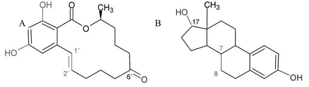
Zearalenone (ZEN) is a phenolic -resorcyclic acid lactone, non-steroidal yet oestrogenic mycotoxin, mainly produced by several Fusarium species, including F. culmorum, F. equiseti and F. graminearum. It can be found as natural contaminant of maize as well as wheat, barley, sorghum and rye. Also vegetable oils produced from corn and wheat contain considerable amounts of ZEN.
The main toxic effect of ZEN is related to its oestrogenic potential, due to its structural similarity to 17- (Fig. 1-3). ZEN is capable of binding the -estradiol, estriol andestrone (Zinedine et al., 2007). Besides ZEN, other related compounds can be generated in vivo upon reduction, s - and
- -ZEL, respectively). Surprisingly ZEL results in
greater ER binding capacity and oestrogenic potential than the parent compound.
Figure 1-3. Structural analogies between zearalenone (A) and 17- (B). Chemical groups interacting with estrogen receptor are highlighted in red, blue and green
-resembles that of C3 i
-allowing those molecules to be more flexible and to undergo some conformational changes leading to a better interaction with ER receptor. Also hydroxyl groups in C4 and C2 contribute to the bindin
latter in increasing ZEN estrogenic potential (Shier et al., 2001).
Acute toxic effects of ZEN are many and vary according to reproductive status (prepuberal, cycling or pregnant) of the affected animal and gender. ZEN is able to decrease fertility, cause sterility, induce precocious puberty and persistent oestrus. Symptoms include inflammation of reproductive organs, enlarged reproductive tracts in females and atrophy of the seminal vesicles and testes in males. ZEN was also found to be hematotoxic, genotoxic, hepatotoxic, to cause liver lesions leading to cancer (Zinedine et al., 2007).
1.1.4. Trichothecenes
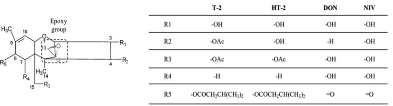
Trichothecenes are a large group of sequisterpenoid sharing a common rigid tetracyclic ring. More than 190 compounds have been isolated and characterized (Rocha et al., 2005). Fusarium, Myrothecium, Spicellum, Stachybotrys, Cephalosporium, Trichoderma, and Trichothecium are the main producing fungal genera. Trichothecenes are classified in four groups, namely A, B, C and D, according to their substitutions on the core structure (Fig. 1-4). Type A (e.g., T-2 and HT-2) trichothecenes do not posess a carbonyl group substitution in C-8, while type B (e.g. deoxynivalenol, DON and nivalenol, NIV) do. Type C trichothecenes (e.g., crotocin and baccharin) possess an additional epoxy ring C-7 and C-8, or between C-9 and C-10; type D trichothecenes (e.g., satratoxin and roridin), contain a macrocyclic ring between C-4 and C-1 (Desjardins et al., 2007).
Type A and B are the most important ones with respect to occurrence and toxicity (Fig. 1-5), with T-2 toxin being the most toxic (Miller, 2002). DON and NIV display also phytotoxicity, thus they can be classified both as mycotoxins and phytotoxins.
Trichothecenes cause a wide variety of toxic effect in animals, including growth retardation, reduced ovarian function and reproductive disorders, immunocompromization, feed refusal and vomiting. At cellular level, they induce apoptosis, inhibition of protein, nucleic acids synthesis and membrane structure alteration (Arunachalam et al., 2013).
The main determinant of toxicity is the 12,13-epoxy ring. In addition, the presence of hydroxyl or acetyl groups at appropriate positions on the
Figure 1-4. Trichothecenes general structure
Figure 1-5. Chemical structure of type A and B trichothecenes. Table shows substituent groups of the most important trichothecenes analogues
trichothecene core; the presence of substituents on C-15 and C-4, and of the side groups, define the degree of toxicity (Cundliffe et at., 1977; Karlovsky et al., 2011). 1.1.5. Ochratoxins
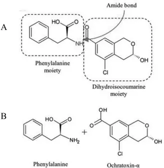
Ochratoxins are a group of mycotoxins mainly produced by Aspergillus ochraceus, A. carbonarius, A. niger and Penicillium verrucousm. OTA is the most important representative and can be defined as a
7-carboxy-5-chloro-8hydroxy-3,4-dihydro-3-R- ,
-phenylalanine (Phe) by an amide bond.
Ochratoxin B and C are OTA derivatives and are formed upon dechlorination and esterification, respectively.
from the cleavage of the amide bond and the release of the Phe moiety (Fig. 1-6). on-toxic compound, with a 10-times shorter elimination half-life than OTA (Koszegi et al., 2011).
Figure 1-6. Chemical structure of ochratoxin A and derivatives. A) Structural determinants of OTA. The amide bond hydrolyzed by the main degrading pathway is OTA
A
OTA is toxic and carcinogenic to animals and possibly carcinogenic to humans (IARC, 1993). Kidney is the main target organ, but also immune system suppression has been described as an important adverse effect. At cellular levels, it is responsible for the inhibition of protein synthesis, the formation of DNA adducts, indirect oxidative DNA damage and epigenetic modifications.
OTA exposure in humans has been correlated with the outbreaks of several nephropathies, of which the most known is the Balkanic endemic syndrome ( ), although a direct carcinogenic effect in humans has not been yet verified.
OTA is a common contaminant of cereals, dried fruits, grape juices, coffee and fermented products such as wine and beer. The carry over in kidney, blood and muscular tissue of pigs and milk and dairy was also reported (Völkel et al., 2011).
1.2. Prevention and reduction strategies
Mycotoxin contamination of food commodities depends upon several environmental and physiological factors governing the secondary metabolism of the producing fungus and can occur both in field and during storage. Thus, it is difficult to predict and almost impossible to avoid their occurrence in food commodities.
Managing mycotoxin risk is a complex task and requires an integrated strategy, covering both the pre and post-harvest.
1.2.1. Prevention in pre-harvest
Mycotoxin prevention in pre-harvest starts with the application of the good agricultural practice (GAPs) which include the choice of resistant varieties, correct soil management and the application of appropriate agronomical practices and use of biocontrol agents. They aim at reducing the extent of fungal contamination, thus mycotoxin contamination risk.
Plants show different susceptibility to fungal infection. Specific genes are known to regulate metabolic pathways for new or increased resistance systems governing the initial fungal infection (type I resistance) or its spread (type II resistance). Also phenotypic traits (height, dwarfing, lodging susceptibility, flower opening, anthers type) contribute to plant resistance to fungal infection (Steiner et al., 2017).
The correct soil management aims at reducing fungal contamination and interrupt the cycles of infection. Correct timing of fungicide application allows to control fungal contamination. However, the presence of different species, with different fungicide susceptibility and the non-homogenous phenological development of plants might reduce the efficiency of the treatment (Wegulo et al., 2015).
Tillage to bury host crop residues and crop rotation with non-susceptible cops reduce fungal infection frequency and intensity. Other important factors in reducing fungal infection are a correct fertilizer and water management. Early harvesting helps in reducing mycotoxin production, since less time is available for fungi to produce toxins.
The use of biocontrol agents is another important strategy which can be used to control mycotoxin producing fungi in an environmentally friendly way. There are several mechanisms through which they act: they are able to compete, produce antifungal substances, parasitize the mycotoxigenic fungi or promote plant defence systems (Nguyen et al., 2017)
Predictive models are also in use to forecast mycotoxin contamination in crops and plan the most effective risk management strategies. They were developed implementing data from variables having a significant impact on the possible mycotoxin production (crop type, environmental conditions, fungal infection cycle) into mathematical models. A correct prediction is a powerful supporting tool to apply the correct intervention strategies to counteract mycotoxin contamination and minimize the risk (Battilani, 2016).
1.2.2. Post-harvest reduction strategies
Pre-harvest strategies are only able to mitigate mycotoxin contamination, which can also occur during storage. Post-harvest strategies have to be implemented within the pre-harvest ones to reduce mycotoxin contamination and to improve the safety of food and feed commodities. They can be divided into physical, chemical or biological methods.
Desired process characteristics should i) be effective (ii) produce non-toxic metabolites (iii) not alter the nutrient profile and technological production and if possible, (iv) destroy fungal spores (Grenier, et al., 2014).
It is noteworthy mentioning that, to date, in Europe the detoxification of commodities intended for human consumption exceeding regulatory limits is not allowed (Commission Regulation EC number 1881/2006). However, these methods can be applied to commodities intended for animal consumption (Commission Regulation EC 786/2015).
Physical methods: physical methods include the removal of highly contaminated fractions from raw materials (by sorting, cleaning, milling, dehulling), the application of heat, the use of and cold plasma application.
Sorting is the first step to drastically reduce mycotoxin contamination. In fact, it is well known to occur heterogeneously, with hot spots of high contamination in fine materials, broken kernels and dust. DON, FB1, T-2 and HT-2 toxin are more
frequently associated with the pericarp layers and bran, thus cleaning, dehulling and milling leads to their removal from refined grains and accumulation in cereal by-products. (Fleurat-Lessard, 2017).
Toxins are generally heat stable within the range of food-processing temperatures (80 121 °C), however higher temperatures, like in roasting and extrusion processes were reported to reduce mycotoxins in nuts and maize (Kabak, 2008).
Cold plasma appears to be an innovative method for mycotoxins reduction with minimal effect on the nutritional value of food. Degradation of compounds in solid materials by cold plasma is restricted to thin surface layers, where mycotoxins are present the most, and occurs at room temperature. However, degradation was shown to be strongly dependent on mycotoxin structure and food matrix assayed (Bosch et al., 2017).
The use of adsorbents is very popular in feed production. The principle is that bioavailability of mycotoxins is reduced because of their binding to mineral, organic or biological material. The most common mineral adsorbents are activated charcoal, bentonites, silicates and yeast cell walls. The major drawback is the limited efficacy towards trichothecenes and the lack of specificity, which leads to unspecific binding of nutrients and antibiotics (Jarda et al., 2011).
Chemical methods: several chemical compounds among acids, bases, oxidising, chlorination or reducing agents, were shown to reduce, destroy and inactivate mycotoxins. AFB1, FB1 and trichothecenes degradation by ammoniation was
reported (Kabak et al., 2007). However, despite the efficacy, the application of chemicals is limited because it can be impractical, unsafe, expensive and unfavourable. Chemical treatments require harsh conditions, which can generate toxic residues and drastically reduce the nutritional, sensory and functional properties of the product (Jalili, 2015).
Biological methods: recently, biological degradation with microorganisms or their enzyme arose great interest in the scientific community. Many organisms were reported to degrade AFs, DON and ZEN among fungi and bacteria (Vanhoutte et al., 2016) and many enzymes were isolated and characterize for mycotoxins degradation capability (Loi et al., 2017). The use of enzymes allows to use mild condition to obtain a specific, likely irreversible reaction, with minimal impact on the sensory and nutritional quality of raw matrices.
It is important to discriminate among biodegradation and bio-detoxification. The biodegradation leads to the formation of new products, with no regards to their toxicity, which can also be equal or higher than that of the parent compound -ZOL, AFM1). Through the biodetoxification, the toxic compound is
transformed into a non-toxic, or significantly less toxic compound (e.g. HFB1,
OT ). The biotransformation of mycotoxins implies that a biodetoxification process is performed (EFSA, 2009).
Among the many identified enzymes capable of degrading or detoxifying mycotoxins, laccases (LC) deserve particular attention because they are green catalysts, thus environmental friendly, extremely versatile, and their ability to degrade several toxins, such as AFs and ZEA, was already reported (Alberts et al., 2009, Banu et al, 2014).
2. Laccases
2.1. Overview, occurrence and physiological role of laccase enzymes
Laccases (LCs) (benzenediol: oxygen oxidoreductases, EC 1.10.3.2) represent the largest subgroup of blue multicopper oxidases (MCO). LCs are copper containing enzymes which catalyse the oxidation of mono and ortho-diphenols, thiols and anilines to the corresponding quinones concurrently to the four-electron reduction of oxygen to water (Giardina et al., 2015). LCs were discovered in the early 1880s, in the sap of the Japanese lacquer tree Rhus vernicifera and since then they have been extensively studied (Yoshida, 1883). They are ubiquitous enzymes, as they have been identified in fungi (Hatakka, 1994), plants (Mayer, 1987), bacteria (Enguita et al., 2003) and insects as well. Fungal LCs are secreted by most white-rot basidiomycetes during lignin biodegradation, however they are also involved in melanin-like pigment synthesis, as well as in the bio-detoxification of harmful compounds or humus turnover in soil (Lisova et al., 2012).
Other important roles are played by laccases in chitin and lignin synthesis (in insects and higher plants, respectively). Lastly, in bacteria, they may be involved in pigmentation, resistance of spores and pathogenesis (Cañas and Camarero, 2010).
Fungi and in particular white rot fungi are the main LCs producers and have been extensively studied for many biotechnological applications from a molecular and biochemical point of view. Among white rot fungi the genus Pleurotus is one of the most studied because it is a safe, edible mushroom, easy to be cultivated and one of the most important genus from a commercial point of view. Originally it was supposed that the variability of LC proteins was due to the differential post traduction modification of the same gene product. Instead, many laccase genes and relative cDNA copies were identified and characterized during the last 20 years. Great variability exists among the same genus; in Pleurotus genus, 12 different complete gene sequences have been described for Pleurotus ostreatus, 5
for Pleurotus sajor caju and 8 for Pleurotus eryngii (https://www.ncbi.nlm.nih.gov/, accessed on 08/11/17).
LC genes expression can be divided into constitutive (e.g. Lacc3 and Lacc12 of P. ostreatus; Lac3 of P. sajor caju) or inducible (e.g. Lacc2 and Lacc10 of P. ostreatus; Lac1, Lac2 and Lac4 of P. sajor caju). Constitutive LCs are evenly expressed in changing environmental conditions, the latter are expressed or significantly more expressed in response to environmental stimuli or specific molecules. The promoters of these genes contain a high number of motifs that are sensitive to components present in the wheat straw extract, such as xenobiotic response elements (XRE) and metal-responsive elements (MRE) (Castanera et al., 2012).
Physiological mechanisms occurring during mycelia development can also modulate the relative expression levels of laccase isoenzymes. Some isoforms have been observed during the lag and exponential phases of fungal fermentation and should be involved in substrate degradation, whilst other isoforms have been detected in the stationary phase and should be involved in mushroom morphogenesis and pigmentation processes (Piscitelli et al., 2011). In P. pulmonarius (formely known as P. sajor caju) three different laccase isoenzymes are produced by the fungus in presence of different aromatic compounds (Zucca et al., 2011), while in P. ostreatus Lacc2 and Lacc10 resulted upregulated when wheat straw extract is added to the culture media (Castanera et al., 2012).
2.2. Molecular structure and catalytic activity
The crystal structures of LCs from several fungal species were fully resolved, e.g. LC from the ascomycete Melanocarpus albomyces (Hakulinen et al., 2002) and the basidiomycetes C. cinerea (Ducros et al., 1998), Trametes versicolor (Piontek et al., 2002; Bertrand et al., 2002), Rigidoporus lignosus (Garavaglia et al., 2004), Cerrena maxima (Lyashenko et al., 2006) and Lentinus (Panus) tigrinus (Ferraroni et al., 2007).
LCs are usually monomers, although some dimers and tetramers exist (Lisova et al., 2012; Molitoris and Reinhammar, 1975). They are generally glycosylated, with 10 to 30% of glycosylation which may have a role in the protection of the enzyme from proteolysis and correct folding (Maestre-Reyna et al., 2015). Their isoelectric point is usually acidic, ranging between 3 and 6.
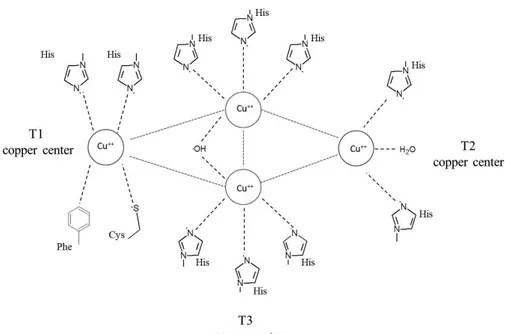
LC owes its blue color to the presence of one to four copper ions named T1 and T2/T3, coordinated by amino acid residues in two conserved regions (Fig. 2-1). T1 site has only three conserved ligands, two histidines and one cysteine which coordinate one copper ion in a trigonal planar geometry, whereas one axial ligand is usually variable and greatly contributes to the determination of the redox potential of the enzyme (Strong and Claus, 2011). This axial ligand is methionine in the bacterial laccases (CotA), and leucine or phenylalanine in fungal laccases; mutations of these two residues leads to the decrease of the oxidation potential (Kumar et al., 2003).
T2 and T3 copper ions form a trinuclear cluster, where molecular oxygen is reduced to water. The T2 copper is coordinated by two histidines and one water molecule; and each of the two T3 copper ions by three histidines. Some variants from this general scheme do exist.
The 12 amino acid residues in the enzymes serving as the copper ligands are located within four conserved regions which are named L1, L2, L3 and L4. The eight histidines that serve as ligands of the trinuclear cluster occur in a highly conserved pattern of four HXH motifs. In one of these motifs, X is the cysteine bound to the T1 copper, while each of the histidine is bound to one of the two T3 coppers. Intraprotein homologies between the conserved L1 and L3 and between L2 and L4 suggest the occurrence of duplication events.
Some LC variants lack the T1 copper and are often referred to as the yellow LCs, as they show no characteristic absorption band around 600 nm (Leontievsky et al., 1997).
The T1 copper is characterized by a strong absorption around 600 nm, whereas the T2 copper exhibits only weak absorption in the visible region. The T2 site is electron paramagnetic resonance (EPR)-active, whereas the two copper ions of the T3 site are EPR-silent due to an antiferromagnetic coupling mediated by a bridging ligand (Ferraroni et al., 2007).
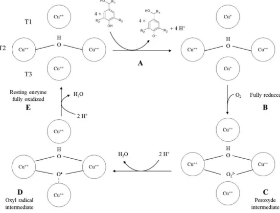
In contrast to most enzymes, which are generally substrate specific, LC is able to oxidise a wide variety of substrates, ranging from phenols to polyphenols, anilines and benzenthiols. The catalytic activity of laccase can be divided into several steps shown in Fig. 2-2.
Firstly, the phenolic substrate is oxidized by the resting, fully oxidized enzyme into a phenoxyradical (Fig. 2-2, A). In particular, due to its high redox potential (ca. +790 mV) type 1 copper is the site where substrate oxidation takes place. The extracted electrons are then transferred, probably through a strongly conserved His-Cys-His tripeptide motif, to the T2/T3 site. Once four substrates have been oxidized and the electrons have been transferred from the T1 copper to the
trinuclear cluster, the enzyme switches to its fully reduced state (Fig. 2-2,B). Molecular oxygen binds to the trinuclear cluster to form a peroxide intermediate (Fig. 2-2,C). The presence of a conserved aspartate residue lowers the reduction potential of T2 and T3 copper, allowing them to donate an electron to the oxygen molecule. It results in a peroxide bridge. The peroxide intermediate then decays through the O-O bond cleavage.
The cleavage may proceed through a proton unassisted pathway at high pH and a proton-assisted pathway at low pH, the latter being supported by a conserved glutamate residue.
The resulting hydroxide lowers the reduction potential of the T2, facilitating electron transfer to the peroxide which turns into an oxyl radical intermediate (Fig. 2-2,D). In the absence of excess reductant, the radical intermediate decays slowly to the resting, fully oxidized enzyme (Fig. 2-2,E) (Jones and Solomon, 2015).
Since no hydrogen peroxide is detected during laccase activity, it seems reasonable to suppose that a four-electron oxidation of oxygen to water occurs. As the oxidation of the substrate is a one electron reaction, laccase oxidizes four substrates in order to reduce oxygen to water. Thus, the trinuclear cluster is able to store electrons coming from each of the four substrates. In the case of substituted compounds, the reaction can be accompanied by partial demethylations and dehalogenations.
LC activity is strongly dependent on its redox potential and surprisingly it is not limited to phenolic substrates. Factors affecting metal-protein redox potential are varied and of great complexity. Many aspects such as solvation, metal ligand interactions, intramolecular electrostatic interactions, and/or protein folding restrictions (governing the position and orientation of the ligands) can modulate the redox potential (E0) values of these enzymes (Frasconi et al., 2010). Above all,
E0 is strongly governed by the geometry at the T1 Cu center (Kenzom et al., 2014).
LC can be divided in to low and high redox potential laccases, as their E0 can
be as low as +400 or as high as +800mV. High redox LC are able to oxidise a wide variety of substrates, whose redox potential is lower or similar to that of the enzyme. The redox potential of phenolic substrates (0.5 0.9 V, at acid pH) and enzyme (0.6 0.8 V) is similar therefore a fast and efficient oxidation occurs. However, 2,4,6-tri(But)phenol is not oxidized by LC due to steric hindrances, proving that not all polyphenols are suitable LC substrates ( Acunzo et al., 2006). Low redox laccases have a limited range of substrates that can be oxidized, however this limit can be overcome by the use of redox mediators in the so-called Laccase Mediator System (LMS).
2.3. Laccase mediator system
Among the fungal oxidoreductive enzymes, laccases have the lowest redox potential (usually less than +0.8V); however, target compounds with high molecular weight or high redox potential can still be indirectly oxidized. As a
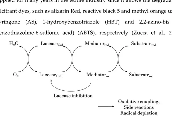
matter of fact, LC oxidation can be mediated by low molecular weight molecules which act as electron shuttles from the enzyme to the target substrate. This system, to which we refer as LMS, is naturally used by white rot fungi to degrade lignin, which is a high molecular weight polymer, as well as in industrial application, mostly in paper pulp and textile industry (Bugg et al., 2015).
The enzyme acts towards a low molecular weight redox mediator, which is then oxidized. The oxidized mediator performs a non-enzymatic oxidation of the substrate, by using mechanisms that may be unavailable to laccase, thereby explaining the possibility to oxidise non-phenolic substrates, and widening the usefulness of a purely enzymatic method. The catalytic cycle is closed when oxygen is reduced to water by LC (Fig. 2-3). LMS is a powerful tool which has been applied for many years in the textile industry since it allows the degradation of recalcitrant dyes, such as alizarin Red, reactive black 5 and methyl orange using acetosyringone (AS), 1-hydroxybenzotriazole (HBT) and 2,2-azino-bis (3-ethylbenzothiazoline-6-sulfonic acid) (ABTS), respectively (Zucca et al., 2011;
Wang et al., 2011; Telke et al., 2011).
LMS allows to generate a compound with enhanced oxidative capacity and reduced steric hindrance with respect to that of LC itself. The oxidized mediator
has a higher E0 and can easily react with other compounds without the steric
restriction of an enzyme active site.
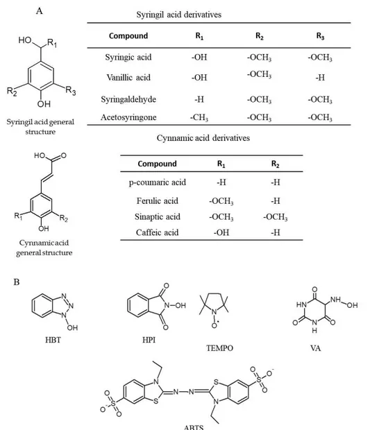
More than 20 compounds of different origin and structure, including heterocycles, >N OH compounds, syringil and cynnamic acid derivatives have been used as LC mediators and their mechanism of oxidation studied (Baiocco et al., 2003; Brogioni et al., 2008).
Simple and substituted phenols deriving both from fungal metabolism or lignin depolymerisation are efficiently oxidized by LC and they are the most used natural redox mediators. Syringil acid derivatives include syringic acid, vanillic acid, syringaldehyde (SA) and AS, while cynnamic acid derivatives comprise p-coumaric acid (pCA), ferulic acid (FA), synaptic acid and caffeic acid.
Artificial compounds include HBT, N-hydroxyphtalimide (HPI), (2,2,6,6-tetramethylpiperidin- 1-yl)oxyl (TEMPO), violuric acid (VA), and ABTS. The structures of representative laccase mediators are shown in Fig. 2-4.
The rate-determining step in LMS is the oxidation of the mediator, thus the electron transfer to the T1 copper centre. LC and mediator type, i.e. their E0, is a
crucial parameter that governs the kinetics of oxidation: the higher LC E0, or the
Substrate oxidation by the oxidized mediator can occur by multiple mechanisms according to the type of mediator used. Substrate oxidation may occur by hydrogen atom transfer (HAT), single electron transfer (ET) or following a ionic route. This variety of mechanism is another strong point of LMS, because a selective oxidation can be performed by choosing the right mediator.
An ideal mediator should be a small-size molecule, able to quickly create a stable radical (i.e. electrochemically stable), easily regenerated, not consumed in unwanted side reactions such as polymerization or enzyme inactivation (Fig 2-3).
Figure 2-4. Chemical structures of the major natural (A) and artificial (B) redox mediators. hydroxybenzotriazole (HBT), N-hydroxyphtalimide (HPI), (2,2,6,6-tetramethylpiperidin- 1-yl)oxyl (TEMPO), violuric acid (VA), and 2,2-azino-bis (3-ethylbenzothiazoline-6-sulfonic acid) (ABTS).
In view of an industrial application, the natural origin, availability and low cost are also features of great importance (Canas and Camarereo, 2010).
Electron transfer mediated mechanism
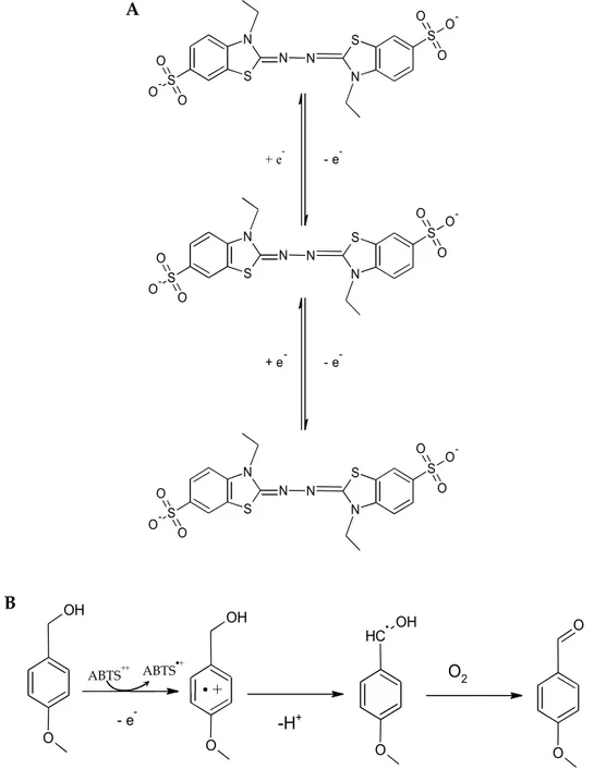
The first synthetic mediator to be discovered for the oxidation of both lignin and non-phenolic compounds was ABTS (Burbonnais and Paice, 1990). ABTS is oxidized by LC into the green radical cation ABTS +, which then undergoes
disproportion to restore ABTS and generate the dication radical ABTS 2+ (Fig.
2-5,A) (Rochefort et al., 2003). The dication radical is stable and has a greater E0 than
the single radical cation (0.91V compared to 0.47V, respectively). Both redox states of ABTS are stable and reversible (Morozova et al., 2007) and are shown in Fig. 2-5,A. Substrate oxidation by ABTS 2+ follows an electron transfer mechanism, thus a
single electron is abstracted with subsequent proton loss from the substrate (Fig. 2-5,B).
Radical hydrogen atom mechanism
Natural phenols and N-OH type mediators oxidize the substrate through a radical route. Upon LC oxidation, those compounds are transformed into reactive phenoxy or nitroxy radicals, which remove a hydrogen atom from the substrate, thus regenerating itself and producing another radical (Fig. 2-6). In this case, the rate of HAT route, therefore mediator reactivity, is strongly dependent on the bond dissociation energy rather than on the electrochemical potential of the final substrate.
Phenoxy radicals can also undergo further enzyme-independent reactions such as homo- and hetero-coupling to form dimeric, oligomeric, polymeric, or cross-coupling product, and in some cases, aromatic ring opening reactions (Hashmi et al., 2016). Coupling and crosslinking reactions are exploited in organic synthesis and grafting processes (Wells et al., 2006). Among natural phenols, metoxy-substituted ones (i.e. acetosyringone, syringaldehyde or synaptic acid) proved to be extremely effective in removing industrial dyes (Camarero et al., 2005) because electron-donating substitutes, like methoxide, lower phenol
Figure 2-5. ABTS oxidation by laccase (A) and electron transfer mechanism (B)
- e -OH O OH O + C H OH O O O -H+ O2 ABTS ABTS S N N S N N S O O O -S O O O -S N N S N N S O O O -S O O O -S N N S N N S O O O -S O O O -+ e -+ e -- e -- e -A B
electrochemical potential. Moreover, ortho-substituted phenols are endowed with increased lifetime and less susceptibility to 5
2010). HAT route is the only mechanism available for N-OH type mediators like HBT, HPI VA.
Despite their efficacy, artificial N-OH mediators can be toxic and expensive, so their use in industrial application has been limited (Canas et al., 2010).
Ionic route
TEMPO is a stable N-oxyl radical known to be a highly selective catalyst in the oxidation of alcohols. Its role as laccase mediator was also reported (Fabbrini et al., 2001). Upon oxidation by LC, TEMPO is transformed into a oxoammonium ion (Fig. 2-7), which is a relatively strong oxidant (E0 = 0.75 V).
Within LMS, TEMPO is supposed to proceed through an ionic mechanism, which was well described for alcohol oxidation to aldehyde. A nucleophilic attack of the substrate onto the nitrogen atom of the oxoammonium takes place leading
Figure 2-6. Hydrogen atom transfer mechanism. 4-methoxy benzyl alcohol is oxidized by phenoxy and
N-rearrangements proceeds via aldehyde formation
N OH
N+ O LC
to a transient adduct. Cleavage of the -C-H bond and deprotonation leads to the reduced form of TEMPO and an aldehyde (Fig. 2-8).
Unlike TEMPO, other transient N-oxyl radicals, generated by LC from the oxidation of N-OH mediators such as HBT and HPI, react by an HAT route. The oxidation of those N-oxyl mediators to the corresponding oxoammonium ion would occur at a too high redox potential (>1.3V), unattainable by LC.
In the case of TEMPO, the HAT mechanism only occurs at a very slow rate because it is enthalpically unfavoured. The enthalpic variation for the removal of a hydrogen atom would be too high, owing to the weak O-H bond that would be formed. Since the oxidation of TEMPO to oxoammonium ion is fast, the ionic mechanism takes over (Fabbrini et al., 2001).
2.4. Laccase applications
LC applications are widespread in view of its green feature and possibility to selectively extend its activity with redox mediators. The radical mechanism followed by many redox mediators activates both anabolic and catabolic pathways and can be exploited to produce new compounds, to degrade already existent ones and to develop redox biosensors. LMS have been applied in the pulp and
Figure 2-8. Ionic mechanism. Oxidation of the model compound 4-methoxybenzyl alcohol by tempo follows an ionic route. Electrophylic oxygen athom of 4-methoxybenzyl alcohol attacks electrondeficient nitrogen in TEMPO (1), leading to a transient abduct (2), which is then deprotonated to 4-methoxybenzyl aldehyde and the reduced form of TEMPO (3).
paper industry, textile, organic synthesis, environmental bioremediation, food production, pharmaceuticals and nano-biotechnology. Many of those applications are patented and used at industrial level (Kunamneni, et al., 2008).
Delignification of lignin and pulp bleaching for paper production by means of LC has been exploited in substitution of conventional and polluting chlorine-based methods.
Bioremediation with LMS is an expanding field. Many important environmental pollutants can be degraded by means of LMS treatment, such as polycyclic aromatic hydrocarbons (Collins et al., 2006) sulphonamide antibiotics (Shi et al., 2014), industrial dyes (Zucca et al., 2011) chloro-phenols and other phenol derivatives from industrial wastes (Zhang et al., 2008).
Phenols are natural antioxidants, but at very high concentration can be toxic to microorganisms, including bacteria and yeasts, plants and the marine environment. Detoxification of phenols can be achieved using LC and applied for the detoxification of olive mill wastewater (Tsioulpas et al., 2002) as well as of vegetable biomasses prior ethanol production by yeasts (Jurado et al., 2009). The presence of phenols is also undesirable in some food products and their reduction stabilizes colour and reduces haze in juices, wines and beers (Osma et al. 2010).
LC applications in food industry are not limited to phenol stabilization. LC and LMS were reported to crosslink proteins, create olysaccharide polyphenol or protein polyphenol polysaccharide conjugates to improve food texture, heat resistance, emulsifying properties, stability and viscosity (Liu et al., 2017; Zeeb et al., 2017) in dairy, bakery and meat products.
Coupling reactions, oxidation of sulphur compounds, hydroxylamines and alcohol to aldehyde are exploited in organic synthesis for the production of chemicals, antibiotics (Mikolasch et al., 2008) or grafted and functionalized materials in a green and environmental friendly way (Wells et al., 2006).
2.5. Laccase as tool for mycotoxins bioremediation in feed, food and bioenergy supply chains
Mycotoxin bioremediation with enzymes is emerging as a new and innovative strategy to reduce or remove those harmful compounds. The evidence that white rot fungi could degrade mycotoxins has been already reported, but only recently the degrading activity has been directly ascribed to LC enzyme (Loi et al., 2016). Certainly, when the whole microorganism is employed, several other oxidoreductive enzymes participate together with LC in the degradation process. Indeed, other oxidases were reported to degrade AFs (Yehia et al., 2014; Wang et al., 2011), ZEN (Yu et al., 2012) and DON (Ito et al., 2013).
LC has been recently reported to degrade structurally diverse toxins, such as AFs (Alberts et al., 2009; Loi et al 2016) and ZEN (Banu et al., 2013). In addition, purified enzyme was reported to efficiently degrade toxins only in presence of a redox mediator.
LC application in the field of mycotoxins bioremediation is appealing, especially in view of its environmental friendly feature, versatility and broad range of substrate oxidation.
Although no detoxification process for food exceeding regulatory limits is allowed in EU, acceptability criteria for detoxification processes applied to products intended for animal feed were set in May 2015 (Commission regulation (EU) 2015/786).
Enzyme application in feed is not a new topic, actually it dates back to 1920s with a commercial preparation from Aspergillus orizae, used in poultry diets (Clickner et al., 1925). Several feed enzymes are currently in use to increase nutritional value and micronutrient availability of feed, such as phytases,
- - amylases and polygalacturonidases.
Mandatory requirements for the application of any degrading enzyme are i) solid supporting information about production, safety and efficacy (both in vitro
and in vivo) and ii) non-alteration of feed nutritional and organoleptic features (Commission regulation (EU) 2015/786).
So far only one enzyme, the fumonisin esterase FumZyme® by Biomin
(Holding GmbH), was patented and approved for all avian species and pigs upon and extensive evaluation by the Panel on Additives and Products or Substances used in Animal Feed (FEEDAP) (EFSA, 2014; EFSA 2016). The fumonisin esterase is also part of a combined product, Mycofix®, which exploits a trichothecenes,
zearalenone and ochratoxin degradaing microorganisms as well as an aflatoxin adsorbent in a unique all in one formulation.
No regulation on mycotoxin occurrence in biofuel industries exists, although mycotoxins accumulation in by-products has been reported. During bioethanol production, mycotoxins are not detected in the alcoholic fractions but accumulate in the spent grain products. Since of the biomass is transformed into alcohol and another into CO2, mycotoxins accumulate by three folds in dried
solubles (DDGS) which are then used as feed or fertilizers (Pinotti et al., 2016). Thus, mycotoxin risk is driven back to the food and feed supply chains. Biofuel production efficiency might also be lowered by mycotoxin contamination, as some of them can be toxic and reduce the growth of
microorganisms or fermentation rates Moreover, the
manipulation of such contaminated products is also a concerning risk for operators.
Vegetable biomass pre-treatment could be easily implemented in the biofuel industry, where it is usually used to remove lignin and reduce phenol content (Galbe et al., 2002).
Nonetheless, no implementation of laccase or any other degrading enzyme in the biofuel industries has been yet developed.
References
Alberts, J.F., Gelderblom, W.C., Botha, A., Van, Zyl. W.H. (2009). Degradation of aflatoxin B1 by fungal laccase enzymes. International Journal of Food Microbiology.
135, 47 52
Arunachalam, C., & Doohan, F. M. (2013). Trichothecene toxicity in eukaryotes: Cellular and molecular mechanisms in plants and animals. Toxicology letters, 217(2), 149-158.
Baiocco, P., Barreca, A. M., Fabbrini, M., Galli, C., & Gentili, P. (2003). Promoting laccase activity towards non-phenolic substrates: a mechanistic investigation with some laccase mediator systems. Organic & biomolecular chemistry, 1, 191-197. Banu, I., Lupu, A., & Aprodu, I. (2013). Degradation of Zearalenone by Laccase enzyme. Scientific Study & Research. Chemistry & Chemical Engineering, Biotechnology, Food Industry, 14, 79.
Battilani, P., Costa, L. G., Dossena, A., Gullino, M. L., Marchelli, R., Galaverna, G., Gualla, A. (2009). Scientific information on mycotoxins and natural plant toxicants. Efsa Supporting Publications, 6.
Bennett, J.W., Klich, M. (2003). Mycotoxins. Clinical Microbiology reviews. 497 516 Bertrand, T., Jolivalt, C., Briozzo, P., Caminade, E., Joly, N., Madzak, C., Mougin, C. (2002). Crystal structure of a four-copper laccase com-plexed with an arylamine: insights into substrate recognition and correlation with kinetics. Biochemistry. 41, 7325 7333
Blount, W. Turkeys, 9, 52-55.
Bosch, L., Pfohl, K., Avramidis, G., Wieneke, S., Viöl, W., Karlovsky P. (2017). Plasma-Based Degradation of Mycotoxins Produced by Fusarium, Aspergillus and Alternaria Species. Toxins. 9, 97.
Bourbonnais, R., Paice, M.G. (1990). Oxidation of non-phenolic substrates: an expanded role for laccase in lignin biodegradation. FEBS letters. 267, 99-102.
Brogioni, B., Biglino, D., Sinicropi, A., Reijerse, E.J., Giardina, P., Sannia, G., Pogni, R. (2008). Characterization of radical intermediates in laccase-mediator systems. A multifrequency EPR, ENDOR and DFT/PCM investigation. Physical Chemistry Chemical Physics. 10, 7284-7292.
Bugg, T.D., Rahmanpour, R. (2015). Enzymatic conversion of lignin into renewable chemicals. Current opinion in chemical biology, 29, 10-17.
Camarero, S., Ibarra, D., Martínez, M.J., Martínez, Á.T. (2005). Lignin-derived compounds as efficient laccase mediators for decolorization of different types of recalcitrant dyes. Applied and environmental microbiology 71, 1775-1784.
Cañas, A.I., Camarero, S. (2010). Laccases and their natural mediators: Biotechnological tools for sustainable eco-friendly processes. Biotechnology Advances, 28, 694 705
Castanera, R., Pérez, G., Omarini, A., Alfaro, M., Pisabarro, A.G., Faraco, V., Amore, A., Ramírez, L. (2012). Transcriptional and Enzymatic Profiling of Pleurotus ostreatus Laccase Genes in Submerged and Solid-State Fermentation Cultures. Applied Environmental Microbiology. 8, 4037-45
Clickner, F.H., Follwell, E.H. (1925) Aspergillus
Orizae to poultry feeding. Poultry Science. 5:241 247.
Commission Recommendation of 17 August 2006 on the presence of deoxynivalenol, zearalenone, ochratoxin A, T-2 and HT-2 and fumonisins in products intended for animal feeding (2006/576/EC). Off J Eur Union, L. 229: 7 9. Commission Recommendation of 27 March 2013 on the presence of T-2 and HT-2 toxin in cereals and cereal products (2013/165/EU). Off J Eur Union L, 91, 12-15. Commission Regulation (EC) 2015/786 defining acceptability criteria for detoxification processes applied to products intended for animal feed. Off J Eur Union 125:10 14
Commission Regulation (EC) No 1881/2006 of 19 December 2006 setting maximum levels for certain contaminants in foodstuff. 2006R1881-EN-01.09. 2014-014.001-1. Cundliffe, E., Davies, J.E. (1977). Inhibition of initiation, elongation, and termination of eukaryotic protein synthesis by trichothecene fungal toxins. Antimicrobial Agents and Chemotherapeutics. 11, 491 499.
, F., Galli, C., Gentili, P., Sergi, F. (2006). Mechanistic and steric issues in the oxidation of phenolic and non-phenolic compounds by laccase or laccase-mediator systems. The case of bifunctional substrates. New Journal of Chemistry. 30, 583-591.
d'Acunzo, F., Galli, C., Masci, B. (2002). Oxidation of phenols by laccase and The FEBS Journal. 269, 5330-5335.
Desjardins, A.E., McCormick, S.P., Appell, M. (2007)
relationships of trichothecene toxins in an Arabidopsis thaliana leaf assay. Journal of agricultural and food chemistry. 55, 6487-6492.
Ducros, V., Brzozowski, A.M., Wilson, K.S., Brown, S.H., Østergaard, P., Schneider, P., Yaver, D.S., Pedersen, A.H., Davies, GJ. (1998). Crystal structure of the type-2 Cu depleted laccase from Coprinus cinereus at 2.2 Å resolution. Nature Structural and Molecular Biology. 5, 310 316.
Eaton, D.L., Gallagher, E.P. (1994). Mechanisms of aflatoxin carcinogenesis. Annual Reviews in Pharmacology and Toxicology. 34, 135 172