Drugs with Transmission-Blocking Activity
Sarah D’Alessandro,aYolanda Corbett,aDenise P. Ilboudo,aPaola Misiano,aNisha Dahiya,bSolomon M. Abay,b* Annette Habluetzel,b Romualdo Grande,cMaria R. Gismondo,cKoen J. Dechering,dKarin M. J. Koolen,dRobert W. Sauerwein,eDonatella Taramelli,a Nicoletta Basilico,fSilvia Parapinia
Dipartimento di Scienze Farmacologiche e Biomolecolari, Università degli Studi di Milano, Milan, Italya
; Scuola di Scienze del Farmaco e dei Prodotti della Salute, Università di Camerino, Camerino, Macerata, Italyb
; L. Sacco University Hospital, Milan, Italyc
; TropIQ Health Sciences, Nijmegen, The Netherlandsd
; Department of Medical Microbiology, Radboud University Nijmegen Medical Center, Nijmegen, The Netherlandse
; Dipartimento di Scienze Biomediche, Chirurgiche e Odontoiatriche, Università degli Studi di Milano, Milan, Italyf
The drug target profile proposed by the Medicines for Malaria Venture for a malaria elimination/eradication policy focuses on
molecules active on both asexual and sexual stages of Plasmodium, thus with both curative and transmission-blocking activities.
The aim of the present work was to investigate whether the class of monovalent ionophores, which includes drugs used in
veteri-nary medicine and that were recently proposed as human anticancer agents, meets these requirements. The activity of
salinomy-cin, monensin, and nigericin on Plasmodium falciparum asexual and sexual erythrocytic stages and on the development of the
Plasmodium berghei and P. falciparum mosquito stages is reported here. Gametocytogenesis of the P. falciparum strain 3D7 was
induced in vitro, and gametocytes at stage II and III or stage IV and V of development were treated for different lengths of time
with the ionophores and their viability measured with the parasite lactate dehydrogenase (pLDH) assay. The monovalent
iono-phores efficiently killed both asexual parasites and gametocytes with a nanomolar 50% inhibitory concentration (IC
50).
Salino-mycin showed a fast speed of kill compared to that of standard drugs, and the potency was higher on stage IV and V than on
stage II and III gametocytes. The ionophores inhibited ookinete development and subsequent oocyst formation in the mosquito
midgut, confirming their transmission-blocking activity. Potential toxicity due to hemolysis was excluded, since only infected
and not normal erythrocytes were damaged by ionophores. Our data strongly support the downstream exploration of
monova-lent ionophores for repositioning as new antimalarial and transmission-blocking leads.
I
n 2012, malaria caused 627,000 deaths, and there were 207
mil-lion reported cases (
1
). Among the five species that infect
hu-mans, Plasmodium falciparum is responsible for the majority of
deaths and severe cases. The recommended malaria control
mea-sures include drug treatment, in particular with artemisinin-based
combination therapy (ACT), and protection from the vectors with
insecticide-treated bed nets and indoor residual spraying (
1
).
However, the effectiveness of control tools is seriously threatened
by the emergence and spread of drug and insecticide resistance
(
2
). Even for artemisinins, which until now were safe and effective,
resistance is a growing issue in Asia, particularly on the
Cambo-dia-Thailand border, which is the cradle of antimalarial resistance
(
3
,
4
). New drugs and new lead compounds for antimalarial drug
development are greatly needed (
5
).
In the past few years, the international strategy against malaria
has changed toward malaria elimination and, ultimately,
eradica-tion. The Medicines for Malaria Venture (MMV) has defined four
target candidate profiles (TCPs) that describe the requirements
for novel tools for the control and elimination of malaria (
6
). In
particular, new multistage antimalarial drugs able to kill the liver
or sexual stages of the parasite and/or that are capable of
prevent-ing the parasite development in the mosquito are needed (
5
,
6
).
Gametocytes are the sexual stage of Plasmodium, which appear
concomitantly in the circulation or after the asexual
intraerythro-cytic stage. They undergo five stages of maturation, from I to V.
Stage V gametocytes, when taken up by Anopheles mosquitoes
during a blood meal, become gametes and fuse to form a zygote
(
7
). Subsequently, the zygote transforms into a motile ookinete
and becomes an oocyst, which divides to produce sporozoites that
are ready to restart the cycle. As the stage responsible for
transmis-sion, gametocytes are an essential target for malaria elimination/
eradication, and the identification of gametocytocidal
com-pounds has become an absolute priority.
The strategy of drug repositioning (the usage of existing drugs
for new therapeutic indications) allows a significant reduction in
development costs, time to market, and risks of failure. This is
particularly important for diseases in developing countries, for
which research funds are limited (
8
). Here, we provide evidence in
support of the repurposing of salinomycin and/or other
iono-phores as antimalarial and transmission-blocking agents.
Salino-mycin is a polyether antibiotic isolated from Streptomyces spp. and
a monovalent ionophore for alkali ions with relative K
⫹selectiv-ity, able thus to interfere with mitochondrial functions (
9
).
Sali-Received 19 September 2014 Returned for modification 6 November 2014 Accepted 21 May 2015
Accepted manuscript posted online 8 June 2015
Citation D’Alessandro S, Corbett Y, Ilboudo DP, Misiano P, Dahiya N, Abay SM, Habluetzel A, Grande R, Gismondo MR, Dechering KJ, Koolen KMJ, Sauerwein RW, Taramelli D, Basilico N, Parapini S. 2015. Salinomycin and other ionophores as a new class of antimalarial drugs with transmission-blocking activity. Antimicrob Agents Chemother 59:5135–5144.doi:10.1128/AAC.04332-14.
Address correspondence to Nicoletta Basilico, [email protected]. * Present address: Solomon M. Abay, Addis Ababa University, Addis Ababa, Ethiopia.
Copyright © 2015, American Society for Microbiology. All Rights Reserved.
nomycin was patented in 1974 as an anticoccidial agent and has
been used since then in poultry and other livestock. More recently,
salinomycin and other ionophores, such as monensin and
nigeri-cin (sodium and potassium antiporters, respectively), were found
to inhibit cancer stem cell growth by modulating the Wnt pathway
(
10–12
). This finding has prompted the usage of salinomycin for
compassionate use in a few cancer patients, with promising results
(
13
).
Salinomycin and other ionophores (gramicidin, lasalocid, and
monensin) have already been reported to be active against P.
fal-ciparum parasites (
14–16
), but the potency of this class of
com-pounds as transmission-blocking agents has not yet been fully
investigated. The aim of the present work was to evaluate the
an-timalarial activity of salinomycin, monensin, and nigericin on
both the asexual and transmission stages of P. falciparum.
MATERIALS AND METHODS
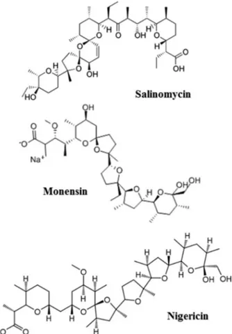
P. falciparum cultures. P. falciparum cultures were carried out according to the method of Trager and Jensen (17), with minor modifications. The P. falciparum strains used in this study are either CQ sensitive (D10, 3D7, and the Ghana isolate) or CQ resistant (W2 and the Burkina Faso isolate). The resistance profile of W2 is well documented in the literature (18), whereas the P. falciparum Burkina Faso isolate is considered resistant, since its 50% inhibitory concentration (IC50) of chloroquine is⬎100 nM, which is commonly accepted as the threshold for resistance (19). All the strains were maintained at 5% hematocrit (human type A-positive red blood cells for D10, W2, and the Burkina Faso and Ghana isolates and O-positive red blood cells for 3D7) in RPMI 1640 medium containing 24 mM sodium bicarbonate (EuroClone; Celbio), with the addition of 0.01% hypoxanthine, 20 mM HEPES, and 2 mM glutamine. All the parasites were cultured in the presence of 1% AlbuMAX II (lipid-rich bovine serum albumin), except the 3D7 strain, which was cultured in the presence of 10% (vol/vol) naturally clotted heat-inactivated O⫹ human serum (In-terstate Blood Bank, Inc.), which ensures constant and high gametocyte production. All the cultures were maintained at 37°C in a standard gas mixture consisting of 1% O2, 5% CO2, and 94% N2. The P. falciparum Ghana and Burkina Faso isolates derived from two different patients with malaria admitted to the Ospedale Sacco (Milan, Italy) returning to Milan from Ghana or Burkina Faso, respectively. The parasites were adapted to grow in culture and are not clonal strains.
Gametocytogenesis was triggered as previously described (20). Briefly, P. falciparum 3D7 asexual parasite cultures were diluted to 0.5% para-sitemia, and the medium was changed daily without the addition of fresh red blood cells (RBC). When a parasitemia of⬎5% was obtained and the parasites were stressed by nutrient deprivation, the cultures were treated for 48 to 72 h with N-acetylglucosamine (NAG) (Sigma-Aldrich) to clear residual asexual parasites. Stage II and III gametocytes were obtained and used for the experiments after 4 days after the addition of NAG to the culture, while stage IV and V gametocytes were used after 7 to 8 days. Gametocyte stages were routinely checked in the Giemsa-stained smears. In vitro P. falciparum drug susceptibility assay. Salinomycin, monensin, and nigericin sodium salts (Sigma-Aldrich) (Fig. 1) were dis-solved in either dimethyl sulfoxide (DMSO) (salinomycin and nigericin) or ethanol (monensin) and then diluted with medium to achieve the required concentrations (final DMSO or ethanol concentration of⬍1%, which is nontoxic to the parasites). The compounds were placed in 96-well plates (EuroClone) and serial dilutions were made in a final volume of 100l/well. Asexual parasite stages derived from asynchronous cul-tures with parasitemia of 1 to 1.5% or gametocyte culcul-tures with para-sitemia of 0.5 to 1% were distributed into the plates (100l/well; final hematocrit, 1%) and incubated for 72 h at 37°C. Chloroquine (CQ) and dihydroartemisinin (DHA) were used as a reference control for the asex-ual stage, and DHA and epoxomicin were used for gametocytes. Three experiments in duplicate were performed with asexual parasites, with at
least two experiments in duplicate or triplicate with gametocytes in each stage (II and III or IV and V). Parasite growth was determined spectro-photometrically by measuring the activity of the parasite lactate dehydro-genase (pLDH), according to a modified version of Makler’s method in control and treated cultures (21). Briefly, the drug-treated culture was resuspended, and 20l/well was transferred to a plate containing 100 l of Malstat reagent (0.11% [vol/vol] Triton-100, 115.7 mM lithiumL-lactate, 30.27 mM Tris, 0.62 mM 3-acetylpyridine adenine dinucleotide [APAD] [Sigma-Aldrich], adjusted to pH 9 with 1 M HCl) and 25l of PES/NBT (1.96 mM nitroblue tetrazolium chloride-0.24 mM phenazine ethosul-fate) to perform the pLDH assay. The plate was read at a wavelength of 650 nm using a microplate reader, Synergy4 (BioTek), and the results were expressed as the 50% inhibitory concentration (IC50).
In the gametocyte assay, the pLDH assay was performed at both 72 and 144 h, as described previously (20). After 72 h of incubation, 150l/well supernatant was collected and checked for hemolysis, and 150l fresh medium was added. Twenty microliters per well of resuspended culture was used to perform the pLDH assay, the plate was incubated for a further 72 h, and the pLDH assay was performed again under the same condi-tions.
In vitro mammalian cell toxicity assay. A long-term cell line of hu-man dermal microvascular endothelial cells (HMEC-1) immortalized by simian virus 40 (SV40) large T antigen was kindly provided by the Centers for Disease Control and Prevention, Atlanta, GA, USA (22). The cells were maintained under standard conditions at 37°C in a 5% CO2incubator in MCDB 131 medium (Gibco-BRL, Paisley, Scotland) supplemented with 10% fetal calf serum (HyClone, Logan, UT, USA), 10 ng/ml epidermal growth factor (PeproTech, Rocky Hill, NY, USA), 1g/ml hydrocorti-sone (Sigma Italia, Milan, Italy), 2 mM glutamine (EuroClone, Pero, It-aly), 100 U/ml penicillin, 100 mg/ml streptomycin (EuroClone), and 20
mM HEPES buffer (pH 7.3) (EuroClone). For the toxicity experiments, HMEC-1 cells at 1.0⫻ 104cells/100l/well were plated in 96-well plates and incubated at 37°C and 5% CO2overnight. The cells were then treated for 72 h with different doses of salinomycin, monensin, or nigericin di-luted as described above (final volume, 200l/well). Three independent experiments were performed in duplicate. The 3-(4,5-dimethyl-2-thia-zolyl)-2,5-diphenyl-2H tetrazolium bromide (MTT) (Sigma) cytotoxicity assay was used to measure cell viability, as described elsewhere (23). Cy-totoxicity was expressed as the 50% inhibitory concentration (IC50).
IC50and selectivity index. The results of the chemosensitivity assays
were expressed as the percent viability compared to the untreated con-trols, calculated with the following formula: 100⫻ ([OD of treated sample ⫺ blank]/[OD of untreated sample⫺ blank]) (OD, optical density). As a blank, uninfected RBCs were used. The percent viability was plotted as a function of drug concentrations, and the curve fitting was obtained by nonlinear regression analysis using a four-parameter logistic method (software Gen5 1.10 provided with the Synergy4 plate reader [Biotek]). The IC50was extrapolated as the dose that induced a 50% inhibition of gametocyte viability.
The selectivity index (SI) was calculated to evaluate the toxicity impact of salinomycin, monensin, or nigericin against normal human cells com-pared to the toxicity against the parasite, and this allows an assessment of the selectivity of these drugs for the parasite. The SI was calculated as the ratio between the cytotoxic IC50s against HMEC-1, calculated as previ-ously described, and the parasitic IC50s against 3D7 gametocytes at stages IV and V for both the time points 72 h and 72 plus 72 h.
Time course experiments. Two different time course protocols were
employed. In the first case, stage IV and V gametocytes were incubated with salinomycin, monensin, or nigericin at 1, 10, or 100 nM for different lengths of time (2, 6, 24, 48, or 72 h), and the pLDH assay was performed at the end of each treatment.
In the second series of experiments, stage IV and V gametocytes were incubated with salinomycin at 1, 10, or 100 nM for different lengths of time (2, 6, 24, 48, or 72 h), the drugs were removed, fresh medium was added, the plate was centrifuged, and the medium was changed again. The pLDH assay was performed 72 h after the onset of the experiment, i.e., 72 h after the addition of the drugs to the cultures.
For both experimental schemes, two independent experiments were performed in triplicate, and the results were expressed as the percent viability compared to that of the untreated controls.
Hemolysis. Gametocyte cultures (about 0.5 and 1% parasitemia),
fresh RBCs, or RBCs kept for 10 days under the same culture conditions as the gametocytes were diluted to 1% hematocrit and treated for 72 h with different doses (1, 10, and 100 nM) of salinomycin, monensin, or nigeri-cin. Hemolysis was evaluated by measuring spectrophotometrically the release of hemoglobin in the supernatants (absorbance at 405 nm, Soret band) and calculating the ratio of untreated to ionophore-treated sam-ples. Similar experiments were performed on mouse RBCs. Blood from uninfected or Plasmodium berghei-infected mice, diluted to 7.5% hemat-ocrit, was incubated for 24 h at 19°C with monensin and salinomycin at doses ranging from 1 to 50 nM. Hemoglobin release in the well superna-tants was measured as described above.
The percentages of hemolysis for both human and mouse RBCs were estimated by referring to a standard curve prepared with serially diluted RBCs, which were lysed with saponin (1%).
Early sporogonic development P. berghei assay. P. berghei CTRPp.GFP,
a strain expressing the green fluorescent protein (GFP) exclusively at early sporogonic stages (zygotes, ookinetes, and early oocysts), was used (kindly provided by R. Sinden, Imperial College, London, United King-dom) to infect BALB/c mice and recover P. berghei gametocytes for the early sporogonic development assay. Experimental animal rearing and handling were in compliance with the Italian legislative decree on the protection of animals used for experimental and other scientific purposes (24) and in full adherence with the European Directive 2010/63/UE (25). The early sporogonic development assay was performed according to
the protocol developed by Delves and colleagues (26), with slight modifi-cations. Salinomycin and monensin were dissolved in DMSO and etha-nol, respectively, and then diluted further to obtain the desired concen-trations with ookinete medium (RPMI 1640 containing 25 mM HEPES, 25 mM sodium bicarbonate, 50 mg/liter hypoxanthine, 100M xan-thurenic acid [pH 7.6 to 8], supplemented with 20% heat-inactivated fetal bovine serum, 50 U/ml penicillin, and 50g/ml streptomycin). All chem-icals were purchased from Sigma-Aldrich. Aliquots (20l) of compounds at a concentration 10 times higher than the desired test concentrations were then added to the microplate wells (96-well plates; Nunc, Denmark) containing 165l of ookinete medium. Subsequently, 15 l of gameto-cytemic blood obtained by cardiac puncture from P. berghei CTRPp.GFP-infected mice were added and the plates incubated at 19°C. DMSO and ethanol at 0.3% were used as solvent controls. After 24 h of incubation, the well contents were diluted (1:550) in another plate, and the zygotes and ookinetes expressing green fluorescent protein were enumerated with the help of an ocular grid with a fluorescence microscope (⫻400 magnifica-tion; Zeiss). Compounds were tested in triplicate in two independent experiments using different gametocyte donor mice. The data are ex-pressed as the percent inhibition of zygote and ookinete formation in drug-treated samples compared to that of the controls.
Luminescent standard membrane feeding assays. Compounds were
serially diluted in DMSO and subsequently in parasite culture medium to reach a final DMSO concentration of 0.1%. Luminescent standard mem-brane feeding assays were performed essentially as described by Stone et al. (27), with the exception that we used an hsp70 promoter instead of the elongation factor 1 alpha (EF-1␣) promoter to drive the expression of the luciferase reporter gene (M. W. Vos, W. J. R. Stone, K. M. J. Koolen, G. van Gemert, B. van Schaijk, R. W. Sauerwein, T. Bousema, and K. J. Decher-ing., unpublished data). Luminescence intensities were analyzed for 24 mosquitoes per sample. To determine the background luminescence lev-els, 24 uninfected mosquitoes were analyzed in parallel. For the determi-nation of oocyst prevalence, mosquitoes were considered infected when the luminescence activity was 5 standard deviations above the average luminescence level observed in the uninfected mosquitoes.
Statistical analysis. The data were expressed as the mean⫾ standard
deviation (SD) and analyzed using a two-tailed Student t test with a level of significance of a P value of⬍0.05 or ⬍0.01.
RESULTS
Ionophores inhibit asexual P. falciparum growth. Salinomycin,
monensin, and nigericin were tested in vitro for antiplasmodial
activity against three CQ-sensitive (D10, 3D7, and the Ghana
iso-late) and two CQ-resistant (W2 and the Burkina Faso isoiso-late)
strains of P. falciparum using CQ and DHA as reference drugs. All
the ionophores displayed a strong inhibition of asexual parasite
growth against all the tested strains (
Table 1
). The activity of
monensin (IC
50range, 0.5 to 1.0 nM) and nigericin (IC
50range,
1.8 to 1.9 nM) against P. falciparum laboratory strains was
supe-rior to that of DHA (IC
50range, 2.1 to 5.6 nM). Against the
CQ-sensitive parasites, the potency of salinomycin was slightly lower
than that of CQ (IC
50range, 22 to 40 versus 11 to 19 nM CQ),
while monensin and nigericin were approximately 10-fold more
active than CQ. Differently from CQ, all the ionophores showed
low-nanomolar activity against the W2 and Burkina Faso strains,
which indicates the absence of cross-resistance.
Ionophores inhibit P. falciparum gametocyte viability.
Us-ing the pLDH assay, we were able to calculate the IC
50for all the
ionophores, even after the first 72 h of incubation. The
iono-phores are far more potent than the reference compound
ep-oxomicin or DHA. Neither compound induced a 50%
reduc-tion in gametocyte viability at the 72-h time point, even at the
highest concentration tested, but required an additional 72 h
without drugs to obtain a
⬎50% inhibition of viability and thus
to calculate the IC
50(
Table 2
) (
20
). Therefore, the ionophores
appear to be the most potent compounds tested so far in our
assay, in that they reduce gametocyte viability over a very short
period, with an IC
50in the nanomolar range.
Selective chemosensitivity of gametocytes in different phases of
their development was previously described (
28
). When the IC
50of salinomycin was determined against stage II and III young
ga-metocytes or stage IV and V mature gaga-metocytes, the results were
significantly different (
Table 2
). After 72 h of treatment,
salino-mycin appeared to be more effective (lower IC
50) on mature than
on young gametocyte stages. After 72 plus 72 h of incubation, the
differences between stages II and III and IV and V remained
sig-nificant. Monensin and nigericin did not show a significant
differ-ence in activity against young versus mature gametocytes.
The IC
50of the ionophores on human endothelial cells
(HMEC-1) was lower than that of DHA, suggesting potential
tox-icity. However, due to the higher activity on mature gametocytes,
the selectivity indices of salinomycin and monensin (25 and 29.8,
respectively) are comparable to that of DHA (19.8), and that of
nigericin was even higher (96.0).
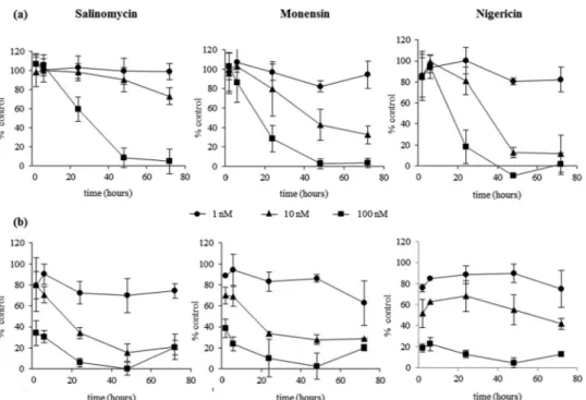
To investigate the time to kill, time course experiments were
performed by treating gametocytes with different doses of
com-pounds for various pulse-inhibitory periods (2, 6, 24, 48, and 72 h)
(
Fig. 2a
and
b
). When the pLDH activity was evaluated
immedi-ately after salinomycin treatment (
Fig. 2a
), the gametocyte
viabil-ity was reduced by 41% with the 100 nM dose only and with a
minimum incubation time of 24 h. Lower doses were ineffective.
Complete inhibition was observed after 48 h. The time-dependent
inhibition of gametocyte viability by monensin or nigericin was
similar to that of salinomycin at 100 nM, at a higher rate than with
the 10 nM dose (at 72 h, inhibition of gametocyte viability was
27% for salinomycin, 68% for monensin, and 88% for nigericin).
In a subsequent series of experiments, the drugs were
re-moved after the different pulse-inhibitory periods, fresh
me-dium was added, and the incubation was continued up to 72 h,
when the pLDH activity was evaluated (
Fig. 2b
). The results
showed that salinomycin at 100 nM already inhibited pLDH
activity by 65% after 2 h, whereas the same percentage of
inhi-bition was obtained at the 10 nM dose only after 24 h. In this set
of experiments, for each dose, the maximal inhibition was
ob-served at 24 h, without any further increase up to 72 h. The
activity of monensin and nigericin was similar to that of
sali-nomycin, with 61% and 82% inhibition, respectively, already
after 2 h at the 100 nM dose.
Hemolytic effect of ionophores is limited to infected RBCs at
doses higher than the IC
50. To study the possible toxic effects of
ionophores on RBCs, hemolysis was measured
spectrophoto-metrically by determining the hemoglobin release in the
super-natants of infected RBCs and, for comparison, of fresh
unin-fected RBCs or cultured uninunin-fected RBCs. The cells were
treated with different doses of ionophores for 72 h. The
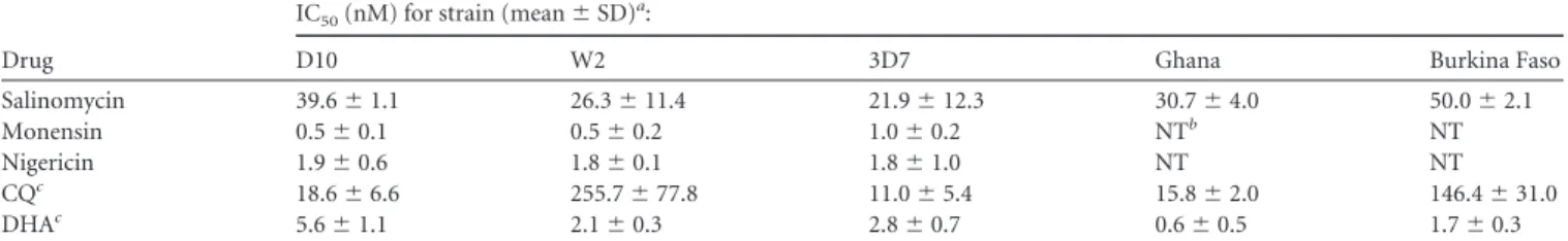
per-TABLE 1 In vitro antimalarial activities of salinomycin, monensin, and nigericin against asexual P. falciparum parasitesDrug
IC50(nM) for strain (mean⫾ SD)a:
D10 W2 3D7 Ghana Burkina Faso
Salinomycin 39.6⫾ 1.1 26.3⫾ 11.4 21.9⫾ 12.3 30.7⫾ 4.0 50.0⫾ 2.1
Monensin 0.5⫾ 0.1 0.5⫾ 0.2 1.0⫾ 0.2 NTb NT
Nigericin 1.9⫾ 0.6 1.8⫾ 0.1 1.8⫾ 1.0 NT NT
CQc 18.6⫾ 6.6 255.7⫾ 77.8 11.0⫾ 5.4 15.8⫾ 2.0 146.4⫾ 31.0
DHAc 5.6⫾ 1.1 2.1⫾ 0.3 2.8⫾ 0.7 0.6⫾ 0.5 1.7⫾ 0.3
aData are from three different experiments performed in duplicate. P. falciparum strains D10 and 3D7 (CQ sensitive), strain W2 (CQ resistant), and two isolates (not clonal) from
different patients coming from Ghana and Burkina Faso were used in this study.
bNT, not tested. c
Chloroquine (CQ) and dihydroartemisinin (DHA) were used as positive controls.
TABLE 2 In vitro antimalarial activities of salinomycin, monensin, and nigericin against different stages of 3D7 P. falciparum gametocytes
Drug Gametocyte stages
IC50(nM) for incubation for (h)a:
SI for incubation for (h)b: HMEC-1 IC50(nM)a 72 72⫹ 72 72 72⫹ 72 Salinomycin II–III 29.7⫾ 9.2c 14.5⫾ 7.4d 157.5⫾ 45.0 IV–V 13.8⫾ 1.4 6.3⫾ 1.7 11.4 25.0 Monensin II–III 4.6⫾ 1.0 1.9⫾ 1.3 169.6⫾ 48.0 IV–V 6.1⫾ 1.1 5.7⫾ 1.1 27.8 29.8 Nigericin II–III 10.6⫾ 4.0 2.7⫾ 1.2 86.4⫾ 31.0 IV–V 9.3⫾ 4.9 0.9⫾ 0.4 9.3 96.0 DHA II–III ⬎500 14.3⫾ 8.7 3,094.8⫾ 316.5 IV–V ⬎500 156.3⫾ 78.3 NAe 19.8 Epoxomicin II–III ⬎100 12.1⫾ 4.5 11.7⫾ 3.1 IV–V ⬎100 6.7⫾ 1.9 NA 1.7
aData are the means⫾ SD from at least two independent experiments in duplicate or triplicate. b
SI, selectivity index, calculated as the IC50HMEC-1/IC50gametocytes at stage IV/V at the indicated incubation time. cP⬍ 0.005, effect of salinomycin on stage II and III versus stage IV and V gametocytes at 72 h.
d
P⬍ 0.005, effect of salinomycin on stage II and III versus stage IV and V gametocytes at 72 plus 72 h.
centage of hemolysis induced on fresh RBCs was
⬍5% in the
control untreated RBCs and in RBCs treated with different
doses of all the ionophores. Both infected and uninfected RBCs
kept at 37°C for 10 days showed a spontaneous hemolysis of
about 15%. However, only in the case of infected RBCs did the
percentages of hemolysis increase in a dose-dependent manner
after ionophore treatment, reaching maximum values of 32, 22,
and 34% after treatment with 100 nM salinomycin, monensin,
and nigericin, respectively. To better compare the hemolytic
effect of the ionophores in the different experimental groups,
the results were expressed as the ratio of the optical density at
405 nm (OD
405) of ionophore-treated RBCs to that of
un-treated RBCs. As shown in
Fig. 3
, the 100 nM dose of
salino-mycin, monensin, or nigericin caused a 3.1-fold, 2.3-fold, or
2.9-fold increase, respectively, in the release of hemoglobin in
the supernatants of treated compared to those of the untreated
gametocyte cultures. The increase in hemoglobin release was
much lower (salinomycin, 1.4-fold; monensin, 1.2-fold;
nige-ricin, 1.3-fold) for cultured uninfected RBCs. Moreover, a dose
close to the IC
50on gametocytes (10 nM) was hemolytic on
gametocyte-infected RBCs but not on normal uninfected
RBCs.
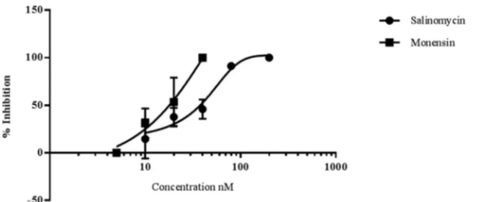
Ionophores inhibit early sporogonic P. berghei development
in vitro. Using an early sporogonic stage-specific GFP reporter
strain of the rodent parasite P. berghei (CTRPp.GFP), the capacity
of gametocytes to undergo in vitro early sporogonic development
(gamete formation, zygote formation, and ookinete maturation)
was examined in the presence of the ionophores.
As illustrated in
Fig. 4
, early sporogonic development was
in-hibited by both ionophores in a dose-dependent manner.
Inhibi-FIG 2 Time course of the activity of salinomycin, monensin, or nigericin on mature gametocytes. Gametocytes were incubated with the ionophores at 1 nM(circles), 10 nM (triangles), or 100 nM (squares) for different lengths of time (2, 6, 24, 48, and 72 h). (a) The pLDH viability assay was performed at the end of each incubation time. (b) The compounds were removed after each incubation time, fresh medium was added, and the pLDH assay was performed at 72 h. The results are the means⫾ SD from two independent experiments in triplicate.
FIG 3 Hemolytic effects of salinomycin (a), monensin (b), and nigericin (c) evaluated by measuring the release of hemoglobin (OD405) in test supernatants. The histograms represent the ratio of the OD of the treated cells to that of the untreated cells. Black bars, gametocyte-infected RBCs; striped bars, fresh RBCs; white bars, normal RBCs kept under the same culture conditions as infected RBCs at 37°C forⱖ7 days. The data are the means ⫾ SD from at least three independent experiments in triplicate.
tion of
ⱖ90% was recorded for salinomycin at 80 nM and for
monensin at 40 nM. From a comparison of the IC
50s, it appeared
that monensin (IC
50, 16.8
⫾ 2.5) was about twice as active as
salinomycin (IC
50, 34.9
⫾ 5.1).
To assess the toxicity of ionophores on infected and uninfected
mouse RBCs, the release of hemoglobin was measured in wells
incubated for 24 h with monensin or salinomycin at
concentra-tions ranging from 1 to 50 nM. No appreciable hemolytic activity
(⬍2%) on uninfected RBCs was observed with monensin or
sali-nomycin at doses between 1 and 10 nM or 1 and 25 nM,
respec-tively. However, a 6- or 8-fold increase in hemolysis was recorded
in wells containing infected RBCs after treatment with monensin
at 10 nM or salinomycin at 25 nM. Given that early sporogonic
development is an extracellular process, it is unlikely that the
ef-fects of ionophores observed on early sporogonic stages and red
blood cells lysis are related.
Ionophores block P. falciparum oocyst development. P.
fal-ciparum stage V gametocytes of an hsp70-GFP:luc reporter strain
were incubated with compounds for 24 h and subsequently fed to
Anopheles stephensi mosquitoes in the standard membrane
feed-ing assays (
27
).
Figure 5
shows that salinomycin and monensin
dose-dependently reduced oocyst intensity in the mosquito
midgut, as indicated by a reduction in luciferase activity. Both
compounds showed IC
50s in the low-nanomolar range (IC
50, 1.9
nM for both drugs). The reduction in oocyst intensity translated
into a reduction in oocyst prevalence or the number of infected
FIG 4 Salinomycin and monensin block early sporogonic-stage development of P. berghei in vitro. The figure illustrates the mean percentages of inhibition andstandard deviations for early sporogonic stages (zygotes and ookinetes) by salinomycin and monensin as a function of the compound concentration indicated on the x axis. The data are from 2 or 3 replicate experiments in triplicate wells, except for the 5 nM and 200 nM doses, which are based on a single experiment.
FIG 5 Salinomycin and monensin block oocyst development in the mosquito. P. falciparum stage V gametocytes of a transgenic hsp70-GFP::luc reporter strain
were incubated for 24 h with different doses of compounds and fed to A. stephensi mosquitoes. (a and b) Luminescence intensities in individual mosquitoes as a function of the compound concentration indicated on the x axis. (c and d) Oocyst prevalence (percentage of infected mosquitoes) as a function of the compound concentration indicated on the x axis.
mosquitoes (
Fig. 5c
and
d
). The IC
50of monensin was 1.3 nM and
that of salinomycin was 18 nM. These data indicate that monensin
and salinomycin effectively block P. falciparum transmission in
the nanomolar range.
DISCUSSION
Repositioning or repurposing of existing drugs for another
appli-cation has already been proposed in the malaria field and applied
to existing libraries against the asexual stages of P. falciparum.
Successful repositioning of drugs significantly benefits the clinical
development pipeline in term of costs and time (
8
). Here, we
demonstrated the antimalarial and transmission-blocking
activi-ties of a specific class of compounds, the monovalent ionophores
(i.e., salinomycin, monensin, and nigericin), polyether antibiotics
that are used in several countries as anticoccidial agents in poultry.
Despite being registered and FDA approved for veterinary use
since 1974, only recently have the ionophores, and salinomycin in
particular, experienced renewed interest as potential human
anti-cancer agents (
10
,
13
).
Our data show that salinomycin, monensin, and nigericin are
very active in vitro against a number of P. falciparum isolates,
including drug-resistant strains, in accordance with other reports
(
16
,
29–33
). Importantly, we also show that these molecules
pos-sess in vitro nanomolar activity against mature P. falciparum stage
IV and V gametocytes and against P. berghei ookinetes, the earliest
development stage in the mosquito vector. These data corroborate
previous findings with salinomycin and monensin on P.
falcipa-rum gametocytes after a large library screening (
15
). The cytocidal
activity against gametocytes and ookinetes apparently leads to a
functional block of oocyst development in the standard
mem-brane feeding assays at concentrations in the nanomolar range.
Monovalent ionophores are known for their activity as
anti-fungal, antiparasitic, and antiviral agents, especially against
mul-tidrug-resistant pathogens. Recent reports highlighted their
activ-ity against Trypanosoma brucei, Toxoplasma gondii, and
cytomegalovirus (
33–36
). Considerable attention has been
de-voted recently to salinomycin and derivatives due to their selective
activity against tumor stem cells (
13
,
37
,
38
). A pilot study has
been conducted in a few cancer patients, under compassionate
use, and salinomycin was shown to induce tumor regression with
minor side effects, which were lower than those of conventional
chemotherapeutic drugs (
13
). In vitro, salinomycin has been
shown to be active against multidrug-resistant cancer cells of
dif-ferent origins by inducing cell cycle arrest in G
1phase (
39
),
inhib-iting cancer cell motility (
40
), modulating the autophagy process
(
41
), or inducing leukemia cell apoptosis via the inhibition of Wnt
signaling (
42
). The inhibition of Wnt signaling also seems to be
involved in the inhibition of cytomegalovirus replication by
sali-nomycin (
35
). In P. falciparum, the autophagy pathway is not yet
completely characterized, but autophagy-related (ATG) proteins
are abundantly expressed in P. falciparum gametocytes, suggesting
that they may play a role in gametocytogenesis (
43
). Since
monen-sin has been shown to induce autophagy and death of T. gondii
(
44
), we cannot exclude a similar effect of ionophores on P.
falcip-arum.
Our data and reports in the literature confirm that the
spec-trum of action of monovalent ionophores against P. falciparum is
quite broad and not stage specific. Here, we demonstrate that
ionophores inhibit the viability of stage IV and V gametocytes, as
already described for all stages of asexual parasites (
14
). In
addi-tion, there is evidence that salinomycin and monensin are effective
against liver stages, both in vitro (HepG2 cells infected by P.
ber-ghei sporozoites) and orally in P. berber-ghei-infected C57BL/6 mice in
vivo (
32
,
33
). Unlike many reference drugs, like DHA (
28
,
45
),
salinomycin was more active on mature (stage IV and V) than
young (stage II and III) gametocytes, which reinforces the
trans-mission-blocking potential of this molecule. Mature gametocytes
(stages IV and V) are metabolically quiescent (no protein
synthe-sis or hemoglobin digestion) and thus relatively more resynthe-sistant
than early stage gametocytes or asexuals (
46
,
47
). Only
prima-quine is effective in reducing late-stage gametocytemia in vivo
(
48
). In that respect, the ionophores, and salinomycin in
particu-lar, compare quite well with the most advanced new antimalarial
drug candidates of the MMV portfolio, including the
spiroin-dolone KAE609 (
49
), the imidazolopiperazine KAF 156, and the
quinolone-3-diarylether ELQ-300 (
50
,
51
). All these compounds
have been shown to inhibit early and late P. falciparum
gameto-cytes at concentrations between 50 and 500 nM.
Ionophores have a relatively fast speed of action. More than
50% inhibition of gametocyte viability was seen at the first 72 h of
the pLDH assay, whereas a longer incubation (72 plus 72 h) was
necessary to observe the same effect with the reference drugs
DHA, epoxomicin (
Table 2
), and methylene blue (
20
). This is
clearly related to the mode of action of the ionophores, but it also
emphasizes the advantages of the dual-time pLDH assay, which
discriminates between fast- and slow-acting compounds. The
minimal contact time required to achieve
⬎50% inhibition of
gametocyte viability was 24 h for salinomycin and monensin at 10
nM and only 2 h for all three compounds at 100 nM. Indeed,
salinomycin showed a similar time course activity on the
intra-erythrocytic parasite Babesia gibsoni, whereas 4 h was reported as
the time for monensin to achieve 50% inhibition of asexual P.
falciparum parasites (
14
,
52
). As far as we know, an activity so fast
has not yet been described for any other molecule (
53
).
Due to the importance of ion balance for RBCs, one of the
potential side effects of Na
⫹and K
⫹monovalent ionophores is the
risk of hemolysis. However, few and controversial data are
re-ported in the literature (
54
). Here, we show that a dose of
iono-phores close to the IC
50for gametocytes (10 nM) was hemolytic
only for gametocyte-infected but not uninfected normal human
RBCs. Similarly, little hemolysis of uninfected mouse erythrocytes
was observed after ionophore treatment in the concentration
range of 10 to 25 nM, whereas on P. berghei-infected RBCs, a 6- to
8-fold increase in hemoglobin release in ionophore-treated
com-pared to untreated samples was observed.
These data highlight the specificity of ionophores for infected
versus normal RBCs. Similar findings were reported by Gumila et
al. (
55
) using asexual parasites and were attributed to the
modifi-cation of the membrane properties of infected RBCs and the
dif-ferent phospholipid and lipid composition of parasite
mem-branes. All ionophores strongly interact with phospholipid
monolayers and facilitate the incorporation and transport of ions
in low-cholesterol-containing membranes typical of asexual P.
falciparum parasites (
55
,
56
). Whether this may also apply to
ga-metocyte-infected RBCs is not known yet. Selectivity and
antipa-rasitic activity might also depend on the ability of monovalent
ionophores to induce K
⫹efflux from the cytosol with a
conse-quent increase in the influx of Na
⫹and water. The parasite
com-partment is characterized by high K
⫹and Ca
2⫹levels and low Na
⫹levels with respect to the infected RBC cytosol, which is poor in K
⫹and rich in Na
⫹(
57
,
58
). This is different from normal RBCs,
which are maintained at very high K
⫹and low Na
⫹concentra-tions by the Na
⫹-K
⫹ATPase pump. Perturbation of the cation
gradient and/or content in infected versus normal RBCs or in the
parasite cytosol may be responsible for the selective antimalarial
effects of ionophores. A similar mechanism has been proposed to
explain the activity of salinomycin against T. brucei and B. gibsoni
(
34
,
52
) and the egress of T. gondii from host cells (
59
).
To consider the potential use of ionophores as antimalarial
drugs, it is of paramount importance to address clinical safety. In
animals, monovalent ionophores can induce considerable
neuro-toxicity and cardioneuro-toxicity after accidental exposure in nontarget
species, such as calves, cats, and horses (
60–62
). No appreciable
risk for humans was demonstrated following the consumption of
products from nontarget animals (
63
). However, preclinical
phar-macological and safety data are scarce. Toxicity in humans has
been reported in cases of accidental high-dose ingestion in people
working with livestock (
64
,
65
). In our assays, salinomycin
ap-peared to be the most toxic for HMEC-1 cells among the
iono-phores tested, but the selectivity index was similar to that of the
antimalarial drug DHA. Salinomycin exhibits an IC
50similar to
that of mefloquine when tested against HepG2 cells, whereas
monensin showed no significant toxicity at the highest tested
con-centration (46
M) (
15
). The narrow therapeutic index of
salino-mycin as a potential anticancer drug has already boosted the
de-velopment of chemically modified derivatives or innovative drug
delivery systems (
66–68
), which should be considered in their
future development as new antimalarials.
The pharmacokinetic properties of salinomycin as an
anticoc-cidial agent have been fully investigated in several animal species.
As a highly lipophilic drug, salinomycin is well absorbed and
dis-tributed throughout the plasma and tissues (especially fat in
chickens) (
66
). In mice, its oral and intravenous bioavailability
was shown to be limited by the presence of the P-glycoprotein; at
a dose of 1 mg/kg of body weight, the peak concentration was
reached after 2 h, and the half-life was approximately 6 h (
69
). So
far, no detailed information is available on salinomycin
pharma-cokinetic properties or plasma levels in humans. In a pilot study of
metastatic cancer patients, the dose of 200
g/kg of intravenous
salinomycin was given every second day for 12 to 14 days; this
protocol was well tolerated and led to partial clinical regression of
heavily pretreated and therapy-resistant cancers (
13
). The
suit-ability of long-term treatment might be an advantage for a
trans-mission-blocking agent, since mature gametocytes persist in the
blood for up to 2 weeks. However, the limited absorption,
distri-bution, metabolism, and excretion (ADME)/Tox data presently
available in animal models suggest that new derivatives with
lon-ger half-lives and improved safety profiles need to be developed.
In conclusion, our data suggest a strong and fast activity of
monovalent ionophores against asexual stages and against mature
gametocytes and ookinetes, resulting in a block of sporogonic
de-velopment in the mosquito. These compounds, being active
against several different P. falciparum life stages, thus appear to be
particularly attractive as potential curative and
transmission-blocking agents, as advocated by the International Malaria
Con-trol Program and by the MMV (
6
).
ACKNOWLEDGMENTS
We thank Pietro Alano and his group for sharing gametocyte culture technology and for helpful suggestions, Daniela Jabes (NAICONS, Milan,
Italy) for providing the molecules that initiated this work, and Laura Galastri, Paola Verducci, and Tiziana Bianchi from AVIS Comunale Mi-lano for providing blood samples for parasite culture.
This work was supported by the Global Health Program of the Bill & Melinda Gates Foundation (grant OPP1040394).
REFERENCES
1. WHO. 2013. World malaria report. World Health Organization, Ge-neva, Switzerland. http://www.who.int/malaria/publications/world _malaria_report_2013/wmr2013_no_profiles.pdf?ua⫽1.
2. White NJ, Pukrittayakamee S, Hien TT, Faiz MA, Mokuolu OA,
Don-dorp AM. 2014. Malaria. Lancet 383:723–735.http://dx.doi.org/10.1016 /S0140-6736(13)60024-0.
3. Dondorp AM, Nosten F, Yi P, Das D, Phyo AP, Tarning J, Lwin KM,
Ariey F, Hanpithakpong W, Lee SJ, Ringwald P, Silamut K, Imwong M, Chotivanich K, Lim P, Herdman T, An SS, Yeung S, Singhasivanon P, Day NP, Lindegardh N, Socheat D, White NJ. 2009. Artemisinin
resis-tance in Plasmodium falciparum malaria. N Engl J Med 361:455– 467.http: //dx.doi.org/10.1056/NEJMoa0808859.
4. White NJ. 2014. Malaria: a molecular marker of artemisinin resis-tance. Lancet 383:1439 –1440. http://dx.doi.org/10.1016/S0140-6736 (14)60656-5.
5. Burrows JN, Burlot E, Campo B, Cherbuin S, Jeanneret S, Leroy D,
Spangenberg T, Waterson D, Wells TN, Willis P. 2014. Antimalarial
drug discovery—the path towards eradication. Parasitology 141:128 –139. http://dx.doi.org/10.1017/S0031182013000826.
6. Burrows JN, van Huijsduijnen RH, Möhrle JJ, Oeuvray C, Wells TN. 2013. Designing the next generation of medicines for malaria control and eradication. Malar J 12:187.http://dx.doi.org/10.1186/1475-2875-12-187. 7. Alano P. 2007. Plasmodium falciparum gametocytes: still many secrets of a hidden life. Mol Microbiol 66:291–302.http://dx.doi.org/10.1111/j.1365 -2958.2007.05904.x.
8. Lotharius J, Gamo-Benito FJ, Angulo-Barturen I, Clark J, Connelly M,
Ferrer-Bazaga S, Parkinson T, Viswanath P, Bandodkar B, Rautela N, Bharath S, Duffy S, Avery VM, Möhrle JJ, Guy RK, Wells T. 2014.
Repositioning: the fast track to new anti-malarial medicines? Malar J 13: 143.http://dx.doi.org/10.1186/1475-2875-13-143.
9. Mitani M, Yamanishi T, Miyazaki Y, O take N. 1976. Salinomycin effects on mitochondrial ion translocation and respiration. Antimicrob Agents Chemother 9:655– 660.http://dx.doi.org/10.1128/AAC.9.4.655. 10. Tang QL, Zhao ZQ, Li JC, Liang Y, Yin JQ, Zou CY, Xie XB, Zeng YX,
Shen JN, Kang T, Wang J. 2011. Salinomycin inhibits osteosarcoma by
targeting its tumor stem cells. Cancer Lett 311:113–121.http://dx.doi.org /10.1016/j.canlet.2011.07.016.
11. Zhou HM, Dong TT, Wang LL, Feng B, Zhao HC, Fan XK, Zheng MH. 2012. Suppression of colorectal cancer metastasis by nigericin through inhibition of epithelial-mesenchymal transition. World J Gastroenterol
18:2640 –2648.http://dx.doi.org/10.3748/wjg.v18.i21.2640.
12. Tumova L, Pombinho AR, Vojtechova M, Stancikova J, Gradl D,
Krausova M, Sloncova E, Horazna M, Kriz V, Machonova O, Jindrich J, Zdrahal Z, Bartunek P, Korinek V. 2014. Monensin inhibits canonical
Wnt signaling in human colorectal cancer cells and suppresses tumor growth in multiple intestinal neoplasia mice. Mol Cancer Ther 13:812– 822.http://dx.doi.org/10.1158/1535-7163.MCT-13-0625.
13. Naujokat C, Steinhart R. 2012. Salinomycin as a drug for targeting hu-man cancer stem cells. J Biomed Biotechnol 2012:950658.
14. Gumila C, Ancelin ML, Delort AM, Jeminet G, Vial HJ. 1997. Charac-terization of the potent in vitro and in vivo antimalarial activities of iono-phore compounds. Antimicrob Agents Chemother 41:523–529. 15. Sun W, Tanaka TQ, Magle CT, Huang W, Southall N, Huang R,
Dehdashti SJ, McKew JC, Williamson KC, Zheng W. 2014. Chemical
signatures and new drug targets for gametocytocidal drug development. Sci Rep 4:3743.
16. Adovelande J, Schrével J. 1996. Carboxylic ionophores in malaria che-motherapy: the effects of monensin and nigericin on Plasmodium falcipa-rum in vitro and Plasmodium vinckei petteri in vivo. Life Sci 59:PL309-PL315.
17. Trager W, Jensen JB. 2005. Human malaria parasites in continuous culture. 1976. J Parasitol 91:484 – 486.http://dx.doi.org/10.1645/0022-33 95(2005)091[0484:HMPICC]2.0.CO;2.
resistance in Plasmodium falciparum. Proc Natl Acad Sci U S A 94:9389 – 9393.http://dx.doi.org/10.1073/pnas.94.17.9389.
19. Ekland EH, Fidock DA. 2008. In vitro evaluations of antimalarial drugs and their relevance to clinical outcomes. Int J Parasitol 38:743–747.http: //dx.doi.org/10.1016/j.ijpara.2008.03.004.
20. D’Alessandro S, Silvestrini F, Dechering K, Corbett Y, Parapini S,
Timmerman M, Galastri L, Basilico N, Sauerwein R, Alano P, Taramelli D. 2013. A Plasmodium falciparum screening assay for anti-gametocyte
drugs based on parasite lactate dehydrogenase detection. J Antimicrob Chemother 68:2048 –2058.http://dx.doi.org/10.1093/jac/dkt165. 21. Makler MT, Ries JM, Williams JA, Bancroft JE, Piper RC, Gibbins BL,
Hinrichs DJ. 1993. Parasite lactate dehydrogenase as an assay for
Plasmo-dium falciparum drug sensitivity. Am J Trop Med Hyg 48:739 –741. 22. Ades EW, Candal FJ, Swerlick RA, George VG, Summers S, Bosse DC,
Lawley TJ. 1992. HMEC-1: establishment of an immortalized human
microvascular endothelial cell line. J Investig Dermatol 99:683– 690.http: //dx.doi.org/10.1111/1523-1747.ep12613748.
23. D’Alessandro S, Gelati M, Basilico N, Parati EA, Haynes RK, Taramelli D. 2007. Differential effects on angiogenesis of two antimalarial compounds, dihydroartemisinin and artemisone: implications for embryotoxicity. Toxi-cology 241:66 –74.http://dx.doi.org/10.1016/j.tox.2007.08.084.
24. Gazzetta Ufficiale. 2014. Protection of animals used for experimental and other scientific purposes. Decreto legislativo 4 marzo 2014, no. 26. Gazzetta Ufficiale, Rome, Italy.http://www.gazzettaufficiale.it/eli/id/2014 /03/14/14G00036/sg. (In Italian.)
25. European Parliament and Council. 2010. Directive 2010/63/EU of the European Parliament and of the Council of 22 September 2010 on the protection of animals used for scientific purposes. European Parliament and Council, Brussels, Belgium.http://eur-lex.europa.eu/legal-content /EN/TXT/?uri⫽celex:32010L0063.
26. Delves MJ, Ramakrishnan C, Blagborough AM, Leroy D, Wells TN,
Sinden RE. 2012. A high-throughput assay for the identification of
ma-larial transmission-blocking drugs and vaccines. Int J Parasitol 42:999 – 1006.http://dx.doi.org/10.1016/j.ijpara.2012.08.009.
27. Stone WJ, Churcher TS, Graumans W, van Gemert GJ, Vos MW, Lanke
KH, van de Vegte-Bolmer MG, Siebelink-Stoter R, Dechering KJ, Vaughan AM, Camargo N, Kappe SH, Sauerwein RW, Bousema T.
2014. A scalable assessment of Plasmodium falciparum transmission in the standard membrane-feeding assay, using transgenic parasites expressing green fluorescent protein-luciferase. J Infect Dis 210:1456 –1463.http://dx .doi.org/10.1093/infdis/jiu271.
28. Lucantoni L, Avery V. 2012. Whole-cell in vitro screening for gametocy-tocidal compounds. Future Med Chem 4:2337–2360.http://dx.doi.org/10 .4155/fmc.12.188.
29. Mahmoudi N, de Julián-Ortiz JV, Ciceron L, Gálvez J, Mazier D, Danis
M, Derouin F, García-Domenech R. 2006. Identification of new
antima-larial drugs by linear discriminant analysis and topological virtual screen-ing. J Antimicrob Chemother 57:489 – 497.http://dx.doi.org/10.1093/jac /dki470.
30. Yuan J, Cheng KC, Johnson RL, Huang R, Pattaradilokrat S, Liu A,
Guha R, Fidock DA, Inglese J, Wellems TE, Austin CP, Su XZ. 2011.
Chemical genomic profiling for antimalarial therapies, response signa-tures, and molecular targets. Science 333:724 –729.http://dx.doi.org/10 .1126/science.1205216.
31. Gumila C, Ancelin ML, Jeminet G, Delort AM, Miquel G, Vial HJ. 1996. Differential in vitro activities of ionophore compounds against Plasmo-dium falciparum and mammalian cells. Antimicrob Agents Chemother
40:602– 608.
32. Derbyshire ER, Prudêncio M, Mota MM, Clardy J. 2012. Liver-stage malaria parasites vulnerable to diverse chemical scaffolds. Proc Natl Acad Sci U S A 109:8511– 8516.http://dx.doi.org/10.1073/pnas.1118370109. 33. Leitao R, Rodriguez A. 2010. Inhibition of Plasmodium sporozoites
in-fection by targeting the host cell. Exp Parasitol 126:273–277.http://dx.doi .org/10.1016/j.exppara.2010.05.012.
34. Steverding D, Sexton DW. 2013. Trypanocidal activity of salinomycin is due to sodium influx followed by cell swelling. Parasit Vectors 6:78. 35. Kapoor A, He R, Venkatadri R, Forman M, Arav-Boger R. 2013. Wnt
modulating agents inhibit human cytomegalovirus replication. Antimi-crob Agents Chemother 57:2761–2767.http://dx.doi.org/10.1128/AAC .00029-13.
36. Lavine MD, Arrizabalaga G. 2011. The antibiotic monensin causes cell cycle disruption of Toxoplasma gondii mediated through the DNA repair
enzyme TgMSH-1. Antimicrob Agents Chemother 55:745–755.http://dx .doi.org/10.1128/AAC.01092-10.
37. Gupta PB, Onder TT, Jiang G, Tao K, Kuperwasser C, Weinberg RA,
Lander ES. 2009. Identification of selective inhibitors of cancer stem cells
by high-throughput screening. Cell 138:645– 659.http://dx.doi.org/10 .1016/j.cell.2009.06.034.
38. Antoszczak M, Huczyn´ski A. 2015. Anticancer activity of polyether iono-phone-salinomycin. Anticancer Agents Med Chem 15:575–591. 39. Koo KH, Kim H, Bae YK, Kim K, Park BK, Lee CH, Kim YN. 2013.
Salinomycin induces cell death via inactivation of Stat3 and downregula-tion of Skp2. Cell Death Dis 4:e693.http://dx.doi.org/10.1038/cddis.2013 .223.
40. Kopp F, Hermawan A, Oak PS, Herrmann A, Wagner E, Roidl A. 2014. Salinomycin treatment reduces metastatic tumor burden by hampering cancer cell migration. Mol Cancer 13:16.http://dx.doi.org/10.1186/1476 -4598-13-16.
41. Verdoodt B, Vogt M, Schmitz I, Liffers ST, Tannapfel A,
Mirmoham-madsadegh A. 2012. Salinomycin induces autophagy in colon and breast
cancer cells with concomitant generation of reactive oxygen species. PLoS One 7:e44132.http://dx.doi.org/10.1371/journal.pone.0044132. 42. Lu D, Choi MY, Yu J, Castro JE, Kipps TJ, Carson DA. 2011.
Salino-mycin inhibits Wnt signaling and selectively induces apoptosis in chronic lymphocytic leukemia cells. Proc Natl Acad Sci U S A 108:13253–13257. http://dx.doi.org/10.1073/pnas.1110431108.
43. Cervantes S, Bunnik EM, Saraf A, Conner CM, Escalante A, Sardiu ME,
Ponts N, Prudhomme J, Florens L, Le Roch KG. 2014. The
multifunc-tional autophagy pathway in the human malaria parasite, Plasmodium falciparum. Autophagy 10:80 –92.http://dx.doi.org/10.4161/auto.26743. 44. Lavine MD, Arrizabalaga G. 2012. Analysis of monensin sensitivity in
Toxoplasma gondii reveals autophagy as a mechanism for drug induced death. PLoS One 7:e42107.http://dx.doi.org/10.1371/journal.pone.004 2107.
45. Adjalley SH, Johnston GL, Li T, Eastman RT, Ekland EH, Eappen AG,
Richman A, Sim BK, Lee MC, Hoffman SL, Fidock DA. 2011.
Quanti-tative assessment of Plasmodium falciparum sexual development reveals potent transmission-blocking activity by methylene blue. Proc Natl Acad Sci U S A 108:E1214-E1223.http://dx.doi.org/10.1073/pnas.1112037108. 46. Baker DA. 2010. Malaria gametocytogenesis. Mol Biochem Parasitol 172:
57– 65.http://dx.doi.org/10.1016/j.molbiopara.2010.03.019.
47. Peatey CL, Leroy D, Gardiner DL, Trenholme KR. 2012. Anti-malarial drugs: how effective are they against Plasmodium falciparum gametocytes? Malar J 11:34.http://dx.doi.org/10.1186/1475-2875-11-34.
48. Chotivanich K, Sattabongkot J, Udomsangpetch R, Looareesuwan S,
Day NP, Coleman RE, White NJ. 2006. Transmission-blocking activities
of quinine, primaquine, and artesunate. Antimicrob Agents Chemother
50:1927–1930.http://dx.doi.org/10.1128/AAC.01472-05.
49. van Pelt-Koops JC, Pett HE, Graumans W, van der Vegte-Bolmer M,
van Gemert GJ, Rottmann M, Yeung BK, Diagana TT, Sauerwein RW.
2012. The spiroindolone drug candidate NITD609 potently inhibits ga-metocytogenesis and blocks Plasmodium falciparum transmission to anopheles mosquito vector. Antimicrob Agents Chemother 56:3544 – 3548.http://dx.doi.org/10.1128/AAC.06377-11.
50. Nilsen A, LaCrue AN, White KL, Forquer IP, Cross RM, Marfurt J,
Mather MW, Delves MJ, Shackleford DM, Saenz FE, Morrisey JM, Steuten J, Mutka T, Li Y, Wirjanata G, Ryan E, Duffy S, Kelly JX, Sebayang BF, Zeeman AM, Noviyanti R, Sinden RE, Kocken CH, Price RN, Avery VM, Angulo-Barturen I, Jiménez-Díaz MB, Ferrer S, Her-reros E, Sanz LM, Gamo FJ, Bathurst I, Burrows JN, Siegl P, Guy RK, Winter RW, Vaidya AB, Charman SA, Kyle DE, Manetsch R, Riscoe MK. 2013. Quinolone-3-diarylethers: a new class of antimalarial drug. Sci
Transl Med 5:177ra137.
51. Kuhen KL, Chatterjee AK, Rottmann M, Gagaring K, Borboa R,
Buen-viaje J, Chen Z, Francek C, Wu T, Nagle A, Barnes SW, Plouffe D, Lee MC, Fidock DA, Graumans W, van de Vegte-Bolmer M, van Gemert GJ, Wirjanata G, Sebayang B, Marfurt J, Russell B, Suwanarusk R, Price RN, Nosten F, Tungtaeng A, Gettayacamin M, Sattabongkot J, Taylor J, Walker JR, Tully D, Patra KP, Flannery EL, Vinetz JM, Renia L, Sauerwein RW, Winzeler EA, Glynne RJ, Diagana TT. 2014. KAF156 is
an antimalarial clinical candidate with potential for use in prophylaxis, treatment and prevention of disease transmission. Antimicrob Agents Chemother 58:5060 –5067.http://dx.doi.org/10.1128/AAC.02727-13. 52. Yamasaki M, Nakamura K, Tamura N, Hwang SJ, Yoshikawa M, Sasaki
mechanisms of action of ionophorous antibiotics valinomycin and salino-mycin-Na on Babesia gibsoni in vitro. J Parasitol 95:1532–1538.http://dx .doi.org/10.1645/GE-2036.1.
53. Bolscher JM, Koolen KM, van Gemert GJ, van de Vegte-Bolmer MG,
Bousema T, Leroy D, Sauerwein RW, Dechering KJ. 2015. A
combina-tion of new screening assays for prioritizacombina-tion of transmission-blocking antimalarials reveals distinct dynamics of marketed and experimental drugs. J Antimicrob Chemother 70:1357–1366.http://dx.doi.org/10.1093 /jac/dkv003.
54. Bissinger R, Malik A, Jilani K, Lang F. 2014. Triggering of erythrocyte cell membrane scrambling by salinomycin. Basic Clin Pharmacol Toxicol
115:396 – 402.http://dx.doi.org/10.1111/bcpt.12250.
55. Gumila C, Miquel G, Seta P, Ancelin ML, Delort AM, Jeminet G, Vial HJ. 1999. Ionophore-phospholipid interactions in langmuir films in relation to ionophore selectivity toward Plasmodium-infected erythrocytes. J Colloid Interface Sci 218:377–387.http://dx.doi.org/10.1006/jcis.1999.6432. 56. Omodeo-Salè F, Motti A, Basilico N, Parapini S, Olliaro P, Taramelli D.
2003. Accelerated senescence of human erythrocytes cultured with Plas-modium falciparum. Blood 102:705–711.http://dx.doi.org/10.1182/blood -2002-08-2437.
57. Ginsburg H, Handeli S, Friedman S, Gorodetsky R, Krugliak M. 1986. Effects of red blood cell potassium and hypertonicity on the growth of Plasmodium falciparum in culture. Z Parasitenkd 72:185–199.http://dx .doi.org/10.1007/BF00931146.
58. Kirk K, Lehane AM. 2014. Membrane transport in the malaria parasite and its host erythrocyte. Biochem J 457:1–18.http://dx.doi.org/10.1042 /BJ20131007.
59. Fruth IA, Arrizabalaga G. 2007. Toxoplasma gondii: induction of egress by the potassium ionophore nigericin. Int J Parasitol 37:1559 –1567.http: //dx.doi.org/10.1016/j.ijpara.2007.05.010.
60. Pakozdy A, Challande-Kathman I, Doherr M, Cizinauskas S, Wheeler
SJ, Oevermann A, Jaggy A. 2010. Retrospective study of salinomycin
toxicosis in 66 cats. Vet Med Int 2010:147142.http://dx.doi.org/10.4061 /2010/147142.
61. Holliman A, Howie F, Payne J, Scholes S. 2011. Salinomycin toxicity in dairy calves. Vet Rec 169:561.http://dx.doi.org/10.1136/vr.d7423. 62. Aleman M, Magdesian KG, Peterson TS, Galey FD. 2007. Salinomycin
toxicosis in horses. J Am Vet Med Assoc 230:1822–1826.http://dx.doi.org /10.2460/javma.230.12.1822.
63. Dorne JL, Fernández-Cruz ML, Bertelsen U, Renshaw DW, Peltonen K,
Anadon A, Feil A, Sanders P, Wester P, Fink-Gremmels J. 2013. Risk
assessment of coccidostatics during feed cross-contamination: animal and human health aspects. Toxicol Appl Pharmacol 270:196 –208.http://dx .doi.org/10.1016/j.taap.2010.12.014.
64. Story P, Doube A. 2004. A case of human poisoning by salinomycin, an agricultural antibiotic. N Z Med J 117:U799.
65. Kouyoumdjian JA, Morita MP, Sato AK, Pissolatti AF. 2001. Fatal rhabdomyolysis after acute sodium monensin (Rumensin) toxicity: case report. Arq Neuropsiquiatr 59:596 –598.http://dx.doi.org/10.1590/S0004 -282X2001000400022.
66. Zhou S, Wang F, Wong ET, Fonkem E, Hsieh TC, Wu JM, Wu E. 2013. Salinomycin: a novel anti-cancer agent with known anti-coccidial activi-ties. Curr Med Chem 20:4095– 4101.http://dx.doi.org/10.2174/15672050 113109990199.
67. Antoszczak M, Popiel K, Stefan´ska J, Wietrzyk J, Maj E, Janczak J,
Michalska G, Brzezinski B, Huczyn´ski A. 2014. Synthesis, cytotoxicity
and antibacterial activity of new esters of polyether antibiotic– salinomycin. Eur J Med Chem 76:435– 444.http://dx.doi.org/10.1016/j .ejmech.2014.02.031.
68. Surolia R, Pachauri M, Ghosh PC. 2012. Preparation and characteriza-tion of monensin loaded PLGA nanoparticles: in vitro anti-malarial activ-ity against Plasmodium falciparum. J Biomed Nanotechnol 8:172–181. 69. Lagas JS, Sparidans RW, van Waterschoot RA, Wagenaar E, Beijnen JH,
Schinkel AH. 2008. P-glycoprotein limits oral availability, brain
penetra-tion, and toxicity of an anionic drug, the antibiotic salinomycin. Antimi-crob Agents Chemother 52:1034 –1039.http://dx.doi.org/10.1128/AAC .01041-07.