Università degli Studi di Ferrara
DOTTORATO DI RICERCA IN
SCIENZE CHIMICHE
CICLO XXVIICOORDINATORE Prof. Bignozzi Carlo Alberto
Biophysical mechanisms of
membrane perturbation and signal
transduction produced
by proteins and peptides
Settore Scientifico Disciplinare BIO/09
Dottorando Tutore
Dott.ssa Fasoli Anna Prof.ssa Rampi Maria Anita
Co-Tutore Prof. Rispoli Giorgio
I
Preface
My primary research interest is focused on the field of cellular electrical activity, ranging from the ion channels that generates it, up to the study of intracellular processes regulating it, and new generation of drugs. For this purpose, during my Ph.D. I have learnt and improved different cutting-edge techniques, i.e. the patch-clamp technique, the fluorescence imaging, and the synthesis and use of model membranes. Moreover, to explore particular aspects of these molecular mechanisms and to overcome the issues raised during the investigations, non-conventional strategies were employed, even requiring the development of specific devices not commercially available.
In summary, my Ph.D. thesis is focused on two projects: the biophysical characterization of a particular class of membrane active peptides, and the modulation of visual phototransduction in vertebrate cones.
In the first project, I investigated the mechanism of membrane perturbation of cell-penetrating and antimicrobial peptides using the patch-clamp technique. Cell-penetrating peptides (CPPs) are short peptides that are able to cross the cell membrane via energy-dependent and/or independent mechanisms, with low toxicity and without the use of specific receptors (Bechara and Sagan, 2013). This ability is preserved even when CPPs are conjugated with a large cargo, thus representing an innovative pharmacological tool for the diffusion of large and hydrophilic drugs into the cells (Stewart et al., 2008). Despite the mechanism of cellular uptake is still debated in literature, it has been proved that it can occur by either direct translocation (Derossi et al., 1996; Vivès et al., 1997) or endocytosis (Richard et al., 2003; Duchardt et al., 2007). In the latter case, though, the cargo-peptide complex often remains trapped inside the endocytic vesicles and is not able to reach its therapeutic target (Lindgren et al., 2000). A possible solution to this problem could be found in another class of small peptides, similar to CPPs, i.e. the antimicrobial peptides (AMPs). AMPs are 12-50 amino acids long peptides, which represent an essential part in the innate immune system in most organisms. Indeed, they are among the first defensive molecules released during infections and their activity is direct thorough the membrane of bacteria, causing its destruction and consequently the death of the pathogen (Reddy et al., 2004). Therefore, the ability of AMPs to disrupt biological membranes could be exploited to improve the CPPs escape from the endocytic vesicles in addition to, of course, their application as a novel class of antibiotics. The idea is to conjugate the CPP with a molecule that possess an antimicrobial activity, which can destroy the vesicle membrane, and help the complex to reach its target once it has been internalized in the cell.
II
On this ground, the first project I carried out regards the study of a novel chimeric peptide, CM18
-Tat11, composed of the antimicrobial peptide CM18 (a cecropin-mellitin hybrid peptide) linked to
the cell-penetrating peptide Tat11 (derived from the basic domain of HIV-1 Tat protein). In
particular, I investigated the membrane perturbing activity of this peptide (and of its elements) using the patch-clamp technique and operating under strictly physiological conditions. This study has been carried out by recording the ion current flowing through the channels formed by these peptides (if any), once inserted in the membrane of Chinese hamster ovary (CHO) cells. In these experiments, the peptides were applied to (and removed from) the extracellular CHO membrane in ~50 ms with a computer-controlled microperfusion system. Therefore, besides assessing ion channel characteristics, the dynamics of pore formation and disaggregation could be precisely evaluated as well. I found that CM18-Tat11 produces a large and irreversible plasma membrane
lysis, at concentration where CM18 and Tat11 give instead a nearly reversible membrane
permeabilization and no perturbation, respectively. Furthermore, using the same method, I studied the biophysical characteristic of another antimicrobial peptide, called CM12, which
sequence was obtained from the optimization of CM18 (Maccari et al., 2013). When applied on
CHO, CM12 and CM18 produce voltage-independent membrane permeabilization, and no
single-channel events were detected at low peptides concentration. These results indicate that both peptides form pores according to a toroidal model, in which the lipid layer bends continuously through the pore so that the core is formed by both lipid head groups and the peptides. Finally, I have studied the single channels properties generated by the pore-forming peptide alamethicin (Alm) F50/5 and its [L-Glu(OMe)7,18,19] analog inserted in a natural membrane and in giant
unilamellar vesicles (GUVs). The possibility to compare the channel activity in the precisely controlled lipid environment of GUVs, with the one recorded in a natural membrane, will open new possibilities in the biophysical characterization of the pores.
The second project of this thesis is focused on the study of the physiological role of the calcium sensor GCAP3 (guanylate cyclase activated protein 3) in the phototransduction cascade in zebrafish. I pursued this study simulating the over expressions and the knockdown of this protein, through the delivery of zGCAP3, or of its monoclonal antibody, into zebrafish cone cytoplasm, while recording their photorensponses with the patch-clamp technique. The proteins were administered inside the cone via the patch pipette thanks to an intracellular perfusion system developed in this thesis. This system allows the delivery of exogenous molecules inside the cell with a controlled timing, by expelling them with a small teflon tube inserted into the pipette lumen controlled by a microperfusion apparatus. Results indicated that the increase in the concentration in zGCAP3 did not altered significantly the light response, while the perfusion with the antibody anti-zGCAP3 caused the progressive fall of the dark current, together with the progressive slowing down of the flash response kinetics. The surprising lack of an effect of zGCAP3 incorporation,
III suggests that the endogenous number of zGCAP3 is saturating, therefore any further increase of this sensor is ineffective. However, the effects of the antibody can be explained as an inhibition of the target enzyme of zGCAP3, which is the guanylate cyclase (GC).
Finally, no experiments mentioned above would have been accomplished without the development of a “pressure-polishing” system, which makes it possible to modify the geometry of the patch-clamp pipette. The pipette shank (the final part of the pipette) is, in fact, very long and tapered, thus generating a high resistance to the passage of ions and molecules, and making very difficult to perfuse efficiently the cell with the internal perfusion. The pressure polishing setup I developed enlarged the patch pipette shank, using a calibrated combination of heat and air pressure. These pipettes minimized errors in membrane potential control and allowed the insertion of teflon tubes in the pipette lumen very close to its tip.
IV
Table of Contents
Preface ... I Table of Contents ... IV Abbreviations ... VII 1. Introduction ... 1Part I Membrane Active Peptides: Biophysical Characterization in Natural and Model Membranes ... 2
1.1 Cell-Penetrating Peptides ... 2
1.1.1 Classification of CPPs ... 3
1.1.2 Mechanism of Action of CPPs ... 3
1.1.3 Applications in Drug Delivery... 6
1.2 Antimicrobial Peptides ... 9
1.2.1 Classification of AMPs ... 10
1.2.2 Mechanisms of Action of AMPs ... 11
1.2.3 AMPs vs Multidrug-Resistant Bacteria... 12
1.3 Aim of This Work (pt. I): Mechanism of Action of Membrane Active Peptides ... 14
1.3.1 CM18-Tat11: a Chimeric CPP with Membrane-Disruptive Properties ... 14
1.3.2 The Rational Designed Peptide CM12 ... 15
1.3.3 Alamethicin F50/5 and its Analogue [L- Glu(OMe)7,18,19] ... 16
Part II Real-Time Modulation of zGCAP in Zebrafish Green Cones ... 17
1.4 Visual Perception ... 17
1.5 Retina ... 17
1.5.1 Functional Architecture of Photoreceptors ... 18
1.6 Phototransduction ... 21
1.6.1 The Dark Current ... 21
1.6.2 Enzymatic Cascade Activation ... 22
1.6.3 Enzymatic Cascade Inactivation ... 23
1.6.4 Ca2+-Mediated Light Adaptation ... 24
1.7 Aim of This Work (pt. II): Role of zGCAP3 in Green-Sensitive Cones ... 27
2. Materials and Methods ... 29
V
2.1.1 AMPs and CPPs ... 30
2.1.2 zGCAP3 and IgG1 Monoclonal Antibodies ... 30
2.2 Solutions ... 31 2.3 Cell Preparation ... 31 2.3.1 CHO Culture ... 31 2.3.2 Photoreceptor Isolation ... 32 2.3.3 Cell Viewing ... 32 2.4 GUVs Electroformation ... 33
2.5 The Patch-Clamp Technique ... 33
2.5.1 Patch-Clamp Configurations ... 34
2.5.2 Patch-Clamp Setup and Data Analysis ... 35
2.6 Pressure Polish Technique ... 36
2.6.1 Pressure Polish Setup ... 36
2.6.2 Pressure Polish Procedure ... 37
2.6.3 Intracellular Perfusion ... 38
2.7 Fast Extracellular Perfusion ... 41
2.8 Light Stimulation and Calibration ... 42
2.9 Fluorescence Imaging ... 43
3. Results and Discussions ... 45
Part I Membrane Active Peptides: Biophysical Characterization in Natural and Model Membranes ... 46
3.1 Electrical Properties of CHO-K1 Membrane ... 46
3.2 Peptide Activity Properties ... 47
3.3 Membrane-Disruptive Properties of CM18-Tat11 ... 50
3.3.1 Isolated CM18 and Tat11 Peptides ... 51
3.3.2 Comparison between CM18-Tat11, CM18, and Tat11... 52
3.4 Membrane-Disruptive Properties of CM12 ... 55
3.5 Single-Channel Properties of F50/5 and [L- Glu(OMe)7,18,19] ... 57
3.6 Discussion ... 59
Part II Real-Time Modulation of zGCAP in Zebrafish Green Cones ... 61
3.7 Recording Stability and Response Waveform ... 61
3.8 Rising Phase Kinetics ... 64
3.9 Light Adaptation ... 64
3.10 Modulation of zGCAP3 in Single Cone Cells ... 66
3.10.1 Up-Regulation of zGCAP3 ... 66
VI
3.11 Discussion ... 68 Conclusions ... 70 References ... 72
VII
Abbreviations
Aib α-aminoisobutyric acid
Alm Alamethicin
AMP Antimicrobial peptide
ATP Adenosine triphosphate
cGMP Guanosine 3’,5’-cyclic adenosine monophosphate
CHO Chinese hamster ovary
CM Cecropin-melittin
CPP Cell-penetrating peptide
DMEM Dulbecco’s Modified Eagle’s Medium
DNA Deoxyribonucleic acid
GC Guanylate cyclase
GCAP Guanylate cyclase activating protein
GCL Ganglion cell layer
GDP Guanosine diphosphate
GTP Guanosine triphosphate
GUV Giant unilamellar vesicle
HEPES N-[2-hydroxyethyl]piperazine-N’-[2ethasulfonic acid]
HIV Human immunodeficiency virus Imax Steady-state current
INL Inner nuclear layer
IPL Inner plexiform layer
IS Inner segment
ITO Indium tin oxide
LWS Long wavelength sensitive
MCPP Multivalent cell-penetrating peptide
MWS Medium wavelength sensitive
ONL Outer nuclear layer
OPL Outer plexiform layer
OS Outer segment
pAnt Antennapedia peptide
PBS Phosphate buffered saline
VIII
Ra Access resistance
Rm Membrane resistance
Rh Rhodopsin (inactive)
Rh* Rhodopsin (active)
RPE Retinal pigment epithelium
siRNA Small interfering ribonucleic acid
SWS Short wavelength sensitive τa Activation time constant
τd Deactivation time constant
Tat Trans-activator of transcription
UV Ultraviolet Vh Holding potential
2
Part I
Membrane Active Peptides:
Biophysical Characterization in Natural and Model
Membranes
1.1 Cell-Penetrating Peptides
Cell-penetrating Peptides (CPPs) are short peptides that have the ability to translocate across the cell membrane in a non-disruptive way, overcoming the semipermeable nature of biological membranes. Since their discovery, more than twenty years ago, it was clear that a deeper knowledge of CPPs could have been very useful in many ways. First of all, studying these peptides can help to shed light on the prime mechanism of cellular entry across the plasma membrane; but CPPs represent also an innovative tool for the delivery of different molecules inside the cells. Historically, the first observation that some proteins were able to pass thorough the membrane was made in 1988, when Frankel and Pabo were developing an assay to measure the activity of the Tat protein from the Human Immunodeficiency Virus 1 (HIV-1). They observed an unexpected result: purified Tat, a regulatory protein that trans-activate genes expressed in HIV genome, was taken up by cells in culture without using any loading method (Frankel and Pabo, 1988). In the same year, another group made the same observations (Green and Loewenstein, 1988), and both works were published simultaneously. Later, a new polypeptide, pAnt, was discovered, derived from the Antennapedia transcription factor of Drosophila melanogaster, that is able to penetrate neurons and regulate their morphological differentiation (Joliot et al., 1991).
The interesting activity of these proteins, and of their transduction domain, stimulated researchers to find the shortest amino acid sequence necessary for the uptake. This led to the identification of the first CPPs: Tat peptide, that corresponds to the basic domain of HIV-1 Tat protein (Vivès et al., 1997) and penetratin, corresponding to the third helix of the Antennapedia homeodomain (Derossi et al., 1994). Nowadays, several peptides showing the same penetrating activity have been discovered and studied, but this field of research is relatively young, and there are a lot of question still unanswered. In particular, the mechanism through which CPPs operates remains subject of controversy in the literature. Moreover, many efforts are made in order to exploit the ability to cross the membrane for the internalization of drugs, and more generally molecules, called cargoes, into the cell. In the following sections, a brief overview on CPPs will be given, firstly
3 presenting a general classification of these peptides, followed by the most accepted theories on the mechanism of internalization and their pharmaceutical use in drug delivery.
1.1.1 Classification of CPPs
Despite their low number of amino acids, usually less than 30, CPPs differ considerably in terms of composition and 3D structure. According to the physical-chemical properties of their sequences, it is possible to identify three main classes of CPPs: hydrophobic, amphipathic and cationic peptides. Most of CPPs have a net positive charge, but also anionic and neutral peptides are present in this large family.
Recently, several groups have proposed a clearer overview, based not only on the primary sequence of the CPP, but also on the conformation arranged by the peptide when is in contact with the cell membrane (Fernández-Carneado et al., 2004; Deshayes et al., 2005). Here, amphipathic peptides are also subdivided in primary and secondary amphipathic peptides. Primary amphipathic peptides, such as trasportan (Pooga et al., 1998) and Pep-1 (Morris et al., 2001), contain typically more than 20 amino acids, and alternate hydrophobic and cationic domains in their primary sequence. On the other hand, peptides that exhibit their amphipathic characteristic only through a change in their secondary structure are called secondary amphipathic CPPs. They are usually smaller, and upon interaction with lipids assume the classic α-helical or β-sheet conformations, which allow the positioning of hydrophobic and hydrophilic residues in separate faces of the molecule and are essential for the penetrating activity. In this group are present penetratin (Derossi et al., 1996), proline-rich CPPs, and the model amphipathic peptide (Oehlke et al., 1998). Finally, there are non-amphipathic CPPs whose sequence is very hydrophobic or presents mostly cationic amino acid. This class includes Tat peptide (Vivès et al., 1997) and polyarginine peptides (e.g. R9) (Futaki et al., 2001; Tünnemann et al., 2008), and they do not
require a specific 3D-structure for uptake (Eiríksdóttir et al., 2010).
Another simple classification is based on the origin of the peptide (Table 1.1), and in this case we can distinguish protein-derived peptides, chimeric peptides (formed by the fusion of two natural sequences), and synthetic peptides rationally designed starting from natural sequences (especially antimicrobial peptides).
1.1.2 Mechanism of Action of CPPs
A common feature of CPPs is that they are able to penetrate the cell membrane at low micromolar concentrations, both in vivo and in vitro, with very low toxicity (Mäe and Langel, 2006; Järver et al., 2010). However, as previously mentioned, in literature there is no consensus about the mechanism of cellular internalization. Of course, the wide structural diversity among these
4
peptides results in different modes and levels of uptake, but also the type of cargo carried by the CPPs, cell type, and the experimental conditions can affect profoundly their uptake mechanism. The first studies on penetratin, Tat, and R9 indicated that the internalization of these peptides is
receptor-independent and not significantly suppressed by depletion of the intracellular ATP, by inhibitors of endocytosis, or by low temperatures (Derossi et al., 1996; Vivès et al., 1997; Futaki et al., 2001; Suzuki et al., 2002). Based on these results, it was commonly accepted that these peptides were entering the cells via direct translocation through the membrane lipid bilayer. Not long after, though, Lebleu and colleagues discovered that most of the methods used in those experiment, especially the cell fixation with methanol prior to fluorescence assays, led to artifactual results (Richard et al., 2003). In this way, they re-evaluated the mechanism of uptake based on endocytosis and, from then, the research was mainly directed on that field. Pharmacologically, in fact, endocytosis is the most interesting mechanism of internalization, since applications of CPPs focused on import of macromolecular cargo, and endocytosis is the preferred route for cargo uptake (Tünnemann et al., 2006). Nowadays, both energy-independent translocation and endocytosis are commonly accepted, even for the same CPP, with the latter believed to be the most common mechanism at low peptide concentrations (Holm and Langel, 2005; Nakase et al., 2009).
Direct Translocation
Direct translocation via energy-independent mechanisms may include different pathways such as inverted micelle formation (Derossi et al., 1996), pore formation (Herce et al., 2009), carpet-like perturbation (Piantavigna et al., 2011), and the
membrane thinning model (Herce and Garcia, 2007). In any case, the first step in all these mechanisms is the interaction of the CPP’s positive charge with the membrane components that are negatively charged. The subsequent destabilization of the membrane is often associated with peptide folding, and vary a lot depending on the peptide sequence and concentration, as well as the lipid/protein composition of the membrane. In the “inverted micelle” model (Figure 1.1), initially proposed for pAnt (Berlose et al., 1996), the interaction of the peptides with the membrane slightly disrupts the lipid bilayer, leading to the
5 micelles). The CPPs remain confined in the hydrophilic core of the micelles, until the inverse process occurs and the peptides are released in the intracellular environment.
However, given the limited space inside the micelles, this mechanism is not likely to occur for macromolecular complexes (e.g. CPP-cargo conjugates), but admits only the uptake of small hydrophilic peptides. The “pore formation” and the “carpet-like” model are analogue to those mechanisms of membrane perturbation used to explain the activity of AMPs, and will be further described in detail in this thesis (see Mechanisms of Action of AMPs).
Finally, since direct translocation occurs only when the CPPs reach a threshold concentration on the membrane, all these mechanisms requires the cell membrane to face high concentration of the peptides and extensive destabilization. These results are in conflict with the features of many CPPs, i.e. low cytotoxicity and low-concentration efficacy; therefore, none of the models described above can give a complete explanation of the data experimentally obtained, suggesting that alternative pathways should participate in peptide translocation.
Endocytosis
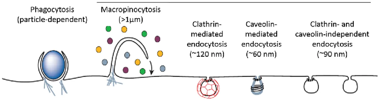
Endocytosis is an energy-dependent process that triggers the uptake of molecules (or even other cells) of different type and weight (Conner and Schmid, 2003). This is not a single mechanism; in fact, two different pathways can be recognized: phagocytosis and pinocytosis. The first one occurs only in specialized cells (e.g. macrophages) and, of course, is not involved in CPPs mechanism of action. On the contrary, pinocytosis is common among many eukaryotic cells and seems to play an important role in the internalization of CPPs, especially when they are conjugated with large cargoes (Lundin et al., 2008). There are four basic modes of pinocytosis: macropinocytosis, clathrin-dependent endocytosis (CDE), endocytosis mediated by caveolae, and clathrin and caveolae-independent endocytosis (Figure 1.2). Macropinocytosis is a non-specific process that leads to the formation of large vesicles, called macropinosomes (> 1 µm), generated by a membrane ruffling that surrounds and gradually internalizes part of the extracellular fluid and all the molecules dissolved inside it (Swanson and Watts, 1995). In CDE, the formation of the vesicle is preceded by the interaction of a ligand with specific transmembrane receptors, which cause invagination of a clathrin-coated piece of membrane. A receptor binding is also required in the endocytosis mediated by caveolae, but while the clathrin-coated vesicles are about 120 nm in diameter, caveolin-coated do not exceed 50-60 nm (Mayor and Pagano, 2007). Finally, clathrin and caveolae-independent endocytosis group several mechanisms that are related to the internalization of microdomains present on the cell surface, called ‘rafts’ (Conner and Schmid, 2003).
It is now evident that CPPs do not enter the cells via a single favorite method; indeed, the mechanism used strongly depends on their attached cargo (Maiolo et al., 2005). For instance, Tat
6
peptide uses a clathrin and caveole-independent endocytosis when conjugated to a protein (Fittipaldi et al., 2003), but clathrin-dependent endocytosis when conjugated to a fluorophore (Richard et al., 2005). Furthermore, it is important to emphasize that all endocytic processes end with the enclosure of the CPP and its linked cargo into lipid vesicles, where they can remain for extended periods of time and undergo degradation. In order to exploit CPPs for clinical applications, an efficient vesicular escape is therefore needed. At the present, several studies have the primary goal to overcome this problem, and some of them will be introduced in the next section, dedicated to the therapeutic applications of CPPs.
Figure 1.2 Different endocytic pathways. Phagocytosis is limited to specialized cells (e.g. macrophages); CPPs can enter the cells by using all pinocytosis mechanisms (Conner and Schmid, 2003).
1.1.3 Applications in Drug Delivery
As mentioned before, the ability of CPPs to translocate across the cellular membrane represents a major breakthrough for the introduction of drugs and large molecules into the cells for therapeutic purposes. At the moment, we are witnessing a dramatic acceleration in the development of novel therapeutic molecules such as proteins, peptides, nanoparticles and numerous nucleic acids: oligonucleotides, small interfering RNA (siRNA), plasmid and viral genes. Unfortunately, due to their physicochemical properties, these compounds exhibit several issues like poor stability in vivo and lack of cellular uptake and specificity. Therefore, the optimization of their intracellular delivery is one of the major priorities, and CPPs seems to be excellent candidates for this goal, offering several advantages over other tested vectors like liposomes or nanotubes (Veldhoen et al., 2006; Cirillo et al., 2014; Mallick and Choi, 2014).
Indeed, many studies present promising results in CPP-mediated intracellular delivery of many drugs, both in vitro and in vivo, including antisense oligonucleotides (Astriab-Fisher et al., 2002), antibodies (Morris et al., 2001), peptides and proteins (Snyder and Dowdy, 2005; Gros et al., 2006), and even contrast agents for in vivo imaging (Kersemans et al., 2008). The most representative applications of CPPs are reported in Table 1.1.
7 Table 1.1
Most representative CPPs: origins and applications.
CPP Origin Cargo types
Protein-derived
Tat11 HIV-Tat protein (48-58) protein/peptide/siRNA/liposome/nanoparticles Penetratin Antennapedia (43-58) peptide/siRNA/liposome
Chimeric-peptides
Pep-1 Trp-rich motif/SV40 T-antigen protein/peptide
MPG HIV-gp41/SV40 T-antigen siRNA/ODN/plasmid
Transportan Galanin/Mastoparan protein/PNA/siRNA
Synthetic-peptides
Polyarginine Based on Tat peptide protein/peptide/siRNa/ODN
MAP small molecule/plasmid
ODN, oligodeoxynucleotide; PNA, peptide-nucleic acid.
Covalent or Non-Covalent?
Among the numerous attempts in using CPPs as drug carriers, we can distinguish two principal strategies. The first one is the covalent strategy, which requires the formation of a covalent bond between the CPP and the cargo, achieved by either chemical cross-linking (Zorko and Langel, 2005; Lundin et al., 2008) or by cloning and expression of recombinant conjugates (Münst et al., 2009; Heffernan et al., 2012). Despite all the advantages offered by covalent technology (high reproducibility and stoichiometry control), the synthetic covalent bond can alter the biological activity of the cargo, thus resulting in a loss of its function. This problem especially concern siRNA and oligonucleotides conjugates (Meade and Dowdy, 2007; Juliano et al., 2008), for which non-covalent strategies appear to be a better solution (Simeoni et al., 2003). In this case, in fact, the cargo molecules do not undergo any chemical modifications, since they are associated with the CPPs only through electrostatic and hydrophobic interactions (Morris et al., 2001; Simeoni et al., 2003; Gros et al., 2006).
Overcoming Endosomal Entrapment
In order to exert its pharmacological activity, it is important for the cargo to reach its specific target. To ensure this, the passage through the cellular membrane is not the only limiting step. Indeed, CPP-cargo conjugates that enter the cell following one of the endocytic pathways are often sequestered into vesicles, keeping them from reaching their intracellular target. In addition, along with the endosomal membrane barrier, trapped macromolecules have to face a pH decrease and the attack by hydrolytic enzymes (Lee et al., 2008).
In recent years, many research groups proposed several solution to enhance endosomal escape (Erazo-Oliveras et al., 2012). One approach suggests the use of multivalent CPPs (MCPPs), which consist of creating an oligomer with multiple copies of the same CPP, in order to increase its local concentration when it interacts with membranes (Kawamura et al., 2006; Hassane et al., 2009).
8
MCPPs can be obtained by attaching two or more CPP monomers with protein highly branched molecules (Juliano et al., 2008), scaffolds (Sheldon et al., 1995) or simple disulphide bridges (Rudolph et al., 2004). Despite their encouraging increase in activity, the complex and expensive methods for the synthesis of MCPPs is an important limitation. Moreover, it is possible that similar compounds could act as immunogens, producing an unpleasant immune response (Schwarze et al., 1999).
Another approach that has been proposed to increase endosomal escape is based on the addition of chromophores that, upon activation with an appropriate light stimulus, are able to disrupt the vesicle membrane. Here, CPPs are linked to fluorophores (Maiolo et al., 2004) as well as to photosensitizers (Choi et al., 2006; Zhao et al., 2011), but in several experiments a significant phototoxic activity is observed, causing cell damage and apoptosis (Choi et al., 2006; Oliveira et al., 2007).
The endosomal entrapment of CPP-cargo complexes can be overcome also with the help of other specific peptides. For instance, dependent membrane-active peptides (PMAPs) are pH-sensitive peptides that become active only in acid environment (El-Sayed et al., 2009). This activity is extremely advantageous in this context, since the pH in the endosome lumen gradually drops as maturation occurs (Schmid et al., 1989; Serresi et al., 2009). An example of PMAP is the HA2 fusion peptide, which has been proved to increase endosomal escape of several cargoes when associated with CPPs such as Tat peptide (Kumar et al., 2007; Lee et al., 2011) and penetratin (Lundberg et al., 2007). Another class of peptides that received researchers’ attention is the antimicrobial peptides (AMPs) family. These small peptides possess membrane-perturbing activity (see Section 1.2, dedicated to AMPs); therefore, they might be able to permeabilize the endosome membrane leading to the release of its contents.
The work of this thesis is in part dedicated to the study of an AMP-CPP hybrid, called CM18-Tat11,
synthetized by the group of prof. Beltram of NEST laboratory (Scuola Normale Superiore and
Istituto Nanoscienze-CNR, Pisa): the characteristics of this novel peptide will be described in details
9
1.2 Antimicrobial Peptides
The first discovery of substances that possess antimicrobial activity dates back to the first half of the nineteenth century, when many compounds isolated from normal tissues and body fluids were found to kill both Gram-positive and Gram-negative bacteria (Skarnes and Watson, 1957). Nowadays, we know that those molecules are evolutionally conserved oligopeptides that play an important role in the innate immune response. To date, the antimicrobial peptide (AMP) database contains 2480 peptides (http://aps.unmc.edu/AP/main.html, last update: Dec 2014) which have been identified in several species ranging from bacteria to mammals, including humans and plants (Wang and Wang, 2004).
AMPs are typically short sequences (between 12 and 50 amino acids), with a net positive charge that range from +2 to +9. In addition to their broad spectrum activity against both Gram-positive and Gram-negative bacteria, some AMPs are also lethal for parasites, fungi, yeasts, and viruses (Zasloff, 2002). Furthermore, a promising anti-tumorigenic activity of some AMPs has also been observed (Papo and Shai, 2005; Hoskin and Ramamoorthy, 2008; Schweizer, 2009). Some early observations also suggest that AMP function is not exclusively limited to innate immunity. Some peptides, e.g. defensins, seem to modulate angiogenesis, promote inflammatory response and stimulate adaptive immune system by inducing cytokine secretions, recruiting dendritic cells and macrophages (Suarez-Carmona et al., 2014). All these properties make AMPs a promising resource in the development of new drugs, not only as new antibiotics, but also against tumors and many other diseases (Reddy et al., 2004).
The mechanism of action used by several AMPs in their antimicrobial activity is based on the formation of transmembrane pores on the bacterial membrane, altering its permeability and leading to bacteria’s death (Figure 1.3). For this reason, and given their evolutionary conserved sequences, AMPs provide a simple model in understanding the molecular basis of peptide-lipid interactions in membranes, as well as the structure-function relationships of ion channels, ion pumps and exchangers (Marsh, 1996). A better knowledge of such mechanisms could then be exploited to build custom molecules with a wide range of applications in biotechnology, like ion channel modulators (Hille, 2001), biosensors for several analytes (Aili and Stevens, 2010), and may provide also a pharmacological approach for the treatment of channelopathies (Wilde, 2008). In the latter case, a synthetic channel could incorporated in cells expressing an aberrant ion channel and restore the physiological ion flow. Encouraging results have been obtained in human epithelia of the cystic fibrosis patients with a defective chloride channel (Wallace et al., 2000).
10
1.2.1 Classification of AMPs
As for CPPs, AMPs can also be classified in different ways according to their origin, amino acid sequence and structure. From a structural point of view, we can distinguish two major groups: α-helical peptides and β-sheet peptides. The first group includes cecropins (the first AMP identified) and magainins, and more in general linear cationic peptides, with less than 40 amino acids, that lack cysteine residues (Brogden, 2005). They are disordered in aqueous solutions, but have the tendency to form highly amphipathic helix in organic solvents and lipid vesicles (Marion et al., 1988; Bechinger, 1999). In this case, the folded peptide exhibits its hydrophilic amino acids residues along one side of the helix and its hydrophobic amino acids residues along the opposite side, thus allowing the contact with both the lipids and the aqueous environment. In the second group are present anionic and cationic AMPs with cysteine in their sequence and form one or more disulfide bridge that maintain the β-sheet structure of the peptide (Dhople et al., 2006). The most representative AMPs in this group are defensins, derived from humans and several mammals, and protegrin. Other peptides not included in this simple classification are cyclic AMPs and cationic peptides enriched for specific amino acids like proline, arginine or tryptophan (Reddy et al., 2004).
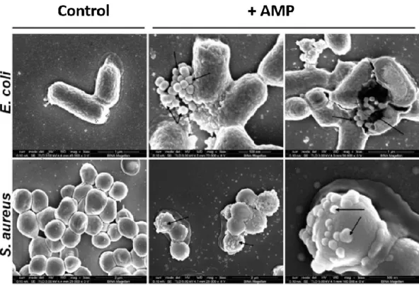
Figure 1.3 Scanning electron microscopy of two different bacterial strains untreated (control), and treated with AMPs (+ AMP). Upon peptide interaction, bacterial membranes appear corrugated and show small protruding blebs (arrows) (modified from Bi et al., 2014).
11
1.2.2 Mechanisms of Action of AMPs
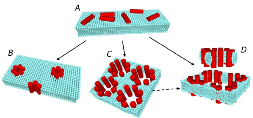
In literature, two different strategies are proposed for the antimicrobial activity of AMPs. The most validate mechanism is the previously mentioned formation of transmembrane pore, which cause an extensive membrane rupture leading to the lysis and death of the microbial cells. However, recent studies revealed that AMPs might have also intracellular targets, including DNA, RNA, and protein synthesis, as well as enzymatic processes essential for bacteria metabolism (Lee and Lee, 2014; Choi et al., 2015). These mechanisms are not fully understand yet, thus more researches are required on this argument. On the other hand, the membrane-disrupting activity of AMPs has been deeply investigated in the last years. By now, there are three main models able to describe the AMPs modes of membrane perturbation (Figure 1.4): “barrel-stave”, “toroidal” and “carpet” model (Brogden, 2005).
Barrel-stave Model
In the barrel-stave model, the peptides initially bind in parallel on the membrane surface due to an electrostatic interaction (Figure 1.4 A). As the peptide monomers accumulates on the membrane, they insert perpendicularly in the membrane and bind together around a central pore, arranging like the staves of a barrel (Figure 1.4 B). In this structure, the peptide helices are organized in a way that the hydrophobic residues interact with the lipid core of the membrane, while the hydrophilic segments face the aqueous lumen of the pore. According to this mechanism, when a peptide monomer inserts in the bilayer, it recruits other peptides to form the aggregates, acting in a cooperative way. One of the most representative peptide acting with this mechanism is alamethicin (Baumann and Mueller, 1974).
Toroidal Model
The toroidal pore differs from the barrel-stave one in that the peptide monomers do not aggregate with each other, but rather interact with the polar head groups of the lipids, even when the peptides are arranged vertically in the membrane. The interaction between the peptides and the phospholipid head groups is stronger than in the case of the barrel-stave model, so that the lipid monolayer is forced to bend back on itself, connecting one side to the other of the membrane, in a toroidal shape manner. Therefore, the pore walls are formed by lipids and peptides, and not by peptides only as in the barrel-stave model (Figure 1.4 C). This particular model was first proposed to describe the pores formed by the peptide magainin (Ludtke et al., 1996; Matsuzaki et al., 1996).
Carpet Model
In the carpet mechanism, the strong electrostatic interaction between the peptides and the phospholipid head groups leads to a peptide-induce membrane ‘carpeting’ effect. Once the peptides have reach a threshold concentration on the membrane, they assemble to form
12
transiently toroidal pores, allowing more and more peptides to cross the membrane and eventually cover the other membrane leaflet, so that the bilayer is rapidly and extremely perturbed and disintegrated. It is also possible that entire fragments of the membrane, called micelles, are detached from the membrane itself in a detergent-like manner (Figure 1.4 D). Of course, similarly to the case of CPPs, the same AMP do not act following exclusively one of the models described above. Indeed, it has been demonstrated that temperature, peptide concentration, the presence of metal ions and other environmental conditions might change the activity of the same AMP from one mechanism of action to another (Brogden, 2005; Noshiro et al., 2010).
1.2.3 AMPs vs Multidrug-Resistant Bacteria
Since the discovery of penicillin, antibiotics are considered one of the major discoveries of modern medicine. Unfortunately, the dramatic increase in antibiotic-resistant bacterial strains causes a loss of efficacy of these broadly used drugs, with negative consequences in the healthcare system along with social and economic implications (Huttner et al., 2013; Rossolini et al., 2014). Furthermore, the multidrug-resistant infections that in the past years were limited to hospital or large communities, are now spreading all over the world. Thus, innovative strategies for the development of new treatments are urgently needed.
Among all the ongoing attempts to find a solution to the so called “antibiotic resistance crisis”, AMPs represent an encouraging resource for future drug development. The reason is that all the strategies developed by microorganisms in order to overwhelm antibiotics are entirely intracellular mechanisms: 1) alteration of the antibiotic target (Wilson, 2014); 2) antibiotic
Figure 1.4 Mechanisms of peptide-induced membrane permeabilization. After adhering on the external face of the membrane (A), the peptide could insert in the membrane according to a barrel stave (B), toroidal (C), or carpet mechanism (D).
13 degradation by specific enzymes (Wright, 2005); 3) antibiotic expulsion thorough membrane pumps (Li and Nikaido, 2004). On the other hand, the effect of the AMPs against the bacterial membrane is driven by an aspecific interaction with the peptides and the double layer. Since modifications in the physical structure of the membrane are very difficult to occur, the development of an AMP resistance is very unlikely.
14
1.3 Aim of This Work (pt. I):
Mechanism of Action of Membrane Active Peptides
In literature, several strategies are described to assess the mechanism of AMPs activity. A simple and direct method is the observation via microscopy techniques of the microbial cell integrity after the application of AMPs (Kalfa et al., 2001; Yenugu et al., 2004). To study the biophysical interactions between peptides and membrane, a large variety of techniques are also employed, such as nuclear magnetic resonance, surface plasmon resonance (Aquila et al., 2013), circular dichroism and electron paramagnetic spectroscopy (Pistolesi et al., 2007). The long-term goal of these researches is the investigation at a molecular level of the lipid-peptide interaction, from orientation to conformational changes. Moreover, since pore formation is the AMPs favorite mechanism of action, a widely used experiment to assess their membrane permeabilization is the measure of the leakage of a fluorescent molecule previously confined into phospholipid vesicles (Orioni et al., 2009). However, none of these studies addresses directly the problem of the kinetics of pore formation when peptides assemble on the plasma membrane of a living cell.
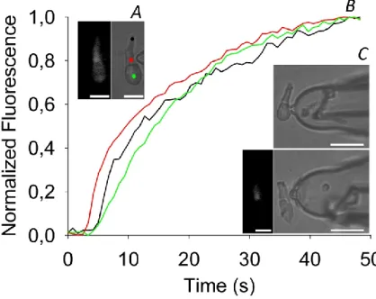
The dynamics of the formation of the AMP pore and its biophysical characteristics are investigated in this thesis under strict physiological conditions. For this purpose, the peptides are applied on the membrane of a living cell, and the ionic current that flows through the pore formed by these peptides is simultaneously recorded by using the patch-clamp technique (described in Materials
and Methods). The first part of my Ph.D. work was focused on membrane active peptides is
dedicated to the biophysical study of the mechanism of membrane perturbation of two peptides: the novel chimeric peptide CM18-Tat11 (and its isolated parts) and the rational designed AMP CM12.
1.3.1 CM
18-Tat
11: a Chimeric CPP with Membrane-Disruptive Properties
The arginine-rich Tat peptide (Tat11, residues 47-57 of HIV-1 Tat protein) is able to stimulate an
efficient endocytosis of a large variety of cargoes into a wide range of target cells (Brooks et al., 2005). These properties, plus the low toxicity for eukaryotic cells, make this peptide an excellent resource in drug delivery. Unfortunately, all reports show that molecules taken up by Tat11
-mediated endocytosis undergo endosomal entrapment, limiting their intracellular availability. To overcome this problem, it was proposed to link Tat11 with a sequence that possess
membrane-disruptive properties. An attempt was made with the fusogenic peptide HA2, derived from the influenza-virus hemagglutinin protein (Skehel et al., 2001). In this case, despite a slight enhancement of endosomal entrapment was observed (Wadia et al., 2004), still more than 99% of the Tat-cargo complexes remains sequestered into vesicles (Kaplan et al., 2005).
Another resource of molecules that are able to disrupt the phospholipid barrier comes from the AMPs. In this thesis, the biophysical characteristics of a novel hybrid peptide formed by the CPP
15 Tat11 and the hybrid AMP, called CM18,are studied. Among all the AMPs included in this large class
of peptides, Cecropin-A/Melittin (CM) hybrids raised particular interest because they are among the shortest and most effective peptides with membrane-permeabilization properties. Cecropin-A is a linear, naturally occurring, cationic peptide initially isolated from the hemolymph of the silk moth Hyalophora cecropia (Steiner et al., 1981). Its 37 amino acids sequence can be divided in two segments: a cationic, amphipathic N-terminal domain and a hydrophobic C-terminal domain. Cecropin has shown to be active against both Gram-negative and Gram-positive bacteria, as well as against some fungi and tumor cell lines (Andreu et al., 1985). Moreover, cecropin-A possess a relatively slight hemolytic activity and exhibit low toxicity toward normal eukaryotic cells (Boman et al., 1989; Wade et al., 1992). Melittin, in contrast, is extremely hemolytic and has a stronger, broad-spectrum antimicrobial activity. This 26-residue peptide is isolated from the venom of the honey bee Apis mellifera, and contains, as cecropin-A, two distinct cationic and hydrophobic domains but in reverse order (Boman et al., 1989; Andreu et al., 1992). The cecropin-melittin hybrid are constructed combining different domains of these two peptides, with the intent to find the shortest sequence that display a remarkable antimicrobial activity, yet lacking the melittin-associated hemolytic properties. In particular, CM18 hybrid (sequence: KWKLFKKIGAVLKVLTTG) is
synthesized by combining the first seven residues of cecropin-A with residues 2-12 of melittin. Despite the recent results that indicate an efficient uptake and endosomal escape of cargoes associated with the chimeric CM18-Tat11 (Salomone et al., 2012), its mechanisms of membrane
permeabilization are still unknown. In this thesis, a biophysical characterization of the membrane activity of CM18-Tat11 and its isolated domains is given, providing some suggestions about their
mechanism of action.
1.3.2 The Rational Designed Peptide CM
12Despite their promising properties, the use of some natural AMPs for therapeutic applications turned out to be problematic, especially due to their high toxicity and vulnerability to proteolytic enzymes (Aoki and Ueda, 2013). For this reason, in recent years several AMP sequences have been improved thanks to a computer-aided optimization (Fjell et al., 2012). Along with the enhancement of the activity of preexisting peptides, the computational approach is exploited also for the identification and design of new sequences that possess an antimicrobial activity and at the same time lack all the unfavorable properties of natural peptides (Kaltofen et al., 2014; Maccari et al., 2015). Part of this thesis work is dedicated to the study of the mechanism of action of a rational designed AMP, called CM12, selected and synthetized by Maccari at the Center of
16
1.3.3 Alamethicin F50/5 and its Analogue [L- Glu(OMe)
7,18,19]
Alamethicins (Alm) are a family of pore-forming peptides produced by the fungus Trichoderma
viride (Meyer and Reusser, 1967; Reusser, 1967). Since their sequences contain the
non-proteogenic amino acid α-aminoisobutyric acid (Aib), they are called peptaibolic peptides. Alamethicins exhibit broad biological activities, including antibacterial effects against both Gram-positive and Gram-negative bacteria (Meyer and Reusser, 1967; Jen et al., 1987; Béven et al., 1998). It was demonstrated that these peptides are able to form voltage-dependent pores into biological membranes (Fox and Richards, 1982); and different mechanisms of pore formation were proposed to describe alamethicin mode of action. Currently, the most accepted one is the barrel-stave model, firstly proposed by Fox and Richards (Fox and Richards, 1982; Mathew and Balaram, 1983).
Part of this thesis work is focused on the study of the single channel properties formed in natural and model membranes by the synthesized Alm F50/5 peptaibol, which is the major component of the neutral fraction of the alamethicin, and its synthetic analog [L- Glu(OMe)7,18,19]. The latter
peptide was obtained by substitution of specific amino acids from the primary sequence of Alm F50/5. Specifically, glutamine residues at positions 7, 18, and 19 have been replaced with side-chain esterified glutamic acid residues, since glutamine was found to play a major role in pore formation (Fox and Richards, 1982; Molle et al., 1996). Both peptides were inserted in a cell plasma membrane (of the rod outer segment of the frog retina, recorded in whole-cell) and in membrane-patches excised from giant unilamellar vesicles (GUVs). The possibility to compare the channel activity in the precisely controlled lipid environment of GUVs, with the channel activity recorded in a natural membrane with the same ionic electrochemical gradients, will open new possibilities in the biophysical characterization of the pores.
17
Part II
Real-Time Modulation of zGCAP in Zebrafish Green Cones
1.4 Visual Perception
Light perception is one of the most important environmental stimuli for all organisms. Other than being fundamental for the generation and preservation of life on Earth, light is, in most animal, the responsible of many physiological functions including circadian rhythms, defense, food-hunting and other important behavioral reactions. Even though simple life form as Protozoa are sensible to light stimuli, the first draft of the vision function occurred in animals only with the appearance of particular photosensitive cells: the photoreceptors. Initially spread all over the body surface, as in many Invertebrates nowadays, the photoreceptors organized later in small formations called ocelli, which represent the first form of a specialized structure dedicated to vision. A crucial step in evolution that allowed the transition from the simple detection of light to real vision is the development of the eye. This organ is composed of transparent accessory structures, with converging lens characteristics, which allow the detection of a highly detailed final image. The image (real, reduced, and inverted) is focused on a layer of neuronal cells, including photoreceptors, arranged like a mosaic to form the retina. Here, the photoreceptors, and other retinal neurons connected with them, translate the light signal into an electrical signal that travel to the brain via axons in the optic nerve (for a complete review, see Masland, 2012).
1.5 Retina
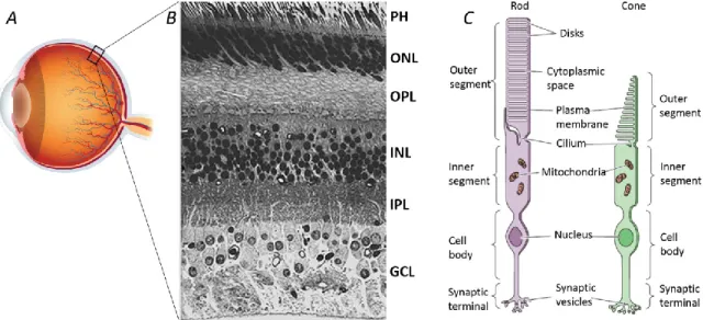
The retina is a thin layer of cells lining the inner surface of the optic bulb (Figure 1.5A), and it is actually considered a part of the central nervous system. On the inner side of the eye, the retina is in direct contact with the vitreous humor, while on the other side lies in front of the retinal pigment epithelium (RPE) and the underlying choroid. The RPE is a layer composed of cells that synthetize melanin, a black pigment that captures all the light not absorbed by the retina. This prevents the backscattering of the light towards the receptors and avoids any image detriment. All vertebrate retinae are composed of five types of neurons: photoreceptors, bipolar cells, ganglion cells, horizontal cells, and amacrine cells. The cell bodies and their processes (long branches that arise from the cell body) are organized in distinct layers, clearly visible at a microscopic examination of a vertical section of the retina (Figure 1.5B) (Cajal, 1893; Dowling and
18
Boycott, 1966). The cell bodies of the photoreceptors are contained in the outer nuclear layer (ONL) while in the inner nuclear layer (INL) there are cell bodies of the bipolar, horizontal and amacrine cells. Ganglion cells bodies are located in the ganglion cell layer (GCL), where also some displaced amacrine cells can be found (Dowling, 1970). In other two different layers, called inner plexiform and outer plexiform layers (IPL and OPL, respectively), are positioned the neuron processes, and these are the areas where synaptic contacts between cells occur. Contrary to what can be expected, photoreceptors are not facing directly the interior of the eye, but lie in the deepest layer against the RPE. As a result, light must pass through the other non-light-sensitive layers of cells as well as the retina vasculature before reaching the photoreceptors. The reason for this counterintuitive organization of the retina lies in the special relationship that exist among the photoreceptors and the RPE. Other than absorbing the excess light, RPE is also essential for the preservation and turnover of photoreceptors and for the restoration of the light-sensitive molecules after they have been exposed to light (Young, 1967, 1971; Bok and Young, 1972; Anderson et al., 1978).
Figure 1.5 Structure of the retina and photoreceptor cell types. (A) Eye picture; (B) Cross section of the retina (400x), PH: photoreceptors layer, ONL: outer nuclear layer, OPL: outer plexiform layer, INL: inner nuclear layer, IPL: inner plexiform layer, GCL, ganglion cell layer; (C) Rod and cone structure.
1.5.1 Functional Architecture of Photoreceptors
Photoreceptors are a class of highly specialized cells responsible for the transformation of a light stimulus into an electrical stimulus, which can reach the brain through the optic nerve. There are two basic types of photoreceptors, distinguished by shape: rods and cones (Figure 1.5C). In addition to their different morphology, they also carry out different vision functions (better explained in the following paragraphs); nonetheless, they share several characteristics. Both rods and cones have an outer segment (OS), slim rod-shaped for the former, and robust conical-shaped
19 for the latter, separated from the inner segment (IS) by a thin cilium (Anderson and Fisher, 1976). Notably, the OS of both types of photoreceptors are entirely filled with a succession of hundreds of flattened discs, disposed one on the other, that originate from invaginations of the plasma membrane (Cohen, 1960, 1964; Besharse and Pfenninger, 1980). The discs are continuously produced from the base of the OS, in the region of the cilium, and while in cones they remain attached to one side of the OS membrane, they completely separate in rods, becoming free-floating discs inside the OS membrane (Steinberg et al., 1980). In these membrane discs are embedded many (thousands) of photosensitive molecules, also called photopigments, which are different in rods and cones, and consists of two parts: a protein called opsin and a chromophore, called retinal (Bowmaker et al., 1978; Bowmaker and Dartnall, 1980). The opsin molecule is synthetized in the IS and binds to the membrane discs at the cilium, whereas the retinal (derived from the vitamin A) is synthetized in the RPE and delivered to the discs via specific carrier molecules (Adler and Martin, 1982; Chader, 1989). The IS is almost entirely occupied with mitochondria, and contains also ribosomes and other cytoplasmic organelles, except for the nucleus that takes place below the IS, in the cell body. Finally, at the lower extremity of the photoreceptor there is an enlargement of the membrane, rich in synaptic vesicles, that forms synapse in contact with second order neurons.
Rods and Dim-Light Vision
The rods are photoreceptor cells that contain only one type of visual pigment, rhodopsin. They are extremely sensitive, up to the single-photon level, with a peak at ~500 nm (Brown and Wald, 1964) and are therefore responsible for scotopic vision, i.e. in dim light conditions, as at night. In order to maximize quantum catch, a very high number of visual pigments is needed, thus explaining the dense packing of the discs in the rod OS. Nevertheless, the price for this high is the much slower photoresponse compared with the one of cones (Baylor, 1987), a poor resolution of the image and no perception of colors (MacLeod, 1972). As a result, rod function provides vision at light conditions even below the level of starlight, but uncolored and with very low spatial resolution.
Cones: Acuity and Color Vision
As illumination increases, the participation of cones in the vision function become more and more important. The cone system is in fact responsible for the perception of higher intensities of light, the so-called photopic vision, and also possess a high spatial resolution due to the high cone density that provides a better visual acuity. Furthermore, cones are responsible for color perception, and can be divided in different classes depending on the type of opsin (the visual pigment of cones) they express. Each of these opsins differ from the others in the sensitivity to light of different wavelengths, and for this reason are referred to as short, medium, and long wavelength sensitive cones (SWS, MWS, and LWS, respectively). More precisely, SWS-cones are
20
known to peak around 420 nm (blue region of the spectra), MWS-cones peak around 530 nm (green), while LWS-cones are maximally sensitive to wavelength of about 560 nm (red) (Bowmaker and Dartnall, 1980; Bowmaker et al., 1980). Cones are therefore known as blue-, green-, and red-sensitive cones and it has been shown that are morphologically different from each other, allowing their identification simply by optical microscopy (Sherry et al., 1998). Even though the most notorious color identification is based on trichromatic vision (as in humans), most mammalian species are merely dichromatic, while many other vertebrates are tetrachromatic, as they possess a fourth type of cone sensitive to ultraviolet light (UV-cones) (Bowmaker et al., 1980; Branchek and Bremiller, 1984). UV-cones were found among several species of reptiles (Liebman and Granda, 1971), amphibians (Sherry et al., 1998), birds (Bowmaker, 1980), and fishes, including zebrafish (Robinson et al., 1993).
21
1.6 Phototransduction
Despite the different physiological function of rods and cones (because of their different morphology, response times, wavelength sensitivity, and adaptation mechanism), several phototransduction proteins are similar, or even the same, in any type of photoreceptors. Indeed, it has been proved that they evolve from common ancestors (Hisatomi and Tokunaga, 2002). For this reason, the cascade of intracellular events following light absorption by the visual pigments, named phototransduction, is comparable in all vertebrates (Lamb, 2013). Contrary to what happens in most sensory systems, the stimulation of a photoreceptor do not elicit action potentials; rather, light absorption causes only a graded change in membrane potential, corresponding in a change in the rate of neurotransmitter release onto postsynaptic neurons. Another surprising element is that the light-induced activation of the photoreceptor leads to a hyperpolarization of its membrane potential, rather than a depolarization, as it occurs in many other sensory neurons. All the biochemical events involved in the phototransduction have been deeply studied in the last four decades, and many of them are now well-characterized (for a complete review, see Kawamura and Tachibanaki, 2008). In the following sections, an overall picture of the most important steps in the phototransduction pathway will be given.
1.6.1 The Dark Current
In the dark-adapted state, the photoreceptors are maintained in a depolarized state by a flux of Na+ and Ca2+ ions that enter into the cells through specific ligand gated channels located in the OS
membrane (Hagins et al., 1970). The open- or closed-states of these channels are regulated by the intracellular level of the nucleotide cyclic guanosine monophosphate (cGMP) (Fesenko et al., 1985; Yau and Nakatani, 1985b). In particular, in darkness, the high cytoplasmic concentration of cGMP keeps the channels open, and Na+ and Ca2+ ions are free to enter into the OS. This steady inward
current is balanced by an outward K+ current that flows through non-gated K+-selective channels
that are confined in the IS membrane (Bader et al., 1982; Barnes and Hille, 1989). Furthermore, in the IS membrane are confined several Na+/K+-ATPase pumps that are responsible for the
maintenance of stable intracellular concentration of Na+ and K+ by pumping out Na+ ions and
pumping in K+ ions (Zuckerman, 1973). The resulting circulating current stabilizes around -40 mV
and is called “dark current” (Hagins et al., 1970). In the synaptic terminals, this relatively depolarized membrane potential generates the opening of voltage-sensitive Ca2+ channels, to
which corresponds the release of glutamate (the photoreceptor neurotransmitter) toward the postsynaptic cell (Sarantis et al., 1988; Copenhagen and Jahr, 1989; Ayoub and Copenhagen, 1991). Light activates an enzymatic cascade which ends with the hydrolysis of cGMP. This causes
22
the closure of the cGMP-gated channels, and in turn the reduction of the influx of Na+ and Ca2+
ions, the hyperpolarization of the cell, and finally the reduction of glutamate release at the synaptic terminal.
1.6.2 Enzymatic Cascade Activation
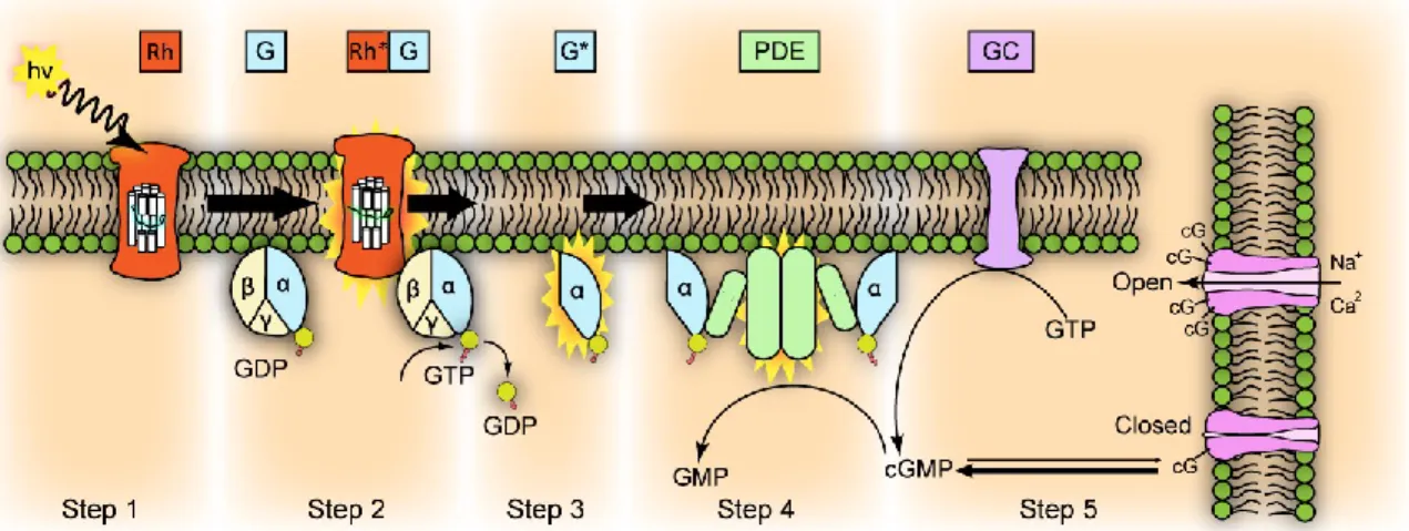
Since rod cells are easier to purify compared to cones, the best-characterized phototransduction pathway is so far the one occurring in rods (Figure 1.6), in which the visual pigment is the rhodopsin. Upon photon absorption, the 11-cis retinal, which is the light-sensitive moiety of inactivated rhodopsin (Rh), isomerizes to a more stable all-trans configuration in less than 20 ps, and triggers a conformational change of the entire molecule, that becomes enzymatically active (Rh*). In this state, Rh* catalyzes the activation of a G-protein, also known as transducing (G). Transducin is a heterotrimer whose α subunit bounds non-covalently to a molecule of GDP (guanosine diphosphate) when inactive. Upon interaction with Rh*, α subunit exchanges GDP with GTP, dissociates from transducin, and activates the cGMP phosphodiesterase (PDE). This enzyme causes the hydrolysis of cGMP into 5’-GMP: the fall of the cytoplasmic concentration of cGMP causes the closure of a number of cGMP-gated channels proportional to the amount of light initially absorbed.
Figure 1.6 Phototransduction cascade activation by light.
The dark current sustained in the OS by the entrance of Na+ and Ca2+ ions is therefore reduced,
and the change in membrane potential can be detected by electrophysiological recordings performed on single isolated cells (Figure 1.7). This membrane hyperpolarization leads to the closure of the voltage-gated Ca2+ channels, and the intracellular Ca2+ concentration drops, due to
23 decrease in Ca2+ concentration reduces or terminates the glutamate release at the synaptic
terminal, which is detected by the postsynaptic neuron. The signal is further processed by other neurons present in the higher layers of the retina, and subsequently sent to the brain.
This first step of the phototransduction cascade is characterized by an enormous level of amplification. A single rhodopsin molecule, when activated, can diffuse within the disc membrane and activate hundreds of transducin, each one of which stimulates one PDE enzyme. Each PDE in turn is able to hydrolyze over a thousand of cGMP molecules per second, leading to a final hydrolyzation of more than 105 cGMP molecules per second for a single Rh* (Leskov et al., 2000).
1.6.3 Enzymatic Cascade Inactivation
In order to maintain enough light sensitivity to respond to subsequent stimuli, it is essential for the photoreceptor to recover from its active state. This implies that all the molecules activated during the cascade need to be inactivated, and the cGMP concentration has to be restored. Transducin possess GTPase activity and is able to inactivate itself by hydrolyzing the bound GTP molecule into GDP, thus switching off also the PDE activity. At the same time Rh* becomes a target for phosphorylation by a rhodopsin kinase, which phosphorylates the C-terminus of Rh*. The phosphorylated Rh* then interacts with the protein arrestin, leading to its rapid inactivation and causing the dissociation of the all-trans retinal from the rhodopsin. In order to be restored into its 11-cis active form, the all-trans retinal is reduced to all-trans retinol, transported out of the OS and converted in the RPE by appropriate enzymes (Lamb and Pugh, 2004).
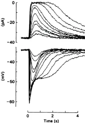
Figure 1.7 Electrical response of an isolated rod photoreceptor.
Upper panel: The transient suppression of the current entering the outer segment in response to 20 ms flashes of light of progressively increasing intensity, is recorded with a suction pipette;
Lower panel: simultaneous voltage
recording with an intracellular microelectrode impaling the inner segment.
24
Theoretically, once the PDE activity has been reduced, the restoration of the dark levels of cGMP would occur thanks to the basal activity of GC. In this case, however, the recovery rate would be too slow compare with the photoresponse kinetics: indeed, more mechanisms that regulate the transduction cascade must be involved. It is now well established that the responsible for the modulation of the photoresponse cascade is primarily, if not exclusively, the change of Ca2+
intracellular free concentration (Rispoli, 1998; Fain et al., 2001; Nakatani et al., 2002).
1.6.4 Ca
2+-Mediated Light Adaptation
In order to adjust their sensitivity to different light intensities, photoreceptors are endowed with several mechanisms of light adaptation. In particular, these processes operate to increase the sensitivity of the transduction cascade in low-light conditions; whereas at higher levels of illumination the sensitivity decreases, preventing the receptors from saturating. The concentration of Ca2+ ions in the OS appears to play a key role in the light adaptation of photoreceptors, and it is
also involved in the shut-off mechanisms described above (Fain et al., 2001; Nakatani et al., 2002). The most important Ca2+-sensor proteins involved in these mechanisms are recoverin (Rec),
calmodulin, and guanylate cyclase-activating proteins (GCAPs). Recoverin plays a role in the inactivation of Rh*, since it activates the rhodopsin kinase when Ca2+ concentration decreases.
Calmodulin, on the other hand, modulates the cGMP-gated channels. In darkness, when OS free-Ca2+ is high, Ca2+-calmodulin is bound to the channels, and their affinity for cGMP is relatively low.
As Ca2+ concentration decreases in light, calmodulin unbind Ca2+ and dissociates from the channels
increasing their sensitivity to cGMP. Low Ca2+ concentration induces also the GCAPs to activate
another enzyme, the guanylate cyclase (GC), thus promoting the cGMP recovery when the light stimulation is ceased. The regulation mediated by GCAP is the most powerful negative-feedback mechanisms triggered by Ca2+ in photoreceptors, since it has an effect on the photoresponse
amplitude, on the recovery phase of the photoresponse, and also improve the signal-to-noise characteristics of the rod.
Guanylate Cyclase-Activating Proteins (GCAPs)
GCAPs are members of the neuronal calcium sensor protein family, a group of Ca2+-binding
proteins that are mainly expressed in neurons, where they mediate a wide range of Ca2+
-dependent physiological responses (Burgoyne, 2007). The first observation that low Ca2+
concentrations in the OS stimulate the synthesis of cGMP was made in 1982 by Lolley and Racz, who conclude that Ca2+ has an inhibitory effect on GC (Lolley and Racz, 1982). Later, Koch and
Stryer discovered that the activation of GC is not directly controlled by Ca2+ ions, rather is mediated
25 after several years of efforts by Gorczyca et al., and was called guanylate cyclase-activated protein, or GCAP (Gorczyca et al., 1994).
From a structural point of view, GCAP are small proteins of approximately 200 amino acids, and contain four Ca2+-binding domains (of which the first is non-functional) which are termed EF-hand
motifs (Figure 1.8) (Ermilov et al., 2001). GCAPs are expressed in photoreceptors of several vertebrates, and many isoforms have been specifically found in human, bovine, mice, chicken, fish, and amphibians (Palczewski et al., 1994; Subbaraya et al., 1994; Frins et al., 1996; Imanishi et al., 2004).
Figure 1.8
Cartoon representation of GCAP1 with three Ca2+ bound. The N-terminal helix is red, N-terminal domain (EF-1 and EF-2) is orange, terminal domain (EF-3 and EF-4) is yellow, kinked C-terminal helix is green, and the Ca2+ ions are shown as dark green space-filling spheres (modified from Stephen et al., 2007).
The physiological role of GCAPs in the modulation of the photoresponse has been deeply studied during the last years. Mendez and Chen generated a transgenic mouse strain having the genes of the isoforms GCAP1 and GCAP2 knocked-out (GCAPs-/-) (Mendez and Chen, 2002). By comparing
the flash responses recorded from rods of normal mice (GCAPs+/+) with the ones in GCAPs-/-, they
discovered that responses triggered by flashes of same intensity were larger and slower in GCAPs -/-. This indicates that the prevention of GCAP regulation in photoreceptors makes them more
sensitive to light and unable to quickly recover their dark state following light stimuli, and adapt to background illumination.
Zebrafish GCAP
Within the last decade, the zebrafish (Danio rerio) has become a common and valuable vertebrate model in many branches of neuroscience, including embryology (Kimmel, 1993), developmental neuroscience (Barinaga, 1990), genetic analysis (Driever et al., 1994), and, of course, visual neuroscience (Bilotta and Saszik, 2001). The zebrafish retina is endowed with one type of rod cell