Facoltà di Agraria, Dipartimento di Scienze e Tecnologie Agroalimentari (AGR/15)
CORSO DI DOTTORATO DI RICERCA Biotecnologia Degli Alimenti - XXIII ciclo
Removal of unstable proteins from white wine by
immobilized acid protease
Coordinatore: Prof. Marco Esti Firma
Tutor: Prof. Marco Esti Firma
Dottoranda: Ilaria Benucci Firma
ABSTRACT
This PhD thesis research project was aimed at assessing and optimizing different immobilization procedures of pineapple stem bromelain, in order to develop an innovative biotechnological technique, alternative to bentonite fining, useful to removal selectively unstable proteins from white wines.
Stem bromelain activity was assessed on a suitable synthetic substrate at a reference pH value (3.2), this being the average minimum pH value of wine. Protease was covalently immobilized on different supports by various procedures and the best biocatalyst was chosen measuring immobilization percentage, kinetic parameters and half-life (in model wine buffer). Moreover, the influence on free and immobilized protease activity of potential inhibitors naturally present in wine, such as ethanol, tannins and sulphur dioxide (SO2) over the average range concentration of wine, was investigated. Finally a
kinetic study was carried out using 6 artisan and unrefined white wines, spiced with the synthetic substrate, in order to compare catalytic properties of free and immobilized bromelain. Immobilized protease activity, then, was tested in these wines both on total proteins and on unstable ones.
Covalent immobilization reduced bromelain catalytic properties. All kinds of procedures applied at pH 7 allowed the highest immobilization yield. Nevertheless, biocatalysts immobilized at pH 3.2 showed the best catalytic performance. Stem bromelain was successfully immobilized on chitosan beads without glutaraldehyde at pH 3.2, obtaining the most interesting and food-safe biocatalyst, which was used for all other experiments.
Inhibition study proved that all the inhibitors tested resulted to be reversible for stem bromelain activity. Nevertheless, the immobilized enzyme was lesser affect respect to the free one. Free sulphur dioxide was the strongest inhibitor, being a mixed-type for free stem bromelain and an uncompetitive inhibitor for the immobilized one.
Kinetic study of free and immobilized protease in different artisan and unrefined white wines, showed that the catalytic efficiency of immobilized one, as measured with
respect to the synthetic substrate, paralleled that on wine proteins, varied depending on the different inhibitors content in the tested wines.
In terms of turbidity haze, bromelain immobilized on chitosan beads exerted its useful proteolytic activity on unstable white wine proteins in almost the same way whatever their nature and content, in a 24-h treatment.
RIASSUNTO
Questa tesi di dottorato ha avuto per oggetto l’applicazione e l’ottimizzazione di differenti procedure per l’immobilizzazione di bromelina estratta da gambo d’ananas, al fine di sviluppare una biotecnologia innovativa, alternativa al trattamento con bentonite, in grado di rimuovere selettivamente le proteine instabili presenti nei vini bianchi. L’attività della bromelina è stata valutata impiegando un substrato sintetico ad un pH di riferimento (3.2), considerato il valore minimo medio di pH del vino. La proteasi è stata immobilizzata covalentemente su differenti supporti applicando varie procedure e, fra i biocatalizzatori ottenuti, è stato individuato quello migliore valutando la resa di immobilizzazione, i parametri cinetici e l’emivita in vino modello. E’ stata inoltre studiata l’influenza sull’attività della proteasi libera ed immobilizzata di potenziali inibitori naturalmente presenti nel vino, quali etanolo, tannini e anidride solforosa (SO2), testandone l’effetto nel range di concentrazione tipica del vino. Le proprietà
catalitiche della bromelina libera ed immobilizzata sono state, infine, confrontate mediante uno studio cinetico, condotto in 6 differenti vini bianchi artigianali, arricchiti con il substrato sintetico. In tali vini è stata poi valutata l’attività della proteasi immobilizzata, sia sulle proteine totali che su quelle instabili.
Dai risultati ottenuti è emerso che l’immobilizzazione covalente ha inciso negativamente sulle proprietà catalitiche della bromelina, indipendentemente dal pH di immobilizzazione (3.2 o 7). Fra le procedure impiegate, quelle applicate a pH 7 hanno permesso di ottenere le più alte rese d’immobilizzazione, anche se le migliori performance catalitiche sono state rilevate per tutti i biocatalizzatori immobilizzati a pH 3.2. La bromelina da gambo d’ananas è stata immobilizzata con successo su sfere di chitosano a pH 3.2 senza l’impiego di glutaraldeide, ottenendo così un biocatalizzatore food-safe che, risultando il più interessante dal punto di vista catalitico, è stato impiegato per i successivi esperimenti.
Lo studio di inibizione ha dimostrato che l’attività della proteasi è inibita in modo reversibile da tutti i composti testati e che l’immobilizzazione ha reso l’enzima più resistente alla loro azione. L’anidride solforosa ha ridotto notevolmente l’attività
catalitica della bromelina risultando, fra i composti testati, il più forte degli inibitori. La sua azione inibente è risultata di tipo misto nei confronti della bromelina libera e acompetitiva nei confronti di quella immobilizzata.
Dallo studio cinetico condotto nei vini bianchi artigianali, è emerso che la proteasi immobilizzata esercita un’efficace azione catalitica sul substrato sintetico, così come nei confronti delle proteine presenti nei vini testati, in modo diverso a seconda del loro differente contenuto in inibitori.
In seguito ad un trattamento di 24-h, la bromelina immobilizzata su sfere di chitosano è risultata inoltre utile nel rimuovere le proteine instabili presenti nei vini bianchi in esame, indipendentemente dalla loro natura e dal loro contenuto.
Nomenclature
ε molar absorptivity (mM-1 cm-1) A.S. specific activity (I.U. mg-1 of protein) IY immobilization yield (%)
Vmax maximum velocity at which enzyme catalyze reaction (I.U. mg-1)
KM Michaelis-Menten constant (µM)
kcat (Vmax/[E]tot), turnover number (min-1)
Ka (kcat/KM), affinity constant (min-1µM-1)
Ki inhibition constant
Ki’ inhibition constant
Et amount of enzyme added to the assay (µl)
BSA Bovine Serum Albumin
E free enzyme
ES enzyme-substrate complex
I inhibitor
EI enzyme- inhibitor complex
ESI enzyme-substrate- inhibitor complex
Vmax(app) apparent maximum velocity at which enzyme catalyze reaction in
presence of an inhibitor (I.U. mg-1)
KM(app) apparent Michaelis-Menten constant in presence of an inhibitor (µM)
kcat(app) (Vmax(app)/[E]tot), apparent turnover number in presence of an inhibitor
Contens
ABSTRACT ... I
RIASSUNTO III
Nomenclature V
INTRODUCTION ...1
CHAPTER 1: White wine proteins...3
1.1 Proteins: function and structure... 3
1.2 Origin of wine proteins... 4
1.3 Haze in bottled white wine ... 7
1.4 Proteins responsible for wine turbidity... 9
1.5 Pathogenesis-related proteins ... 10
1.6 Protein fining treatments ... 11
CHAPTER 2: Enzyme immobilization...14
2.1 Enzymes in food industry ... 14
2.2 Enzyme immobilization... 16
2.3. Supports for enzyme immobilization ... 18
2.3.1. Silica materials ... 20
2.3.2. Epoxy-activated acrylic resins... 21
2.3.3. Chitin- and chitosan-based materials... 22
2.4. Immobilized enzymes applications ... 25
CHAPTER 3: Bromelain from pineapple stem ...31
3.1 Proteases ... 31
3.2 Catalytic mechanism of cysteine proteases ... 32
3.3 Selection of bromelain from pineapple stem ... 33
3.4 Bromelain in pineapple plant... 34
3.5 Bromelain extraction from pineapple plant ... 35
CHAPTER 4: Materials and methods...42
4.1 Enzyme and chemicals ... 42
4.2 Buffer preparation... 42
4.3 Stem bromelain immobilization ... 44
4.3.1 Immobilization supports ... 44
4.3.2 Immobilization procedures ... 44
4.3.3 Determination of immobilization yield ... 45
4.4 Bromelain activity assay... 46
4.4.1 Selection of synthetic substrate to test bromelain activity at wine pH... 46
4.4.2 Bromelain assay optimisation... 47
4.5 Half-life determination ... 47
4.6 Kinetic studies ... 47
4.6.1 Kinetic study of free bromelain in different buffer solution ... 47
4.6.2 Kinetic study of free and immobilized bromelain in model and real wines.. 48
4.6.3 Determination of kinetic parameters ... 48
4.7 Inhibition study of free and immobilized bromelain ... 48
4.7.1 Competitive inhibition... 50
4.7.2 Uncompetitive inhibition... 51
4.7.3 Mixed-type inhibition ... 52
4.8 Total phenol content of tannin preparations ... 53
4.9 Stabilization of wine proteins using immobilized bromelain ... 53
4.9.1 Total protein content... 53
4.9.2 Protein heat stability test ... 54
4.9.3 Wine composition... 55
CHAPTER 5: Results and discussion...56
5.1 Selection of synthetic substrate to test bromelain activity at wine pH... 56
5.2 Optimisation of assay conditions... 57
5.3 Kinetic study of free bromelain in different buffer solution ... 58
5.4 Kinetic study of immobilized bromelain in model wine ... 62
5.5 Inhibition study of free stem bromelain ... 64
5.5.1 Inhibitory effect of ethanol on free stem bromelain activity ... 65
5.5.2.1 Total phenolic content of tannins preparations ... 67
5.5.2.2 Skin and seed grape tannins... 68
5.5.2.3 Gallic and ellagic tannins ... 70
5.5.3 Inhibitory effect of free SO2 on free stem bromelain activity ... 72
5.6 Inhibition study of immobilized stem bromelain ... 75
5.6.1 Inhibitory effect of ethanol on immobilized stem bromelain activity ... 75
5.6.2 Inhibitory effect of tannins on immobilized stem bromelain activity ... 77
5.6.2.1 Skin and seed grape tannins... 77
5.6.2.2 Gallic and ellagic tannins ... 79
5.6.3 Inhibitory effect of free SO2 on immobilized stem bromelain activity ... 80
5.7 Kinetic study of free and immobilized bromelain in real wines... 82
5.8 Immobilized bromelain activity toward total and unstable wine proteins ... 85
Conclusions ...87
INTRODUCTION
Among the different nitrogenous substances present in white wines, proteins, in spite of their small concentration, varying from about 10 to 500 mg l-1 (Sauvage et al., 2010; Batista et al., 2009; Ferreira et al., 2002), are of primary importance for the colloidal stability and clarity of white wine (Batista et al., 2009; Waters et al., 1991). Haze or deposit formation in bottled wines is due to protein aggregation during storage and is a common defect of commercial wines, this make them unacceptable to consumers. The traditional method applied to stabilize white wine against haze formation is based on bentonite fining. The bentonite particles are negatively charge and interact electrostatically with the wine proteins, allowing their removal (Ferreira et al., 2002). Though effective, this non-specific treatment generates several problems because of the non-specific adsorption properties of bentonite.
To the best of our knowledge, several studies have been carried out to remove unstable wine proteins, using different procedures, such as ultrafiltration (Hsu et al., 1987; Peri et al., 1988; Flores et al., 1990) and flash pasteurization (Hsu & Heatherbell, 1987b). No other techniques have been so far developed, since their application under standard winemaking conditions is neither viable economically nor qualitatively.
For these reasons, increasing attention is given today to the development of alternative practices for white wine protein stabilization, which would maintain wine quality, reducing costs and being more sustainable (Waters et al., 2005).
The main aims of this PhD thesis were the assessment and the optimization of different immobilization procedures of pineapple stem bromelain, in order to develop an innovative biotechnological technique, alternative to bentonite fining, useful to removal selectively unstable proteins from white wines.
In this PhD thesis a six-step experimental procedure was set up by performing in sequence the following activities:
i) assessment of stem bromelain activity on a suitable synthetic substrate at a reference pH value, this being the average minimum pH value of wine (3.2); ii) determination of free protease kinetic, both in reference buffer (McIlvaine)
iii) different immobilization procedures were applied and the best biocatalyst was chosen measuring immobilization percentage, kinetic parameters and half-life (in model wine buffer);
iv) the influence on free and immobilized protease activity of potential inhibitors naturally present in wine, such as ethanol, tannins and sulphur dioxide (SO2) over the average range concentration of wine, was
investigated;
v) a kinetic study was carried out in different wines, using synthetic substrate, in order to compare catalytic properties of free and immobilized bromelain; vi) immobilized bromelain activity was studied in unstable white wines to test
CHAPTER 1: White wine proteins
1.1 Proteins: function and structure
Proteins are macromolecules, defined as linear polymers of amino acids linked by peptide bonds. The main subunit of a protein is an amino acid, which consists of a central carbon atom (Cα), an amino group (NH2), a hydrogen atom (H), a carboxyl
group (COOH), and a side chain (R), which are bound to the α-Carbon atom, as shown in Figure 1.
Figure 1: Amino acid structure, consisting of amino group (NH2), carboxyl group (COOH),
hydrogen atom (H) and side chain (R).
In nature, there are twenty different types of amino acids, which can be group, according to the structure of their side chain, into different classes: apolar, polar, uncharged and charged.
Four levels of structure are usually distinguished: primary, secondary, tertiary and for some proteins also quaternary structure. The primary structure is the sequence of amino acids in the polypeptide chain. The secondary structure is the bending and hydrogen bonding of a protein backbone to form repeating patterns. The elements of the secondary structure are subdivided into α-helices and β-sheets (Figure 2). The way in which twist or bends of whole polypeptide are folded together is called tertiary structure (Figure 2). The association of proteins by non-covalent bonds in order to form larger units is termed quaternary structure. Many globular proteins (monomers) form dimmers,
α-Carbon atom Carboxyl group Side chain Αmino group C C H R N O OH H H
trimers or larger aggregates. Secondary, tertiary and quaternary structures together determine the conformation of the protein.
The proteins in general are amphoteric molecules, since they contain both acidic and basic moieties, and amphiphilic molecules, since they contain also both hydrophilic and hydrophobic moieties.
Figure 2: Crystal structure of uncleaved chicken egg albumin at 1.95 Å resolution (Stein et al., 1991).
Hydrophilic amino acid side groups reside predominantly at the surface of protein, while hydrophobic ones in the core of the structure. This chemical particularity allows them to be absorbed on a wide range of different surfaces (Martin, 2003; Engel, 2004; Rezwan, 2005).
1.2 Origin of wine proteins
Wine proteins have long been considered as a mixture of grape proteins and proteins from autolyzed yeast.
Yeasts may affect wine protein composition in two ways: by transferring proteins to wine during their autolysis and/or by the action of their exocellular protease, that hydrolyze must proteins (Feuillat et al., 1980). More recently, Ferreira et al. (2000)
employed immunological methods to show that at least the vast majority of polypeptides present in wines derive entirely from the grape pulp.
Wines contain polypeptides ranging in molecular mass from 9 to 62 kDa and isoelectric points from 3 to 9 (Brissonet & Maujean, 1993; Lamikanra & Inyang, 1988). However, the vast majority of wine proteins exhibit low molecular masses (20-30 kDa) and low isoelectric points (4.1<pI<5.8), possessing a net positive charge at wine pH values (Brissonet & Maujean, 1993; Ferreira et al., 2000). A simple denaturing electrophoretic analysis suggests that wine total protein fraction is mainly composed of only a few polypeptides. However, a more detailed examination reveals the presence of many tens of polypeptides with similar molecular masses but subtle differences in electric charge (Monteiro et al., 2001). Immunological and N-terminal sequencing experiments revealed that these polypeptides are structurally related, possibly deriving from one or a few common precursors synthesized during grape formation, which may have undergone limited proteolysis in the later stages of grape maturation or during vinification, to generate the variety of distinct polypeptides present in wines (Monteiro et al., 2001).
Colby was the first to point out, in 1896, that the grape variety, the environment where the grapes are grown, terroir, and the fermentation process are the three main factors affecting wines stability. The variations in protein content detected among wines are primarily caused by factors that influence the total nitrogen in grapes, such as climate, soil and the variety itself (Bayly & Berg, 1967). Terroir and biotic stresses influence not only the quantity but also the quality of proteins synthesized during grape development and maturation. Dorrestein et al., (1995) obtained variety specific chromatograms when the protein profiles of four different varietal wines were analysed by FPLC ion exchange chromatography. Other studies showed that fruit maturity also relates to the irregular occurrence of protein clouding in wines (Murphey et al., 1989). Indeed, the soluble proteins in juice and wine increase with increasing grape maturity (Murphey et al., 1989). Protein synthesis proceeds rapidly after veraison and parallels the rapid accumulation of sugars (Luis, 1983). Nevertheless, the proteins present in wine do not correspond to a representative fraction of the pulp proteins, since most of these are lost during winemaking (Ferreira et al., 2000). It is well known that protein configuration is subjected to several modifications during winemaking, as suggested by wine proteins
electrophoretic bands. In fact, while their molecular masses are the same of those found in the juices, their isoelectric points differ (Lamikanra & Inyang, 1988; Murphey et al., 1989; Pueyo et al., 1993). Fermentation is primarily responsible for the difference between grape juice and wine protein content (Murphey et al., 1989). The lower protein levels typically found in wine are mainly due to proteolysis and denaturation of the grape proteins during fermentation, caused by proteases and changes in the pH, respectively (Bayly & Berg, 1967; Feuillat et al., 1980; Murphey et al., 1989). In fact, the proteins that end up in wine are those that are highly resistant to proteolysis and to the low pH values characteristic of this beverage (Waters et al., 1992). Moreover, during vinification, part of the soluble grape proteins precipitate via interaction with tannins (Figure 3), (Somers & Ziemelis, 1973).
Figure 3: Proteins precipitation via interaction with tannins.
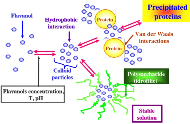
In addition, it has been estimated that approximately one-half of total wine protein is bound to grape phenols via different interaction, as described in Figure 4.
Proteina Protein Proteina Protein
Polysaccharide
(idrofilic)
Hydrophobic
Hydrophobic
interaction
interaction
Flavanol
Flavanol
Colloid
Colloid
particles
particles
Stable
Stable
solution
solution
Flavanols concentration,
T, pH
Proteina Protein Proteina Protein Proteina Protein Proteina ProteinPolysaccharide
(idrofilic)
Polysaccharide
(idrofilic)
Hydrophobic
Hydrophobic
interaction
interaction
Hydrophobic
Hydrophobic
interaction
interaction
Flavanol
Flavanol
Flavanol
Flavanol
Colloid
Colloid
particles
particles
Stable
Stable
solution
solution
Stable
Stable
solution
solution
Flavanols concentration,
T, pH
Flavanols concentration,
T, pH
Flavanols concentration,
T, pH
Precipitated
Precipitated
proteins
proteins
Precipitated
Precipitated
proteins
proteins
Van der Waals
Figure 4: Interaction between wine proteins and polyphenols.
1.3 Haze in bottled white wine
Limpidity (translucency) is of vital importance to wine quality because this visual attribute make the first impression on the consumer (Ferreira et al., 2004). For these
H H H H O O O N N N N OH O H O O H OH O H OH O H O O H OH O H H O O H N N N N N O O H H OH O H O O H OH O H O O H H O N N NH+ O O H O O H OH O H O Protein Protein (Proline) Protein Flavan Flavan Hydrophobic interaction Ionic bond 3 Hydrogen bond
reason, protein precipitation in bottled white wines reduces their commercial value, indicating that they are unstable and therefore unacceptable for sale (Bayly & Berg, 1967; Hsu & Heatherbell, 1987a; Waters et al., 1991, 1992).
The measurement of limpidity is related with the turbidity, which corresponding to the optical phenomenon known as the Tyndall effect, caused by the presence of particles in suspension that deflect light from its normal path (Ribéreau-Gayon et al., 2000), (Figure 5).
Figure 5: Example of protein haze formation in white wine. On the left side a white wine protein unstable and on the right side a white wine clarified and stabilized.
It is possible to find different types of hazes in bottled wine, which can have microbiological or chemical origin. The haze induced by microorganism can occur due to the action of spoilage yeast (Zygosaccharomyces and Brettanomyces) and bacteria (lactic acid bacteria, acetic acid bacteria and Bacillus species), causing off-flavours and changes in wine quality (Rankine & Pilone, 1973; Van de Waters, 1985). The haze induced by chemical factors can occur by crystallization of metal salts (iron and copper), including potassium bitartrate (Dunsford & Boulton, 1981; Rodriguez-Clemente et al., 1990; Lubbers et al., 1993) and calcium tartrate (Clark et al., 1988). Polysaccharides and polyphenols are also involved in the induced-haziness, producing color changes and precipitates (Somers & Ziemelis, 1985; Siebert et al., 1996; Waters et al., 1996; Pellerin et al., 1994).
The most abundant haze problem in bottled white wine is still due to precipitation of unstable proteins (Waters et al., 2005), that is considered one of the most common non-microbial defects of commercial wines (Bayly & Berg, 1967; Hsu & Heatherbell, 1987a; Waters et al., 1992). Slow denaturation of wine proteins, resulting from unfavourable storage conditions, probably originate protein aggregation and flocculation into a hazy suspension, leading to the appearance of a haze or deposit in bottled wine.
1.4 Proteins responsible for wine turbidity
Despite significant advances in wine proteins research, the involved factors and the precise molecular mechanism of protein haze formation in bottled white wines, remain largely to be elucidated.
For a number of years, the study of protein haze formation in bottled white wines was essentially focused on the proteins themselves. It was initially proposed that wine instability is solely related to its protein content as reported by Somers & Ziemelis (1973) and Anelli (1977). Supporting this hypothesis is the study of Koch and Sajak (1959), who used paper electrophoresis to show that wine contains two major protein fractions, both of which decrease upon heat treatment.
However, other studies have shown that protein instability does not correlate well with the wine total protein content, and, therefore, the potential of wine to form haze is not predictable from its protein concentration (Berg & Akioshi, 1961; Moretti & Berg, 1965). Afterwards, Bayly & Berg (1967) supported this assertion demonstrating the different heat sensitivity of proteins in must. Therefore, two alternative hypotheses have been advanced to explain the insolubility of proteins in wine:
a) individual proteins behave differently in their sensitivity to heat denaturation, contributing differentially to haze formation; only part of the protein mixture is responsible for instability rather than the entire protein content;
b) although protein-dependent, the development of turbidity in wines is controlled by one or more factors of non-protein origin (Ferreira et al., 2002).
There is also conflicting evidence in the literature as to which proteins are responsible for haze and deposit formation. Thus, some reports suggest that the lower molecular
mass, lower pI proteins are the major and most important fractions contributing to protein instability in wines (Hsu & Heatherbell, 1987a, 1987b; Hsu et al., 1987; Mesrob et al., 1983). Other studies indicate that the lower molecular mass and higher pI (Heatherbell et al., 1984; Lee, 1986; Ngaba & Heatherbell, 1981) or the higher molecular mass proteins contribute mostly to heat instability. Other investigations revealed that all the major wine protein fractions are present in wine hazes and all have been shown to be heat unstable (Waters, 1991; Waters & Høj, 1999; Waters et al., 1990, 1991, 1992).
Recently, the attention of wine researchers moved towards compounds of non-protein nature. Indeed, the observations that wines are essentially composed of identical sets of polypeptides, identified as PR proteins, and that the haze forming wine proteins are PR proteins apparently similar in wines vinified from different grape varieties (Dawes et al., 1994; Ferreira et al., 2000; Monteiro et al., 2001; Pueyo et al., 1993; Waters et al., 1992, 1996), support the view that protein insolubility is not determined by the protein molecules themselves. It, probably, depends on some other non-protein factor, such as polyphenols, wine pH and the presence of polysaccharides. In other words, the presence of PR proteins in wine is certainly a pre-requisite for haze formation, even if it results affected by factors of non-protein origin (Ferreira et al., 2002).
1.5 Pathogenesis-related proteins
Pathogenesis-related proteins (PRs) are now widely considered as a rich source of allergens. The PRs are encoded by the plant genome and induced specifically in response to infections by pathogens such as fungi, bacteria, viruses, or by adverse environmental factors (Stintzi et al., 1993; Edreva, 2005; Kiba et al., 2007). These proteins include chitinases, thaumatinlike proteins and osmotins (Monteiro et al., 2001; Waters et al., 1998), which are particularly stable under winemaking conditions (low pH, proteolysis), passing selectively into the wine. The actual pattern of polypeptides that accumulate in mature grapes and wines is determined by the environmental and pathological conditions that prevail during vegetative growth (Ferreira et al., 2004). Infection with common grapevine pathogens or skin contact, such as occurs during
transport of mechanically harvested fruit, results in enhanced concentrations of PR proteins in wine (Cilindre et al., 2007; Waters et al., 2005).
Pathogenesis-related proteins do not constitute a super-family of proteins but represent a collection of unrelated protein families, which contribute to the plant defence system. Today, PRs are classified into 14 families. Many important plant food allergens are homologues to proteins that are members of PR families (Van Loon & Van Strien, 1999; Hoffmann-Sommergruber, 2002). The family 5 of PRs comprises unique proteins with diverse functions. Because of the sequence homologies between PR-5 proteins and thaumatin, an intensely sweet tasting protein isolated from the fruits of the West African rain forest shrub Thaumatococcus daniellii, members of this family of proteins are referred to as thaumatin-like proteins (TLPs). TLPs can be classified into three groups, (i) those produced in response to pathogen infection, (ii) those produced in response to osmotic stress, also called osmotins, and (iii) antifungal proteins present in cereal seeds (Breiteneder, 2004).
1.6 Protein fining treatments
The term ‘fining’ is used in winemaking to describe the deliberate addition of an adsorptive compound that is followed by the settling or precipitation of partially soluble components from wine.
The mechanisms of fining wine are based on the charge cancellation between the suspended particles and the fining agent ones, allowing the colloid suspension to agglomerate and flocculate by gravity. Other mechanism can be the absorption of the suspended particles on the surface of the fining agent.
Commonly, fining agents used in winemaking are proteins of animal origin, such as egg albumin, blood albumin, casein, isinglass, gelatins or plant proteins sourced from cereals and legumes.
Nevertheless, the traditional method of stabilizing white wine against haze formation is based on bentonite fining. This material is a montmorillonite clay, which contains exchangeable cations. The adsorption of wine proteins onto bentonite is due to the cationic exchange capacity of this clay, depending on the amount of displacement of aluminium ions by sodium, calcium, or magnesium ions (Figure 6).
Bentonite, carrying a net negative charge at the wine pH, interacts electrostatically with the positively charged wine proteins, producing their flocculation (Hsu & Heatherbell, 1987a; Lamikanra & Inyang, 1988).
The relationship between wine pH and protein isoelectric point governs the cationic nature of the wine proteins and thus their tendency to be adsorbed electrostatically by the negatively charged plate surfaces of bentonite (Bayly & Berg, 1967; Hsu & Heatherbell, 1987a; Moretti & Berg, 1965; Somers & Ziemelis, 1973).
Figure 6: Bentonite structure.
However, bentonite is not equally effective in removing all proteins from wine. Various studies showed that different protein fractions may require distinct bentonite concentrations for removal (Hsu & Heatherbell, 1987a; Murphey et al., 1989; Paetzold et al., 1990). Dawes et al. (1994) observed that the amount of protein depletion correlates approximately linearly with the level of bentonite added. The amount of bentonite required to stabilize wines has increased over the last 10-20 years. Doses of 0.2–0.4 g l-1 bentonite were generally enough to stabilize wines. However, nowadays, to ensure a secure stabilization, doses of 0.8–1 g l-1 bentonite are often employed (Hsu & Heatherbell, 1987a; Paetzold et al., 1990).
As a cation exchanger, bentonite adsorption is not specific for proteins, removing other charged species or aggregates involved as aroma and flavour compounds, inducing
significant aroma loss and, occasionally, colour alteration (Cabaroglu et al., 2002). Bentonite fining also causes substantial volume losses (between 3% and 10%) (Hoj et al., 2001) and the disposal of spent bentonites constitute a non-negligible source of waste. Finally, bentonite handling is also of concern on account of occupational health and safety issues (Sauvage et al., 2010).
Several alternative techniques to bentonite fining, as well as the use of others fining agents, to removal haze-forming wine proteins have been studied. Some alternative techniques studied have been ultrafiltration (Hsu et al., 1987; Flores, et al., 1990), flash pasteurization (Pocock et al., 2003) and use of others adsorbents materials such as resin (Sarmento et al., 2000) and metal oxides (Pashova et al., 2004a; Pashova et al., 2004b; Salazar et al., 2006; Salazar et al., 2007).
The addition of glycoproteins, such as mannoproteins (Waters et al., 1991; Waters et al., 1994a; Waters et al., 1994b), of polysaccharides extracted from seaweeds (Cabello-Pasini et al., 2005), as well the use of immobilized phenolic compound, such as proanthocyanidins (Weetall et al., 1984; Powers et al., 1988), has been tested as alternative to reduce the protein haze in white wine. The results obtained to date have not been successful on industrial scale and, thus, bentonite is still the only commercially acceptable practical solution to avoid protein haze in bottled white wines (Waters et al., 2005).
CHAPTER 2: Enzyme immobilization
2.1 Enzymes in food industry
Enzymes exhibit a number of features that make their use advantageous as compared to conventional chemical catalysts (Krajewska, 2004).
Enzymes have a high level of catalytic efficiency, often far superior to chemical catalysts, and a high degree of specificity that allows them to discriminate not only between reactions but also between substrates (substrate specificity), similar parts of molecules (regio-specificity) and between optical isomers (stereo-specificity). These specificities warrant that the catalyzed reaction is not perturbed by side-reactions, resulting in the production of one wanted end-product, whereas production of undesirable by-products is eliminated. This provides substantially higher reaction yields reducing material costs (Bullock, 1995; Woodley, 1992; Van de Velde et al., 2002). In addition, enzymes generally operate at mild conditions of temperature, pressure and pH with reaction rates of the order of those achieved by chemical catalysts at more extreme conditions, resulting in substantial process energy savings and reducing manufacturing costs (Krajewska, 2004).
For many thousands of years, man has used naturally occurring microorganisms, bacteria, yeasts and their enzymes to make foods such as bread, cheese, beer and wine. In bread-making, for example, amylase is used to break down flour into soluble sugars, which are transformed by yeast into alcohol and carbon dioxide, making the bread rise. Today, enzymes are used for an increasing range of applications in food industries, such as bakery, cheese making, starch processing, production of fruit juices and other drinks (Table 1), in order to improve food texture, appearance and nutritional value, generating desirable flavours and aromas.
Like all proteins, enzymes can cause allergic reactions when people have been sensitized through exposure to large quantities. For this reason, enzyme immobilization results useful in order to allow the prevention of protein contamination in food products (Krajewska, 2004).
Table 1: Enzyme applications in food industry.
Market Enzyme Purpose / function
Rennet (protease) Coagulation in cheese production
Lactase Hydrolysis of lactose to give lactose-free milk products
Protease Hydrolysis of whey proteins
Dairy
Catalase Removal of hydrogen peroxide
Brewing Cellulase, beta-glucanase, alpha-amylase, protease, maltogenic amylase
For liquefaction, clarification and to supplement malt enzymes
Alcool
production Amyloglucosidase Conversion of starch to sugar Alpha-amylase
Breakdown of starch, maltose production
Amyloglycosidase Saccharification
Maltogen amylase Delays process by which bread becomes stale
Protease Breakdown of proteins
Pentosanase Breakdown of pentosan, leading to reduced gluten production
Baking
Glucose oxidase Stability of dough
Pectinase Increase of yield and juice clarification
Glucose oxidase Oxygen removal
Wine and fruit juice
Beta-glucanases
Meat Protease Meat tenderising
Starch
Alpha amylase,
glucoamylase, hemicellulase, maltogenic amylase, glucose isomerase
Modification and conversion (to dextrose or high fructose syrups)
2.2 Enzyme immobilization
In addition to the unquestionable advantages, there exist a number of practical problems in the use of enzymes. To these belong: the high cost of their isolation and purification, the instability of their structures, once they are isolated from their natural environments, and enzymes sensitivity both to process conditions other than to trace levels of substances that can act as inhibitors, resulting in enzymes short operational lifetimes. Moreover, unlike conventional heterogeneous chemical catalysts, most enzymes operate dissolved in water in homogeneous catalysis systems, contaminating the product. Consequently, they cannot be recovered in the active form from reaction mixtures for further uses.
Several methods have been proposed to overcome these limitations, one of the most successful is enzyme immobilization (Krajewska, 2004, Tischer & Wedekind, 1999; Scouten et al., 1995). Immobilization is achieved by fixing enzyme to or within solid supports. By mimicking the natural mode of occurrence in living cells, where enzymes for the most cases are attached to cellular membranes, the systems stabilize the structure of enzymes, hence their activities (Figure 7).
Figure 7: Different types of enzymatic reactions. Enzyme catalysis has commonly been studied in solution phase (a). For practical reasons, enzymes can be immobilized on a polymeric
support, which is also well documented (b), (Laurent et al., 2008).
Thus, as compared to free enzymes in solution, immobilized ones are more robust and resistant to environmental changes (Krajewska, 2004).
In details, enzyme immobilization provides the following advantages:
• enhanced stability;
• easy separation from the reaction mixture;
• possible modulation of the catalytic properties;
• prevention of protein contamination in the product;
• prevention of microbial contaminations.
However, regardless of its nature or preparation, an immobilized enzyme, by definition, has to perform two essential functions, namely:
i. the non-catalytic functions (NCFs), that are designed to aid separation (e.g. isolation of catalysts from the application environment, reuse of the catalysts and particularly control of the process);
ii. the catalytic functions (CFs), that are designed to convert the targeting compounds (or substrates) within a desired time and space (Cao, 2005).
Enzymes can be immobilized by a variety of methods, which may be broadly classified as physical, where weak interactions between support and enzyme exist, and chemical, where covalent bonds are formed with the enzyme (Krajewska, 2004; Tischer & Wedekind, 1999; Scouten et al., 1995; Bullock, 1995; Woodley, 1992), as described in Figure 8.
Figure 8: Schematic classification of immobilization methods.
To the physical methods belong: (i) containment of an enzyme within a membrane reactor, (ii) adsorption (physical, ionic) on a water-insoluble matrix, (iii) inclusion (or gel entrapment), (iv) microencapsulation with a solid membrane, (v) microencapsulation with a liquid membrane, and (vi) formation of enzymatic Langmuir-Blodgett films. The chemical immobilization methods include: (i) covalent attachment to a water-insoluble matrix, (ii) crosslinking with use of a multifunctional, low
molecular weight reagent, and (iii) co-crosslinking with other neutral substances, e.g. proteins. All these approaches are a compromise between maintaining high catalytic activity while achieving the advantages of immobilization. Numerous other methods, which are combinations of the ones listed or original and specific of a given support or enzyme, have been devised. However, no single method and support is best for all enzymes and their applications. Besides, all of the methods present advantages and drawbacks. Adsorption is simple, cheap and effective but frequently reversible; covalent attachment and cross-linking are effective and durable, but expensive and easily worsening the enzyme performance; in membrane reactor-confinment, entrapment and microencapsulations generate diffusional problems (Krajewska, 2004). Consequently, as a rule the optimal immobilization conditions for a chosen enzyme are found empirically by a process of trials, in order to ensure the highest activity retention, operational stability and durability. Nevertheless, immobilization affects enzyme performance (Krajewska, 2004; Tischer & Wedekind, 1999; Scouten et al., 1995; Bullock, 1995; Woodley, 1992). Immobilized enzyme, usually, presents both lower activity and higher Michaelis constant respect to the free one. These alterations result from enzyme structural changes, due to the immobilization procedure application and from the creation of a microenvironment, different from the bulk solution, in which enzyme works. The latter is strongly dependent on the reaction taking place, the nature of the support and on the design of the reactor. Furthermore, being two phase systems, the immobilized enzyme systems suffer from inevitable mass transfer limitations, producing unfavorable effects on their overall catalytic performances. These, however, may be reduced by applying appropriate reactor designs (Krajewska, 2004).
2.3. Supports for enzyme immobilization
The properties of immobilized enzymes depend both on enzyme and support material properties (Krajewska, 2004; Tischer & Wedekind, 1999). In Figure 9, the most used immobilization supports are schematized.
Figure 9: The most used immobilization supports (Cao, 2005).
Silica and derivatives. (I) Silanized silica and (II) PEI-coated silica; Mesoporous zeolite: (III) SAB-15, (IV) MPTMS, (V) VTES, (VI) TSBN-COOH, (VII) APTES, (VIII) PTMS; hydrophobic carriers: (IX) no spacer, (X) amino dextran as spacer, (XI) aldehyde dextran as spacer, (XII) amino PEG as spacer, (XIII) BSA as spacer; polysaccharide-based carriers: (XIV) DEAE-sephadex, (XV) TEAE-Cellulose, (XVI) Ecetola-cellulose; functionalized Eupergit C: (XVII) Eupergit C; (XVIII) DAE-Eupergit C, (XIX) dodecyl-Eupergit C, (XX) IDA-Eupergit C, (XXI) Cu++-IDA-Eupergit C.
Although it is recognized that there is no universal support for all enzymes and their applications, a number of desirable characteristics should be common to any material considered for immobilizing enzymes. These include: high affinity to proteins, availability of reactive functional groups for direct reactions with enzymes and, for chemical modifications, hydrophilicity, mechanical stability, rigidity and regenerability. Understandably, for food, pharmaceutical, medical and agricultural applications, non-toxicity and biocompatibility of the materials are also required. Furthermore, to respond to the growing public health and environmental awareness, the materials should be biodegradable, economical and inexpensive (Krajewska, 2004). In the last decades, numerous organic and inorganic materials have been proposed as carriers for enzyme immobilization, among these, silica materials, epoxy-activated acrylic resins and chitosan have been largely studied for a wide range of industrial applications.
2.3.1. Silica materials
Among various inorganic carriers, silica materials, have been widely used as the stationary phases for HPLC. Recently, mesoporous silica materials have gathered a significant attention in both academic and industrial areas because of its well-ordered and adjustable pore structures as well as abundant surface silanol groups. Their large surface area and their high pore volume allow the reusability of immobilized enzymes, enhancing their activity, selectivity as well as operational stability. For these reasons silica materials have been largely applied for enzyme immobilization.
Recently, a wide range of enzyme has been immobilized on silica materials. For the first time, Wainer and co-workers immobilized trypsin on silica particles modified by hydrophilic polymers and glutaraldehyde in sequence (Ma et al., 2009). Bonneil et al. proposed an immobilized trypsin reactor with porous glass beads (80–120 mesh; 700Å average pore size) as carrier, bonding trypsin via diisothiocyanate (Bonneil et al., 2000; Bonneil & Waldron, 2000). Later, the immobilization of lipase on several kinds of mesoporous silica has been well investigated (Gao et al., 2010; Lee et al., 2005; Hartmann, 2005; Kim et al., 2007; Li et al., 2009a). Moreover, it has been reported that the pore-size of support materials, especially the ratio pore-size vs enzyme molecule size, may greatly influence the enzyme adsorption (Lu et al., 2008; Kang et al., 2007)
and the ideal size of pores was found to be 3–5 times of the protein size (Serra et al., 2008; Gritti and Guiochon, 2007).
2.3.2. Epoxy-activated acrylic resins
The commercial products EUPERGIT C and EUPERGIT C 250 L are amongst the most extensively studied matrixes for enzyme immobilization (Boller et al., 2002). Both resins are microporous, epoxy-activated acrylic beads with a diameter of 100-250 µm. They differ in the content of oxirane groups and in their porosity. While EUPERGIT C has an average pore size of r = 10 nm and an oxirane density of 600 µmol/g dry beads, EUPERGIT C 250 L has larger pores (r = 100 nm) and a lower oxirane density (300
µmol/g dry beads).
Immobilization of enzymes on these resins is rapid and easy both at laboratory scale and at industrial scale. There is no need for additional reagents or special equipment. As a standard procedure, the enzyme is simply dissolved in aqueous buffer and, after addition of the EUPERGIT beads, left to stand at room temperature or at 4 °C for 12-72 h.
Guisan et al. have proposed a two-step binding mechanism for this process (Mateo et al., 2000). It is assumed that, in the first step, the enzyme is physically adsorbed on the carrier by hydrophobic interactions. This brings amino and thiol groups on the surface of the enzyme in close proximity to the oxirane groups of the carrier. In the second step they react with the oxirane groups by nucleophilic attack. In this way, very stable C-N and C-S bonds are formed. Lasch and Janowski showed that there was no detectable protein leakage from Azocasein-EUPERGIT C conjugates once residual, non-covalently bound protein had been removed (Lasch & Janowski, 1988).
The high density of oxirane groups on the surface of the support promotes “multipoint attachment”. It is assumed that such multipoint attachment leads to an increase in conformational stability of the enzyme and hence improves longterm operational stability of the immobilized biocatalyst. Other advantages of covalent immobilization onto EUPERGIT are increased thermostability, immunisation against aldehydes, and increased stability against denaturing polar organic solvents. Due to its compatibility
with a wide range of different enzymes, it is also frequently used for the immobilization of enzymes in academic and industrial research. Different classes of enzymes, such as oxidoreductases (E.C.1), transferases (E.C. 2), hydrolases (E.C. 3), and lyases (E.C. 4), have been successfully immobilized on these supports (Boller et al., 2002).
2.3.3. Chitin- and chitosan-based materials
Of the many carriers that have been considered and studied for immobilizing enzymes, organic or inorganic, natural or synthetic, chitin and chitosan are of interest since their characteristics. Chitin and chitosan are natural polyaminosaccharides (Krajewska, 2004; Peter, 1995; Krajewska, 1991). Chitin is one of the world’s most plentiful, renewable organic resources; it is the major constituent of crustaceans shell, insects exoskeleton and fungi cell walls, providing strength and stability. Chitin is estimated to be synthesized and degraded in the biosphere in the vast amount of at least 10 Gt each year. Chemically, chitin is composed of β (1 → 4) linked 2-acetamido-2-deoxy-β -D-glucose units (or N-acetyl-d-glucosamine) (Peter, 1995), forming a long chain linear polymer (Figure 10).
Chitosan, the principal derivative of chitin, is obtained by N-deacetylation to a varying extent that is characterized by the degree of deacetylation, and is consequently a copolymer of N-acetyl-D-glucosamine and D-glucosamine. Chitin and chitosan can be chemically considered as analogues of cellulose, in which the hydroxyl at carbon-2 has been replaced by acetamido and amino groups, respectively. Chitosan is insoluble in water, but the presence of amino groups renders it soluble in acidic solutions below pH about 6.5. Chitin and chitosan are not single chemical entities, but vary in composition depending on the origin and manufacture process. Chitosan can be defined as chitin sufficiently deacetylated to form soluble amine salts, the degree of deacetylation necessary to obtain a soluble product being 80–85% or higher (Krajewska, 2004). Commercially, chitin and chitosan are obtained at a relatively low cost from shells of shellfish (mainly crabs, shrimps and lobsters), wastes of the seafood processing industry (Hudson & Smith, 1998; Tharanathan & Kittur, 2003). Basically, the process consists of deproteinization of the raw shell material with a dilute NaOH solution and decalcification with a dilute HCl solution. To result in chitosan, the obtained chitin is
subjected to N-deacetylation by treatment with a 40–45% NaOH solution, followed by purification procedures. Thus, production and utilization of chitosan constitutes an economically attractive means of crustacean shell wastes disposal sought worldwide. Chitosan possesses distinct chemical and biological properties (Peter, 1995; Krajewska, 1991). In its linear polyglucosamine chains of high molecular weight, chitosan has reactive amino and hydroxyl groups, amenable to chemical modifications (Peter, 1995; Tharanathan & Kittur, 2003; Dutta et al., 2002).
Figure 10: Chitin and chitosan structures.
Additionally, amino groups make chitosan a cationic polyelectrolyte (pKa ≈ 6.5), one of the few found in nature. This basicity gives chitosan singular properties: chitosan is soluble in aqueous acidic media at pH < 6.5 and when dissolved it possesses high positive charge on –NH3+ groups, it adheres to negatively charged surfaces, it
aggregates with polyanionic compounds, and it chelates heavy metal ions. Both the solubility in acidic solutions and aggregation with polyanions impart chitosan with excellent gel-forming properties. Along with unique biological properties that include biocompatibility, biodegradability to harmless products, non-toxicity, physiological inertness, remarkable affinity to proteins, hemostatic, fungistatic, antitumoral and anticholesteremic properties. Chitin and chitosan, as yet underutilized, offer an extraordinary potential in a broad spectrum of applications which are predicted to grow rapidly once the standardized chitinous materials become available. Crucially, as biodegradable polymers chitin/chitosan materials are eco-friendly, safe for humans and
Chitin
Chitosan
the natural environment. Increasingly over the last decade chitin- and chitosan based materials have been examined and a number of potential products have been developed for areas such as (Peter, 1995; Dutta et al., 2002; No & Meyers, 2000) wastewater treatment (removal of heavy metal ions, flocculation/coagulation of dyes and proteins, membrane purification processes), the food industry (anticholesterol and fat binding, preservative, packaging material, animal feed additive), agriculture (seed and fertilizer coating, controlled agrochemical release), pulp and paper industry (surface treatment, photographic paper), cosmetics and toiletries (moisturizer, body creams, bath lotion). Moreover, owing to the unparalleled biological properties, the most exciting uses of chitin/chitosan-based materials are those in the area of medicine and biotechnology (Khor, 2002; Paul & Sharma, 2000; Singla & Chawla, 2001). In medicine they may be employed as bacteriostatic and fungistatic agents, drug delivery vehicles, drug controlled release systems, artificial cells, wound healing ointments/dressings, haemodialysis membranes, contact lenses, artificial skin, surgical sutures and for tissue engineering. In biotechnology, on the other hand, they may find application as chromatographic matrices, membranes for membrane separations, and notably as enzyme/cell immobilization supports.
As enzyme immobilization supports chitin- and chitosan based materials are used in the form of powders, flakes and gels of different geometrical configurations. Chitin/chitosan powders and flakes are available as commercial products among others from Sigma-Aldrich and chitosan gel beads (Chitopearl) from Fuji Spinning Co. Ltd. (Tokyo, Japan). Otherwise the chitinous supports are laboratory manufactured. Preparation of chitosan gels is promoted by the fact that chitosan dissolves readily in dilute solutions of most organic acids, including formic, acetic, tartaric and citric acids, to form viscous solutions that precipitate upon an increase in pH and by formation of water-insoluble ionotropic complexes with anionic polyelectrolytes. In this way, chitosan gels in the form of beads, membranes, coatings, capsules, fibres, hollow fibres and sponges can be manufactured (Krajewska, 2004).
2.4. Immobilized enzymes applications
To date, several immobilized enzyme-based processes have been implemented on a larger scale, mainly in food industry, where they replace free enzyme-catalyzed processes, in chemical and pharmaceutical manufactures, where asymmetric synthesis or resolution of enantiomers to produce optically pure products are involved (Bullock, 1995; van de Velde et al., 2002; Wiseman, 1993). A selection of currently used immobilized-enzyme processes, in the approximate order of the decreasing scale of manufacture, is given in Table 2.
Areas of present and potential future applications of immobilized enzyme systems other than industrial include: laboratory scale organic synthesis, analytical and medical applications (Bullock, 1995; van de Velde et al., 2002; Scouten et al., 1995). In analytical applications immobilized enzymes are used chiefly in biosensors (Wilson & Hu, 2000; Krajewska, 2004) and to a lesser extent, in diagnostic test strips. Biosensors are constructed by integrating biological sensing systems, e.g. enzyme(s), with transducers. These obtain a chemical signal produced by the interaction of the biological system with an analyte and transduce it into a measurable response. Different kinds of transducers have been employed in biosensors, viz-potentiometric, amperometric, conductometric, thermometric, optical and piezo-electric, most of the current research being placed on the first two. Enzymes for the most cases are immobilized either directly on a transducer’s working tip or in/on a polymer membrane tightly wrapping it up. In principle, due to enzyme specificity and sensitivity biosensors can be tailored for nearly any target analyte, and these can be both enzyme substrates and enzyme inhibitors. Advantageously, their determination is performed without special preparation of the sample. Meeting the demand for practical, cost-effective and portable analytical devices, enzyme-based biosensors have enormous potential as useful tools in medicine, environmental in situ and real time monitoring, bioprocess and food control, and in biomedical and pharmaceutical analysis. Their use, impaired as yet by not quite satisfactory reliability, is predicted to become widely accepted once their storage and operational stabilities have been improved. The most extensively studied enzymes for the application in enzyme-based biosensors are presented in Table 3.
An overview of enzymes immobilized on chitin- and chitosan-based materials for applcation in food industry, is presented in Table 4.
Table 2: Some of the most important industrial applications of immobilized enzyme systems (Krajewska, 2004).
Enzyme (EC number) Substrate Product
Glucose isomerase (5.3.1.5) Glucose Fructose (high-fructose corn syrup)
β-Galactosidase (3.2.1.23) Lactose Glucose and galactose (lactose-free milk and whey)
Lipase (3.1.1.3) Triglycerides Cocoa butter substitutes
Acrylonitrile Acrylamide
3-Cyanopyridine Nicotinamide Nitrile hydratase (4.2.1.84)
Adiponitrile 5-Cyanovaleramide
Aminoacylase (3.5.1.14) D, L-Aminoacids L-Amino acids (methionine, alanine, phenylalanine, tryptophan, valine) Raffinase (3.2.1.22) Raffinose Galactose and sucrose (raffinose-free solutions)
Invertase (3.2.1.26) Sucrose Glucose/fructose mixture (invert sugar)
Aspartate ammonia-lyase (4.3.1.1) Ammonia + fumaric acid L-Aspartic acid (used for production of synthetic sweetener aspartame)
Thermolysin (3.4.24.27) Peptides Aspartame
Glucoamylase (3.2.1.3) Starch D-Glucose
Papain (3.4.22.2) Proteins Removal of “chill haze” in beers
Hydantoinase (3.5.2.2) D,L-Amino acid hydantoins D,L-Amino acids
Penicillin amidase (3.5.1.11) Penicillins G and V 6-Aminopenicillanic acid
(precursor of semi-synthetic penicillins, e.g. ampicillin)
Table 3: Some of the most frequently studied enzymes for enzyme-based biosensors (Krajewska, 2004).
Enzyme (EC number) Substrate Application
Glucose oxidase (1.1.3.4) Glucose Diagnosis and treatment of diabetes, food science, biotechnology Horseradish peroxidase (1.11.1.7) H2O2 Biological and industrial applications, inhibition-based determination
of heavy metal ions and pesticides
Lactate oxidase (1.13.12.4) Lactate Sports medicine, critical care, food science, biotechnology
Tyrosinase (1.14.18.1) Phenols, polyphenols Determination of phenolic compounds in foods, inhibition-based determination of carbamate pesticides
Glutamate oxidase (1.4.3.11) Glutamate Food science, biotechnology
Urease (3.5.1.5) Urea Medical diagnosis, artificial kidney, environmental monitoring Alcohol dehydrogenase (1.1.1.1) Ethanol Food science, biotechnology
Acetylcholinesterase (3.1.1.7) Acetylcholine, acetylthiocholine
Inhibition-based determination of organophosphorus and carbamate pesticides
Choline oxidase (1.1.3.17) Choline Enzyme used in conjunction with acetycholinesterase Lactate dehydrogenase (1.1.1.27) Lactate Sports medicine, critical care, food science, biotechnology Cholesterol oxidase (1.1.3.6) Cholesterol Medical applications
Penicillinase (3.5.2.6) Penicillins Pharmaceutical applications
Table 4: Applications of enzymes immobilized on chitin- and chitosan-based materials in food industry (Krajewska, 2004).
Enzyme (EC number) Application Support Immobilization
α-Amylase (3.2.1.1) Hydrolysis of starch for glucose syrup Chitin powder III
β-Amylase (3.2.1.2) Production of high maltose syrup from starch Chitosan beads I
α-L-Arabinofuranosidase (3.2.1.55) Aromatization of musts, alcoholic beverages and fruit juices Chitosan powder I, II , III
Catalase (1.11.1.6) Removal of H2O2 from food Chitosan powder I, IV, II
Cellulase (3.2.1.4) Decrease in viscosity of fruit/vegetable juices Chitin powder IV
α-Galactosidase (3.2.1.22) Raffinose hydrolysis in beet molasses Chitin powder IV Chitin powder III Chitosan powder III Chitosan beads I, III
β-Galactosidase (3.2.1.23) Hydrolysis of lactose (lactose-free dairy products)
Chitosan precipitate II Chitin powder III Chitosan magnetite beads I Chitosan powder I Glucoamylase (3.2.1.3) Hydrolysis of starch (ethanol production)
Chitosan beads I, III
β-Glucosidase (3.2.1.21) Wine making and juice processing Chitosan powder III, II
β-Glycosidase (3.2.1.group) Cellobiose hydrolysis for glucose production Chitosan powder II Chitosan powder I, III, IV Chitosan microbeads V Chitosan-organosilane
particles
I Invertase (3.2.1.26) Hydrolysis of sucrose (production of invert sugar)
Chitosan-magnetite beads
I
Isoamylase (3.2.1.68) Hydrolysis of starch (glucose and maltose) Chitin powder III
Lysozyme (3.2.1.17) Cheesemaking Chitosan powder I
Neutral proteinase (3.4.24.28) Hydrolysis of soybean protein Chitosan precipitate II
Papain (3.4.22.2) Removal of “chill haze” in beers Chitin powder II
Pectin lyase (4.2.2.10) Reduction of fruit/vegetable juices’ viscosity Chitin powder III Chitin powder III Proteases (3.4.groups) Casein hydrolysate debittering
Chitin film II
Chitin powder III
Chitosan-magnetite particles
IV
Chitosan powder I, III Pullulanase (3.2.1.41) Hydrolysis of starch (glucose/maltose syrup)
Chitin powder II Chitosan powder III, II
α-L-Rhamnopyranosidase (3.2.1.40) Aromatization of musts, alcoholic beverages and fruit juices
Chitosan particles V Chitin powder and
colloidal chitin
III, I
Chitosan precipitate III Tannase (3.1.1.20) Hydrolysis of tea tannins
Chitosan-triphosphate beads
V
Transglutaminase (2.3.2.13) Deamidation of food proteins Chitosan beads III
Trypsin (3.4.21.4) Hydrolysis of proteins Chitosan-magnetite
particles I Chitosan-PVA capsules V Chitosan-PGMA precipitate I Chitosan-coated alginate beads V Urease (3.5.1.5) Removal of urea from beverages and food
Chitosan-organosilane particles
I
Immobilizations are presented as five techniques: (I) adsorption of enzyme on support; (II) adsorption of enzyme on support followed by cross-linking with glutaraldehyde (reticulation); (III) covalent binding of enzyme to glutaraldehyde-activated support; (IV) covalent binding of enzyme to support activated with agents other than glutaraldehyde; (V) gel inclusion.
CHAPTER 3: Bromelain from pineapple stem
3.1 Proteases
The large family of peptide-bond-cleaving hydrolases, the peptidases (= proteases, EC 3.4), can be classified as endopeptidases (= proteinases, EC 3.4.21-99) and exopeptidases (EC 3.4.11-19), according to the point at which they break the peptide chain (Otto & Schirmeister, 1997). These enzymes can be ordered further, according to the reactive groups at the active site involved in catalysis, into serine (EC 3.4.21), aspartic (EC 3.4.23), metallo (EC 3.4.24) and cysteine-endopeptidases (EC 3.4.22), as shown in Figure 11.
Figure 11: Reactive groups at the active site of serine a), aspartic b), metallo c) and cysteine peptidases d).
Cysteine proteases have been found in viruses and prokaryotes as well as in higher organisms such as plants and mammals, including humans. Mammalian cysteine
d) His Cys His Glu His H2O Zn2+ c) Ser 221 His 64 Asp 32 Asn 155 a) b) Asp Asp d) His Cys d) His Cys His Cys His Glu His H2O Zn2+ c) His Glu His H2O Zn2+ His Glu His H2O Zn2+ c) Ser 221 His 64 Asp 32 Asn 155 a) Ser 221 His 64 Asp 32 Asn 155 Ser 221 His 64 Asp 32 Asn 155 a) b) Asp Asp
proteinases fall into two classes: caspases (Rzychon et al., 2004; Barrett et al., 1998) and the papain superfamily comprising the papain family, calpains and bleomycin hydrolases (Otto & Schirmeister, 1997; Barrett et al., 1998; McGrath et al., 1999). Cysteine proteinases participate in varied biological processes. The cathepsins alone are involved in protein breakdown in lysosomes, antigen presentation, proteolytic processing of proenzymes and prohormones, fertilization, cell proliferation, differentiation and apoptosis (Chapman et al., 1997; Grzelakowska-Sztabert, 1998; Berdowska & Siewiński, 2000).
3.2 Catalytic mechanism of cysteine proteases
The proteases of this group are most commonly exemplified by papain, being a well described plant enzyme isolated from the latex of Carica papaya fruit (Rzychon et al., 2004).
The proteolytic activity of all cysteine proteases arises from the presence of the catalytic Cys and His residues in the enzyme active centre. In the case of papain-like cysteine proteinases, the catalytic centre is complemented with Asn that ensures an orientation of the His imidazole ring optimal for successive stages of hydrolysis. The crucial step of the catalytic process involves formation of a reactive thiolate/imidazolium ion pair (Cys-S–/His-Im+), which results from proton transfer between Cys-25 and His-159 (papain numbering). In principle, the thiolate anion attacks the carbonyl carbon of the scissile peptide bond, and the double bond between the carbon and the oxygen converts into a single one (Figure 12A). The oxygen assumes a negative net charge allowing formation of the first tetrahedral transition state. The oxyanion is stabilized by hydrogen bonding to the NH groups of Gln-19 side chain and Cys-25 backbone, which is likely to result in the formation of an oxyanion hole (Figure 12B) (Menard et al., 1991; Menard et al., 1995; Harrison et al., 1997; Otto & Schirmeister, 1997). Subsequent rotation of the His residue enables proton transfer from the imidazolium cation to the nitrogen of the peptide bond being hydrolyzed, and cleavage occurs. The newly formed substrate amine is hydrogen bonded to His-159, whereas the substrate carboxylic part is linked to Cys-25 via a thioester bond, forming acyl enzyme (Figure 12C). The next reaction step involves dissociation of the aminic part of the substrate and its replacement with a water
molecule. The imidazole nitrogen contributes to polarization of the water molecule that in turn attacks the carbonyl carbon of acyl enzyme (Figure 12D). This is followed by formation of the second tetrahedral intermediate (Figure 12E). In the final step, thioester deacylation leads to reconstruction of the carboxyl group in the hydrolyzed substrate, which is concerted with the release of an active enzyme (Figure 12F) (Menard et al., 1991; Otto & Schirmeister, 1997; Rzychon et al., 2004).
Figure 12: Catalytic mechanism of cysteine proteinases as exemplified by papain (Rzychon et al., 2004).
3.3 Selection of bromelain from pineapple stem
The most extensively investigated plant cysteine protease is papain (EC 3.4.22.2) from the latex of Carica papaya fruit. Papain is a monomeric polypeptide consisting of a chain of 212 amino acid residues with three disulfide bridges (Cys22-Cys63, Cys56-Cys95, Cys153-Cys200). Other plant cysteine proteases whose known fragment sequences indicate a relationship to papain are: chymopapains A and B from Carica