Volume 2009, Article ID 476897,5pages doi:10.1155/2009/476897
Clinical Study
Oral Contraceptives after Myomectomy: A Short Term Trial
Stefano Luisi, Valentina Ciani, Massimo Gabbanini, Sofia Sollazzi,
Michela Torricelli, Francesco Calonaci, and Felice Petraglia
Obstetrics and Gynecology Unit, Department of Pediatrics, Gynecology, and Reproductive Medicine, University of Siena, 53100 Siena, Italy
Correspondence should be addressed to Felice Petraglia,[email protected]
Received 5 February 2009; Revised 4 May 2009; Accepted 4 June 2009 Recommended by Anil K. Agarwal
Following myomectomy the rate of fertility is restored and pregnancy may be attempted with a good outcome. In the present study a 3 month treatment with OCs in a group of women after a myomectomy was evaluated. The drug compliance and side effects, the benefits of OC in order to reduce symptoms, to increase post-surgical hemoglobin levels and to avoid an early pregnancy after myomectomy were analyzed. A group of women (n=55) each with myoma≥5 cm was recruited: they presented menorrhagia, pelvic pain, dyspareunia and dysmenorrhae. After laparotomic myomectomy the women were divided into 3 groups. Group 1: women (n=16) treated with pill A (15 mcg of ethynilestradiol + 60 mcg of gestodene); group 2: women (n=23) treated with pill B (20 mcg of ethynilestradiol + 100 mcg of levonorgestrel); group 3: women (n=16) treated with a placebo (oral calcium). After three months from myomectomy and treatment patients in each group reported a reduced menorrhagia, dismenorrhea and pelvic pain. Serum haemoglobin levels increased in all women (P < .05). No pregnancy occurred in any group and the compliance was good. A post surgery treatment by using oral contraceptives guarentees pregnancy prevention, associated with reduction of pain, and improvement of haematologic conditions.
Copyright © 2009 Stefano Luisi et al. This is an open access article distributed under the Creative Commons Attribution License, which permits unrestricted use, distribution, and reproduction in any medium, provided the original work is properly cited.
1. Introduction
Uterine leiomyomata is a major source of morbidity among
women of reproductive age [1]. They are benign tumors of
smooth muscle, commonly referred to as fibroids. Despite the importance of these tumors, little is still known about
their epidemiology or aetiology [2]. Epidemiologic studies
suggest that risk is inversely associated with age at menarche, parity and age at first birth and positively associated with
years since last term birth [3], so the incidence of
leiomy-omata rises through out the reproductive years [2,4], while
the incidence of surgery is lowest in the menopausal years
[4,5]. This pattern suggests a dependence on ovarian steroid
hormones, but the role of these agents in the etiology of
uterine leiomyomata remains unclear [6,7]. Uterine fibroids
cause a variety of symptoms, such as menorrhagia, pelvic pain, infertility, reproductive dysfunction (like pregnancy loss and pregnancy complications), but sometimes they
are completely asymptomatic [8]. The effect of
estrogen-progestin oral contraceptives (OCs) on the volume of uterine leiomyomata is not well characterized.
OCs may play a role in the development or growth of
leiomyomata [9] and a significantly elevated risk among
women who first used OCs at ages 13–16 years compared
with non-OCs users has been shown [1]. On the other
hand, the little available epidemiologic data have suggested
a protective effect of OCs in the risk of fibroids [10, 11].
Low-dose OC use provides the benefit of a reduction in the duration of menstrual bleeding, with resultant improve-ment in haemoglobin levels, without increasing uterine
size [12]. Antiprogesterone which induces ovarian acyclicity
also decreases size of leiomyomata, thus antiprogesterone may provide a novel mode of management of leiomyomata
[13].
Therefore, the association between oral contraceptive and the risk of uterine fibroids is still unclear. Uter-ine leiomyomata is the fifth cause of hospitalization for gynecologic conditions unrelated to pregnancy in women aged 15–44 and the primary indications for hysterectomy
among women of all ages [14]. The range of alternatives
to hysterectomy includes medical regimens (levonorgestrel-releasing intrauterine system), a wide range of endometrial
Table 1: Distribution of patients according to age, dysmenorrhoea, menometrorrhagia and pelvic pain at previous surgery.
Variable Patients (n◦) % Age 30–35 years 9 16 35–40 years 42 77 >40 years 4 7 Presence of symptoms 48 87 Dysmenorrhoea 8 16 Menometrorrhagia 36 75 Pelvic pain 22 45 Absence of symptoms 7 13
ablative techniques, and where fibroids are the primary pathology myomectomy and uterine artery embolization.
Abdominal myomectomy is an effective surgical alter-native to hysterectomy one for therapy of symptomatic
uterine fibroids, it preserves fertility and femininity [15].
Myomectomy is associated with a favourable outcome in infertile women, particularly if no other complication
variable is present [16]. The location of the myomata may
play an important role in determining infertility. Both large intramural and subserous myoma are thought to interfere with conception and reduce the effectiveness of
assisted reproduction cycles [17]. The size of the myoma
may represent another important prognostic factor, 5 cm in diameter being the size limit which appears to justify myomectomy. Restoration of fertility after myomectomy has been reported, with pregnancy rates ranging between 44 and
52% [18].
The time to postmyomectomy conception is short, with
∼80% of pregnancies occurring during the first year
follow-ing surgery, however, in the first months after a myomectomy
a gestation the risk of uterine rupture is higher [17].
In the present study a 3-month treatment with OCs in a group of women after a myomectomy was evaluated. In addition, the drug compliance and side effects, the benefits of OC in order to reduce symptoms, to increase post-surgical hemoglobin levels and to avoid an early pregnancy after myomectomy was analyzed. Moreover, we investigated the risk of uterine leiomyomata in relation to short-term contraceptive use.
2. Materials and Methods
The study was approved by the Institutional Review Board of the Academic Health Center of Siena, and an informed consent was obtained from each participant.
A group of 55 women (aged 30–45, BMI 22–25) with ultrasound and a histologically confirmed diagnosis of uterine fibroids was recruited from September 2006 to April 2007. All the women presented unexplained infertility and had only one large intramural or subserosal myoma
mea-suring≥5 cm and underwent on laparotomic myomectomy
(these myomas were not as suitable for the laparoscopic approach). None of the subjects had taken any medications
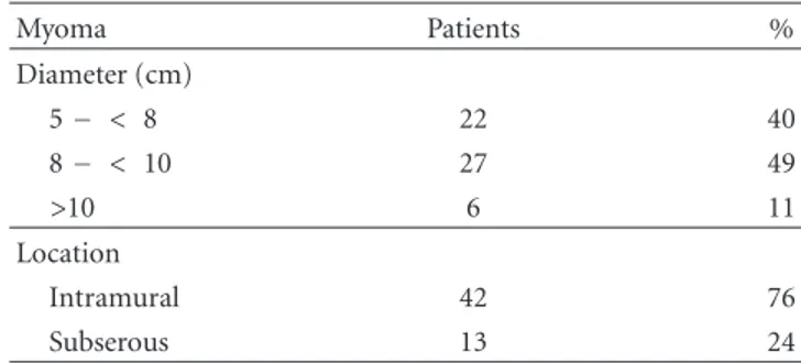
Table 2: Distribution of myomas according to location and diameter. Myoma Patients % Diameter (cm) 5− < 8 22 40 8− < 10 27 49 >10 6 11 Location Intramural 42 76 Subserous 13 24
for at least 3-months before the study. Most of them presented a variety of female reproductive problems and symptoms, such as menorrhagia (excessive uterine bleeding occurring at regular intervals or prolonged uterine bleeding
lasting more than seven days) [19] or irregular bleeding (n=
36), pelvic pain (n =22), dyspareunia and dysmenorrhoea
(n=15).
To investigate the outcome, women were instructed to keep (i) a diary of menstrual bleeding (number of days of menstrual flow and total number of pads/tampons used), rating the blood loss on a visual analog scale from zero (no blood loss) to 10 (gushing-type bleeding); (ii) the presence of side effects and overall satisfaction with treatment was rated on a five-level scale (very satisfied, satisfied, uncertain, dissatisfied, and very dissatisfied); (iii) a questionnaire for the assessment of pain symptoms (dysmenorrhea, dyspareunia and pelvic pain) by using a 10 points Visual Analog Scale
(VAS) [20].
The clinical characteristics of the women enrolled in the
study are shown inTable 1, while the characteristics of the
myomas are shown inTable 2.
Exclusion criteria were von Willebrand’s disease or coag-ulopathies (known or suspected), or a history of hormone-dependent malignancies; known or a history of deep-vein thrombosis, active thrombophlebitis, thromboembolic dis-order, or cerebrovascular accident, myocardial infarction or ischemic heart disease, untreated hypertension, liver disease, any endocrine disorder other than controlled thyroid disease, and a smoking habit of one pack or more of cigarettes per day.
After laparotomic myomectomy and a normal postop-eration time (5 days) without post-operative complications, the women were divided into three randomized groups according to the treatment:
(i) group 1: treated with pill A (n = 16) (15 mcg of
ethynilestradiol + 60 mcg of gestodene) (Arianna, Bayer Schering, Berlin, Germany);
(ii) group 2: treated with pill B (n = 23) (20 mcg of
ethynilestradiol + 100 mcg of levonorgestrel) (Mira-nova, Bayer Schering, Berlin, Germany);
(iii) group 3: treated with placebo (n = 16) (oral
After myomectomy Before 0 7 14 Se rum h ae mog lob in (mg/dl) (a) After myomectomy Before 0 7 14 Se rum h ae mog lob in (mg/dl) (b) After myomectomy Before 0 7 14 Se rum h ae mog lob in (mg/dl) (c)
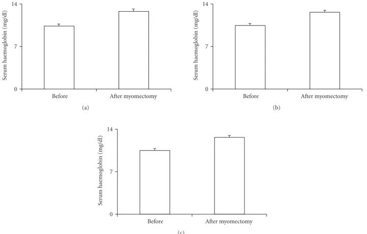
Figure 1: Serum haemoglobin concentration after treatment in the three groups. Group 1: women treated with 15 mcg of ethynilestradiol + 60 mcg of gestodene; group 2: women treated with 20 mcg of ethynilestradiol + 100 mcg of levonorgestrel; group 3: women treated with a placebo.∗=P < .05.
We used two different pill formulations to compare if
there were any differences between second (levonorgestrel)
and third (gestodene) generation progestin.
Since it was a blind study to each group was said to use barrier contraception in order to avoid pregnancy. All the women were controlled in the following 3-months after surgery.
Before and after 3-months a pelvic transvaginal ultra-sound (LAB 70 ESAOTE SpA, Genova, Italy) evaluation was done, and peripheral blood was drawn to measure red and white blood cell count, hematocrit, hemoglobin, and platelets.
The serum results are expressed as a mean of ±SE,
and differences between groups were assessed by using the unpaired t-test. Probability values of less than .05 were considered statistically significant.
3. Results
After the treatment the three groups of patients showed
reduced menorrhagia, dysmenorrhoea, pelvic pain (P < .01)
and increased serum haemoglobin concentration (P < .05)
(Figure 1). The haemoglobin levels showed to be about the same in the three groups after myomectomy.
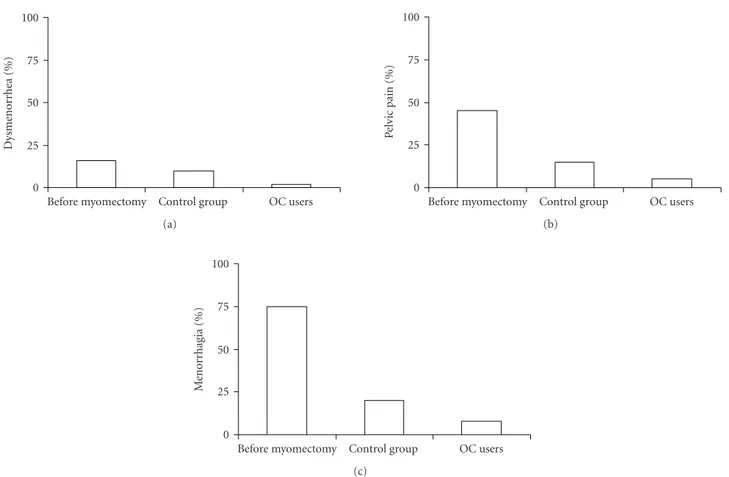
During the transvaginal pelvic ultrasound no gyneco-logical complications were noted in any of the subjects. A
reduction of symptoms occurred in OCs users faster than in
the control group (Figure 2). No pregnancy was reported in
any of the patients.
No relevant side effects (spotting, bloating, acne, mood
swing, weight gain or loss, headaches, breast pain) were registered in any of the groups after 3-months of treatment.
4. Discussion
The present results showed that a 3-month treatment with OCs after myomectomy is associated with regular menstrual cycles, increased serum haemoglobin concentration without pelvic pain, dysmenorrhoea, and risk of pregnancy.
Myomectomy is frequently performed to preserve or increase fertility, although the risk of future uterine rupture is a major concern of any surgery to the uterus. For this reason it is necessary to avoid pregnancy in the first months after the
surgery when the risk is higher [21]. Women recruited in our
present study were>30 years and each had a intramural or
subserosal myoma. The rationale of the OC treatment was to give a contraceptive cover at least in the first 3-months after myomectomy.
In women with menstrual disorders, such as menorrha-gia, the OC treatment reduces the menstrual blood loss and increases the serum haemoglobin concentration, reducing the incidence of anemia. Oral contraceptives represent also
OC users Control group Before myomectomy 0 25 50 75 100 Dysmenor rhea (%) (a) OC users Control group Before myomectomy 0 25 50 75 100 P el vic pain (%) (b) OC users Control group Before myomectomy 0 25 50 75 100 Me n o rr h ag ia (% ) (c)
Figure 2: Improvement of symptoms (dysmenorrhoea, pelvic pain, menorrhagia) after myomectomy and OCs use.
the main medication used to treat dysmenorrhea. Although several mechanisms underlying the dysmenorrheal pain relief attained by OC use have been reported, suppression of prostaglandin (PG) synthesis, which leads to reduced uterine contractions, represents one of the most probable pathways
[22].
The effect of OCs on myomas is still not fully clear.
Evidence showed that both reproductive factors and oral contraceptive use at a young age, influence the risk of uterine
leiomyomata among premenopausal women [23]. In a case
report, a 45-year-old woman, with a symptomatic uterine leiomyoma, presented a reduced myoma volume after the
discontinuation of OCs [24].
To the contrary after a 1-year study Friedman et al. showed that in most women with leiomyomas, low-dose OC use provides the noncontraceptive benefit of a reduction in the duration of menstrual flow, with resultant improvement
in hematocrit, without increasing uterine size [25].
The prolonged use of the last generation of oral con-traceptives does not increase the uterine myoma volume and furthermore it produces a noteworthy reduction in the duration of menstrual flow with consequent increase in
hematocrit [26].
We chose a short-term trial since the majority of women undergoing myomectomy despite of hysterectomy for fibroids want a pregnancy in the immediate postoperative period. We suggested them the use of the pill to improve symptoms and menstrual bleeding. Moreover, we wanted to
underlined the rapid improvement of the clinical condition during the first 3-months after surgery in association with oral contraceptive.
Therefore, an oral contraceptive post-surgery treatment for at least 3-months guarantees the prevention of pregnancy, allowing a faster improvement of the clinical and haemato-logic conditions, without increasing the risk of recurrency.
Condensation. The use of a low-dose of estro-progestins
after myomectomy guarantees the prevention of pregnancy, allowing a faster improvement of the clinical and haemato-logic conditions.
References
[1] L. M. Marshall, D. Spiegelman, M. B. Goldman, et al., “A prospective study of reproductive factors and oral contracep-tive use in relation to the risk of uterine leiomyomata,” Fertility
and Sterility, vol. 70, no. 3, pp. 432–439, 1998.
[2] R. K. Ross, M. C. Pike, M. P. Vessey, D. Bull, D. Yeates, and J. T. Casagrande, “Risk factors for uterine fibroids: reduced risk associated with oral contraceptives,” British Medical Journal, vol. 293, no. 6543, pp. 359–362, 1986.
[3] F. Parazzini, C. La Vecchia, E. Negri, G. Cecchetti, and L. Fedele, “Epidemiologic characteristics of women with uterine fibroids: a case-control study,” Obstetrics and Gynecology, vol. 72, no. 6, pp. 853–857, 1988.
[4] L. M. Marshall, D. Spiegelman, R. L. Barbieri, et al., “Variation in the incidence of uterine leiomyoma among premenopausal
women by age and race,” Obstetrics and Gynecology, vol. 90, no. 6, pp. 967–973, 1997.
[5] I. Romieu, A. M. Walker, and S. Jick, “Determinants of uterine fibroids,” Post Marketing Surveillance, vol. 5, no. 2, pp. 119– 133, 1991.
[6] R. K. Yantiss, P. B. Clement, and R. H. Young, “Neoplastic and pre-neoplastic changes in gastrointestinal endometriosis: a study of 17 cases,” American Journal of Surgical Pathology, vol. 24, no. 4, pp. 513–524, 2000.
[7] M. A. Lumsden and E. M. Wallace, “Clinical presentation of uterine fibroids,” Bailliere’s Clinical Obstetrics and
Gynaecol-ogy, vol. 12, no. 2, pp. 177–195, 1998.
[8] J. Andersen and R. L. Barbieri, “Abnormal gene expression in uterine leiomyomas,” Journal of the Society for Gynecologic
Investigation, vol. 2, no. 5, pp. 663–672, 1995.
[9] A. H. John and R. Martin, “Growth of leiomyomata with estrogen—progestogen therapy,” Journal of Reproductive
Medicine for the Obstetrician and Gynecologist, vol. 6, no. 2, pp.
56–58, 1971.
[10] F. Parazzini, E. Negri, C. La Vecchia, L. Fedele, M. Rabaiotti, and L. Luchini, “Oral contraceptive use and risk of uterine fibroids,” Obstetrics and Gynecology, vol. 79, no. 3, pp. 430– 433, 1992.
[11] H. Ratner, “Risk factors for uterine fibroids: reduced risk associated with oral contraceptives,” British Medical Journal, vol. 293, p. 1027, 1986.
[12] A. J. Friedman and P. P. Thomas, “Does low-dose combination oral contraceptive use affect uterine size or menstrual flow in premenopausal women with leiomyomas?” Obstetrics and
Gynecology, vol. 85, no. 4, pp. 631–635, 1995.
[13] A. A. Murphy, A. J. Morales, L. M. Kettel, and S. S. C. Yen, “Regression of uterine leiomyomata to the antiprogesterone RU486: dose-response effect,” Fertility and Sterility, vol. 64, no. 1, pp. 187–190, 1995.
[14] C. M. Farquhar and C. A. Steiner, “Hysterectomy rates in the United States 1990–1997,” Obstetrics and Gynecology, vol. 99, no. 2, pp. 229–234, 2002.
[15] N. S. Banu and I. T. Manyonda, “Alternative medical and surgical options to hysterectomy,” Best Practice and Research:
Clinical Obstetrics and Gynaecology, vol. 19, no. 3, pp. 431–
449, 2005.
[16] G. Connolly, M. Doyle, T. Barrett, P. Byrne, M. De Mello, and R. F. Harrison, “Fertility after abdominal myomectomy,”
Journal of Obstetrics and Gynaecology, vol. 20, no. 4, pp. 418–
420, 2000.
[17] D. W. Stovall, S. B. Parrish, B. J. Van Voorhis, S. J. Hahn, A. E. T. Sparks, and C. H. Syrop, “Uterine leiomyomas reduce the efficacy of assisted reproduction cycles: results of a matched follow-up study,” Human Reproduction, vol. 13, no. 1, pp. 192– 197, 1998.
[18] C. Chapron and J.-B. Dubuisson, “Laparoscopic treatment of deep endometriosis located on the uterosacral ligaments,”
Human Reproduction, vol. 11, no. 4, pp. 868–873, 1996.
[19] B. S. Apgar, A. H. Kaufman, U. George-Nwogu, and A. Kittendorf, “Treatment of menorrhagia,” American Family
Physician, vol. 75, no. 12, pp. 1813–1819, 2007.
[20] C. Larroy, “Comparing visual-analog and numeric scales for assessing menstrual pain,” Behavioral Medicine, vol. 27, no. 4, pp. 179–181, 2002.
[21] M. Lieng, O. Istre, and A. Langebrekke, “Uterine rupture after laparoscopic myomectomy,” Journal of the American
Association of Gynecologic Laparoscopists, vol. 11, no. 1, pp. 92–
93, 2004.
[22] A. R. Davis, C. Westhoff, K. O’Connell, and N. Gallagher, “Oral contraceptives for dysmenorrhea in adolescent girls: a randomized trial,” Obstetrics and Gynecology, vol. 106, no. 1, pp. 97–104, 2005.
[23] L. M. Marshall, D. Spiegelman, M. B. Goldman, et al., “A prospective study of reproductive factors and oral contracep-tive use in relation to the risk of uterine leiomyomata,” Fertility
and Sterility, vol. 70, no. 3, pp. 432–439, 1998.
[24] R. L. Barbieri, “Reduction in the size of a uterine leiomyoma following discontinuation of an estrogen-progestin contracep-tive,” Gynecologic and Obstetric Investigation, vol. 43, no. 4, pp. 276–277, 1997.
[25] A. J. Friedman and P. P. Thomas, “Does low-dose combination oral contraceptive use affect uterine size or menstrual flow in premenopausal women with leiomyomas?” Obstetrics and
Gynecology, vol. 85, no. 4, pp. 631–635, 1995.
[26] G. Larsson, I. Milsom, G. Lindstedt, and G. Rybo, “The influence of a low-dose combined oral contraceptive on menstrual blood loss and iron status,” Contraception, vol. 46, no. 4, pp. 327–334, 1992.
Submit your manuscripts at
http://www.hindawi.com
Stem Cells
International
Hindawi Publishing Corporationhttp://www.hindawi.com Volume 2014
Hindawi Publishing Corporation
http://www.hindawi.com Volume 2014
INFLAMMATION
Hindawi Publishing Corporation
http://www.hindawi.com Volume 2014
Behavioural
Neurology
Endocrinology
International Journal ofHindawi Publishing Corporation
http://www.hindawi.com Volume 2014
Hindawi Publishing Corporation
http://www.hindawi.com Volume 2014
Disease Markers
Hindawi Publishing Corporation
http://www.hindawi.com Volume 2014
BioMed
Research International
Oncology
Journal ofHindawi Publishing Corporation
http://www.hindawi.com Volume 2014
Hindawi Publishing Corporation
http://www.hindawi.com Volume 2014
Oxidative Medicine and Cellular Longevity
Hindawi Publishing Corporation
http://www.hindawi.com Volume 2014
PPAR Research
The Scientific
World Journal
Hindawi Publishing Corporation
http://www.hindawi.com Volume 2014
Immunology Research
Hindawi Publishing Corporation
http://www.hindawi.com Volume 2014
Journal of
Obesity
Journal ofHindawi Publishing Corporation
http://www.hindawi.com Volume 2014
Hindawi Publishing Corporation
http://www.hindawi.com Volume 2014
Computational and Mathematical Methods in Medicine
Ophthalmology
Journal ofHindawi Publishing Corporation
http://www.hindawi.com Volume 2014
Diabetes Research
Journal ofHindawi Publishing Corporation
http://www.hindawi.com Volume 2014
Hindawi Publishing Corporation
http://www.hindawi.com Volume 2014
Research and Treatment
AIDS
Hindawi Publishing Corporation
http://www.hindawi.com Volume 2014
Gastroenterology Research and Practice
Hindawi Publishing Corporation
http://www.hindawi.com Volume 2014