In Vivo
and
In Vitro
Evidence for Placental DNA Damage
in Preeclampsia
Serkalem Tadesse1,2, Dawit Kidane3, Seth Guller4, Tianmeng Luo2, Nicholas G. Norwitz4, Felice Arcuri5, Paolo Toti6, Errol R. Norwitz1,2*
1 Department of Obstetrics & Gynecology, Tufts Medical Center, Boston, Massachusetts, United States of America, 2 Mother Infant Research Institute (MIRI), Tufts University School of Medicine, Boston, Massachusetts, United States of America,3 Department of Therapeutic Radiology and Genetics, Yale University School of Medicine, New Haven, Connecticut, United States of America,4 Department of Obstetrics, Gynecology & Reproductive Sciences, Yale University School of Medicine, New Haven, Connecticut, United States of America, 5 Department of Molecular and Developmental Medicine, University of Siena, Siena, Italy, 6 Department of Medical Biotechnologies, University of Siena, Siena, Italy
Abstract
Preeclampsia (PE) is an idiopathic multisystem disease affecting 5–7% of pregnant women. Placental oxidative stress is a characteristic feature of PE and occurs when the production of reactive oxygen species (ROS) within the placenta overwhelms the intrinsic anti-oxidant defenses. We hypothesize that excessive oxidative DNA damage at the fetal-maternal interface coupled with a defective DNA damage/repair response is causally related to PE. Here we demonstrate that cH2AX (a sensitive marker of DNA damage) is expressed in the maternal decidua but not trophoblast of normal placentas, and that expression is significantly higher in PE placental tissues in vivo. Using primary in vitro cultures of maternal decidual stromal cells (DSCs) and fetal cytotrophoblast cells (CTs), we show an increase in cH2AX foci in DSCs cultured with vs without H2O2
(70.6% vs 11.6%; P,0.0001) or under hypoxia-reperfusion vs normoxia (20- vs 3-fold; P = 0.01); no foci were seen in CTs. We further demonstrate that Base Excision Repair (BER) intermediates are significantly increased in DSCs (not CTs) under these same conditions. Our data show that DNA damage is significantly more common in PE placentas, and that this DNA damage is localized to the maternal and not fetal side of the placenta. CTs may be selectively resistant to DNA damage in an effort to protect the fetus.
Citation: Tadesse S, Kidane D, Guller S, Luo T, Norwitz NG, et al. (2014) In Vivo and In Vitro Evidence for Placental DNA Damage in Preeclampsia. PLoS ONE 9(1): e86791. doi:10.1371/journal.pone.0086791
Editor: Hai-Yan Lin, Chinese Academy of Sciences, China
Received August 14, 2013; Accepted December 14, 2013; Published January 22, 2014
Copyright: ß 2014 Tadesse et al. This is an open-access article distributed under the terms of the Creative Commons Attribution License, which permits unrestricted use, distribution, and reproduction in any medium, provided the original author and source are credited.
Funding: This work was supported in part by the NIH/NICHD-sponsored Reproductive Scientist Development Program (to ERN) and March of Dimes (21-FY05-1250 to ERN). The funders had no role in study design, data collection and analysis, decision to publish, or preparation of the manuscript.
Competing Interests: The authors have declared that no competing interests exist. * E-mail: [email protected]
Introduction
Preeclampsia (PE) is an idiopathic multisystem disease affecting 5–7% of pregnant women. It is characterized clinically by new-onset hypertension and proteinuria after 20 weeks of gestation [1]. While the exact pathophysiology of PE remains unclear, there is evidence to suggest that such processes as abnormal trophoblast invasion, placental ischemia, and an imbalance in circulating vasoactive regulatory factors are implicated leading to generalized endothelial injury and vasospasm [2]. Excessive placental oxida-tive stress appears to be an important inciting event [3,4].
The high oxygen demand of the mother and fetus increases oxidative metabolism and boosts free radical generation during pregnancy [5]. Mitochondrial superoxide anion (O22) production
is a critical source of oxidative stress within the placenta, and contributes to the overall increase in maternal and placental lipid peroxidation seen in pregnancy [6–8]. In healthy pregnancies, a balance is maintained between lipid peroxides and anti-oxidative processes. In contrast, PE pregnancies are characterized by an imbalance in these processes leading to an increase in oxidative stress [6–8]. Oxygen concentrations at the fetal-maternal interface fluctuate during pregnancy as a consequence of the vascular remodeling within the tissues of the uterus [9]. Infection/
inflammation, intense tissue remodeling, and changes in vascular perfusion generate reactive oxygen species (ROS)–including O22,
hydroxyl radical, and hydrogen peroxide (H2O2)–all of which are
capable of damaging nucleic acids, proteins, and lipids if levels of these ROS overwhelm the intracellular anti-oxidative defenses [10].
DNA damage induced by ROS is repaired by a series of specific and non-specific repair mechanisms [11,12]. Base Excision Repair (BER) is one of the major DNA repair pathways in eukaryotic cells. It is initiated by DNA glycosylases, enzymes that recognize and cleave the damaged bases thereby creating sites along the DNA strand that are temporarily devoid of a base, known as apurinic/apyrimidinic (AP) sites [11]. These AP sites are then further processed by DNA glycosylases family members with AP-lyase or AP-endonuclease activity (such as APE1), which fill in the single nucleotide gap and seal over the hole in the DNA strand to complete the repair reaction [13]. Importantly, until they are fully repaired, these AP sites are both cytotoxic and mutagenic [14–16]. Therefore, although these DNA repair mechanisms are critical for the maintenance of genomic stability, they generate intermediates that can exacerbate DNA damage. High numbers of AP sites can therefore overwhelm the BER machinery leading to further DNA damage [17,18].
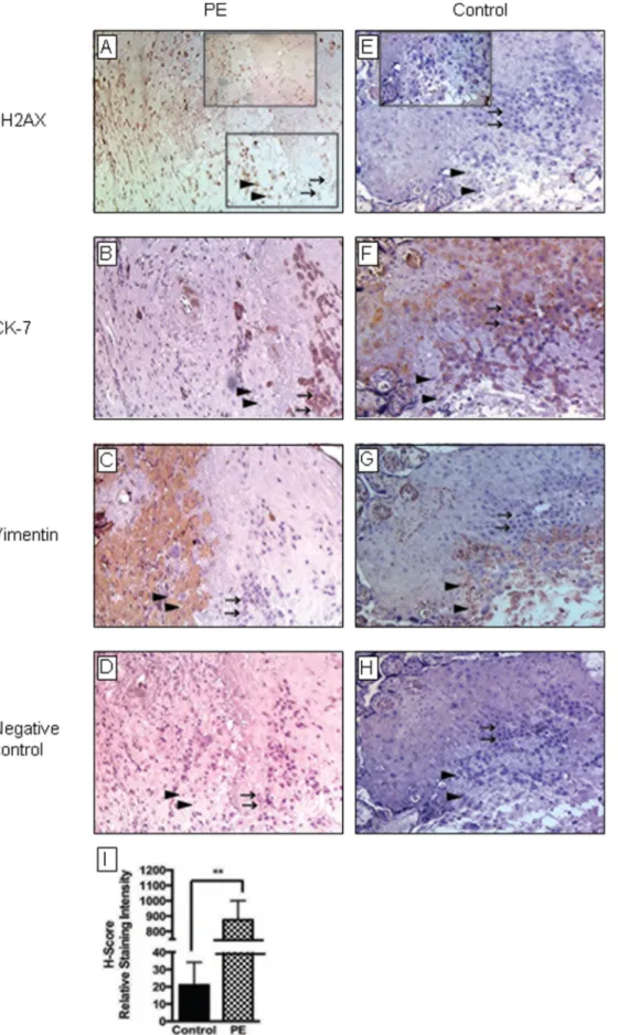
Figure 1. Immunohistochemical analysis of placental tissues forin vivoevidence of DNA damage. Placental tissues were collected from women with preeclampsia [PE] [A–D] and gestational age-matched normotensive controls (n = 10 for each) [E–H]. Tissues were prepared and labeled with antibodies against cH2AX (a biomarker of DNA damage [A,E]), cytokeratin 7 (CK-7) (which stains trophoblast cells, see arrows [B,F]), and vimentin
Traditional concepts hold that ROS-mediated DNA damage occurs randomly with a tissue, which then triggers activation of the BER pathway to remove and replace the damaged bases to minimize their potentially pathogenic consequences. However, emerging evidence suggests that ROS-mediated DNA damage is not random. Rather it may be localized to one or more specific cell populations within any given tissue. We hypothesize that this may be true also in the placenta. Exactly how the balance between ROS-induced DNA damage and repair is maintained at the fetal-maternal interface is not known, but the association between high levels of circulating markers of DNA damage and PE has been previously reported [19]. In this study, we hypothesize that ROS-mediated DNA damage may lead to targeted injury to specific cell populations at the fetal-maternal interface, which may have important implications for the pathogenesis of PE.
Materials and Methods Reagents
Cell culture media (DMEM/F12), estradiol (E2),
medroxypro-gesterone acetate (MPA), HEPES, BSA, FeSO4, ZnSO4, CuSO4,
ascorbic acid were purchased from Sigma-Aldrich (St. Louis, MO). Penicillin and streptomycin, L-Glutamine, sodium pyruvate, and trypsin were purchased from Invitrogen (Burlington, Canada). Collagenase-DNase and protease inhibitors (complete cocktail) for tissue digestion were from Roche (Montreal, Canada). Reagents for sodium dodecyl sulfate-polyacrylamide gels (SDS-PAGE) were supplied by Bio-Rad (Bio-Rad Laboratories, Hercules, CA). Enhanced chemiluminescence kit was purchased from Thermo Fisher Scientific, (Rockford, IL). Insulin/transferrin/selenium and epidermal growth factor were purchased from BD Biosciences (San Jose, CA). Fetal bovine serum (FBS) and charcoal-stripped calf serum (SCS) was purchased from Gemini Biosciences (West Sacramento, CA). Primary antibodies included: anti-histone H2A.X (D17A3) XPH rabbit mAb (#7631) and a-tubulin (#2144) from Cell Signaling (Danvers, MA); anti-vimentin (Clone V9) and anti-cytokeratin-7 (Clone OV-TL 12/30) from DakoCy-tomation (Carpinteria, CA); and anti-CD45 mAb, anti-CD9 mAb, and isotype-matched control antibodies from R&D Systems (Minneapolis, MN). Goat anti-mouse IgG immunomagnetic beads (Dynabeads) were purchased from Dynal (Oslo, Norway). Secondary antibodies such as horseradish peroxidase (HRP)-conjugated, FITC-conjugated and biotin-conjugated anti-mouse and anti-rabbit antibodies were purchased from Jackson Immu-noResearch Laboratories (West Grove, PA). For flow cytometry, BD Biosciences supplied FITC-conjugated goat anti-mouse IgG (Bedford, MA). For immunohistochemistry (IHC) and immuno-cytochemistry (ICC), avidin-blocking kit, avidin-biotin-peroxidase kit with 3,3-diaminobenzidine tetrahydrochloride dehydrate (DAB) and Vectashield mounting medium with DAPI (H-1500) were purchased from Vector Laboratories (Burlingame, CA). For RT-qPCR, pre-validated human primers were from TaqMan Assay-On-Demand (Applied Biosystems, Foster City, CA), includ-ing cH2AX (Hs00266783_s1) and 18S (Hs99999901_s1).
DNA-ZOL, Trizol, SuperScript II reverse transcriptase, and oligo dT
were purchased from Invitrogen (Carlsbad, CA).
Subjects and Specimens
Tissues for IHC were collected from women with or without PE and prepared as previously described [20,21]. In all cases, intra-amniotic infection (IAI) was excluded on the basis of clinical criteria (absence of fever, maternal and/or fetal tachycardia, uterine tenderness, and foul lochia) as well as routine laboratory investigations (white cell count) and the absence of chorioamni-onitis at the time of histological examination by a single placental pathologist (P.T.) who was blinded to the clinical status of the women. Collection of tissues for this study was approved by the Human Investigations Committee (Institutional Review Board) of Yale University School of Medicine in New Haven, CT, and the Institutional Review Board of the University of Siena in Siena, Italy. Written patient consent was waived by the approving Institutional Review Boards.
Immunohistochemical Studies
Serial sections (4 mm) of paraffin-embedded tissues were cut, de-paraffinized, rehydrated, and washed in Tris-buffered saline (20 mmol/L Tris-HCl, 150 mmol/L NaCl, pH 7.6). Antigen retrieval was performed in citrate buffer (10 mmol/L, pH 6.0) under slow boil for 8 min. Endogenous peroxidase activity was quenched by incubating the slides in 3% H2O2for 15 min. The
slides were then incubated in 10% normal donkey serum diluted in PBS/0.1% Tween-20 with an avidin blocking kit for 1 h at room temperature (RT) in a humidifying chamber. After blotting off excess serum, slides were incubated overnight at 4uC with primary antibodies prepared in PBS/0.1% Tween-20 and biotin blocking buffer. Thereafter, slides were washed and incubated with secondary antibody for 30 min at RT. After further washing, the antigen-antibody complex was detected using an avidin-biotin-peroxidase kit with DAB as the chromogen. Slides were counterstained with hematoxylin (Sigma, St Louis, MO) and mounted. Negative controls for each section were prepared by substituting pre-immune serum for the corresponding pre-immune antibody.
H-Score
cH2AX immunostaining intensity was evaluated semi-quanti-tatively using AxioVision and the corresponding digital image processing software (Carl Zeiss Micro Imaging, Thornwood, NY) as previously described [20,21] according to the following scale: 0 (no staining), 1 (weak but detectable staining), 2 (moderate staining), or 3 (intense staining). In brief, the H-SCORE was determined by calculating the sum of the percentage of cells that stain at each intensity scale and multiplying that value by the weighted intensity scale using the following formula: HSCOR-E = Sii X Piwhere ‘‘i’’ represents an intensity score and ‘‘Pi’’ is its
corresponding percentage of cells. In each slide, 5 fields and at least 100 cells per field were evaluated under a light microscope (6200 magnification). Scoring was performed by a single investigator (S.T.) who was blinded to the clinical status of the women. Results are reported as mean6SEM from a minimum of 5 separate readings from 3 separate tissue sections.
Isolation and Purification of CTs
Primary cultures of CTs were isolated from human term placentas and cultured using established methodology [20,21]. (which stains decidual cells, see arrow heads [C,G]) as described in the Materials and Methods. Negative controls [D,H] included saline in place of first antibody. Representative serial tissue sections are shown. cH2AX labeling (see arrow heads inA) was more intense in PE tissues than normotensive controls. The intensity of staining in decidua cells was quantified using the H-SCORE system (measured in relative staining intensity) and the data analyzed using GraphPad Prism software. Data are mean6SEM from a minimum of 3 separate experiments performed in triplicate; **P,0.001[I]. doi:10.1371/journal.pone.0086791.g001
Figure 2. c-H2AX focus formation in cultured decidual and cytotrophoblast cellsin vitro. Term decidual stromal cells (DSCs) [A–H] were isolated, purified, and cultured without [A–D] or with [E–H] H2O2100mM for 1 h to generate excess ROS. Thereafter, cells were fixed and stained with
DAPI (to identify cell nuclei [A,E]), vimentin (to identify decidual cells [B,F]), and cH2AX (a biomarker of DNA damage [C,G]). Representative immunocytochemical images are shown. In each case, a merged image of the individual DAPI, vimentin, and cH2AX images was generated by computer analysis [D,H]. Identical experiments were carried out with term cytotrophoblast cells (CTs) [I–P], but using cytokeratin 7 (CK-7) (which stains trophoblast cells [J,N]) in place of vimentin. All images were taken using a Zeiss confocal microscope at 636 magnification. Quantification of cH2AX staining was based on the number of positive cells for cH2AX foci in the treated vs untreated groups (see arrows). Many more DSCs stained
Briefly, villous placental tissue was minced, sequentially digested, and filtered through a stainless steel sieve. The filtrate was centrifuged and resuspended in FBS-containing medium to stop the digestion. The supernatant was then separated on a 4-layered discontinuous Percoll gradient, and the enriched cell fraction subjected to immunopurification by negative selection using incubations with antibodies mentioned above conjugated to immunomagnetic beads. Thereafter, contaminating immune and fibroblast cells were magnetically separated from the negative cell fraction, and the unbound cells were collected, washed, and cultured in a 95% air/5% CO2 incubator at 37uC in DMEM
supplemented with 10% FBS. The purified term trophoblast cell populations were characterized by flow cytometry as previously described [22]. The immunomagnetic microsphere purification resulted in CTs that contained ,1% contaminating vimentin-positive fibroblasts and ,0.4% contaminating CD45-vimentin-positive immune cells (data not shown).
Isolation of DSCs
Term DSCs were prepared as previously described [21,23–25]. In brief, decidual tissues were scraped from the maternal surface of the chorion, minced, and digested in Ham’s F-10 medium containing 10% SCS and 25 mg/mL Collagenase-DNase for 75 min. The digested tissue was passed through a 23 gauge needle to dissociate remaining cell clusters, purified on a Percoll gradient, grown to confluence in a 95% air/5% CO2incubator at 37uC,
and passaged until ICC revealed that DSCs were more than 99% pure (vimentin-positive) and free of contaminating CD45-positive and cytokeratin-positive cells (data not shown).
Experimental Incubations for CTs and DSCs
A total of 76106 CTs were plated in T25 flasks (Falcon) and cultured at 37uC in a 95% air/5% CO2incubator. For decidual
experiments, 56105DSCs were also plated in T25 and grown to confluency. They were then treated with 1028mol/L estradiol (E2)
and 1027mol/L medroxyprogesterone acetate (MPA) for 7 days [21,23–25]. Thereafter, DSC cultures were washed twice with HBSS to remove residual serum and switched to a serum-free
defined medium containing insulin/transferrin/selenium, 5 mmol/L FeSO4, 0.5 mmol/L ZnSO4, 1 nmol/L CuSO4,
50 mg/L ascorbic acid, and 50 ng/mL epidermal growth factor. For the hypoxia/reperfusion (HPX/R) experiments, cells were cultured under ,2% O2 for 24 h and then with fresh media
equilibrated at 21% O2/5% CO2 for an additional 6 h. Cells
cultured at 21% O2/5% CO2for 24–36 h will serve as a normoxic
(NMX) control [26,27]. In a separate set of experiments, cells were cultured under NMX for 1 h with or without H2O2(100 mM) to
generate excess ROS. These experimental paradigms were chosen based on prior dose-response and time-course experiments [21,23–25].
Western Blot
Protein was extracted from whole cell lysates from each treatment group as previously described [28,29]. Equal amounts of protein (30 mg) were then loaded on precast 4%–15% SDS-PAGE gels under reducing conditions and transferred to a nitrocellulose membrane. Membranes were incubated overnight with primary antibody in 5% milk in Tris-buffered saline with Tween-20 at 4uC and then secondary antibody coupled with HRP (1:5000–10000) in 5% milk at RT for 1 h. Positive signals were positive for cH2AX foci after as compared with before H2O2treatment (70.6% [228/323 cells] vs 11.6% [147/1267 cells], respectively; P,0.0001), while
CTs showed low foci formation with or without H2O2 treatment (6.0% [108/1800] vs 4.0% [142/3550], respectively; P = 0.319) [Q]. Similarly,
significantly more DSCs stained positive for cH2AX foci when cultured under HPX/R vs NMX conditions (24.4% vs 2.9%, respectively; P,0.0001), whereas the response in CTs was less dramatic (3.3% vs 0.1%, respectively; P,0.001)[R]. All data were analyzed using GraphPad Prism software. Values are expressed in mean6SEM percent of cells with cH2AX foci from a minimum of 3 separate experiments performed in triplicate. **P,0.001, ****P,0.0001, ns = non-significant.
doi:10.1371/journal.pone.0086791.g002
Figure 3. Western blot analysis. To investigate the effect of H2O2
treatment on cH2AX protein expression, Western blot analysis was performed on protein extracted from decidual stromal cells (DSCs) and cytotrophoblast cells (CTs) cultured with or without H2O2100mM for
1 h as described in the Materials and Methods. Representative Western blots are shown, including a-tubulin control.
doi:10.1371/journal.pone.0086791.g003
Figure 4. DNA apurinic/apyrimidinic (AP) sites as a measure of DNA damage in cultured decidual and cytotrophoblast cells in vitro. Term decidual stromal cells (DSCs) and cytotrophoblast cells (CTs) were isolated, purified, and cultured with or without H2O2100mM
for 1 h as described in the Materials and Methods. The effect of H2O2
treatment on the number of DNA AP sites was estimated using a commercial ELISA-like assay that utilizes an aldehyde reactive probe (ARP) (Abcam, Cambridge, MA). Results are expressed as fold change in the number of DNA AP sites per 105 nucleotides. **P,0.001,
****P,0.0001.
doi:10.1371/journal.pone.0086791.g004
detected by an enhanced chemiluminescence kit according to the manufacturer instruction. The density of the band was measured using NIH Image J software, and the relative signals were quantified as the ratio of cH2AX phosphorylated to a-tubulin.
Nuclear Protein Localization in CTs and DSCs Using Confocal Microscopy
To localize cH2AX staining, CTs and DSCs were grown on chamber slides and fixed with methanol:acetic acid (3:1 ratio) for 15 min at 220uC. Cells were then permeabilized in PBS containing 0.5% Triton X-100 for 10 min at RT, incubated with anti-cH2AX antibody for 1 h at RT, and staining detected with a secondary FITC-conjugated antibody. Antibody dilutions and washes were performed in PBS. Finally, chamber slides were mounted in Vectashield mounting medium with DAPI and visualized using a Zeiss LSM 510 META confocal microscope processed by Zeiss LSM software (Carl Zeiss, Oberlocken, Germany).
DNA Damage Analysis by Detection of AP Sites
To determine if AP sites represent the major class of DNA damage induced by H2O2treatment, we measured the number of
DNA AP sites per nucleotide in cultured DSCs and CTs with a previously described ELISA-like assay that utilizes an aldehyde reactive probe (ARP) and has been shown not to introduce additional AP sites [30–32]. In brief, term CTs and DSCs treated with/without H2O2 were washed three times with PBS and
genomic DNA prepared using DNAZOLreagents. The DNA was
then immobilized on a 96-well plate with DNA binding solution, incubated with streptavidin-conjugated HRP, and rinsed with washing buffer. After adding 100 mL of substrate solution to each well and incubating the 96-well plate at 37uC for 1 h as recommended by the manufacturer, the enzymatic activity of HRP was detected calorimetrically by measuring absorbance at 450 nm. The number of AP sites was calculated based upon a standard curve generated using ARP standard DNA solutions according to the manufacturer’s protocol (Abcam, Cambridge, MA).
Statistical Analysis
Data were evaluated using GraphPad (GraphPad Software Inc., San Diego, CA) and SigmaStat (Jandel Scientific Corp., San Rafael, CA). All data sets were subjected to normality testing. H-SCORE data sets were analyzed using the Holm-Sidak test for both pair-wise comparisons and comparisons versus control groups. These data are reported as mean6SEM. Normally distributed results were analyzed by ANOVA and Student’s t-test and expressed as mean6SEM? In vitro assays were performed in triplicate and experiments were performed a minimum of three times to verify results. Statistical differences are reported.
Results
Oxidative DNA Damage is Enhanced in PE Placentas in vivo
To determine whether oxidative DNA damage is present in the placenta and whether this is altered in the setting of PE, we performed IHC for cH2AX in placental tissues from patients with PE (n = 10) vs gestational age-matched normotensive controls (n = 10). Results demonstrate that placentas from PE patients have significantly higher expression of cH2AX vs normotensive controls (H-SCORE: 875.1673.9 vs 21.167.8; mean6SEM; P,0.001) (Fig. 1). Indeed, while low levels of cH2AX immunoreactivity was
detected in control tissues (Fig. 1A–1D), high levels were evident in PE placentas and appeared to be localized to the maternal cells of the decidua, which also stained positive for vimentin (Fig. 1C and 1G). In contrast, cH2AX immunostaining was very low in cytokeratin 7-positive trophoblast cells in both cases and controls (Fig. 1B and 1F).
cH2AX Foci Formation in CTs and DSCS in vitro
Phosphorylation of the histone protein H2AX in response to DNA damage results in the formation of discrete cH2AX foci at the sites of DNA double-strand breaks [33]. In an effort to recapitulate our in vivo placental findings, we exposed DSCs and CTs to either 100 mM H2O2for 1 h (to generate excess ROS) or
HPX/R as described, then fixed the cells and stained them with anti-cH2AX antibody. Results showed a significant increase in the number of cells staining positive for cH2AX when DSCs cells were treated as opposed to untreated with H2O2 (70.6% vs 11.6%,
respectively; P,0.0001) (Fig. 2A–2H and Fig. 2Q). In contrast, cH2AX focus formation was low in CTs regardless of whether or not the cells were treated with H2O2(6.0% vs 4.0%, respectively;
P = 0.319) (Fig. 2I–2P and Fig. 2Q). To better understand the upstream events that may lead to excess ROS production and DNA damage at the fetal-maternal interface, DSCs and CTs were treated under conditions of HPX/R or NMX as described. The percentage of cells showing cH2AX foci were significantly increased in both DSCs and CTs following HPX/R, but the effect was far more dramatic in DSCs (24.4% vs 2.9%, respectively; P,0.0001) than in CTs (3.3% vs 0.1%, respectively; P,0.001). Western blot analysis confirmed an increase in cH2AX protein expression (approximately 2- to 3-fold) following H2O2
treatment for 1 h in DSCs, but not CTs (Fig. 3).
Effect of Oxidative Stress on DNA AP Sites in DSCs and CTs in vitro
To determine the amount of AP sites, genomic DNA was isolated from DSCs and CTs treated with or without 100 mM H2O2for 1 h as described using a method previously shown not to
introduce additional AP sites [30–32]. In response to H2O2
treatment, the number of DNA AP sites increased two-fold in both DSCs (4.7 vs 9.4 AP sites/105nucleotides, respectively; P,0.0001) and CTs (1.8 vs 4.9 AP sites/105 nucleotides, respectively; P,0.001) (Fig. 4). Interestingly, the basal number of DNA AP sites was three-fold higher in DSCs than in CTs (4.7 vs 1.8 AP sites/105 nucleotides, respectively; P,0.001). Therefore, while BER intermediates are generated on both sides of the fetal-maternal interface in response to oxidative stress, they appear to ‘accumulate more rapidly on the maternal side, which may lead to more DNA damage in the decidua.
Discussion
The integrity of the fetal-maternal interface is critical for the survival of the conceptus. There is substantial evidence to suggest that failure of the trophoblast to adequately invade the maternal tissues of the uterus and remodel the maternal vasculature in the early second trimester leads to placental dysfunction and PE [34]. High oxygen tension at the fetal-maternal interface may lead to excessive ROS generation, which may, in turn, lead to DNA damage and interfere with trophoblast invasion and placentation. In this study, we demonstrate that DNA double-strand breaks (as evidenced by cH2AX staining) is significantly more common in the placentas of women with PE compared with gestational age-matched normotensive controls, and this increase appears to be localized in vivo to the cells of the maternal decidua and not the
trophoblast (Fig. 1). Consistent with this observation, we further demonstrate that cH2AX protein expression is increased in cultured DSCs (but not CTs) in response to treatment with H2O2
to generate excess ROS (Fig. 2 and 3).
The phosphorylated form of the histone protein H2AX (cH2AX) is a sensitive marker of DNA double-strand breaks and repair. Prior studies in different model systems have shown that cH2AX can be visualized by immunocytochemistry of cell nuclei and chromosomes [35,36]. We confirm that this is true also of the placenta, and we further demonstrate that the DNA damage is not randomly distributed among different placental cell population, but appears to be limited to the decidual cells. An up-regulation of DNA damage-related genes (especially ribonucleotide reductase 2) within the decidua has been shown, in a mouse model, to promote uterine cell proliferation and decidualization in early pregnancy [37]. Whether this is true also in human pregnancy is not known, although several studies have suggested that the process of decidualization in humans confers protection against oxidative stress-induced cell damage [38] and represses signal transduction pathways that promote oxidative stress-mediated cell death [39]. Regardless, excessive DNA damage at the fetal-maternal interface is likely to be pathogenic.
Although both DSCs and CTs retain the ability to generate DNA AP sites (a marker of ongoing DNA damage and repair) in response to 1 h stimulation with excess ROS, the number of DNA AP sites under both basal and stimulated conditions was three-fold higher in DSCs than CTs (Fig. 4). Why is the DNA damage/repair response more robust in DSCs? Alternatively, why is it that CTs are more resistant to cH2AX focus formation at the site of oxidative DNA damage? It may indicate, as has been demon-strated for other tissues and cells types [40,41], that DNA BER capabilities are not equivalent between the two cell populations. Since the DNA intermediates in the BER repair pathway are themselves cytotoxic and can lead to genomic instability, a complex series of steps exists within most cells to hand off these toxic intermediates from one enzyme to the next in a coordinated,
sequential fashion so that the intermediates are sequestered and DNA damage is limited [42]. One explanation is that CTs may be able to achieve this coordinated sequence of events more effectively than DSCs thereby limiting DNA damage. In this way and consistent with our hypothesis, CTs may be selectively resistant to DNA damage in an effort to protect the fetus.
In summary, the current study demonstrates that oxidative stress-induced DNA damage and repair is present in higher amounts in the placentas of women with PE vs gestational age-matched normotensive controls, and appears to be clustered in the genomic DNA of DSCs, but not CTs. These in vivo findings could be recapitulated in vitro using conditions of excess ROS and HPX/ R. These data suggest that CTs may be selectively resistant to ROS-induced DNA damage. The biological importance of oxidative DNA damage remains to be determined, but it is reasonable to suggest that such DNA damage could affect the transcriptional regulation of placental genes involved in implan-tation and placenimplan-tation and/or cell fate decisions (apoptosis) thereby contributing to the pathogenesis of PE. Further studies (including the assessment of other BER-related pathways under both basal and stimulated conditions and the possible role of genetic polymorphisms in DNA damage/repair genes) are needed to confirm these observations and to fully understand the relationship between the DNA damage/repair pathways and PE.
Acknowledgments
We would like to thank Zhonghua Tang for his assistance with this research.
Author Contributions
Conceived and designed the experiments: ST DK SG FA PT ERN. Performed the experiments: ST DK TL NGN. Analyzed the data: ST DK SG TL ERN. Contributed reagents/materials/analysis tools: ST SG FA PT. Wrote the paper: ST DK SG TL NGN FA PT ERN.
References
1. Walker JJ (2000) Pre-eclampsia. Lancet 356: 1260–1265.
2. Roberts JM, Redman CW (1993) Pre-eclampsia: more than pregnancy-induced hypertension. Lancet 341: 1447–1451.
3. Aplin JD (2000) Hypoxia and human placental development. J Clin Invest 105: 559–560.
4. Kingdom JC, Kaufmann P (1997) Oxygen and placental villous development: origins of fetal hypoxia. Placenta 18: 613–621; discussion 623–616. 5. Wisdom SJ, Wilson R, McKillop JH, Walker JJ (1991) Antioxidant systems in
normal pregnancy and in pregnancy-induced hypertension. Am J Obstet Gynecol 165: 1701–1704.
6. Wang Y, Walsh SW (1998) Placental mitochondria as a source of oxidative stress in pre-eclampsia. Placenta 19: 581–586.
7. Kaur G, Mishra S, Sehgal A, Prasad R (2008) Alterations in lipid peroxidation and antioxidant status in pregnancy with preeclampsia. Mol Cell Biochem 313: 37–44.
8. Poranen AK, Ekblad U, Uotila P, Ahotupa M (1996) Lipid peroxidation and antioxidants in normal and pre-eclamptic pregnancies. Placenta 17: 401–405. 9. Brosens JJ, Parker MG, McIndoe A, Pijnenborg R, Brosens IA (2009) A role for
menstruation in preconditioning the uterus for successful pregnancy. Am J Obstet Gynecol 200: 615 e611–616.
10. Valko M, Leibfritz D, Moncol J, Cronin MT, Mazur M, et al. (2007) Free radicals and antioxidants in normal physiological functions and human disease. Int J Biochem Cell Biol 39: 44–84.
11. Barnes DE, Lindahl T, Sedgwick B (1993) DNA repair. Curr Opin Cell Biol 5: 424–433.
12. Demple B, Harrison L (1994) Repair of oxidative damage to DNA: enzymology and biology. Annu Rev Biochem 63: 915–948.
13. Fortini P, Pascucci B, Parlanti E, D’Errico M, Simonelli V, et al. (2003) The base excision repair: mechanisms and its relevance for cancer susceptibility. Biochimie 85: 1053–1071.
14. Gentil A, Cabral-Neto JB, Mariage-Samson R, Margot A, Imbach JL, et al. (1992) Mutagenicity of a unique apurinic/apyrimidinic site in mammalian cells. J Mol Biol 227: 981–984.
15. Guillet M, Boiteux S (2003) Origin of endogenous DNA abasic sites in Saccharomyces cerevisiae. Mol Cell Biol 23: 8386–8394.
16. Loeb LA, Preston BD (1986) Mutagenesis by apurinic/apyrimidinic sites. Annu Rev Genet 20: 201–230.
17. Ding SZ, O’Hara AM, Denning TL, Dirden-Kramer B, Mifflin RC, et al. (2004) Helicobacter pylori and H2O2 increase AP endonuclease-1/redox factor-1 expression in human gastric epithelial cells. Gastroenterology 127: 845–858. 18. Loeb LA (1985) Apurinic sites as mutagenic intermediates. Cell 40: 483–484. 19. Takagi Y, Nikaido T, Toki T, Kita N, Kanai M, et al. (2004) Levels of oxidative
stress and redox-related molecules in the placenta in preeclampsia and fetal growth restriction. Virchows Arch 444: 49–55.
20. Budwit-Novotny DA, McCarty KS, Cox EB, Soper JT, Mutch DG, et al. (1986) Immunohistochemical analyses of estrogen receptor in endometrial adenocar-cinoma using a monoclonal antibody. Cancer Res 46: 5419–5425.
21. Snegovskikh VV, Schatz F, Arcuri F, Toti P, Kayisli UA, et al. (2009) Intra-amniotic infection upregulates decidual cell vascular endothelial growth factor (VEGF) and neuropilin-1 and -2 expression: implications for infection-related preterm birth. Reprod Sci 16: 767–780.
22. Kliman HJ, Nestler JE, Sermasi E, Sanger JM, Strauss JF, 3rd (1986) Purification, characterization, and in vitro differentiation of cytotrophoblasts from human term placentae. Endocrinology 118: 1567–1582.
23. Rosen T, Schatz F, Kuczynski E, Lam H, Koo AB, et al. (2002) Thrombin-enhanced matrix metalloproteinase-1 expression: a mechanism linking placental abruption with premature rupture of the membranes. J Matern Fetal Neonatal Med 11: 11–17.
24. Lockwood CJ, Arcuri F, Toti P, Felice CD, Krikun G, et al. (2006) Tumor necrosis factor-alpha and interleukin-1beta regulate interleukin-8 expression in third trimester decidual cells: implications for the genesis of chorioamnionitis. Am J Pathol 169: 1294–1302.
25. Norwitz ER, Snegovskikh V, Schatz F, Foyouzi N, Rahman M, et al. (2007) Progestin inhibits and thrombin stimulates the plasminogen activator/inhibitor system in term decidual stromal cells: implications for parturition. Am J Obstet Gynecol 196: 382 e381–388.
26. Jessmon P, Kilburn BA, Romero R, Leach RE, Armant DR (2010) Function-specific intracellular signaling pathways downstream of heparin-binding EGF-like growth factor utilized by human trophoblasts. Biol Reprod 82: 921–929. 27. Heazell AEP, Moll S.J., Jones C.J.P., Baker P.N. and Crocker I.P. (2007)
Formation of Syncytial Knots is Increased by Hyperoxia, Hypoxia and Reactive Oxygen Species. Placenta 28, Supplement A, Trophoblast Research, Vol. 21 S33–S40.
28. Parast MM, Yu H, Ciric A, Salata MW, Davis V, et al. (2009) PPARgamma regulates trophoblast proliferation and promotes labyrinthine trilineage differentiation. PLoS One 4: e8055.
29. Tadesse S, Luo G, Park JS, Kim BJ, Snegovskikh VV, et al. (2011) Intra-amniotic infection upregulates neutrophil gelatinase-associated lipocalin (NGAL) expression at the maternal-fetal interface at term: implications for infection-related preterm birth. Reprod Sci 18: 713–722.
30. Kubo K, Ide H, Wallace SS, Kow YW (1992) A novel, sensitive, and specific assay for abasic sites, the most commonly produced DNA lesion. Biochemistry 31: 3703–3708.
31. Kow YW, Dare A (2000) Detection of abasic sites and oxidative DNA base damage using an ELISA-like assay. Methods 22: 164–169.
32. Kurisu S, Miya T, Terato H, Masaoka A, Ohyama Y, et al. (2001) Quantitation of DNA damage by an aldehyde reactive probe (ARP). Nucleic Acids Res Suppl: 45–46.
33. Rogakou EP, Pilch DR, Orr AH, Ivanova VS, Bonner WM (1998) DNA double-stranded breaks induce histone H2AX phosphorylation on serine 139. J Biol Chem 273: 5858–5868.
34. Sharma S, Norris WE, Kalkunte S (2010) Beyond the threshold: an etiological bridge between hypoxia and immunity in preeclampsia. J Reprod Immunol 85: 112–116.
35. Rothkamm K, Kruger I, Thompson LH, Lobrich M (2003) Pathways of DNA double-strand break repair during the mammalian cell cycle. Mol Cell Biol 23: 5706–5715.
36. Rogakou EP, Boon C, Redon C, Bonner WM (1999) Megabase chromatin domains involved in DNA double-strand breaks in vivo. J Cell Biol 146: 905– 916.
37. Lei W, Feng XH, Deng WB, Ni H, Zhang ZR, et al. (2012) Progesterone and DNA damage encourage uterine cell proliferation and decidualization through up-regulating ribonucleotide reductase 2 expression during early pregnancy in mice. J Biol Chem 287: 15174–15192.
38. Kajihara T, Tochigi H, Uchino S, Itakura A, Brosens JJ, et al. (2011) Differential effects of urinary and recombinant chorionic gonadotropin on oxidative stress responses in decidualizing human endometrial stromal cells. Placenta 32: 592– 597.
39. Kajihara T, Jones M, Fusi L, Takano M, Feroze-Zaidi F, et al. (2006) Differential expression of FOXO1 and FOXO3a confers resistance to oxidative cell death upon endometrial decidualization. Mol Endocrinol 20: 2444–2455. 40. Duguid JR, Eble JN, Wilson TM, Kelley MR (1995) Differential cellular and
subcellular expression of the human multifunctional apurinic/apyrimidinic endonuclease (APE/ref-1) DNA repair enzyme. Cancer Res 55: 6097–6102. 41. Wilson TM, Rivkees SA, Deutsch WA, Kelley MR (1996) Differential expression
of the apurinic/apyrimidinic endonuclease (APE/ref-1) multifunctional DNA base excision repair gene during fetal development and in adult rat brain and testis. Mutat Res 362: 237–248.
42. Wilson SH, Kunkel TA (2000) Passing the baton in base excision repair. Nat Struct Biol 7: 176–178.