Danny Porath,* and Yossi Paltiel*
DOI: 10.1002/smll.201804557
magnetic memory devices are among the leading technologies for meeting this demand.[1–4] Spintronics technology is already industrialized and widely used in devices such as hard disk drives and magnetic random access memory.[4,5] All spintronic devices require the use of mag-netic materials as the memory storage medium and for generating spin polarized currents. The use of ferromagnets (FMs) for producing the spin current creates size reduction limitations and poses complex materials requirements.[3–5]
The size reduction limitation arises from the physicochemical nature of the materials, specifically FMs, whose mag-netic properties are comprised from their magnetic domains at smaller sizes.[6] Typically, magnetic domains in a ferro-magnet are micrometer in size, so that reducing the size to ≈50 nm scale cre-ates a single magnetic domain with high coercivity. The exact location of the transition from multiple domain to single domain of Fe3O4 depends on both size and shape. For example, in cuboidal particles the critical diameter for forming a multidomain structure has been theoretically The rapid growth in demand for data and the emerging applications of Big
Data require the increase of memory capacity. Magnetic memory devices are among the leading technologies for meeting this demand; however, they rely on the use of ferromagnets that creates size reduction limitations and poses complex materials requirements. Usually magnetic memory sizes are limited to 30–50 nm. Reducing the size even further, to the ≈10–20 nm scale, destabilizes the magnetization and its magnetic orientation becomes susceptible to thermal fluctuations and stray magnetic fields. In the present work, it is shown that 10 nm single domain ferromagnetism can be achieved. Using asymmetric adsorption of chiral molecules, superparamagnetic iron oxide nanoparticles become ferromagnetic with an average coercive field of ≈80 Oe. The asymmetric adsorption of molecules stabilizes the magnetization direction at room temperature and the orientation is found to depend on the handedness of the chiral molecules. These studies point to a novel method for the miniaturization of ferromagnets (down to ≈10 nm) using established synthetic protocols.
The ORCID identification number(s) for the author(s) of this article can be found under https://doi.org/10.1002/smll.201804557.
The rapid growth in demand for data (such as videos, pictures, and audio) and the emerging applications of Big Data require the increase of memory capacity, as well as faster and more energy efficient nonvolatile memory devices. Spintronic-based
G. Koplovitz, Dr. S. Yochelis, Prof. Y. Paltiel Department of Applied Physics
Hebrew University of Jerusalem Jerusalem 91904, Israel E-mail: [email protected]
G. Koplovitz, Dr. S. Yochelis, Dr. D. Rotem, Prof. D. Porath, Prof. Y. Paltiel
Center for Nanoscience and Nanotechnology Hebrew University of Jerusalem
Jerusalem 91904, Israel
G. Koplovitz, Dr. D. Rotem, Prof. D. Porath Institute of Chemistry
Hebrew University of Jerusalem Jerusalem 91904, Israel
E-mail: [email protected]
Dr. G. Leitus
Department of Chemical Research Support Weizmann Institute of Science
Rehovot 76100, Israel
S. Ghosh, B. P. Bloom, Prof. D. H. Waldeck Department of Chemistry
University of Pittsburgh Pittsburgh, PA 15260, USA F. Vischio, Dr. M. Striccoli
Institute for Chemical and Physical Processes (IPCF) – National Council of Researches CNR
Via Orabona 4, Bari 70126, Italy Dr. E. Fanizza
Department of Chemistry University of Bari
Via Orabona 4, Bari 70126, Italy Prof. R. Naaman
Department of Chemical and Biological Physics Weizmann Institute of Science
www.advancedsciencenews.com www.small-journal.com
and experimentally determined to be 76 nm[7] and for spherical Fe3O4 nanoparticles the theoretical critical diameter was found to be 128 nm.[8] Reducing the size of the material even further destabilizes the magnetization and its magnetic orientation becomes susceptible to thermal fluctuations and weak stray magnetic fields. At small enough sizes, the material enters the superparamagnetic regime, ≈10–20 nm scale, with zero coer-civity and no magnetic remanence (Figure 1a,b).[9] Thus, the reduction in size of magnetic memory devices and magnetic reading heads is usually limited to a 30–50 nm size regime.[10]
In recent works, it was shown that simple spintronic devices without magnetic layers could be realized using the chiral-induced spin selectivity (CISS) effect.[11,12] The CISS effect provides an easy and efficient way to generate a spin polar-ized current that is either parallel or antiparallel to the elec-trons’ velocity. In addition, it correlates with the handedness of the chiral molecules. Using the CISS effect, new types of simple spintronic memory devices and a nanoscale memristor have been demonstrated.[13–16]
The adsorption of chiral molecules on a metal surface has been shown to change the metal’s magnetic properties[17–20] and also switch the magnetization direction of a soft FM in a way that depends on the handedness of the chiral molecules.[21] This effect was named the magnetism induced by proximity of adsorbed chiral molecules, and it arises from the CISS effect, i.e., the spin-dependent electron fluctuations in chiral mole-cules. For right-handed (left-handed) chiral molecules electrons with a preferred spin up (down) move upward through the molecule and electrons with a preferred spin down (up) move downward through the molecule (Figure 1c). In a semiclassical picture, placing chiral molecules with these charge oscillations in contact with a metal induces a magnetic moment in the metal by spin torque transfer.[22–24] In a more quantum picture,
the proximity induced wavefunction delocalization of surface states occurs upon surface modification,[25,26] and the wavefunc-tion penetrawavefunc-tion into the chiral molecules is spin dependent because of the CISS effect. This phenomenon induces a non-symmetric density of states in the material, which is equiva-lent to magnetization of a ferromagnetic. Note that these works show that chiral molecules can reorder magnetization or induce paramagnetism. However, no reports have claimed that superparamagnetism can be changed to ferromagnetism.
In the present work, we show that chiral molecules can change the magnetic properties of 10 nm iron oxide nanopar-ticles from superparamagnetic to ferromagnetic, thus realizing 10 nm chiral induced ferromagnetic iron oxide nano particles (CIFIONs). These CIFIONs present ferromagnetic properties even at 10 nm size. The anisotropic placement of the chiral molecules stabilizes the nanoparticles magnetization direction at room temperature and the orientation depends on the hand-edness of the chiral molecules. These studies point to a novel method for the miniaturization of well-defined ferromagnets (≈10 nm) using established synthetic protocols for superpara-magnetic iron oxide nanoparticles (SPIONs).
In this work, we used ≈10 nm spherical SPIONs capped by oleic acid, oleylamine, and 1,2 dodecanediol in a chloro-form solution, which were synthesized following a previously reported procedure.[27,28] Figure 2a shows a representative transmission electron microscopy image of the as-synthesized SPIONs. As a reference system, we also used SPIONs capped by L- tartaric acid molecules in a water solution (Figure S1, Supporting Information).
Right-handed (L) and left-handed (D) α-helix polyalanine (AHPA) were used as the chiral molecules to imprint a magnet-ization on the iron oxide nanoparticles. The chiral molecules were purchased from Sigma-Aldrich Co. LLC, with a sequence Figure 1. Panel (a) shows the generic dependence of a magnetic material (FM in the bulk) on the reduction physical size: multidomain (green), single
domain (blue), and superparamagnetism (red). Panel (b) shows magnetization hysteresis loops for the different domain size behaviors. Panel (c) illustrates the CISS effect in which a right-handed chiral molecule has preference for spin up to move upward and for spin down to move downward.
of 36 amino acids of [H]-CAAAAKAAAAKAAAAKAAAAKAAA AKAAAAKAAAAK-[OH] where C, A, and K are cysteine, ala-nine, and lysine amino acids, respectively. The AHPA mole-cules are terminated with a thiol group from the cysteine amino acid and a carboxylic terminal group through the last lysine residue. The thiol is used to covalently bind the AHPA molecules to metal surfaces such as gold and silver and the car-boxylic group was used to bind and immobilize the SPIONs.
Measurement of the magnetization imprinting effect on the SPIONs by the AHPA molecules was performed with a super-conducting quantum interference device (SQUID) and by using magnetic atomic force microscopy (M-AFM). For both types of measurements, the sample consisted of a monolayer film of SPIONs supported by a self-assembled monolayer (SAM) of L- or D- AHPA, on a 100 nm Ag film. For the M-AFM measure-ment, additional lithography was performed to pattern AHPA-SAM areas with lift-off assisted selective adsorption (Figure 2b outlines this procedure) in order to compare SPIONs with and without chiral molecules in the same M-AFM image. M-AFM measurements were obtained by taking interleaved topography and magnetic interaction images using a cobalt alloy-coated tip (coercive field of ≈300 Oe, K ∼ 2.8 N m−1, F ∼ 75 KHz) which was magnetized either normal (up) or antinormal (down) to the substrate with a 5000 Oe magnetic field. Scanning electron microscopy images taken at the border between an AHPA-SAM (left side) and bare silver (right side) show that a higher particle
density is present on the AHPA-SAM areas (Figure 2c). The higher SPION density is attributed to the carboxylic terminal group of the AHPA molecule.
For the M-AFM measurements, two additional reference samples were prepared. The first reference sample had only SPIONs on Ag (without AHPA). The second reference sample used SPIONs that were coated from all directions with L-tar-trate (or D-tarL-tar-trate) ligands and placed on a mica subsL-tar-trate, to check the importance of symmetry breaking.
Preparation and measurements details of all samples are available in the Supporting Information.
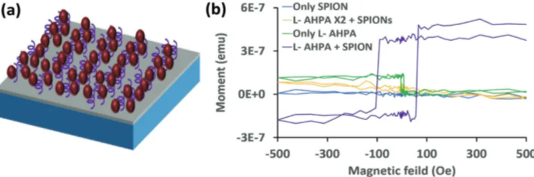
Magnetic moment measurements of SPIONs adsorbed on L-AHPA molecules (Figure 3a) were performed using a SQUID, and the magnetization hysteresis loop was obtained (Figure 3b) by subtracting the response of a clean Ag substrate (see Figure S3 in the Supporting Information).[29] The SPIONs by themselves do not show significant magnetization when deposited on an Ag substrate (blue). The adsorption of chiral molecules on the Ag substrate generates a weak additional magnetism (green), which may arise from chiral imprinting.[21] Interestingly, when the SPIONs are deposited on the L-AHPA-SAM the hysteresis loop inverts and a stronger magnetization is recorded (purple). Despite the intrinsic property of SPIONs to be superparamagnetic, i.e., zero coercivity and no remanence field, upon their adsorption on chiral molecules the SPIONs transform into CIFIONs with a ferromagnetic hysteresis loop Figure 2. a) Transmission electron microscope image of the ≈10 nm SPIONs. b) Illustration of the M-AFM samples patterning of the AHPA-SAM on a 100 nm Ag (gray layer) evaporated on 4 × 4 mm2 Si chip (blue layer). 1-the resist pattern. 2-AHPA-SAM (purple helical molecules) formation. 3-Resist
lift-off, AHPA-SAM was left only where there was no resist. 4-iron oxide nanoparticle (red spheres) adsorption. c) Scanning electron microscope image on the border between an area with AHPA-SAM (left side) and an area without (right side). The iron oxide density is much higher on the AHPA-SAM because of the carboxylic terminal group of the AHPA molecule which binds to iron oxide.
www.advancedsciencenews.com www.small-journal.com
and average coercive field of ≈80 Oe. The hysteresis loop is not symmetric around zero and shows a preference to magnetize the CIFIONs in the upward direction. All measurements were repeated on three samples.
To relate this effect to the adsorption of chiral molecules from one side only, a reference sample with an additional L-AHPA-SAM adsorbed on top of the SPIONs, named L- AHPA X2 (the SPIONs are now embedded between two layers of L-AHPA molecules), was prepared. Figure S2 in the Supporting Information illustrates the preparation. In this case, no significant magnetization was measured (orange); it was similar to the measurement with only SPIONs depos-ited on silver. The hysteresis loop is not symmetric around zero and shows a preference to magnetize the CIFIONs in the upward direction. The flip from upward to downward magneti-zation occurs at ≈–95 Oe, whereas the flip from downward to upward magnetization occurs at ≈60 Oe. In all of the reference samples (without the SPIONs), the hysteresis was below the noise level. It is interesting to note that the magnetic moment seems to be symmetric around the L-AHPA curve. It is clear that the chiral molecules impart some magnetization onto the substrate, which may give rise to the asymmetry around zero magnetic field.
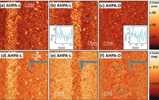
Figure 4 shows the results of the M-AFM measurements. For the L-AHPA (right-handed helix), the SAM was patterned into stripes (topography images are shown in Figure 4a,b and magnetic interaction images are shown in Figure 4d,e for downward and upward magnetization of the M-AFM tip, respectively). When magnetizing the M-AFM tip downward (red arrow), a repulsive magnetic interaction is measured (bright features), and for an upward magnetized tip (blue arrow), an attractive magnetic interaction is observed (dark fea-tures). This behavior demonstrates that the CIFIONs are mag-netized with an upward orientation. For D-AHPA (left-handed helices), the SAM was patterned into squares; the topography image is shown in Figure 4c and the magnetic interaction image for downward magnetization of the M-AFM tip is shown in Figure 4f. In this case, an attractive interaction dominates
which indicates that the CIFIONs are magnetized with a downward orientation. This observation stands in contrast to Figure 4d in which L-APHA CIFION samples, probed with the same tip magnetization, show the opposite behavior. Thus, the chirality of the AHPA SAM controls the orientation of the mag-netization the iron oxide nanoparticles. Similar measurements are presented for an individual nanoparticle in Figure S4 in the Supporting Information.
Two additional reference systems were investigated using M-AFM. The first reference measured the magnetic interac-tion of SPIONs physically adsorbed on an Ag layer without any chiral molecules (Figure 5a–c and see Figure S5 in the Supporting Information for topography cross-section images). These data fail to generate a discernable magnetic response above the background. In the second system, SPIONs were covered on all sides with L-tartaric acid and were measured by the M-AFM method on a mica surface (Figure 5d–f). These nanoparticles displayed a random magnetization direction. Additional images for L- and D-tartaric acid SPIONs on SiO2 and statistical analysis thereof are shown in Figure S6 in the Supporting Information.
The experimental results indicate that the chiral molecules change the magnetic properties of SPIONs from superpara-magnetic, which has zero coercive field and no remanence, to ferromagnetic CIFIONs, which have a nonzero coercive field and display remanence. The SQUID measurements exhibit a significant ferromagnetic hysteresis loop for CIFIONs on an AHPA-SAM with an average coercive field of ≈80 Oe, while the reference measurements of only SPIONs or only AHPA-SAMs show no hysteresis loops and much weaker magnetic signals (Figure 3b). The hysteresis loop is also not symmetric around zero magnetic field, which indicates that magnetization of the CIFIONs in the upward direction requires a lower absolute magnetic field than in the downward direction for the right-handed helical molecules (L-AHPA).
The M-AFM measurements show that the CIFIONs on an L-AHPA SAM have attractive or repulsive magnetic interac-tions with the M-AFM tip when the tip is magnetized in the Figure 3. a) Illustration of the sample for the SQUID measurements: 100 nm Ag (gray layer) evaporated on 4 × 4 mm2 Si chip (blue layer). SAM of
AHPA chiral molecules (purple helical molecules) was adsorbed on the Ag layer and iron oxide nanoparticles (red spheres) were adsorbed on top of the AHPA-SAM. b) SQUID measurements of the magnetization hysteresis loop. The sample of L-AHPA with iron oxide nanoparticles (purple line) shows CIFIONs with a hysteresis loop exhibiting ferromagnetic behavior with an average coercive field of ≈80 Oe. The hysteresis loop is not symmetric around zero and shows a preference to magnetize the CIFIONs in the upward direction. Three reference samples were measured. The first of L-AHPA X2 with chiral molecules adsorbed on both sides of the iron oxide nanoparticles (orange line) display no measurable response. The second of only L-AHPA (without nanoparticles, green line) shows weak magnetic order and no hysteresis is measured. In this case, the adsorption of chiral molecules from one side also seem to generate some small substrate magnetic ordering. The third reference sample of only iron oxide nanoparticles (without AHPA, blue line) presents no measurable response.
down and up orientations, respectively. This dependence is reversed for CIFIONs on a D-AHPA SAM (Figure 4f). These results corroborate the SQUID measurements by showing that the functionalized iron oxide nanoparticles exhibit a stable magnetization, which is robust to thermal fluctuations and dis-plays a significant coercive field and remanence (in contrast to
a nanoparticle with superparamagnetic behavior). The meas-urements also show that the orientation of the CIFION mag-netization is controlled by the chirality of the SAM molecules.
We ascribe the chiral-based magnetization imprinting on the SPIONs to chiral-induced spin selectivity at the chiral mole-cule–iron oxide interface. An electron exchange current flows Figure 4. Panels (a–c) show topography images that were taken and interleaved with the magnetic interaction images (tip distance of 130 nm) in panels
(d–f). Topography and phase color scales are on the respective right sides of the images. The M-AFM tip was magnetized down (red arrow), or up (blue arrows). In panels (d) and (e) an L-AHPA-SAM (right-handed), the substrate, is patterned with lines and shows that the iron oxide nanoparticles are magnetized upward. The tip experiences repulsive interactions for downward tip magnetization and attractive interactions for upward tip magnetiza-tion, as indicated by the bright and dark colors. In panel (f), D-AHPA-SAM (left handed) has a square patterned surface (red squares) and the darker color shows that the iron oxide nanoparticles are magnetized downward due to attractive magnetic interactions with a downward magnetized M-AFM tip. The insets in panels (b) and (c) show topographic cross sections.
Figure 5. a) Illustration of the SPIONs with no chiral molecule reference sample. b,c) Magnetic interaction measurements of SPIONs on Ag without
chiral molecules. There is no measurable signal for both M-AFM tip magnetizations (upward and downward) although there are SPIONs as can be seen in the topography images (insets). d) Illustration of SPION covered, from all directions, with left-handed chirality tartaric acid. e,f) Magnetic interaction measurements of tartaric acid covered SPIONs on a mica substrate. For both tip magnetization (upward and downward), there is a repulsive magnetic interaction, which correlates to the SPIONs seen in the topography images (insets).
www.advancedsciencenews.com www.small-journal.com
between the iron oxide nanoparticles and the Ag substrate by way of the chiral molecules, and the forward and reverse cur-rent are spin filtered by the chiral molecules. The spin prefer-ence is determined by the helicity of the chiral molecule; for right-handed (left-handed) chiral molecules spin up (down) electrons will be preferentially transmitted to the molecule, while spin down (up) electrons will be preferentially transferred down the molecule. This process results in a majority spin up population in the nanoparticle and stabilizes the magnetization in the upward direction (Figure 6a). The opposite magnetiza-tion of the iron oxide nanoparticle is observed when the chi-rality of helical molecules is reversed (Figure 6b). In the case of a SPION covered by chiral molecules from all directions or embedded between two SAMs of the same handedness, the symmetry plane is not broken and no net stable mag-netization is observed; an electron with one type of spin will transfer into the SPION from the bottom interface with chiral molecules, while the interface at the top of the SPION will transfer electrons of the opposite spin type (Figure 6c).
Measurements (both SQUID and M-AFM) of the reference samples of SPIONs without chiral molecules do not show magnetization and substantiate the role of chiral-induced spin selective transport for the magnetization imprinting. The magnetic interaction measurements on chiral tartaric acid coated SPIONs indicate that achieving strong magnetization imprinting requires anisotropic coverage, e.g., a defined orien-tation for spin flow.
Using SQUID and M-AFM measurements, we have shown how ferromagnetic properties can be imprinted on 10 nm SPIONs. This size reduction should enable the scale down of magnetic memory area by an order of magnitude and may prove to be important to magnetic force microscopy as well. These magnetic chiral iron oxide nanoparticles manifest remanence and an average coercive field of ≈80 Oe at room temperature. In addition, the preferred magnetization direction is determined by the chirality of the molecules and their nonsymmetrical place-ment around the nanoparticle. We propose a mechanism for
the chiral imprinting that is based on the well-established CISS effect; namely, magnetism induced by proximity of adsorbed chiral molecules. This explanation suggests that the ferromag-netic properties are imprinted when the chiral molecules are bound on one side of the SPIONs and should disappear when the SPIONs are covered with chiral molecules from two oppo-site faces, or from all directions. Measurements of SPIONs covered with chiral molecules from two or all directions indeed showed no remanence and zero coercive field. The results indi-cate that magnetic-based devices (especially spintronic devices) can be further miniaturized using chiral-based magnetization imprinting and that magnetic nanomaterials could be magnet-ized using chemical adsorption, without an external magnetic field and without applying spin-polarized current.
Supporting Information
Supporting Information is available from the Wiley Online Library or from the author.
Acknowledgements
Y.P. acknowledges the support from the Volkswagen Foundation (No. VW 88 367), the Israel Science Foundation (ISF Grant No. 1248/10), and the MOS Israel. R.N. acknowledges support in part from the European Research Council under the European Union’s Seventh Framework Program (No. FP7/2007-2013)/ERC Grant Agreement No. (338720), the MOS Israel, and the VW Foundation (No. VW 88 367). D.P. acknowledges the Israel Science Foundation (ISF grant 1589/14) and the Minerva Centre for bio-hybrid complex systems. D.P. also thanks the Etta and Paul Schankerman Chair of Molecular Biomedicine. D.H.W. and R.N. acknowledge support by the U.S. Department of Energy; Grant No. ER46430.
Conflict of Interest
The authors declare no conflict of interest.
Figure 6. Panels (a) and (b) show right-handed and left-handed chiral molecules, respectively, linked only to the bottom side of the iron oxide
nano-particle. Because of the CISS effect, spin up (down) electrons are preferentially going into the nanoparticle, while spin down (up) electrons are pref-erentially going out of the nanoparticle creating majority spin up (down) electrons in the nanoparticle. This process creates a net magnetization of the nanoparticle with a defined upward (downward) orientation. Panel (c) shows a right-handed chiral molecule linked to the iron oxide nanoparticle from all directions. In this case, spin up (down) electrons are going into the nanoparticle from the bottom (top) chiral molecule and going out of the nanoparticle from the top (bottom) molecule, resulting in unstable magnetization.
[2] S. Bhatti, R. Sbiaa, A. Hirohata, H. Ohno, S. Fukami, S. N. Piramanayagam, Mater. Today 2017, 20, 530.
[3] C. S. Hwang, Adv. Electron. Mater. 2015, 1, 1400056. [4] Y. Huai, n.d., 8.
[5] T. Kawahara, K. Ito, R. Takemura, H. Ohno, Microelectron. Reliab.
2012, 52, 613.
[6] “Ferromagnetism.” Available at http://hyperphysics.phy-astr.gsu. edu/hbase/Solids/ferro.html.
[7] Q. Li, C. W. Kartikowati, S. Horie, T. Ogi, T. Iwaki, K. Okuyama,
Sci. Rep. 2017, 7, 9894.
[8] D. L. Leslie-Pelecky, R. D. Rieke, Chem. Mater. 1996, 8, 1770. [9] A. S. Teja, P.-Y. Koh, Prog. Cryst. Growth Charact. Mater. 2009, 55, 22. [10] A. Hirohata, K. Takanashi, J. Phys. D: Appl. Phys. 2014, 47, 193001. [11] R. Naaman, D. H. Waldeck, J. Phys. Chem. Lett. 2012, 3, 2178. [12] R. Naaman, D. H. Waldeck, Annu. Rev. Phys. Chem. 2015, 66, 263. [13] O. B. Dor, S. Yochelis, S. P. Mathew, R. Naaman, Y. Paltiel, Nat.
Commun. 2013, 4, 2256.
[14] O. Ben Dor, N. Morali, S. Yochelis, L. T. Baczewski, Y. Paltiel,
Nano Lett. 2014, 14, 6042.
[15] G. Koplovitz, D. Primc, O. Ben Dor, S. Yochelis, D. Rotem, D. Porath, Y. Paltiel, Adv. Mater. 2017, 29, 1606748.
2004, 93, 116801.
[21] O. B. Dor, S. Yochelis, A. Radko, K. Vankayala, E. Capua, A. Capua, S.-H. Yang, L. T. Baczewski, S. S. P. Parkin, R. Naaman, Y. Paltiel,
Nat. Commun. 2017, 8, 14567.
[22] L. Berger, Phys. Rev. B 1996, 54, 9353.
[23] J. C. Slonczewski, J. Magn. Magn. Mater. 1996, 159, L1.
[24] D. C. Ralph, M. D. Stiles, J. Magn. Magn. Mater. 2008, 320, 1190.
[25] M. Kisielewski, A. Maziewski, M. Tekielak, A. Wawro, L. T. Baczewski, Phys. Rev. Lett. 2002, 89, 087203.
[26] M. Kisielewski, A. Maziewski, Z. Kurant, M. Tekielak, A. Wawro, L. T. Baczewski, J. Appl. Phys. 2003, 93, 7628.
[27] N. Depalo, P. Carrieri, R. Comparelli, M. Striccoli, A. Agostiano, L. Bertinetti, C. Innocenti, C. Sangregorio, M. L. Curri, Langmuir
2011, 27, 6962.
[28] N. Depalo, R. M. Iacobazzi, G. Valente, I. Arduino, S. Villa, F. Canepa, V. Laquintana, E. Fanizza, M. Striccoli, A. Cutrignelli, A. Lopedota, L. Porcelli, A. Azzariti, M. Franco, M. L. Curri, N. Denora, Nano Res. 2017, 10, 2431.
[29] Chemical Rubber Company, in CRC Handbook of Chemistry and