R E S E A R C H
Open Access
Multisystemic manifestations in a cohort of
75 classical Ehlers-Danlos syndrome
patients: natural history and nosological
perspectives
Marco Ritelli
1, Marina Venturini
2, Valeria Cinquina
1, Nicola Chiarelli
1and Marina Colombi
1*Abstract
Background: The Ehlers-Danlos syndromes (EDS) are rare connective tissue disorders consisting of 13 subtypes with overlapping features including joint hypermobility, skin and generalized connective tissue fragility. Classical EDS (cEDS) is principally caused by heterozygousCOL5A1 or COL5A2 variants and rarely by the COL1A1 p.(Arg312Cys) substitution. Current major criteria are (1) skin hyperextensibility plus atrophic scars and (2) generalized joint hypermobility (gJHM). Minor criteria include additional mucocutaneous signs, epicanthal folds, gJHM complications, and an affected first-degree relative. Minimal criteria prompting molecular testing are major criterion 1 plus either major criterion 2 or 3 minor criteria. In addition to these features, the clinical picture also involves multiple organ systems, but large-scale cohort studies are still missing. This study aimed to investigate the multisystemic involvement and natural history of cEDS through a cross-sectional study on a cohort of 75 molecularly confirmed patients evaluated from 2010 to 2019 in a tertiary referral center. The diagnostic criteria, additional mucocutaneous, osteoarticular, musculoskeletal,
cardiovascular, gastrointestinal, uro-gynecological, neuropsychiatric, and atopic issues, and facial/ocular features were ascertained, and feature rates compared by sex and age.
Results: Our study confirms that cEDS is mainly characterized by cutaneous and articular involvement, though none of their hallmarks was represented in all cases and suggests a milder multisystemic involvement and a more favorable natural history compared to other EDS subtypes. Abnormal scarring was the most frequent and characteristic sign, skin hyperextensibility and gJHM were less common, all without any sex and age bias; joint instability complications were more recurrent in adults. Some orthopedic features showed a high prevalence, whereas the other issues related to the investigated organ systems were less recurrent with few exceptions and age-related differences.
(Continued on next page)
© The Author(s). 2020 Open Access This article is licensed under a Creative Commons Attribution 4.0 International License, which permits use, sharing, adaptation, distribution and reproduction in any medium or format, as long as you give appropriate credit to the original author(s) and the source, provide a link to the Creative Commons licence, and indicate if changes were made. The images or other third party material in this article are included in the article's Creative Commons licence, unless indicated otherwise in a credit line to the material. If material is not included in the article's Creative Commons licence and your intended use is not permitted by statutory regulation or exceeds the permitted use, you will need to obtain permission directly from the copyright holder. To view a copy of this licence, visithttp://creativecommons.org/licenses/by/4.0/. The Creative Commons Public Domain Dedication waiver (http://creativecommons.org/publicdomain/zero/1.0/) applies to the data made available in this article, unless otherwise stated in a credit line to the data.
* Correspondence:[email protected]
1Division of Biology and Genetics, Department of Molecular and Translational
Medicine, University of Brescia, Viale Europa 11, I-25123 Brescia, Italy Full list of author information is available at the end of the article
(Continued from previous page)
Conclusions: Our findings define the diagnostic relevance of cutaneous and articular features and additional clinical signs associated to cEDS. Furthermore, our data suggest an update of the current EDS nosology concerning scarring that should be considered separately from skin hyperextensibility and that the clinical diagnosis of cEDS may be enhanced by the accurate evaluation of orthopedic manifestations at all ages, faciocutaneous indicators in children, and some acquired traits related to joint instability complications, premature skin aging, and patterning of abnormal scarring in older individuals.
Keywords: Classical Ehlers-Danlos syndrome, Nosology, Atrophic scars, Skin hyperextensibility, Joint hypermobility, Multisystemic involvement, Natural history,COL5A1, COL5A2, COL1A1
Background
Ehlers–Danlos syndromes (EDS) represent a clinically and genetically heterogeneous group of heritable con-nective tissue disorders (HCTDs) sharing a variable combination of skin hyperextensibility, joint hypermobil-ity (JHM), and internal organ and vessel fragilhypermobil-ity. The 2017 international classification of EDS recognizes 13 subtypes that are caused by pathogenic variants in 19 different genes, mainly encoding fibrillar collagens, collagen-modifying proteins, or processing enzymes [1]. The classical (cEDS), vascular (vEDS), and the molecu-larly unsolved hypermobile (hEDS) EDS subtypes ac-count for more than 90% of patients. cEDS (MIM #130000) has an estimated prevalence of 1/20,000 and it is principally caused by heterozygous pathogenic variants in COL5A1 or COL5A2 encoding type V collagen and rarely by the c.934C > T p.(Arg312Cys) missense variant inCOL1A1 encoding type I collagen [1–14].
Although cEDS is recognized since the seventeenth century because of the remarkable cutaneous and articu-lar involvement, it remains poorly defined on clinical grounds and its diagnosis is still based on experts’ opin-ion rather than systematic published data. According to the up-to-date EDS nosology, cEDS should be suspected in the simultaneous presence of skin hyperextensibility plus atrophic scarring (major criterion 1). This com-bined criterion must be present together with the other major criterion, i.e., generalized JHM (gJHM) assessed with the Beighton score (BS) [15], and/or with at least three of the following minor criteria: easy bruising, soft, doughy skin, skin fragility, molluscoid pseudotumors, subcutaneous spheroids, hernia (or a history of thereof), epicanthal folds, JHM complications (sprains, luxation/ subluxation, pain, flexible flatfoot), and family history of a first degree relative who meets clinical criteria [1, 16]. Skin hyperextensibility is formally defined as the stretch-ing of the skin over a standardized cut off in 3 of the fol-lowing areas: 1.5 cm for the distal part of the forearms and the dorsum of the hands and 3 cm for neck, elbows, and knees [1,7,16, 17]. Although atrophic scarring can range in severity, most cEDS patients have very poor wound healing leading to multiple, widened atrophic
scars in different body areas, especially over pressure points and areas prone to trauma [6,7, 16]. In addition to the nosological criteria, the clinical picture of cEDS variably involves multiple organ systems, but observa-tional data in large cohorts of molecularly proven cEDS patients are still limited. In particular, it is reported that cEDS patients may show delayed motor development with mild hypotonia, characteristic facial features, fatigue and muscle cramps, premature rupture of fetal mem-branes, cervical insufficiency during pregnancy, rectal prolapse in early childhood, mild scoliosis, and cardiac/ blood vessel fragility including mitral/tricuspid valve prolapse, and aortic root dilatation. In very rare individ-uals, especially (but not exclusively) in those with the COL1A1 p.(Arg312Cys) missense variant, spontaneous rupture of large arteries, intracranial aneurysms, and arteriovenous fistulae may occur [5–7,9–14,18–26].
A definite diagnosis of cEDS is established by the iden-tification on molecular genetic testing of a heterozygous pathogenic variant in one of the cEDS-associated genes. The largest part of patients carry small COL5A1 patho-genic variants and the majority of these are null alleles (nonsense, frameshift, out-of-frame splice) leading to functional haploinsufficiency; some missense variants, especially within the collagenous domain of the protein, and a few intragenic rearrangements including (multi) exon deletions/duplications are also described [6, 7]. In COL5A2, structural variants (missense, in-frame splice) exerting a dominant negative effect are the most com-mon [4–6, 16]. In patients who fulfill the main clinical features of cEDS, the variant detection rate is about 90% [5, 6]. No clear-cut genotype-phenotype correlations have emerged so far, except thatCOL5A2 variants seem to result in a more severe phenotype, although numbers are still limited. The EDS Leiden Open Variation Data-base (LOVD) [27] contains approximately 200 and 60 unique COL5A1 and COL5A2 pathogenic variants, re-spectively. Until recently, genetic testing was principally based on (serial) single-gene testing by Sanger sequen-cing, i.e., analysis ofCOL5A1 is performed first, followed by COL5A2 analysis and then gene-targeted copy number variant analysis to identify large deletions or
duplications. If no pathogenic variant is found, the search for the recurrent COL1A1 p.(Arg312Cys) variant should be performed [1, 6,16]. As specific types of mu-tations may be lost due to technical limits, negative mo-lecular testing does not exclude the diagnosis of cEDS; however, alternative diagnosis should be considered mainly if the patient’s phenotype is truly doubtful [1]. Indeed, recognition of cEDS is generally not challenging, since most patients present with the typical cutaneous hallmarks and gJHM (BS≥5/9). However, vast intra- and interfamilial variability tells a much wider clinical pres-entation [4–6] and an important overlap with other EDS subtypes and HCTDs [1, 9, 16, 28–30]. Nowadays, mo-lecular analysis, especially for doubtful patients, should rely on next generation sequencing (NGS) technologies, including multigene panels containing at least all EDS-associated genes and/or panels comprising genes of the major HCTDs in differential diagnosis and/or a custom phenotype-focused whole exome analysis [1]. Herein, we present a cross-sectional study on a cohort of 75 cEDS patients from 44 families with a confirmed molecular diagnosis focusing on the diagnostic criteria recognized in the current EDS nosology and on additional mucocu-taneous, osteoarticular, musculoskeletal, cardiovascular, gastrointestinal, uro-gynecological, neuropsychiatric, atopic, and facial/ocular features, in order to investigate the multisystemic involvement and natural history of the disorder.
Patients and methods
Patients’ cohort and evaluated clinical features
Seventy-five patients, 44 index-cases and 31 relatives, with cEDS were evaluated from 2010 to 2019 in a ter-tiary referral center for the diagnosis and management of HCTDs (i.e., “Ehlers-Danlos Syndrome and Inherited Connective Tissue Disorders Outpatient Clinic (CESE D)” at the Spedali Civili University Hospital of Brescia. Until February 2017, all clinical signs included in the Villefranche nosology [15] were evaluated prior to gen-etic testing. After the publication of the revised EDS classification, the new criteria were applied for confirma-tory molecular analysis and clinical features of the previ-ously evaluated patients were reanalyzed according to the novel guidelines. In addition, several previously diag-nosed patients were reevaluated during follow-ups (for number and age at visits see Additional Table1).
Direct physical examination of available patients sys-tematically consisted in different steps. First, facial fea-tures, including frontal and other facial scars, ocular anomalies and other traits, as well as the appearance, lo-cation, and number of scars all over the body were searched and documented by clinical photographs (Figs. 1 and 2). Ocular signs included epicanthal folds, palpebral ptosis, sunken eyes, infraorbital creases,
hypotelorism, hypertelorism/telecanthus, myopia, stra-bismus, xeropthalmia, and elongated/up-slanting palpe-bral fissures (Fig. 2). Additional investigated facial features comprised micro/retrognathia, low-set ears, anteverted nostrils, elongated philtrum, and hypoplastic auricular lobe. Atrophic scars were extremely variable in clinical appearance; in particular, 3 different frequent morphologies were identified and classified as papyrac-eous, hemosiderotic, and cigarette paper (Fig. 1). Scar-ring was designed positive only in patients with multiple, widened scars at the typical areas (knees, elbows, fore-head, shins, chin, etc.), whereas small, isolated, single, or post-surgical scars were not considered representative of cEDS. Second, to asses texture/consistence, the skin was touched at different sites including arms, thorax, hands and legs. Soft/velvety/doughy skin was an entirely sub-jective feeling developed during clinical practice. Third, to assess hyperextensibility, the skin was stretched in specific points comprising upper eyelids, neck, dorsum of hand and forearm, chest, abdomen, elbows, and knees. Skin was considered hyperextensible when overextended according to the standardized cut off values in at least 3 of the areas defined in the 2017 EDS nosology [1]. Further evaluated mucocutaneous features included molluscoid pseudotumors, easy bruising, subcutaneous spheroids, inguinal, umbilical, and incisional hernia, ke-loid formation, gingival inflammation/recession, livedo reticularis, keratosis pilaris/hyperkeratosis of extensor surfaces, xerosis, striae distensae/rubrae, resistance to local anesthetic drugs, acquired cutis laxa/premature skin aging, piezogenic papules of the heels, abnormalities of the uvula, absent/short lingual frenulum, and light blue sclerae. Subsequently, JHM was evaluated according to the BS [15]; in adults with a BS < 5 the presence of historical JHM was routinely investigated with the five-point questionnaire [31].
In addition, we selected a set of clinical features and signs related to almost all organ systems by adapting our clinical checklist routinely used for the assessment of pa-tients with a suspicion of hEDS, in which the multisyste-mic involvement is already well recognized, even though not codified yet in the current nosology [1]. Further assessed musculoskeletal and orthopedic features in-cluded congenital hip dysplasia, sprains, (fixed) disloca-tions, subdislocations, articular pain, back pain, recurrent inflammatory soft-tissue lesions (i.e., bursitis, tendinitis, myofascial pain, carpal tunnel syndrome, epi-condylitis, tenosynovitis, plantar fasciitis), walking diffi-culties, limited walking autonomy, temporomandibular joint dysfunction, high arched/narrow palate, marfanoid habitus (arm span to height ratio > 1.05), arachnodactyly (positive thumb/wrist sign), (non-surgical) pectus exca-vatum/carinatum, scoliosis, dorsal hyperkyphosis, cer-vical spine curvature anomalies, lumbar hyperlordosis/
hypolordosis, minor asymmetry at lower limbs (anatom-ical or functional anisomelia) or other body areas, cubita/genua valga, halluces valgi, and pes planus/cavus. When available, the documented presence of disc her-nias/protrusions, osteopenia/osteoporosis, and spondylo-listhesis was recorded. In patients with osteopenia/ osteoporosis the main primary causes (e.g. hormonal and vitamin D deficiencies, lifestyle, and medications) were investigated. Cardiovascular features that were sys-tematically explored included varicose veins, Raynaud’s phenomenon, acrocyanosis, livedo reticularis, capillary fragility, and recurrent epistaxis or gingival bleeding. When possible, the presence of arterial aneurysms, pro-gressive aortic root dilatation, aortic ectasia, valvular re-gurgitation with hemodynamic involvement, and mitral valve prolapse (MVP) was investigated by cerebral,
thoracic, abdominal magnetic resonance imaging (MRI) and/or heart ultrasound. In patients with extensive easy bruising, routine coagulation tests were performed resulting normal in all. Assessed muscular features included hypo-tonia at birth, muscle hypohypo-tonia, recurrent myalgias and cramps, involuntary muscle contractions, and fibromyalgia. Investigated gastrointestinal features comprised gastro-esophageal reflux, dysphagia, hiatal hernia, food intoler-ances, celiac disease, abdominal pain, defecatory dysfunction, delayed gastric/bowel/colonic transit, dolicho-colon, and visceroptosis. Investigated uro-gynecological fea-tures included disabling dysmenorrhea, meno/ metrorrhagia, post-partum hemorrhage, urinary stress in-continence, cervical insufficiency during pregnancy, and rectal or pelvic prolapse. Assessed neuropsychiatric features were delayed motor development, clumsiness, chronic
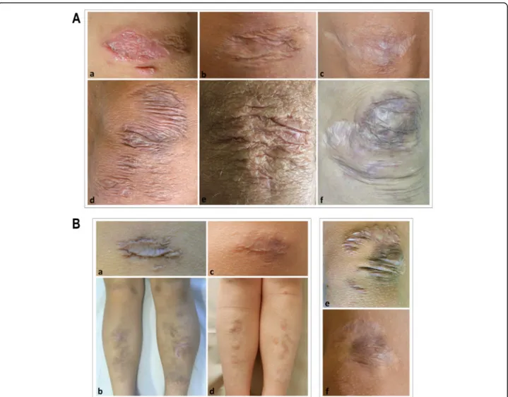
Fig. 1 A) Representative images of different atrophic scars on cEDS patients’ knees. A healing scar in a 6-year-old girl (a); a papyraceous, non-hemosiderotic scar in a 22-year-old female (b); a cigarette paper scar in a 39-year-old female (c); multiple papyraceous, non-hemosiderotic scars in a 40-year-old female (d); multiple papyraceous scars in a 57-year-old male (e); combination of multiple, papyraceous, hemosiderotic scars, and cigarette paper scars in a 34-year-old female (f). B) Evolution of papyraceous, and hemosiderotic scars in 2 children assessed at increasing ages. On the left, a single scar on the knee and multiple scars at the pretibial area in a girl at 6 (a, b) and 11 years of age (c, d); on the right, multiple scars on the knee in a boy at 9 (e) and 12 years of age (f)
fatigue, headache/migraine, impaired memory and concen-tration, paresthesia, cardiovascular dysautonomia, sleep dis-turbances, anxiety/panic/fears, allodynia, depression, and neuropathic pain. Immunological features included allergy/ atopy, rhinitis/rhinoconjunctivitis, asthma, and atopic dermatitis.
Assessment between the presence/absence of investi-gated features and selected dichotomous variables, i.e., age at last examination < or≥ 18 years and gender, was performed with the chi-square test with Yates’s correc-tion or Fisher’s exact test whenever the count was insuf-ficient. Analysis was carried out with the GraphPad Software and considering significant p-values when less than 0.05. In patients who have been evaluated at more than one visit, the 2017 diagnostic criteria were analyzed at the different ages, in order to appraise if they changed over time.
Genetic testing
Molecular analysis was performed on genomic DNA purified from peripheral blood leukocytes by standard
procedures. Most patients were characterized according to our previously reported molecular flow-chart based on (serial) single-gene Sanger sequencing [6]. In particu-lar, all exons and intron-flanking regions of COL5A1 (NM_000093.3, NP_000084.3), COL5A2 (NM_000393.5, NP_000384.2), and exon 14 of COL1A1 (NM_000088.4, NP_000079.2) were amplified by PCR with optimized primer sets (available upon request) followed by bidirec-tional Sanger sequencing with the BigDye Terminator v1.1 Cycle Sequencing kit on an ABI3130XL Genetic Analyzer (Life Technologies). Sequences were analyzed with the Sequencher 5.1 software (www.genecodes.com) and variants annotated by using the Alamut Visual soft-ware version 2.15 (Interactive Biosoftsoft-ware, Sophia Gen-etics). Since 2018, massive parallel sequencing methods were applied in our laboratory and positive results con-firmed by Sanger sequencing. For this study, patients were characterized by using an house NGS panel in-cluding all cEDS-associated genes. Briefly, two panel pools of the custom “connective tissue panel”, CTP (COL5A1, COL5A2, COL1A1, COL1A2, COL3A1,
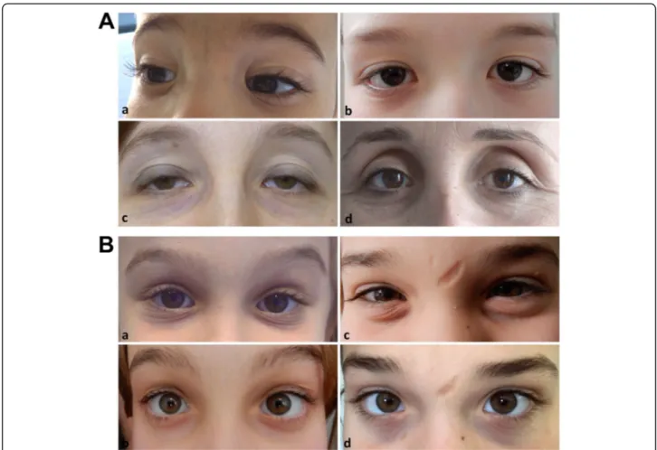
Fig. 2 A) Representative images of characteristic ocular features in cEDS patients from different ages. Epicanthal folds in 2 boys at 3 (a) and 12 years of age (b); palpebral ptosis (c) and sunken eyes (d) in two over 30 years old females. B) Evolution of facial gestalt in 2 children assessed at increasing ages. On the left, infraorbital creases and bluish sclerae in a girl at 6 (a) and 11 years of age (b); on the right, infraorbital creases and facial scars in a girl at 10 (c) and 15 years of age (d)
B3GALT6, B4GALT7, SLC39A13, FKBP14, ADAMTS2, CHST14, DSE, FLNA, ACTA2, MYH11, MYLK, NOTCH1, MFAP5, PRKG1, SLC2A10, TGFBR1, TGFBR2, SMAD2, SMAD3, SMAD4, TGFB2, TGFB3, FBN1) were generated with the AmpliSeq Designer tool (Thermo Fisher Scientific). Panel libraries were gener-ated using the AmpliSeq Library kit 2.0, which includes reagents for generating amplicons with the Ion Ampli-Seq primers and the Ion Xpress™ Barcode Adapters, fol-lowing manufacturer’s protocols (Thermo Fisher Scientific). Library template preparation was performed with the Ion 520 & Ion 530 kit– OT2 on the Ion One-Touch 2 instrument starting from a pool of 15 barcoded libraries (8μl of 100 pM pooled library) and sequenced on the Ion S5 instrument with the Ion 520 chip. Basecal-ling and sequence alignment against hg19 genome as-sembly were performed with the Ion Torrent Suite software v.5.0.2 and genetic variants were identified by the Ion Torrent Variant Caller v.5.0.2.1. Further analyses were performed with the Ion Reporter software 5.6. In patients without a pathogenic variant, deletion/duplica-tion analysis of COL5A1 was performed through Multi-plex Ligation-dependent Probe Amplification (MLPA) analysis with the SALSA MLPA P331 and P332 Probe-Mixes, according to manufacturer’s instructions (MRC-Holland). All novel pathogenic variants were submitted to the EDS LOVD [27].
Results
General and molecular findings
Among the 75 cEDS patients from 44 families described here, 45 were females (60%) and 30 were males (40%) (sex ratio: 1.5). By considering the index-cases, the sex ratio was 1.75 (28 females and 16 males). Forty-nine pa-tients from 31 families were previously published in [6– 8, 12], whereas 18 from 11 families were novel patients. Eight additional affected family members from 2 previ-ously described pedigrees [6, 7] were also reported. Among the 49 earlier published patients that were in-cluded in the present study, 24 were reassessed at least once during scheduled follow-up visits (Additional Table
1). Age at last examination ranged from 3 to 67 years (mean 27; standard deviation [SD] 18.3). In particular, age range was 3 to 67 (mean 27.2; SD 17.8) for females, and 3 to 63 (mean 29.7; SD 19) for males. Patients youn-ger than 18 years old were 31 (41.3%) and 44 (58.7%) pa-tients were adults (≥18 years). In all new families, a COL5A1 pathogenic variant was identified mainly by using the NGS CTP and submitted to the EDS LOVD. In particular, we disclosed a previously reported non-sense mutation [6] and 10 novel variants, i.e., 2 non-sense, 3 small deletions, and 1 multi-exon deletion, all predicted leading to haploinsufficiency, and 2 in-frame exon skipping splice variants, 1 glycine substitution, and
1 intermediate-sized duplication (63 bp) within the col-lagenous domain of the protein and with an estimated dominant negative effect. Detailed clinical and molecular features by single patient and the corresponding LOVD IDs are reported in Additional Table 1. Frequencies of selected clinical features are reported in Table 1, in Figs.3and4, and in Additional Table2.
Diagnostic criteria according to the 2017 EDS nosology and additional mucocutaneous, osteoarticular, and facial/ ocular features
The two major criteria for cEDS, namely marked skin hyperextensibility plus extensive, widened atrophic scar-ring and gJHM, were present respectively in 78.7 and 58.7% of patients at last evaluation without any correl-ation with sex and age (Table1).
Among minor criteria, skin fragility (89.3%), easy bruising (86.7%), and soft doughy skin (78.7%) were the most frequent cutaneous signs; molluscoid pseudotu-mors (29.3%) and spheroids (27.6%) were less common. These features did not show any significant difference between sex and age categories, whereas hernias and epicanthal folds (Fig. 2A) were more frequent in young patients (58.1% vs 20.5 and 32.3% vs 9.1%, respectively). At least one gJHM complication was present in 93.3% of patients, with bilateral pes planus (82.7%) as the most common feature without any correlation with sex and age. A higher prevalence in adults compared to younger individuals was clearly evident for the occurrence of sprains, especially of heels, fingers/wrist, and knees (59.1% vs 25.8%), joint and limb pain (61.4% vs 9.7%), and dislocations (34.1% vs 3.2%) that involved primarily shoulder, digits, hip, radius, and clavicles (Additional Table 1). Subdislocations were also more common in adults (36.4% vs 19.4%), although not statistically signifi-cant. Two or more of gJHM complications were present especially in adults (73.8% vs 35.7%). Fixed dislocations, particularly of shoulders, were sporadic events (6.7%). Likewise, chronic disabling pain was infrequent and pa-tients, mainly adults, used principally nonsteroidal anti-inflammatory drugs or paracetamol on demand, whereas opioid therapy was very uncommon (Additional Table2). Finally, the last minor criterion, namely family history of a first-degree relative meeting clinical criteria, was more common in adults (72.7% vs 32.3%).
Minimal criteria for a cEDS suspicion were present in 59/75 (78.7%) of patients; 49.3% showed both major cri-teria while 29.3% major criterion 1 together with at least three minor criteria; both combinations did not show sex- and age-related differences. Overall, 16/75 patients (21.3%) of our cohort did not fulfill the combined major criterion 1, i.e., 10 for the absence of generalized skin hyperextensibility, 1 for the lack of typical scarring, and 5 for the absence of both features (Additional Table1).
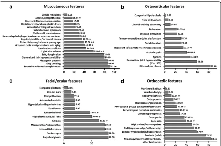
Localized skin hyperextensibility (less than 3 sites) was present in 9/75 patients and the complete absence of skin hyperextensibility in 6/75 (Additional Table2). Concern-ing skin fragility, 92% of patients (69/75) showed exten-sive, widened atrophic scars and the most frequent types were papyraceous (65.3%), hemosiderotic (54.7%), and cigarette paper (40%) (Fig.1A). The rate of hemosiderotic scars was significantly higher in young patients compared to adults (Additional Table 2). Further frequent (> 30%) mucocutaneous features were piezogenic papules of the heels (86.7%), light blue sclerae (58.7%), uvular abnormal-ities (44.1%), acquired cutis laxa/premature skin aging (41.3%), striae distensae/rubrae (40%), and keratosis pilaris/hyperkeratosis of extensor surfaces (30.7%). Among these, light blue sclerae were more common in young fe-males, while acquired cutis laxa, premature skin aging, and striae distensae/rubrae were more recurring in adults, with the latter more frequent in females (Fig.3aand Add-itional Table2).
Further recognized osteoarticular features were recur-rent inflammatory soft-tissue lesions (33.8%), more fre-quently observed in adults (48.8% vs 12.9%), and temporomandibular joint dysfunctions (28.8%). Walking difficulties (22.7%) and limited walking autonomy (10.7%) were rare and occurred more repeatedly in adults, though not statistically significant. Mobile patel-lae were present in 6/75 patients (8%). Congenital hip dysplasia was encountered only in 2 patients as well as spontaneous Achilles tendon rupture (Fig. 3b and Additional Tables1and2).
Apart from epicanthal folds, additional midface/ocular signs were palpebral ptosis and sunken eyes (both 32%) and infraorbital creases (29.3%) (Fig.2). Myopia (25.3%) was more common in adults. The other non-ocular fa-cial features were mostly sporadic observations, except for micro-retrognathia (29.3%) and hypoplastic auricular lobe (20%), the latter with a predominance in young patients (Fig.3cand Additional Table2).
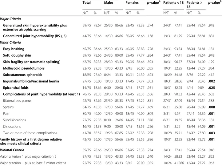
Table 1 Major, minor, and minimal criteria according to the 2017 nosology of classical Ehlers-Danlos syndrome
Total Males Females p-valueA Patients < 18 Patients≥
18 p-value
B
N/T % N/T % N/T % N/T % N/T %
Major Criteria
Generalized skin hyperextensibility plus extensive atrophic scarring
59/75 78.67 26/30 86.66 33/45 73.33 .274 24/31 77.41 35/44 79.54 .948 Generalized joint hypermobility (BS≥ 5) 44/75 58.66 14/30 46.66 30/45 66.66 .138 19/31 61.29 25/44 56.81 .881 Minor Criteria
Easy bruising 65/75 86.66 25/30 83.33 40/45 88.88 .728 29/31 93.54 36/44 81.81 .181 Soft, doughy skin 59/75 78.66 24/30 80.00 35/45 77.77 .954 24/31 77.41 35/44 79.54 .948 Skin fragility (or traumatic splitting) 67/75 89.33 28/30 93.33 39/45 86.66 .593 30/31 96.77 37/44 84.09 .129 Molluscoid pseudotumors 22/75 29.33 13/30 43.33 9/45 20.00 .055 10/31 32.25 12/44 27.27 .834 Subcutaneous spheroids 18/65 27.60 8/24 33.33 10/41 24.39 .623 10/29 34.48 8/36 22.22 .412 Inguinal/umbilical/incisional hernia 27/75 36.00 10/30 33.33 17/45 37.77 .883 18/31 58.06 9/44 20.45 .002 Epicanthal folds 14/75 18.66 6/30 20.00 8/45 17.77 .951 10/31 32.25 4/44 9.09 .025 Complications of joint hypermobility (at least 1) 70/75 93.33 28/30 93.33 42/45 93.33 .636 28/31 90.32 42/44 95.45 .683 Bilateral pes planus 62/75 82.66 25/30 83.33 37/45 82.22 .851 27/31 87.09 35/44 79.54 .588 Sprains 34/75 45.33 17/30 56.66 17/45 37.77 .169 8/31 25.80 26/44 59.09 .008
Pain 30/75 40.00 12/30 40.00 18/45 40.00 .809 3/31 9.67 27.44 61.36 .001
Subdislocations 22/75 29.33 8/30 26.66 14/45 31.11 .876 6/31 19.35 16/44 36.36 .181 Dislocations 16/75 21.33 9/30 30.00 7/45 15.55 .226 1/31 3.22 15/44 34.09 .001 Two or more of these complications 41/70 58.57 19/28 67.85 22/42 52.38 .298 10/28 35.71 31/42 73.80 .003 Family history of a first degree relative
who meets clinical criteria
42/75 56.00 17/30 56.66 25/45 55.55 .886 10/31 32.25 32/44 72.72 .001 Minimal Criteria 59/75 78.66 26/30 86.66 33/45 73.33 .274 24/31 77.41 35/44 79.54 .948 Major criterion 1 plus major criterion 2 37/75 49.33 13/30 43.33 24/45 53.33 .540 14/24 58.33 23/44 52.27 .822 Major criterion 1 plus at least 3 minor criteria 22/75 29.33 13/30 43.33 9/45 20.00 .055 10/24 41.566 12/44 27.27 .963
Abbreviations: N number of patients presenting the investigated feature, T total number of patients in whom the feature was investigated Significantp-values < 0.05 are in bold
A:p-values females vs males
Considering that we reassessed 24 patients during follow-up (approximately 2–5 years after first examination), we had the possibility to evaluate if the major, minor, and min-imal criteria changed over time in an individual single pa-tient. Though the limited time period of observation (range 2–7 years), gJHM displayed an age influence. Indeed, in 14/ 24 patients (8 males and 6 females) a decrease of the BS was observed, and at last examination 5 of these patients (all males) displayed a BS < 5, hence not fulfilling the major criterion 2. The most striking difference was observed in a boy who showed at 10 years of age a BS of 9/9 that 4 years later changed to 2/9. Additional differences included a fe-male that 7 years after the first assessment showed subcuta-neous spheroids, and 2 patients (1 female and 1 male) who displayed after 4 years the presence of multiple molluscoid pseudotumors (Additional Table1). While we did not ob-serve any striking difference concerning skin hyperextensi-bility as well as the other minor criteria, in most patients, especially in adolescence, scars changed over time both in dimension and appearance. For instance, if no further in-jury occurred at the same area, large, thickened, raised, papyraceous, and hemosiderotic scars usually tended to re-solve in smaller, flatter, and less hemosiderotic or even in cigarette paper scars (Fig.1B).
Orthopedic features
In almost all patients of our cohort we systematically evaluated 15 orthopedic features (Fig.3d and Additional Table2). The most recurrent (≥ 50%) were minor
asym-metry at lower limbs or at other body areas (83.6%), mild scoliosis (74.3%), lumbar hyperlordosis/hypolordo-sis (57.9%), cubita/genua valga or halluces valgi (53.3%), and high arched/narrow palate (50%); all these features did not show any apparent sex and age bias. Osteopenia (T-scores between − 1.0 and − 2.4) was identified in 11 (8 females and 3 males) out 21 investigated adults (35.5%), whereas it was absent in 10 examined young in-dividuals. The age range of patients with osteopenia was 29 to 58 (mean 41.5, SD 9.3). Osteoporosis (T-scores above− 2.5) was diagnosed only in 5/21 adults (16.1%, 4 females and 1 male, mean age 38.6, SD 11.4). Vitamin D deficiency was found in 4 patients and cholecalciferol treatment was commenced. Dorsal hyperkyphosis was present in 27.5% of patients without any sex- and age-re-lated difference. A similar frequency was identified also for cervical spine curvature anomalies, but with a higher rate in adults compared to individuals aged below 18 years (36.5% vs 11.1%). Back pain (56.8% vs 12.9%), disk her-nias/protrusions (27.9% vs 3.4%), and spondylolisthesis
Fig. 3 Frequencies of mucocutaneous (a), osteoarticular (b), facial/ocular (c), and orthopedic (d) features in the cEDS patients’ cohort. *presence of statistically significant differences by age; #presence of statistically significant differences by gender (for frequencies andp-values see Additional Table2)
(2.5% vs 0%) were all more common in adults. Finally, non-surgical pectus excavatum/carinatum showed a higher prevalence in males compared to females (40% vs 11.1%) and in 1 of them marfanoid habitus and arachno-dactyly were also evident. Other rare findings comprised short stature, dental malocclusion, radiological vertebral fractures, surgically treated congenital kyphoscoliosis, winged scapulae, ulnar deviation, brachydactyly, hypopla-sia of the upper femur, arthrosis, osteoarthritis, club feet, pes cavus, and hindfoot deformities (Additional Table1).
Cardiovascular features
Apart from easy bruising, the most commonly observed cardiovascular sign in our cohort was MVP (42.4%), which was especially recognized in adults (56.4% vs 22.2), followed by capillary fragility/recurrent epistaxis/ gingival bleeding (41.3%), valvular regurgitation with mild hemodynamic involvement (34.8%), varicose veins (20%), and Raynaud’s phenomenon/acrocyanosis/livedo reticularis (10.6%), all without any significant difference between sex and age categories, with the exception of varicose veins that were more frequent in adults. Non-progressive aortic root dilatation (9.1%) was detected only in adults, namely in 5/27 investigated males and in 1/39 females; as well as aortic ectasia (z score > 2) that was documented in 2 adult males, one patient carried
the COL1A1 c.934C > T p.(Arg312Cys) missense variant and the other aCOL5A1 null allele (Additional Tables1
and2, Fig.4a). Spontaneous rupture of large arteries did not occur in our cohort. Additional sporadic findings included posterior cerebral artery hypoplasia, intracra-nial aneurysm, vertebral artery tortuosity, atrioven-tricular block, tricuspid valve prolapse, interatrial septal aneurysm, bicuspid aortic valve, delayed right ventricular conduction, left ventricular thickening, ventricular arrhythmia, tachycardia with extrasystoles, arteriovenous fistula, and deep venous thrombosis (Additional Table 1).
Muscular features
Among the 5 investigated muscular signs (Additional Table 1 and Fig.4b), the most common issues were re-current myalgias and cramps (41.3%) that affected mainly adults (52.3% vs 25.8%). Hypotonia at birth was infrequent (14.7%) as well as muscle hypotonia of mild degree (6.7%) and involuntary muscle contractions (5.3%). Confirmed fibromyalgia was present only in 1 pa-tient with the most severe form of cEDS, carrying a structural mutation inCOL5A2 (Patient 14 in Additional Table 1), who also suffered from recurrent muscle he-matomas and ruptures.
Fig. 4 Frequencies of cardiovascular (a), muscular (b), gastrointestinal (c), uro-gynecological (d), neuropsychiatric (e), and atopic (f) features in the cEDS patients’ cohort. *presence of statistically significant differences by age; #presence of statistically significant differences by gender (for frequencies and p-values see Additional Table2)
Gastrointestinal features
In almost all individuals of our cohort we assessed 11 gastrointestinal features (Additional Table2and Fig.4c). The most recurrent problem, especially in adults, was gastroesophageal reflux (52.3% vs 22.6%). Treatment with proton pump inhibitors was resolutive in most cases, only 3 patients required fundoplication (Add-itional Table1). Defecatory dysfunction (33.3%), delayed gastric/bowel/colonic transit (26.3%), various food intol-erances, unexplained abdominal pain (both 16%), and dysphagia were less common and did not show any sex and age bias. Sporadic findings comprised hiatal hernia, visceroptosis (gastroptosis, enteroptosis), dolichocolon, confirmed celiac disease, and gastric ulcer (Additional Table 2). Inflammatory bowel disease was not recog-nized. In one patient a postnatal intestinal perforation with meconium peritonitis requiring ileostomy occurred.
Uro-gynecological features
Among females in reproductive age (26 adults and 5 aged below 18 years), disabling dysmenorrhea (22.6%) and meno/metrorrhagias (16.1%) were the most frequent gynecological issues (Additional Table 2 and Fig. 4d). One patient suffered from amenorrhea since the age of 24 (Patient 11 in Additional Table 1). Twenty out of 31 women had at least one pregnancy and 8 of them had multiple pregnancies. Fourteen of the children were af-fected. Multiple miscarriages occurred in 4 patients. Post-partum uterine hemorrhage and a third-degree va-ginal laceration during delivery occurred in 2 and 1 pa-tients, respectively. Premature rupture of membranes with preterm delivery was present in 1 patient with an affected fetus; 16 women had uncomplicated pregnancies and delivery (Additional Table1).
In the entire patients’ cohort, urinary stress incontin-ence was very rare (2.7%). In the most severely affected patient (Patient 14), both rectal and urethral prolapse to-gether with recurrent hemorrhagic cystitis occurred. Further occasional issues were microhematuria, urinary retention, testicular cysts, bilateral mobile testis, pelvic varicocele, cryptorchidism, and bladder diverticula and hypoplasia (Additional Table1).
Neuropsychiatric features
In order to explore the neurological, psychological and emotional dysfunctions in cEDS, we investigated 12 fea-tures (Additional Table 2 and Fig. 4e). The most com-mon problematic was headache/migraine, which was reported by 41.3% of patients without significant sex-and age-related differences, even if it affected more fre-quently adult females. Motor clumsiness was reported by 36.5% of patients as well as chronic fatigue (36%), the latter was more frequent in adults (45.5% vs 22.6%), al-though not statistically significant. Abnormal sensation
of the skin (e.g. formication, tingling, pricking, chilling, burning, and numbness) was described by 25.3% of pa-tients, especially by adults (36.4% vs 9.7%). Paresthesia was most commonly occurring in arms, legs, and abdo-men and was usually painless and transient, except for 5 females who mentioned chronic formication (Additional Table 1). A documented cardiovascular dysautonomia was present in 16% of patients, particularly in adult fe-males, although not reaching a statistic significance. The most recurrent symptom was orthostatic intolerance and in 9 patients tilt-table testing resulted positive. Further associated symptoms were mood swings, fatigue, prob-lems with concentration and memory, gastrointestinal complications, anxiety/panic/fears, and sleep disturbance (Additional Table 1). These latter issues were both re-ported also by patients without cardiovascular dysauto-nomia with an overall frequency of 14.7% without any sex and age bias. Allodynia (13.5%) was more recurrent in adults (20.5% vs 3.3%). The occurrence of either mechanical (especially static), thermal (hot and cold), or movement (joints and/or muscles) allodynia was re-ferred. Delayed motor development, usually associated with primary hypotonia (Additional Table 1), was present in 10.7% of patients without any significant vari-ance between sex and age categories. Chronic depression requiring pharmacological treatment was diagnosed in 3 patients (1 adult female and 2 adolescents), one of the latter showed a self-injurious behavior and attempted suicide (Additional Table 1). Chronic neuropathic pain was present only in the most severely affected patient (Patient 14). Further sporadic features were epileptic sei-zures (2 patients from the same family), mild hearing loss, absence of sphincter control, and developmental coordination disorder affecting fine and gross motor skills (Additional Table1).
Atopic features
In the entire cohort, immunological issues were quite rare (Additional Table 2 and Fig. 4F). Allergy/atopy (32%) was the most recurrent problem followed by re-current rhinitis/rhinoconjunctivitis (14.7%) and con-firmed atopic dermatitis (10.7%). Asthma afflicted only 3 out of 75 patients. The most common allergens were various foods, pollen, animal dander (especially cat hair), and dust mites. Treatment with corticosteroids was reso-lutive in almost all patients and anaphylaxis did never occur. One patient suffered from chronic sinusitis (Add-itional Table1).
Discussion
This study on a cohort of 75 molecularly proven cEDS patients aimed to validate the revised diagnostic criteria and explore the multisystemic involvement of the dis-order by comparing types and incidences of the
numerous clinical manifestations at different ages and with those observed in other EDS subtypes, in order to delineate natural history and assist differential diagnosis. Our results confirm the notion that cEDS is basically characterized by cutaneous and articular involvement, even though none of their hallmarks is represented in 100% of cases, and show that osteoarticular and musculoskeletal complaints as well as gastrointestinal, uro-gynecological, neuropsychiatric, and cardiovascular associated symptoms (and disorders) are not so promin-ent compared to other EDS subtypes.
Albeit the cEDS-specific triad, i.e., widened, atrophic scars (92% of patients and ~ 95% of probands), marked skin hyperextensibility (80% of patients and ~ 82% of probands), and gJHM (BS ≥5, ~ 59% of patients and ~ 66% of probands), is highly predictive for molecular con-firmation of the diagnosis, this combination was ob-served only in ~ 49% of patients and in ~ 55% of probands. Hence, the suspect of cEDS is not always driven by the traditional criteria but is rather gestaltic and based on the overall clinical presentation. Our find-ings offer future perspectives for a revision of the EDS nosology in terms of diagnostic criteria. The observation that in our cohort only ~ 79% of patients and ~ 82% of probands fulfilled the currently defined minimal criteria for a suspicion of the disease, which require the presence of the combined major criterion 1, indicates that their strict application could lead to a lack of diagnoses. Hence, we recommend that atrophic scarring should be considered independently from skin hyperextensibility as an alone-standing major criterion 1. We also propose that minimal criteria prompting genetic testing should be typical cEDS scars (at more than 2 sites) plus either generalized skin hyperextensibility (at least 3 of the currently defined areas and according to their cut-off values) and/or gJHM (BS ≥5), and/or 3 of the cur-rently defined minor criteria, to which orthopedic is-sues may be added. In view of intrafamilial variability, a family history with a documented pathogenic vari-ant is sufficient for genetic testing in individuals not fulfilling the criteria.
These suggestions are supported first by the fact that in our sample abnormal scarring was undoubtedly the most common sign. Although scarring was variable in clinical appearance and affected sites and is shared by other EDS subtypes and HCTDs [1, 9, 28–30, 32–35], scars should basically be considered representative of cEDS. Indeed, in our cohort, scars were typically numer-ous, large, papyracenumer-ous, and hemosiderotic. The findings that the rate of hemosiderotic scars was significantly higher in young patients and that adults often presented small and flat scars suggest an evolving phenotype and natural history of scarring that usually becomes less se-vere in adulthood (Fig. 1) and is likely influenced by
patients’ habits. Defective scarring in cEDS is always sec-ondary to soft tissue traumas, which occur more likely in childhood in exposed areas such as forehead, knees, and elbows, and in those near the underlying bones (e.g. pretibial region), due to skin fragility joined with a de-fective wound healing. The reevaluation of scars in sin-gle individuals over time (Fig. 1) corroborates the concept of an evolving phenotype and suggests that once a cEDS diagnosis is made (or in the presence of a posi-tive family history) and management guidelines for pre-vention of primary manifestations are provided (e.g. avoiding contact sports or wearing protective pads or bandages over forehead, knees, and shins during activ-ities), scarring generally becomes less significant, except for severe cases.
The proposal to modify the current diagnostic criteria also derives from the observation that in 15 out of 16 patients of our cohort who did not satisfy the major cri-terion 1 (and consequently neither the minimal criteria for genetic testing), this was due to the absence of gener-alized skin hyperextensibility. Ten of these patients showed the typical cEDS scars and 5 of them also had an affected family member fulfilling the criteria, thus prompting molecular analysis. Likewise, among the 5 pa-tients who did not show both features of criterion 1, 3 had a first-degree parent meeting the criteria and one an affected mother with positive skin hyperextensibility, al-though without the typical cEDS scars [8]. The most un-certain patient was a sporadic female (Patient 23 in Additional Table 1) who presented only a single, small atrophic scar and localized skin hyperextensibility at el-bows and knees. In addition to this latter sign, the pres-ence of gJHM and its complications and further minor signs prompted molecular analysis that revealed a patho-genic COL5A1 variant. This case highpoints that while molecular diagnosis in patients with a full-blown pheno-type is mainly confirmatory, in those with an incomplete presentation it turns out to be fundamental, since these individuals might not be diagnosed or even be misdiag-nosed. Indeed, considering the clinical overlap not only between the different EDS subtypes but also with other HCTDs [1,9, 28, 30, 32, 35–49], differential diagnosis is not always forthright. Differential diagnosis includes the molecularly unsolved hEDS that shares with cEDS gJHM and more than a few (muco) cutaneous signs; however, hEDS patients usually show a lower degree of scarring and skin hyperextensibility and much more striking gJHM complications [1,7,28,29, 50]. Molluscoid pseu-dotumors and subcutaneous spheroids are highly diag-nostic of cEDS, even if they were rarely observed in our patients’ cohort. Signs of premature skin aging, including acquired cutis laxa of the extremities and skin wrinkling, although less specific, might also help in the differential with hEDS, especially in adults. The additional
mucocutaneous features, including abnormal skin tex-ture, easy bruising, hernias, piezogenic papules, light blue sclerae, uvular abnormalities, striae distensae/ rubrae, keratosis pilaris/hyperkeratosis of extensor sur-faces, absent/short lingual frenulum, resistance to local anesthetic drugs, and gingival inflammation/recession, are almost all shared not only with hEDS but also with other EDS subtypes and HCTDs [28, 29]. As such, the recording of these features at physical examination might reinforce the suspect of systemic disorder, but their low specificity does not attend for confirmatory genetic testing. In cases compatible with an autosomal recessive transmission, differential diagnosis includes the rare classical-like EDS (clEDS) type 1, a.k.a. TNXB defi-ciency [51], and the recently defined clEDS type 2 caused by biallelic variants in AEBP1 [52]. The clEDS type 1 is generally distinguishable from cEDS for the ab-sence of atrophic scarring [28,45,53–57], whereas a more severe multisystemic presentation in clEDS type 2 should assist the differential diagnosis with cEDS [43,52,58–60]. The dermatosparaxis (ADAMTS2) [61], cardiac-valvular (COL1A2) [62–64], kyphoscoliotic (PLOD1, FKBP14) [65,
66], and arthrochalasia (COL1A1, COL1A2) [67, 68] EDS subtypes, also sharing with cEDS several cutaneous and articular issues, are mostly distinguishable for the presence of specific hallmarks [1,9,16].
Concerning the other major criterion of cEDS, gJHM re-mains undeniably characteristic of the disorder, even if in our sample a BS≥5 was identified in less than 60% of pa-tients and it is common to all EDS subtypes and other HCTDs [1,9,28,30,36–39,47,50,69,70]. In our previ-ously published smaller cEDS cohort [7], a lower inci-dence of gJHM was observed in adults compared to younger individuals, suggesting that age affects JHM also in cEDS, as more widely demonstrated in hEDS and in which an age-dependent BS was introduced in the revised diagnostic criteria [1,50,69,71–73]. In the present larger cEDS cohort, no statistically significant difference emerged between age at ascertainment and BS≥5 (~ 57% in adults vs ~ 61% in young individuals), suggesting a dif-ferent natural history of JHM compared to hEDS. Indeed, although patients’ reassessment at different ages showed a decrease of the BS in 12/24 investigated patients, only in 5 males it resulted below the cut-off value of 5/9. A different age influence and disease course in cEDS seems to occur also for joint instability complications, such as disloca-tions, sprains, and soft-tissue injuries. In hEDS, it was pre-viously shown that there were no overt associations between age and their frequency and location, except for soft-tissue injuries that exhibited a higher rate in adults [73]. In our cEDS patients’ cohort, these gJHM
complica-tions as well as articular pain, which in general was of mild degree, were instead all more frequently observed in adult-hood. However, gJHM complications showed an overall
lower rate with the exclusion of pes planus (present in most patients) and recurrent inflammatory soft-tissue in-juries, among which plantar fasciitis, bursitis, and teno-synovitis were particularly frequent. Furthermore, dislocations usually resolved spontaneously and were in general easily managed by the affected individual. Hence, in cEDS, a BS≥ 5 and mild to moderate joint instability complications seem valid markers in adults, especially in those with a minor or naturally diminished cutaneous phenotype (Fig.1). In contrast, gJHM and its related issues might lack both sensitivity and specificity in children, in whom, however, the typical cutaneous hallmark is nearly always plainly manifest. Furthermore, the presence of epi-canthal folds and/or infraorbital creases or an association of the other eye findings and/or non-ocular features plus abnormal facial scars seems to denote a further distinctive gestaltic presentation of young cEDS patients, which usu-ally attenuates or even vanishes over the years (Fig. 2). Musculoskeletal involvement was an interesting finding in our cohort, due to the high prevalence of some related is-sues, thus suggesting that the registration of these features might strengthen the suspect of cEDS, although with a low specificity. This is particularly true for orthopedic fea-tures, considering that in our cohort minor asymmetry at lower limbs and/or other body areas, scoliotic attitude/ mild scoliosis, lumbar hyperlordosis/hypolordosis, valgus deformities of elbows, knees, and feet, and high arched/ narrow palate were all observed with a frequency above 50% without any sex and age bias. Hence, it is reasonable to propose that the presence of 3 or more of these issues might be considered as an additional minor criterion in a future nosology revision. Osteopenia and osteoporosis, dorsal hyperkyphosis, cervical spine curvature anomalies, pectus deformities, disk hernias/protrusions, and spondy-lolisthesis as well as severe back pain (≥ 7 NRS numeric pain rating scale), which in general associated with the presence of radiological vertebral fractures (Additional Table 1), were less frequent but all more recurrent in adults, except for dorsal hyperkyphosis and pectus anom-alies. Osteoarthritis, which has been postulated as a long-term consequence of JHM and related altered joint bio-mechanics [69], was instead only a sporadic finding in our cEDS cohort as well as marfanoid habitus. Our results partly support the still controversial impression that bone quality might be impaired in cEDS [74–78], although undoubtfully less significant compared to other EDS sub-types (e.g. spondylodysplastic, arthrochalasia, kyphoscolio-tic, and classical-like type 2 EDS) [9,43,44,48,49,52,59,
65,68] or skeletal dysplasia [39], and suggests a potential involvement of skeletal fragility in determining a poorer quality of life (QoL) in adult patients. Our findings emphasize a milder phenotype and disease course in cEDS compared to hEDS [50,69] and other EDS subtypes (e.g. clEDS type 1 and 2, kyphoscoliotic, spondylodysplastic,
myopathic, and musculocontractural EDS) [9] also regard-ing muscular and neuropsychiatric involvement. Indeed, in our cohort, apart from myalgias and cramps that af-fected about 50% of adults, primary (causing delayed motor development and clumsiness in few cases) or ac-quired muscle hypotonia/weakness (always of mild de-gree), and involuntary muscle contractions were quite rare; fibromyalgia was only a sporadic observation. These muscular complications, except primary hypotonia, are in-stead very frequent in hEDS and they intensely contribute to the poor QoL in adulthood [50,69], when patients usu-ally show generalization and progressive chronicity of musculoskeletal pain, which is often diagnosed as fibro-myalgia [79], summation of other forms of chronic pain, such as pelvic pain (in women) and migraine as well as ex-acerbation of fatigue, together with additional complaints including paresthesia, mixed functional gastrointestinal disorders, and orthostatic intolerance [71]. In our cEDS cohort, headache/migraine and chronic fatigue were re-ferred respectively by ~ 41% and ~ 36% of cEDS patients, especially by adults even though not statistically signifi-cant, whereas impaired memory and concentration and paresthesia were both more recurrent in adults (~ 40% vs ~ 10%). Cardiovascular dysautonomia as well as allodynia and sleep disturbance were less frequent (~ 15%); 5 adults mentioned chronic pain needing opioid therapy, whereas neuropathic pain was present only in the most severely af-fected patient. While fatigue affects about 1/3 of the gen-eral population [80], the chronic fatigue syndrome, defined as fatigue lasting longer than 6 months, occurs only in ~ 1% of the general population [81,82] and is usu-ally associated with impaired memory, cognitive deficits, muscle and joint pain, headaches, non-restorative sleep, post-exertional illness as well as psychological issues [83– 85]. The discreetly high incidence of chronic fatigue ob-served in our cEDS cohort is in line with previous obser-vations that emphasized that in EDS this issue is more common than in the general population and likely con-tributes to the patients’ functional impairment, psycho-logical distress, and decreased QoL [86–89]. Like in the general population, in cEDS fatigue is multifactorial with contributing factors including articular pain, headache, sleep disturbance, and abnormally low blood pressure often associated with postural orthostatic tachycardia syn-drome (POTS) or neurally mediated hypotension (NMH), nutritional deficiency, medications, and/or allergies (present in 32% of our patients) [80,81,87,90–95]. Cere-brospinal fluid leak, which has been reported in cEDS as a cause of postural hypotension and headache [96], was not diagnosed in our cohort (e.g. increase in headache follow-ing Valsalva maneuver or reduction of headache when the patient takes a prone position) and only few patients with persistent migraine were investigated by imaging tech-niques with negative results. The first evidence for a
possible link between EDS and autonomic dysfunction was published by Rowe et al. [86], who studied eleven pediatric patients (cEDS and hEDS) all showing either POTS or NMH. The concept was later reinforced by fur-ther studies that demonstrated a significant increase of the rate of systemic dysautonomic symptoms in hEDS [31,
97]. In our cEDS cohort, cardiovascular dysautonomia, al-though not particularly frequent, can explain orthostatic intolerance, palpitations, and tachycardia as well as some neurological secondary manifestations including fatigue, mood swings, and memory and concentration troubles. Besides, it has been proposed that in EDS dysautonomia might contribute also to features affecting the gastrointes-tinal and urinary systems (e.g. gut dysmotility and under-active/overactive bladder) as well as to certain psychological traits [98, 99]. Indeed, psychological dys-function and emotional problems (e.g. depression, anxiety, panic, fears, affective disorder, low self-confidence, nega-tive thinking, and hopelessness) are common features among EDS patients [92,100–105]. Some of these issues, which may worsen the pain experience as well as other organ system manifestations (gastrointestinal and auto-nomic), were encountered also in our cohort with anxiety, panic, and/or fears that were referred by ~ 15% of patients. Chronic depression requiring pharmacological treatment was instead rare.
Concerning gastrointestinal involvement, its relevance in EDS and other HCTDs is well recognized [31, 69], whereas, until now, these issues were only slightly inves-tigated in cEDS [106]. A previous study, which intended to explore the relationship between hEDS or gJHM with gastrointestinal disorders, showed that treatment-resistant functional issues such as gastroesophageal re-flux, dysphagia, heartburn, swelling, constipation, recur-rent abdominal pain, and irritable bowel syndrome, might be observed in 1/3 to 3/4 of patients with an in-creasing rate by age [107]. In our cEDS cohort, the most common feature was gastroesophageal reflux, especially in adults (~ 52% vs ~ 22%); however, unlike hEDS, it was mostly responsive to treatment with proton pump inhib-itors and/or nizatidine. Constipation with or without other features of voiding dysfunction was often the ini-tial sign of gastrointestinal involvement, but problems such as hemorrhoids, rectal bleeding, and anal fissures were uncommon and more serious complications such as rectal prolapse or fecal impaction did not occur. Likewise, hiatal hernia, visceroptosis, dolichoco-lon, and celiac disease, for which increased rates were described in hEDS [108, 109], were only sporadic ob-servations in our sample.
In females with EDS, gynecologic complaints, such as mucosal problems with their genital area [69] , heavy menstrual or intermenstrual bleedings [110] and painful intercourse [71, 111] are commonly encountered with
an incidence ranging from 30 to 75%. The frequencies of disabling dysmenorrhea (~ 23%) and meno/metrorrha-gias (~ 16%) observed in our cohort are basically in line with these previous observations, but the high incidence of these issues also in the general population does not allow to draw relevant conclusions. Pelvic floor disorders including urinary stress incontinence, pelvic organ pro-lapse, and other sensory and emptying abnormalities were very uncommon among our affected females. As in the general population, childbirth has a very substantial impact on a woman’s probability of these complaints. Several case-control studies in the past suggested that EDS is associated with pelvic floor disorders [111–113], but most of these studies have not controlled for child-birth history or age and included patients affected by various types of EDS. Furthermore, several additional pregnancy-related complications have been more com-monly reported in women with EDS in some studies [114] but as often, not validated in others [110, 115,
116]. Particularly for cEDS, it was suggested that preg-nancy places both the newborn and the mother at risk for complications, which are assumed to be more fre-quent than in the normal population [16]. However, no comprehensive studies exist, and it is therefore very dif-ficult to quantitate the incidence and related risk of each complication in affected individuals. In our cohort, most women had uncomplicated pregnancies (even multiple), deliveries, and postpartum period. Indeed, miscarriages, premature rupture of membranes, preterm birth, vaginal laceration during delivery, and post-partum uterine hemorrhage were rare or sporadic as well as rectal and urethral prolapse that occurred only in the most severely affected individual. Uterine rupture in the last trimester of pregnancy, which is particularly significant in vEDS as it associates with a high mortality rate (~ 5%) in affected females [46,117,118], did never happen in our patients. Overall, though the small sample size, pregnancy-associated complications seem to be unusual in cEDS. Accurate monitoring of women throughout gestation and in the postpartum period is still recommended, also considering that pregnancy was one of the most import-ant sources of anxiety in our cEDS females.
The vascular aspect of EDS in general remains the major fear for the patients and their clinicians, especially regarding the possibility of arterial aneurysms, dissec-tions and ruptures of medium-sized and large arteries. These life-threatening complications are principally the hallmark of vEDS together with gastrointestinal and uterine fragility, but they may occur also in other EDS subtypes, including rare individuals with a severe form of cEDS [16]. However, an exact estimation of preva-lence and risk of these vascular complaints and possible genotype-phenotype correlations in cEDS are still miss-ing [16, 119]. In particular, either the specific c.934C >
T, p.(Arg312Cys) variant in COL1A1 or COL5A1 vari-ants causing glycine substitutions at the C-terminal end of the triple helix domain have been associated with pro-pensity to severe arterial events in early adulthood, but these assumptions are still a matter of debate due to the limited number of reports [10–14, 22, 25]. In our cEDS cohort, easy bruising, which is present to a variable de-gree in all EDS subtypes, was the most common finding (~ 87%). A bleeding tendency, despite normal coagula-tion status, manifesting either as meno/metrorrhagia and (less frequently) postnatal or peri-operative hemorrhage or as gingival bleeding/recurrent epistaxis (~ 41%) were also common. Varicose veins (~ 20%) and Raynaud’s phenomenon/acrocyanosis/livedo reticularis (~ 11%) were relatively unusual. All these features, except vari-cose veins, did not display any sex and age bias. MVP showed instead a significantly higher incidence in adults (~ 56% vs ~ 22%). Nevertheless, MVP was utmost with-out or of little clinical consequence, valvular regurgita-tion with mild hemodynamic involvement was rare (~ 35%), and none of our patients required surgical inter-vention, in line with earlier reports [16, 20]. Similarly, non-progressive aortic root dilatation was infrequent (~ 9%) as well as aortic ectasia (only 2 adult males) and tri-cuspid valve prolapse and bitri-cuspid aortic valves were sporadic findings, thus suggesting that regular routine echocardiograms to assess for valvular diseases and aor-tic root dilatation may not be strictly necessary unless warranted by presence of symptoms or family history. MVP was formerly considered a common feature of all EDS subtypes and many other HCTDs, but that hap-pened prior to the establishment of the more rigorous criteria for the diagnosis of MVP. Since then, some stud-ies showed no significant increase in the incidence of MVP in EDS compared to the general population [18– 20], whereas others disclosed a higher MVP frequency (28–67%) [120, 121, 122]. Considering the rate of MVP observed in our cEDS cohort and the knowledge that the mitral valve depends upon collagen for its tensile strength and that myxomatous MVP is characterized by disruption of the collagen layer with expansion of glycosaminoglycans within the middle layer of the valve [123], it is reasonable to consider MVP as a poten-tial clue for cEDS, although the true clinical significance is so far unknown. The most noteworthy vascular com-plaints in our cohort were two aortic ectasias, an arterio-venous fistula, an interatrial septal aneurysm and an intracranial aneurysm; all these issues, except one of the two z scores > 2, were recognized in adults harboring a COL5A1 null allele. Spontaneous rupture of large arter-ies was not observed at all and, in particular, neither in the 6 patients with theCOL1A1 p.(Arg312Cys) missense variant nor in 2 individuals with different COL5A1 gly-cine substitutions at the C-terminal domain (Additional
Table 1), which represent variants previously associated with a possible higher risk of vascular events [16]. The fact that i) these 8 patients were diagnosed years ago [12], ii) none of them originally experienced major vas-cular events (1 individual with the COL1A1 variant showed aortic ectasia, uncomplicated tortuous vertebral arteries, and left ventricular thickening), and iii) all underwent periodical vascular surveillance that resulted negative at the time of writing, suggests that the sup-posed genotype-phenotype correlations of these variants should not be considered valid in all cases. The finding that major vascular complaints and/or rupture can be rarely recognized also in patients with differentCOL5A1 variants [5, 21, 23, 24, 26] advises that a cerebral, thor-acic, and abdominal MRI at diagnosis might be consid-ered in a precautionary way for all adult cEDS patients. Once significant changes in arterial caliber are not de-tected and in pediatric patients, a doppler-ultrasound of sopra-aortic branches and abdominal arteries, and a heart doppler-ultrasound with aortic root, arch, and as-cending aorta evaluation every 4–5 years might be enough together with cardiovascular risk factors moni-toring. Overall, despite the few above-mentioned reports and the suggestion for a possible association between type V collagen defects and cardiovascular fragility dem-onstrated in Col5a1 and Col5a2 knock-out mice [124,
125], our results suggest that the risk for severe vascular complications seems to be fairly low in humans harbor-ing a type V collagen defect and the presence of add-itional, yet unknown, genetic modifiers in families with a severe vascular phenotype is reasonable.
Conclusions
In conclusion, we explored the accuracy of the 2017 nos-ology for the cEDS diagnosis in a cohort of patients from different ages and sexes with a defined molecular defect and tried to offer a full picture of the multisystemic in-volvement and some views on the natural history of this condition. Our findings define the value of atrophic scars, skin hyperextensibility, JHM and its complications in selecting patients for molecular testing and the rele-vance of the additional clinical signs associated with cEDS, which should contribute to the rapid ascertain-ment of cEDS patients and facilitate differential diagno-sis. Promptly recognizing among the different EDS subtypes and confirmatory genetic testing is indeed in-creasingly important as these disorders are characterized by different natural histories and prognoses. Moreover, our results suggest the need of a future update of the currently defined diagnostic criteria for cEDS. Additional studies on large cEDS cohorts are expected to confirm the limited multisystemic involvement and rather favor-able natural history of cEDS and to explore possible
genotype-phenotype correlations, which would permit to establish management guidelines.
Supplementary information
Supplementary information accompanies this paper athttps://doi.org/10. 1186/s13023-020-01470-0.
Additional file 1: Table 1. Clinical and molecular features of all cEDS patients.
Additional file 2: Table 2. Multisystemic features in the cEDS patients’ cohort by sex and age.
Abbreviations
BS:Beighton score; cEDS: Classical Ehlers-Danlos syndrome; clEDS: Classical-like Ehlers-Danlos syndrome; CTP: Connective tissue panel; EDS: Ehlers-Danlos syndrome; gJHM: Generalized joint hypermobility; HCTD: Heritable
connective tissue disorder; hEDS: Hypermobile Ehlers-Danlos syndrome; JHM: Joint hypermobility; LOVD: Leiden Open Variation Database; MLPA: Multiplex ligation-dependent probe amplification; MRI: Magnetic resonance imaging; MVP: Mitral valve prolapse; NGS: Next generation sequencing; NMH: Neurally mediated hypotension; POTS: Postural orthostatic tachycardia syndrome; QoL: Quality of life; SD: Standard deviation;
vEDS: Vascular Ehlers-Danlos syndrome Acknowledgements
The authors wish to thank the patients and their families for the cooperation during the diagnostic process and participation in this study and the Fazzo Cusan family for its generous support.
Authors’ contributions
MC and MR conceived the study and coordinated the clinical study to collect the clinical data and assessed the obtained clinical findings. MR and MC drafted the manuscript with substantial support by VC and NC, who organized data contents, reviewed the literature, completed figures and tables and prepared the additional files. Patients were clinically evaluated and recruited with informed consent by MC and MV. The molecular genetic studies, sequence alignments, and annotations of identified sequence alterations were performed by MR, VC, and NC. All authors discussed, read and approved the final manuscript.
Funding
Not applicable. No funding was active on this project. Availability of data and materials
All data generated or analyzed during this study are included in this published article and its Additional files.
Ethics approval and consent to participate
This study was carried out according to the Declaration of Helsinki principles and approved by the local Ethical Committee (Comitato Etico di Brescia, ASST degli Spedali Civili, Brescia, Italia, registration number NP3873). Molecular testing was achieved in the genetic laboratory at the Department of Molecular and Translational Medicine of the University of Brescia in compliance with the Italian legislation on genetic diagnostic tests. The patients (or their parents) signed informed consent for molecular analysis, participation to the study and publication of clinical photographs. Consent for publication
Written consent for publication of clinical data was obtained from all participants (or their parents).
Competing interests
All authors declare that they have no competing interest concerning this work.
Author details
1Division of Biology and Genetics, Department of Molecular and Translational