Department of Physiology and Pharmacology “V. Erspamer”
Ph.D. in Clinical/Experimental Neuroscience and Psychiatry
- XXXII Cycle -
Neurophysiological mechanisms of quiet vigilance in
neurodegenerative diseases: an electroencephalography
research program
Ph.D. thesis
Maria Teresa Pascarelli
Coordinator:
Tutor:
Acknowledgements
I want to express my sincere gratitude to Prof. Claudio Babiloni who first recognized my strong motivation and passion for neuroscience, giving me the possibility to start this great scientific adventure and to grow up during this hard journey. I thank my colleagues for sharing with me all their experience and for letting me feel as part of one big family. Many thanks to Prof. Raffaele Ferri from IRCCS Oasi of Troina who supported my data analysis by the funds attributed by the Italian Ministry of Health. Thanks to the head of the Department of Physiology and Pharmacology “V. Erspamer” Prof. Cristina Limatola and all professors that contributed to enrich my knowledge with their precious lessons. My sincere thanks also go to the members of European Consortium PDWAVES and European Consortium of Dementia with Lewy Body for their strong impact in the works that are part of this PhD thesis. In particular, I want to express my gratitude to Prof. Dag Aarsland who provided me the opportunity to join his team, and who gave me access to the laboratory and research facilities at King’s College of London. Thank my family to support all my life’s choices and for loving me in the way I need.
In the end, thank me for the worth I give in everything I do.
“Without ambition one starts nothing. Without work one finishes nothing. The prize will not be sent to you. You have to win it.” (Ralph Waldo Emerson)
Maria Teresa Pascarelli Sapienza University of Rome October 2019
Index
Executive summary ... 6
Introductory Part ... 11
Cortical arousal system: the role of ascending reticular activating systems (ARASs) ... 11
Ventral pathway ... 12
Aminergic system ... 12
Dorsal pathway ... 14
Three major dementia types (ADD, PDD, DLB) ... 16
Neuropathology ... 17
Clinical features ... 21
Risk factors ... 23
A precursor to dementia: Mild cognitive impairment (MCI) ... 24
Diagnostic criteria for AD ... 25
Diagnostic criteria for PD ... 28
Diagnostic criteria for DLB ... 29
Pre-clinical biomarkers ... 30
Clinical management of dementia ... 33
Methodological Part ... 35
The electroencephalography (EEG) ... 35
Frequency bands ... 36
EEG data acquisition ... 38
Inverse solution method ... 41
Experimental Part ... 43
General aim and objectives of the PhD thesis ... 43
I study ... 45
Abnormalities of cortical neural synchronization mechanisms in patients with dementia due to Alzheimer’s and Lewy body diseases: an EEG study ... 45
Introduction ... 45
Materials and Methods ... 49
Subjects ... 49
EEG recordings and data analysis ... 53
Statistical analysis ... 56
Results ... 57
Correlation analysis ... 60
Classification among Nold, ADD, PDD, and DLB individuals ... 61
Discussion ... 63
The clinical neurophysiological model ... 67
II study ... 69
Abnormal cortical neural synchronization mechanisms in quiet wakefulness are related to motor deficits, cognitive symptoms, and visual hallucinations in Parkinson’s disease patients: an electroencephalographic study ... 69
Introduction ... 69
Materials and Methods ... 71
Subjects ... 71
Diagnostic criteria ... 77
Statistical analysis ... 79
Results ... 81
rsEEG source activities in the Nold, AD, and PD-MMSE(-/+) sub-groups ... 84
rsEEG source activities in the Nold, AD, and PD-UPDRS(-/+) sub-groups ... 87
rsEEG source activities in the Nold, AD, and PD-VH(-/+) sub-groups ... 93
Correlation analysis and classification ... 96
Discussion ... 99
Interactions among the cholinergic, dopaminergic, and serotoninergic ascending systems in PD ... 105
III study ... 107
Abnormal cortical sources of resting state EEG rhythms are related to cognitive deficits, rem sleep behavior disorders, and visual hallucinations in patients with dementia with Lewy Bodies ... 107
Introduction ... 107
Materials and Methods ... 109
Subjects ... 109
Diagnostic criteria ... 117
Statistical analysis ... 117
Results ... 119
rsEEG source activities in the Nold, AD, DLB-VH(-/+) sub-groups ... 122
rsEEG source activities in the Nold, AD, DLB-Flu(-/+) sub-groups ... 127
rsEEG source activities in the Nold, AD, and DLB-RBD(-/+) sub-groups ... 130
Correlation analysis ... 132
Discussion ... 133
Methodological remarks ... 139
Conclusions ... 141
Executive summary
6
Executive summary
In the field of neurophysiological research, the term vigilance refers to a variety of mental states of monitoring of external world during wakefulness, related to activities in brain circuits (Steriade, 1999). These mental states are modulated by the projections of ascending reticular activating systems (ARASs), namely neural circuits mostly composed of sub-systems formed by nuclei and fiber projections (Lin et al., 2011). The ARASs were described in the 1949 by the neurophysiologists Giuseppe Moruzzi and Horace Magoun who first investigated the neural components regulating the brain's sleep-wake mechanisms in cats; but, they were also defined later, by the neuropsychologist Aleksandr Romanovič Lurija, as the components of the “first block of the brain”, regulating arousal and the state of vigilance (Luria, 1973). ARASs are formed by parallel sub-systems running across brainstem, basal forebrain, and diencephalon using dopaminergic, noradrenergic, serotonergic, histaminergic, cholinergic, and glutamatergic neurotransmitters as different contributors to the regulation of general brain arousal and vigilance during wakefulness (Moruzzi and Magoun, 1949).
Experimental studies have shown that high level of the vigilance, associated with a finalized behaviour, is characterized by high activity of ARASs and desynchronization of the cortical electroencephalographic (EEG) signals at frequencies in the alpha (8-12 Hz) and beta (13-30 Hz) bands, associated with the synchronization of frequencies in the theta (4-7 Hz) and gamma (> 30 Hz) bands. On the contrary, when mental activity is at rest, in a quiet vigilance, EEG signals appear characterized by a synchronized activity at alpha rhythms (Moruzzi and Magoun 1949; Buzsaki and Gage 1989).
Buzsaki and colleagueas showed that cholinergic neurons play the main role in the event related variation of vigilance and related changes in on-going EEG activity (Buzsaki et al., 1988). The release of acetilcholine (ACh) from basal forebrain nuclei to the cortex is high during waking as a function of attention load and lowest during sleep. Furthermore, it appears that the activity of brain ACh neurons originating from pons are also involved in sleep-stage when dreams arise (Becchetti and Amadeo 2016). In parallel, dopaminergic neurons modulate motivation and cognitive aspects of sensory information processing as well as the control of motor function via mesocortical-mesolimbic and mesostriatal pathway, respectively (Gratewicke et al., 2015). Malfunctioning of these subcortical cholinergic and dopaminergic neuromodulatory circuits may cause a variety of behavioural and cognitive dysfunctions as those observed in the most common progressive neurodegenerative dementing disorders such as Alzheimer’s (AD),
Executive summary
7 Parkinson’s (PD), and Lewy Bodies diseases (DLB; Berridge et al., 2003; Gratwicke et al., 2015).
AD is the most prevalent type of neurodegenerative dementia, representing 60-80% of cases, and is characterized by accumulation of Aβ1-42 and phospho-tau in the brain, associated with severe deficits in episodic memory in its typical clinical manifestation (Alzheimer’s Association 2016; Alzheimer’s society 2017). A certain level of those cognitive deficits is detectable even before dementia in AD patients, the so-called prodromal stage of amnesic mild cognitive impairment (MCI) where objective disorders of episodic memory (i.e., times, places, associated emotions, contextual knowledge) can be measured by neuropsychological scales. The identification of this prodromal phase of AD lead clinicians and researchers to start immediately interventions to delay the progression of the disease.
PD and DLB represent the 10-15% of the cases of dementia in seniors and are characterized by both frontal executive deficits and motor symptoms (McKeith et al., 2004). PD is mainly characterized by motor symptoms (the three core features are tremor, bradykinesia and rigidity) but recently, cognitive symptoms have caught the attention of the researchers due to the strength by which they manifest in the time (humour disturbance, frontal executive dysfunctions and dementia) (Schneider et al., 2017). Some evidence correlated with motor and cognitive symptoms to the distribution of neuropathological markers of PD (alpha-synuclein intra-cellular inclusions forming the so-called Lewy bodies) in substantia nigra at earlier stages and then in other brain regions including those of cerebral cortex (Braak et al., 2003; Caviness et al., 2016). Unfortunately, there is not an intervention plan to prevent neurodegeneration. DLB is clinically characterized by progressive dementia that is frequently accompanied by parkinsonism and visual hallucinations.
Compared with AD and PD patients, DLB patients have more frequent neuropsychiatric symptoms such as psychosis, depression and apathy, as well as disabling cognitive deficits (i.e. dementia) and abnormalities in sleep behavior (Aarsland et al., 2005; Bostrom et al., 2007a, 2007b; Ricci et al., 2009; Rongve et al., 2010a, 2010b, 2010c). Unfortunately, there is limited information regarding the prodromal DLB state compared with that of AD and PD. The reason of this limitation could be due to the missing definitions of clinical conditions of prodromal DLB.
Due to the fast diffusion of these diseases across aging and the lack of disease-modifying therapies, costs related to the management of AD, PD and DLB patients with dementia are expected to rise near future. Therefore, there is much interest in a better neurobiological and neurophysiological understanding of the prodromal stages of those diseases. This understanding
Executive summary
8 may trigger the validation of instrumental biomarkers of AD, PD and DLB that may probe the function of brain circuits underpinning cognitive functions. These biomarkers may also support decision-making processes underlying clinical management of AD, PD and DLB patients. For the diagnosis of AD in research context, cerebrospinal fluid (CSF) and positron emission tomography (PET) biomarkers of Aβ1-42 and phospho-tau are used (Dubois et al., 2014). A useful diagnostic biomarker of PD and DLB probes striatal dopaminergic function by single-photon emission tomography or PET (McKeith et al., 2017). Unfortunately, CSF and PET markers are invasive and relatively expensive for a serial screening of large elderly population at risk of AD, PD and DLB; therefore, they should be better devoted to a second line screening on high-risk subjects intercepted with non-invasive and more cost-effective procedures. The electroencephalography in resting-state condition (rsEEG) may be a valid candidate to this respect. Indeed, rsEEG markers can give an index of cortical arousal in quiet wakefulness, as revealed by the effects of the administration of a pharmacological agent enhancing vigilance (i.e., modafinil) and sleep deprivation on the regulation of brain rsEEG rhythms in humans (Del Percio et al., 2019).
The aim of this PhD thesis was to improve our understanding of neurophysiological correlates of the quiet vigilance in patients with the most prevalent neurodegenerative dementing disorders such as AD, PD, and DLB. This aim was pursued by three retrospective rsEEG studies developed in the international clinical and EEG databases of High-resolution EEG Laboratory at the Department of Physiology and Pharmacology “V. Erspamer” at Sapienza University of Rome. These studies were developed in cooperation with the Partners of European Consortium of DLB.
In the first study, we tested the hypothesis that resting state eyes-closed electroencephalographic (rsEEG) rhythms might reflect cortical arousal in patients with dementia due to AD (ADD), PD (PDD), and Lewy body disease (DLB). Clinical and rsEEG data of 42 ADD, 42 PDD, 34 DLB, and 40 healthy elderly (Nold) subjects were extracted from our international archive. Demography, education, and Mini Mental State Evaluation score were not different between the patient groups. Individual alpha frequency peak (IAF) determined the delta (< 4 Hz), theta (3-5 Hz), alpha1 (5-7 Hz), alpha2 (7-9 Hz), and alpha3 (9-13 Hz) frequency bands. Fixed beta1 (14-20 Hz), beta2 (20-30 Hz), and gamma (30-40 Hz) frequency bands were also considered. The rsEEG cortical sources were estimated by means of the exact low-resolution brain electromagnetic source tomography and were then classified
Executive summary
9 across individuals, on the basis of the receiver operating characteristic curves. Results were quite interesting at both group and individual levels. At the group level, compared to the Nold subjects, IAF showed marked slowing in the PDD and DLB patients and moderate slowing in the ADD patients. Furthermore, all patient groups over the Nold subjects showed lower posterior alpha 2 source activities. This effect was dramatic in the ADD, marked in the DLB, and moderate in the PDD patients. These groups of patients also showed higher occipital delta source activities, but this effect was dramatic in the PDD, marked in the DLB, and moderate in the ADD patients.
At the individual level, the posterior delta and alpha sources allowed good classification accuracy (approximately 0.85-0.90) between the Nold subjects and patients, and between ADD and PDD patients.
We concluded that in quiet vigilance, delta and alpha sources unveiled different spatial and frequency features of the cortical neural synchronization underpinning brain arousal in ADD, PDD, and DLB patients.
In the second study, we hypothesized that PD patients may show peculiar clinical manifestations related to vigilance (i.e., executive cognitive deficits and visual hallucinations), reflected in rsEEG rhythms. Clinical and rsEEG rhythms in age-, sex-, and education-matched PD (N = 93), AD (N= 70), and Nold (N = 60) subjects were available from the same international archive of the first study. The same methodology for EEG sources estimation was applied as well. Results showed that: (1) compared to the Nold subjects, the AD and PD patients showed higher widespread delta source activities (PD > AD) and lower posterior alpha source activities (AD > PD); (2) the PD patients with the most pronounced motor deficits exhibited very low alpha source activities in widespread cortical regions; (3) the PD patients with the strongest cognitive deficits showed higher delta and alpha source activities in widespread cortical regions; and (4) compared to the PD patients without visual hallucinations, those with visual hallucinations were characterized by higher parieto-occipital alpha sources activities. These results suggest that in PD patients resting in quiet vigilance, abnormalities in cortical neural synchronization at delta and alpha frequencies are differently related to cognitive, motor, and visual hallucinations. Interestingly, parallel PD neuropathological processes may have opposite effects on cortical neural synchronization mechanisms generating cortical alpha rhythms in quiet vigilance, while cortical delta rhythms may be mainly related to neuropathological processes affecting cerebral cognitive systems.
Executive summary
10 The third study tested if cortical sources of rsEEG rhythms may differ as a function of different clinical symptoms in sub-groups of patients with dementia with DLB. Clinical and rsEEG rhythms in age-, sex-, and education-were matched in DLB (N=46), AD (N=60), and Nold (N=20) subjects. Results showed that compared with the Nold subjects, the DLB and AD patients exhibited greater spatially distributed delta source activities (DLB > AD) and lower alpha source activities posteriorly (AD > DLB). In relation to the DLB controls, the DLB patients with (1) rapid eye movement (REM) sleep behavior disorders showed lower delta and alpha source activities in widespread posterior cortical regions; (2) greater cognitive deficits exhibited higher delta source activities posteriorly; (3) visual hallucinations pointed to greater parieto-frontal delta and parietal alpha source activities; (4) cognitive fluctuations manifested higher parietal alpha source activities. These rsEEG results suggest that when prominent, any clinical feature was associated with a different topography of delta and alpha source activities in the DLB patients.
In conclusion, the three studies unveiled specific abnormalities in rsEEG rhythms at delta and alpha frequencies in AD, PD, and DLB patients experiencing quiet vigilance. Interestingly, DLB patients with visual hallucinations showed different abnormalities in cortical delta and alpha rhythms when compared to abnormalities revealed in PD patients with visual hallucinations (second study), thus unveiling different neural substrate and possibly different clinical features of this significant symptom. In the same line, DLB and PD patients with greatest cognitive deficits showed different abnormalities in cortical delta and alpha rhythms. Overall, these effects may represent the neurophysiological correlates of abnormalities in ARASs, cortical arousal, and cholinergic and dopaminergic systems probed by EEG techniques in AD, PD, and DLB patients with indications that the cholinergic systems may mainly affect posterior cortical alpha rhythms of rsEEG activity in AD patients, while dopaminergic systems may mainly impinge on diffuse cortical delta rhythms in DLB and PD patients. These effects were strictly related to clinical manifestations of the mentioned diseases.
Future studies may cross-validate those results in prospective, harmonized rsEEG studies in AD, PD, and DLB patients followed from prodromal to dementia stages of the diseases.
Introductory Part
11
Introductory Part
Cortical arousal system: the role of ascending reticular activating
systems (ARASs)
The first observation of a wake-related structure was that intact brain-stem structures were critical to maintaining arousal as evidenced by EEG recording in cats (Moruzzi and Magoun, 1949). The definition of the ascending reticular activation system (ARASs) was given to indicate the arousal system. It reaches the cortex through: (a) a ventral pathway (basal forebrain cholinergic nuclei and histaminergic neurons of posterior hypothalamus); (b) the aminergic nuclei (the noradrenergic neurons of the locus coeruleus, the serotoninergic neurons of the raphe nuclei); (c) and a dorsal pathway, the thalamic relay (Steriade and McCarley, 1990).
Most recently, a descending pathway to the spinal cord has been discovered to be important for maintaining muscle tone (it also plays a role in the support of the vigilance) (Holstege and Kuypers, 1987).
This introductive chapter about the ARASs (figure 1) aims to show the basic mechanisms involved in the regulation of cortical arousal of quiet a vigilance.
Figure 1. The Image Shows The Main Components Of Reticular Ascending Activating System (Aras) With The Projections (Green Line) To Cerebral Cortex. (Edited And Revised From The Source Www.Accessmedicine.Com).
Introductory Part
12
Ventral pathway
The ventral pathway of ARASs is composed by basal forebrain and posterior hypothalamus. The first is a set of structures important in the production of acetylcholine, which is then distributed widely throughout the brain. The cholinergic neurons tonically discharge during wakefulness (Buzsaki et al., 1988). They can act directly on the cerebral cortex or thalamic reticular neurons suppressing them and generating the spindles of the sleep. The release of acetylcholine from the nuclei of the basal forebrain induces cortical desynchronization and fast EEG rhythms, while deactivation of this area induces cortical synchronization with slow waves activity of EEG rhythms (Casamenti et al., 1986). The activity of the basal forebrain is modulated by the neurons of the lower brain reticular structure (i.e., glutamatergic, noradrenergic, histaminergic and GABAergic). In particular, GABAergic ascending neurons also project to the cortex and play a synergic activity with cholinergic neurons in modulating cortical activation (Freund and Meskenaite, 1992; Jones and Muhlethaler, 1999).
The second structure of the ventral pathway of ARASs is the posterior hypothalamus that has widespread projection connecting with a lot of brain regions involved in the control of the wakefulness (cortex, thalamus, basal forebrain, anterior hypothalamus and brain stem cholinergic and noradrenergic nuclei) (Ford, 1995). It contains different neurotransmitters (dopamine, glutamate, GABA, histamine) and neuropeptides. Most recently, it has gained more importance due to its involvement in neocortical activation (Steininger et al., 1999). Muscimol (GABA-receptor agonist) injections induce sleep in cats (Lin et al., 1992). Furthermore, experimental data have shown the importance of another neurotransmitter that is the histamine. First discovered as a neuromodulator of an immune response, then it has been recognized as a transmitter inducing drowsiness if blocked (Monnier et al., 1967).
Aminergic system
The location and projections of diffuse ascending reticular monoaminergic system, that includes the dopaminergic (DA), noradrenergic (NA), serotonergic (5-HT), and histaminergic (HA) circuitries, were discovered in the 1960s for its important role in psychiatric disorders. Figure 2 shows the main locations of cholinergic and aminergic nuclei with their projections to the cerebral cortex. Inhibition of catecholamine (dopamine and noradrenaline) synthesis induces waking disturbances, on the contrary, an accumulation of them is responsible for waking state and behavioural excitation (Fuxe et al., 2007).
Introductory Part
13 The tonic activity of noradrenergic, dopaminergic and serotoninergic neurons that is observed during the wake, decreases during the sleep. Noradrenergic neurons are also involved in the activation of wake onset as revealed by experiment in noradrenergic neurons of the locus coeruleus in mice (Carter et al., 2009). Some of brainstem noradrenergic neurons are also involved in paradoxical sleep and they modulate cortical arousal as well as cognitive aspects of the waking, such as perception (Cirelli et al., 1996). Dopaminergic neurons play an important role in the control of motor function via mesostriatal pathway as well as motivation and cognitive aspects via mesocortical and mesolimbic pathways that can modulate cortical arousal (Gratewicke et al., 2015). A lesion of dopaminergic cells of periaqueductal grey matter results in increased sleep (Lu et al., 2006). Furthermore, the drug “modafinil” that promotes wake, acts through dopamine D2 receptors in the ventral tegmental area (VTA) (Korotkova et al., 2007). Finally, the serotonin induces inhibition through the action on 5-HT 1 A receptors at postsynaptic level (Jones, 2005) but its synergic activity with acetylcholine may play a role in cortical activation (Dringenberg and Vanderwolf, 1998).
Figure 2. Single components of ARASs (i.e. cholinergic and aminergic networks) regulating wakefulness
Introductory Part
14
Dorsal pathway
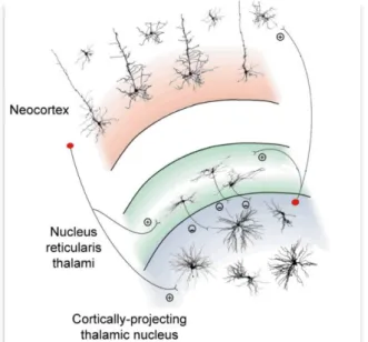
The critical constituent of the ascending reticular activating system is the thalamus. First discovered by Galen in the first century, from the 18th century it is considered the gateway of sensory inputs that receives and sends inputs to the cortex (Bickford et al., 1994; Sherman, 2005) playing an important role in the neuromodulation of the brain. A perithalamic region of reticular nuclei surrounds the thalamus. This region takes the name of reticular thalamus and it is rich in GABAergic neurons. It receives inputs from the fifth and sixth layers of the cortex and from relay thalamus (so defined as they receive excitatory glutamatergic input from peripheral sensory pathways and projects to one or few well-defined cortical areas). The reticular nucleus is important in the control of thalamocortical and corticothalamic connections through the modulation of GABAergic input on the relay thalamus (figure 3). It can be divided into different sections that have connections with thalamus and cortex and different functions (sight, hearing, touch, movement or limbic functions).
Figure 3. Schematic diagram of a cortico-thalamo-cortical module with its most relevant cellular
components and synaptic connections (thalamic interneurons and neocortical neurons other than those in layer 4 and 5/6 have been omitted for clarity). (+) and (−) indicate excitatory and inhibitory synapses, respectively. (Revised from Crunelli and Hughes 2010).
Located in the diencephalon, the thalamus is formed by four components (hypothalamus, epithalamus, ventral thalamus and dorsal-relay thalamus). The most important region is dorsal thalamus, composed by allothalamic and the isothalamic region. The allothalamic region can be divided in: (a) paraventricular region (receiving afferents from the amygdala); (b) the centre-median-parafascicular complex which appears to be a major element in the basal ganglia system
Introductory Part
15 that is involved in motor control (having projections to the striatum); (c) and the intralaminar region that receives inputs from ascending reticular activating system and plays a modulatory role in regulation of cortical arousal.
The isothalamic constitutes 90% or more of the dorsal thalamus and contains the major projections to the cortex. Lesions of these areas could affect cognition. The internal and superior laminae divide the isothalamus into several subregions (regio superior, superregio medio-posterior, superregio basalis and regio inferolateral).
Among the above mentioned subregions: the nucleus medialis receives a large projection from the dorsolateral prefrontal cortex, amygdala, ventral globus pallidus and nigral projections. It projects to prefrontal cortex and it is involved in cognition, sleep-wake cycle and executive function; The pulvinar-lateral posterior complex, which has a lot of reciprocal connections with the associated parieto-occipito-temporal cortical areas, is involved in attention and it is correlated to visual hallucination; the motor thalamus in the infolateral regio transfers information from the substantia nigra (sub-regio dorsalis), the globus pallidus (subregio oralis) and the cerebellum (subregio intermedia) to the prefrontal, supplementary, premotor, motor and somatosensory areas of the cerebral cortex. The motor thalamus receives projections from the nucleus perithalamicus in a specific diffuse manner, which can also contribute to the transfer of motor information (Herrero et al., 2002).
Electrical experiments on brain slices have suggested that thalamic activity is sufficient to alter the cortical state. Thalamocortical neurons are directly excited by stimulation of the brain stem reticular nuclei. During wakefulness, brief activation of the thalamic reticular nucleus is sufficient to evoke thalamic bursts and cortical spindles (Halassa et al., 2011). They receive cholinergic input from the cholinergic reticular nuclei at midbrain pontine junction and aminergic input from locus ceruleus and raphe nuclei as shown by some experiments in rats (Lindvall and Björklund, 1974) but the monominergic neurons seem not be involved in tonic activation processes of thalamocortical systems (Jones and Moore, 1977). On the contrary, the result of local application of a cholinergic agonist is a loss of hyperpolarizing oscillation, the increase of metabolic activity of the thalamic neurons and an EEG desynchronized (Buzsaki et al., 1988).
Introductory Part
16
Three major dementia types (ADD, PDD, DLB)
Alzheimer’s, Parkinson’s and Lewy Body diseases (AD, PD, DLB) are the major neurodegenerative disorders that can progress to dementia (ADD, PDD, DLB).
The ADD represents approximately 60-80%, while PDD and DLB the 10%-15% of all dementia cases in aged people over 65 (Alzheimer’s Association, 2016; Alzheimer’s society 2017). Demented people suffer from a progressive cognitive decline of sufficient magnitude to interfere with normal social or occupational functions, or with usual daily activities (Diagnostic and Statistical Manual of Mental Disorders -DSM).
“Dementia” tends to increase with age (Graves and Kukull, 1994), the reason why, with the rise of expectancy life, dementia is a growing socioeconomic and medical problem.
In 1906 the psychiatrist and neuropathologist Alois Alzheimer gave the name at the most prevalent form of neurodegenerative and age-related dementia in the modern society, the Alzheimer’s disease (AD). In the 2016 Alzheimer’s Disease International’s “World Alzheimer Report” estimated 44 million people worldwide and this number is expected to double in the next years. At the basis of the Alzheimer’s disease there are neurological dis-functions due to the neuron’s death triggered by neuropathological pathway. AD affects patients in different ways, changing the rate of progression for each subject (Weiner et al., 2015).
The most common symptoms of the AD disease are memory loss that disrupts daily life and challenges in planning or solving problems.
In 1984 Kosaka and colleagues described the cytoplasmic inclusions of α-synuclein in the brain- stems of patients affected by Parkinson’s disease. These inclusions were firstly noticed in 1910 by the neurologist Fritz Heinrich Lewy (Lewy et al., 1912) who studied the brain of people showing dementia. In its memory, these cytoplasmic inclusions took the name of Lewy Bodies (LB). The clinical constellations of DLB and PDD include progressive cognitive impairment associated with extrapyramidal motor impairments (parkinsonism), visual hallucinations, and fluctuations of attention and wakefulness. The cognitive domains that are impacted in DLB and PDD overlap substantially, with prominent executive dysfunctions and visual-spatial abnormalities and variable impairment in memory capacities. In DLB, dementia often heralds the onset of illness in advance of parkinsonian motor signs, but by consensus may follow their development up to 1 year from their onset. In contrast, a diagnosis of PDD is made when cognitive impairments develop in the setting of well-established PD (Gomperts, 2016).
Introductory Part
17
Neuropathology
In AD, the symptoms are caused by a progressive loss of cholinergic functions due to neuronal cell death firstly in the hippocampus and then in the cerebral cortex, brain regions involved in thought processing and memory (Selkoe, 1991; Blennow et al., 2006; Mucke, 2009). The neuropathological process that affects the neurons of AD patients, depends on two kinds of protein aggregates: amyloid plaques, also called senile plaques, and tangles of hyper-phosphorylated tau-protein, also called neurofibrillary tangles (Figure 1).
Figure 1. A healthy neuron (left) compared to diseased neuron (right) with the amyloid plaque and
phosphorylated tau (revised illustration Bob Morreale for American Health Assistance Foundation).
Amyloid precursor protein (APP) is a transmembrane protein without known function that is constitutively cleaved into peptides during cell’s metabolism (Haas et al.,1992). The two amyloidogenic forms of amyloid protein (40 or 42 amino acid Aβ peptide) are released after cleavage by β-secretase and γ- secretase enzymes and are usually quickly removed from the brain. However, in the case of overproduction or impaired clearance, Aβ aggregates into extracellular oligomers, fibrillary and, eventually, plaques (Masters et al., 1985). Tau is an intracellular microtubule binding protein that, when hyper-phosphorylated, will cause disassembly of microtubules and thus will impair axonal transport and compromise neuronal and synaptic function (Braak et al., 2003; Iqbal et al., 2005). Weather tangle formation is a cause or a consequence of the disease is still under debate. The dominating hypothesis for the cause of AD is the cascade Aβ hypothesis (Hardy and Selkoe, 2002). According to this hypothesis, abnormal metabolism of the amyloid precursor protein (APP) and the subsequent accumulation of toxic Aβ peptides interferes with the neuron-to-neuron communication at synapses and contributes to cell death. After the cell’s death, the aggregates spread out in the
Introductory Part
18 extracellular space. Tau tangles block the transport of nutrients and other essential molecules inside neurons and are also believed to contribute to cell death.
Apart from the damage to ascending cholinergic projections in Alzheimer's disease, biochemical studies on human brain post mortem have revealed several other neurochemical abnormalities. These include reductions in 5-hydroxytryptamine (5-HT) and 5-HT receptors, and in noradrenaline. Some regions of cerebral cortex show reduced concentrations of GABA (Rossor and Iversen, 1986). Furthermore, evidence exists for both cholinergic and glutamatergic involvement in the aetiology of Alzheimer's disease. The glutamatergic hypothesis links the cognitive decline in patients with Alzheimer's to neuronal damage resulting from overactivation of N-methyl-d-aspartate (NMDA) receptors by glutamate. The sustained low-level activation of NMDA receptors, which are pivotal in learning and memory, may result from deficiencies in glutamate reuptake by astroglial cells in the synaptic cleft (Francis, 2005). Parkinson’s and Lewy Body disease are well characterized by α-synuclein deposition (Jellinger et al., 2009). α-synuclein is a human protein encoded by the gene SNCA. It is predominantly expressed in the neocortex, hippocampus, substantia nigra, thalamus, and cerebellum. In the brain, it is localized in presynaptic terminals where it is involved in neurotransmitters released from synaptic vesicles. If the gene is mutated, the protein forms a stably folded tetramer (aggregates) (Figure 2) (Bartels et al., 2011).
Figure 2. Lewy body in substantia nigra from patients with Parkinson's disease (from the MRC
Introductory Part
19 However, PDD and DLB patients also show AD-type pathology (i.e. cerebral neurofibrillary tangle and Aβ plaques) (Hansen et al., 1998). The pathological substrate seems to follow the Braak stages (a method describing the progressive formation of aggregates from the lower brainstem to the neocortex) (Braak et al., 2003). In PDD, LB are primarily diffuse in the subcortical regions of the brain, predominantly in the midbrain substantia nigra and locus coeruleus (Nussbaum and Ellis, 2003) whereas, DLB is characterized by the presence of Lewy bodies in the subcortical (midbrain) and cortical (frontotemporal) regions of the brain, as well as amyloid plaques. Neurofibrillary tangles are less common in dementia with Lewy bodies (McKeith, 2004).
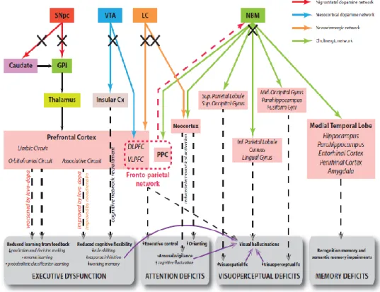
In PDD and DLB different sub-cortical and cortical networks are compromised. In DLB, dopamine via nigral degeneration and acetylcholine via basal forebrain degeneration are the neurochemical systems most consistently affected (Tiraboschi et al., 2000). The neurophysiological model of the interrelation among neurotransmitters and their effects on the functionality of the brain cortex, proposed by Gratwicke et al., 2015 is shown in figure 3.
Figure 3. Hypothetical model of neural circuits malfunctioning in PDD and the corresponding cognitive
deficits. Legend: Cx = cortex; DLPFC = dorsolateral prefrontal cortex; GPi = globus pallidus (internus); PPC = posterior parietal cortex; SNpc = substantia nigra pars compacta; VLPFC = ventrolateral prefrontal cortex; VTA = ventral tegmental area (Gratewike et al., 2015).
Introductory Part
20 In PDD there are evidences about the deficits occurring in dopaminergic nigro-striatal pathway, responsible of motor impairment, as well as cortically projecting dopaminergic neurons in the mesocortical limbic system (Scatton et al., 1983), fronto-striatal network responsible of executive dysfunctions (Middleton and Strick, 2000), loss of neurons in the nucleus basalis of Meynert leading to cortical cholinergic denervation (Whitehouse et al., 1983) and cholinergic cell loss in pedunculopontine in PDD with hallucinations (Hepp et al., 2013). The noradrenergic network, involved in control of the vigilance with its projections from the locus coeruleus to the thalamus, amygdala and cortex is also compromised in Parkinson’s disease (Bertrand et al., 1997).
The altered neurophysiology of motor dysfunctions in PD and DLB, called parkinsonism (a clinical syndrome characterized by tremor, bradykinesia, rigidity, and postural instability), have been clarified.
The motor circuit originates from the precentral and postcentral sensorimotor fields, engages specific portions of the basal ganglia (corpus striatum-caudate nucleus and putamen-, the globus pallidus, the subthalamic nucleus, and the two parts -pars compacta and pars reticularis- of the substantia nigra) and ends back in the precentral motor fields of the frontal lobe.
In healthy subjects there are two pathways involving basal ganglia and the activation of two types of dopamine receptors (D1 and D2) on the striatum by dopaminergic inputs from substantia nigra (pars compacta) (figure 4):
a direct pathway (activation of D1 receptors) involving the neurons projecting from the putamen to the inner part of the Globus Pallidus (GPi) and to the pars reticulata of the Substantia Nigra (SNr), the output nuclei of the BGs. This pathway has an inhibitory effect - GABAergic - directed on the GPi / SNr neurons, thus reducing the excitatory effect of these nuclei on the thalamus, consequently going to promote the movement;
-an indirect pathway (activation of D2 receptors) that connects the putamen with the output nuclei through the external Globus Pallidus (GPe) and the subthalamic nucleus (STN).
The stimulation of this indirect path leads to the inhibition of the GPe, the disinhibition of the STN and the excitement of GPi / SNr. The inhibitory effect on the thalamus is thus improved. The result is a reduction on excitatory glutamatergic input on the cortex and “inhibition” of the movement.
In healthy subjects, the balance between the two pathways allows good motor control. In PD patients, the degeneration of dopaminergic nuclei of the substantia nigra is responsible for unbalance of glutamatergic inputs on motor cortex (Maiti et al., 2017)
Introductory Part
21
Figure 4. The image shows the direct and indirect pathways involved in motor control in healthy
subjects. Legend: Glu=glutamate, DA=dopamine, STN=subthalamic nucleus, SNr=substantia nigra pars reticulate, GPe= globus pallidus externus, GPi= globus pallidus internus (from https://step1.medbullets.com/neurology/113008/basal-ganglia).
Clinical features
Alzheimer’s, Parkinson’s and Lewy Bodies diseases are three complex neurocognitive syndromes influenced by deficits of interrelated cortical (AD, PD, DLB) and subcortical (PD and DLB) networks.
The main clinical symptoms of AD are loss memory and challenges in planning or solving problems probably due to the loss of cholinergic nuclei. However, other many symptoms are very common: difficulty completing familiar tasks at home, at work or at leisure; confusion with time or place; trouble understanding visual images and spatial relationships; new problems with words in speaking or writing; misplacing things and losing the ability to retrace steps; decreased or poor judgment; withdrawal from work or social activities; changes in mood and personality, including apathy and depression (Alzheimer’s Association, 2014). However, the content of these symptoms will vary dramatically from patient to patient, depending upon their premorbid personality, life experiences, and social as well as cultural influences. In this contest, the caregiver plays an important role in the management of the disease even if this role pays high costs in terms of money and quality of life.
Introductory Part
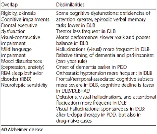
22 PDD and DLB are affected by attentional, executive, memory dysfunctions, mood disturbances and parkinsonism (defined as bradykinesia in combination with rest tremor, rigidity, or both). Most prominent are the executive dysfunctions which term indicates several cognitive abilities, including problem-solving, planning/sequencing, rule-shifting/maintenance, task-switching, manipulation in working memory and response inhibition already present at the onset of Parkinson’s disease (Dubois and Pillon, 1997). The main hypothesis is that these dysfunctions are due to degeneration of fronto-striatal network but, the new evidence, post mortem, have shown a degeneration of the projections of ventral tegmental area (VTA) in the mid brain to the limbic region (mesocortical dopamine network) (Scatton et al., 1983). Executive functions are probably more impaired in PDD than in DLB. Visual symptoms, common in PDD likely due to a reduced metabolism in both dorsal and ventral visual pathway, include visual hallucinations, although they are less common than in DLB. Other non-motor features, including autonomic dysfunctions and sleep disorders may occur according to the severity of dementia, while mood disturbances have a similar frequency as in DLB (Jellinger and Korczyn, 2018). Even if PDD and DLB have many similar clinical aspects, there are differences in timing and in the profile that allow to identify two different clinical syndromes and apply separate diagnostic criteria. Table 1 sums up in the detail, the overlap and the differences between them. Furthermore, before the stage of dementia, there are more evidence clinical features. In Parkinson’s disease, patients primarily suffer from extrapyramidal symptoms that can be followed by slow cognitive impairment, not always progressing in dementia.
In patients with Lewy bodies disease have been identified core clinical features, present already in the first stages of the disease. The core clinical features of DLB have been well described by the DLB Consortium (McKeith et al., 2017) and are summed up in the following: (a) fluctuation: DLB fluctuations occurring as spontaneous alterations in cognition, attention, and arousal. They include incoherent speech, variable attention, or altered consciousness. At least one measure of fluctuation should be documented when applying DLB diagnostic criteria. Fluctuations may also occur in advanced stages of other dementias, so they best predict DLB when they are present early; (b) visual hallucinations: recurrent, complex visual hallucinations occur in up to 80% of patients with DLB and are a frequent clinical sign post to diagnosis. They are typically well-formed, featuring people, children, or animals, sometimes accompanied by related phenomena including passage hallucinations, sense of presence, and visual illusions; (c) parkinsonism: spontaneous parkinsonian features, such as bradykinesia and rigidity, are common in DLB, eventually occurring in over 85%, while rest tremor is less frequent (Fritz et al., 2016). Many DLB patients’ parkinsonism falls short of this, so documentation of only one
Introductory Part
23 of these cardinal features is required; (d) REM sleep behavior disorder:RBD is a parasomnia manifested by recurrent dream enactment behavior that includes movements mimicking dream content and associated with an absence of normal REM sleep atonia. RBD is now included as a core clinical feature because it occurs frequently in autopsy-confirmed cases compared with none-DLB (76% vs 4%). Conditions mimicking RBD are common in people with dementia, e. g., confusional awakenings, severe obstructive sleep apnea, and periodic limb movements, all of which must be excluded by careful supplementary questioning to avoid a false-positive diagnosis.
Table 1 shows the clinical overlap and dissimilarities between dementia with Lewy bodies and
Parkinson’s disease with dementia (PDD) (Jellinger and Korczyn, 2018).
Risk factors
The identification of the risk factors could help to improve the monitoring of those patients with higher probability to get ill. However, the AD, PD and DLB are multifactorial diseases with both familiar/genetic risk factors for AD and sporadic factors for PD and DLB, both sporadic genetic risk due to co-morbidity of aging-diseases and lifestyle quality. The occurrence of the AD due to familial/genetic risk factors came in the early aging, before 65 years, while the AD due to sporadic (not from the birth) genetic mutation has a late-onset occurrence, later 65 years. In less of 1%, the cases of the AD depend on three genes mutation (Bekris et al., 2010). More specifically, these three genes are those encoding for the amyloid precursor protein (APP) and the genes for the presenilin 1 and presenilin 2 proteins (regulating the action of the enzyme
γ-Introductory Part
24 secretase, seen in the above paragraph). The transmission of these genes is autosomal dominant that means the presence of almost one copy (allele) of these three genes mutated from the birth (inherited from a parent) will be responsible for the early onset of the AD. The mutation of presenilin 2 induces the development of the AD with 95% of probability (Goldman et al., 2011). Concerning the PD and DLB, different sporadic genetic factors may be involved in the
etiopathology of PDD and DLB: mutations in SNCA, the gene of α-synuclein as well as
mutations in GBA (glucocerebrosidase), and MAPT (microtubule-associated protein tau) may be responsible on the onset of the two diseases (McKheit et al., 2017) even if the pathological
mechanisms are not yet clarified. Other risk factors to consider for all three diseases are: (a)
aging; (b) APOE-ε4 Gene: the APOE gene provides the blueprint for a protein that transports cholesterol in the bloodstream. Everyone inherits one of three forms of the APOE gene (ε2, ε3 or ε4 ) from each parent. The ε3 form is the most common, and the ε2 form the least common. Those who inherit one copy of the ε4 form have a three-fold higher risk of developing AD than those without the ε4 form, while those who inherit two copies of the ε4 form have an 8- to 12-fold higher risk (Alzheimer’s Drug Discovery Foundation, 2015; Loy et al., 2014). In addition, those with the ε4 form are more likely to develop AD at a younger age (Spinney, 2014). Having the ε2 form may decrease one’s risk compared with having the ε3 form. While the above-mentioned risk-factors are inevitable, other risk factors can be modified to reduce risk of cognitive decline and dementia; (c) Cardiovascular Disease Risk Factors: the factors that increase the risk of cardiovascular disease are also associated with a higher risk of dementia such as smoking (Beydoun et al., 2014), obesity in midlife (Rönnemaa et al., 2001) and diabetes (Gudala et al., 2013). Midlife hypertension (Rönnemaa et al., 2001) and midlife high cholesterol are also implicated as risk factors for dementia; (d) education: According to the “cognitive reserves” hypothesis, having more years of education builds a “cognitive reserve” that enables individuals to better compensate for changes in the brain that could result in symptoms of AD or PD or DLB (Stern Y., 2012); (e) traumatic Brain Injury (TBI): compared with no TBI, moderate TBI is associated with twice the risk of developing Ad, PD or DLB, and severe TBI with 4.5 times the risk (Plassman et al., 2000).
A precursor to dementia: Mild cognitive impairment (MCI)
Mild cognitive impairment (MCI) is considered a clinical state showing objective impairment on neuropsychological tests, but not fulfil the clinical criteria for dementia (Flicker et al., 1991; Petersen et al., 2001). Individuals with MCI convert to dementia at a rate of approximately
5-Introductory Part
25 10% per year in contrast to 1-2% in healthy controls (Petersen, 2011), and it is under debate if MCI may represent a prodromal stage of AD or other forms of dementia (Petersen et al., 2014). As such, MCI represents an important group for the early identification of those at risk of developing dementia, as well as for implementing early treatment options. The revised National Institute on Aging-Alzheimer’s Association (NIA-AA) diagnostic criteria (Albert et al., 2011) defined two main subtypes of MCI: (1) amnestic MCI (aMCI), defined by the presence of episodic memory impairment, and (2) non-amnestic MCI (naMCI), defined by impairment in one or more non-memory cognitive domains. The impairment can further be classified as single domain (impairment restricted on one cognitive domain), or multiple domains (impairment in two or more cognitive domains). For example, 70-90% of aMCI patients who progress to dementia show clinical signs of AD (Petersen et al., 2009; 2001), and naMCI patients may progress to others dementing conditions such as Lewy body dementia, frontotemporal dementia, or vascular dementia (Jack et al., 2009; Petersen et al., 2009). The current consensus is that most MCI patients who convert in AD mostly exhibit an impairment in episodic memory, but other cognitive domains may also be impaired (Albert et al., 2011). The neuropathological features of aMCI include the presence of neurofibrillary tangles in the medial temporal lobes, diffuse cortical amyloid deposition, synaptic loss, and degeneration of the cholinergic system (Drago et al., 2011; Mufson et al., 2012; Stephan et al., 2012). Furthermore, MCI patients show a significant neuronal loss in the entorhinal cortex and hippocampus (Mufson et al., 2012; Stephan et al., 2012). There is some evidence that levels of amyloid deposition may be related to cognitive function in healthy older adults without cognitive impairment and in MCI patients, but not in AD patients (Villemagne et al., 2008), which may be related to the rapid accumulation of amyloid in the early and preclinical phases of the disease followed by a plateau in the later stages (Masdeu et al., 2012). Therefore, the correlation between the amyloid deposition and cognitive impairment remains unclear but emerging evidence is suggestive of a greater relationship as described in the following paragraphs.
Diagnostic criteria for AD
The diagnostic criteria of Alzheimer’s disease are still under debate. Only the brain autopsy, after death, can confirm the diagnosis due to the abnormal accumulation of neurofibrillary tangle and Aβ protein in the whole brain. However, mental and behavioral tests and physical examinations allow physicians to make an accurate diagnosis of AD in 90 percent of cases (American Health Assistance Foundation, AHAF, 2010). Neuropsychological examinations
Introductory Part
26 may be used to identify cognitive symptoms. The most commonly administered tests are the Mini-Mental State Exam (MMSE) (the score from 0 to 30, where a score under 12 indicates severe dementia) and some cognitive scales of Alzheimer’s Disease Assessment (ADAS cog; Skinner et al., 2013). AD patient’s scores typically decrease 2 to 4 points every year (Alzheimer’s Association, 2014). The actually challenge is to define diagnostic criteria able to identify the disease in the early stage, before the manifestation of clinical symptoms for a quick intervention and preventing the fast neurodegenerative process. MMSE offers only a marginal assessment of some cognitive abilities which are considered important both for AD and other types of dementia and, furthermore, it doesn’t result sensitive to the cases of very mild cognitive impairment. The ADAS results deficient in test-retest reliability and the performance of the patients seems to be influenced by other factors, such as their mood at the time of the task. Such neuropsychological assessment, therefore, seems to be not able to discriminate AD risk in a preclinical phase (in cognitively healthy elderly subjects) and the potential conversion from a preclinical form to AD or from MCI to AD (Lin et al., 2011). Thus, an accurate diagnosis of AD results to be very complex, also because of the presence, in elderly subjects, of comorbidities that might contribute to the cognitive impairment. Besides the diagnostic criteria of the Diagnostical and Statistical Manual of Mental Disorders (DSM-V), the diagnosis of AD patients is also based on the parameters of National Institute of Neurological and Communicative Disorders and Stroke-Alzheimer Disease and Related Disorders Association (NINCDS_ADRDA- Mc Khan et al., 1984). According to these criteria, AD can be classified as “well-defined AD” (clinical diagnosis with histological confirm), “probable AD” (typical clinical syndrome without histological confirm) and “possible AD” (atypical clinical characteristics that seem not to have an alternative diagnosis without histological confirm). Although the great sensitivity and specificity of the NINCDS-ADRDA criteria, AD cases are very often diagnosed in an advanced stage of cognitive impairment (Knopman et al., 2001). Dubois et al. (2007) proposed the concept of “prodromal AD” and exhibited new additional criteria for the detection of the different phases of the disease: the main core of this assumption is based on the presence of several impairments of episodic memory which, together with a possible presence of diagnostic biomarkers, identify AD in the entire course of the clinical disease’s spectrum. The first criterion, the criterion A, specifies that a deficit of episodic memory, assessed with specific tests, must be present. The presence of a biological mark is established by the criterion B (structural image), the criterion C (cerebrospinal fluid), the criterion D (molecular image), or the criterion E (dominant mutation in the family). The new criteria highlight the importance of neuroimaging techniques in the diagnostic procedures of
Introductory Part
27 AD and consider also preclinical and early phases of the disease. In 2011, the National Institute on Aging (NIA) and the Alzheimer’s Association proposed revised criteria and guidelines for diagnosing Alzheimer’s disease (Sperling et al., 2011; Albert et al., 2011). These criteria and guidelines update diagnostic criteria and guidelines published in 1984 by the ADRDA (McKhann et al., 1984) and incorporate three notable changes: (1) they identify two stages of Alzheimer’s disease: mild cognitive impairment (MCI) due to Alzheimer’s disease and dementia due to Alzheimer’s disease; (2) they propose criteria for a preclinical phase of Alzheimer’s disease, occurring before symptoms such as memory loss develop; (3) they incorporate biomarker tests.
In 2014, Dubois and colleagues from the International Working group (IWG) proposed a method to simplify the diagnosis for AD. They indicated an appropriate clinical AD phenotype (typical such as with evidence of a specific episodic memory profile characterized by a low free recall and reduction of hippocampal volume; or atypical, less frequent but well defined clinical phenotypic variants of non-amnestic focal cortical syndromes, including logopenic aphasia, posterior cortical atrophy, and frontal variant AD) and a pathophysiological biomarker. They proposed topographical biomarkers of the disease, such as volumetric MRI to identify hippocampal atrophy and fluorodeoxyglucose PET for the accumulation of Aβ-amyloid in the brain to better monitor the course of the disease. Furthermore, they proposed a revision for typical AD: “A research diagnosis of typical AD can be made in the presence of an amnestic syndrome of the hippocampal type that can be associated with various cognitive or behavioral changes, and at least one of the following changes indicative of in-vivo Alzheimer’s pathology: a CSF profile consisting of decreased Aβ1–42 levels together with increased T-tau or P-tau concentrations, or an increased retention on amyloid tracer PET”. The most innovative aspect of the 2007 criteria was the first introduction of biomarkers into the core diagnostic framework. Finally, the last diagnostic criteria have been proposed by Jack and colleagues in 2018 of the National Institute on Aging—Alzheimer’s Association (NIA-AA) Research Framework. Alzheimer’s disease is defined based on specific pathological processes, deviating from the approach based on the observation of clinical symptoms and the deterioration of cognitive and functional functions. This new theoretical framework defines the disease based on the in vivo presence of specific toxic proteins, by biochemical analysis or neuroimaging methods. According to the authors, the biological definition of AD is independent of the clinical phenotype (typically the presence of memory deficits) but based on the A / T / N scheme, where "A +" and "T +" denote the pathological state of beta-amyloid protein (Aβ) and phosphorylated
Introductory Part
28 tau, respectively, while "N +" refers to the presence of neurodegeneration (e.g. hypometabolism or cortical atrophy). According to this new definition, AD is referred to in a continuum, in which the alterations of the different proteins define the state of disease. An "A-" individual will be considered not affected by AD (or with non-AD type pathological changes, if "T +" and / or "N +"), while an individual with a pathological state limited to the Aβ protein ("A +", "T-" and "N-") will be in a condition that the authors define as AD pathological change, i.e. at an early stage of illness. The diagnosis of AD can instead be applied together with the presence of a pathological state of "A +" and "T +". An "N +" state (evidence of neurodegeneration) does not support the diagnosis but is an indicator of the disease stage.
Diagnostic criteria for PD
According to the guideline described by Movement Disorder Society (MDS) Task Force (Emre et al., 2007), the diagnosis of dementia associated with Parkinson's disease is possible if the patient shows the following core and associate cognitive/behavioral features:
Core features: (a) diagnosis of Parkinson's disease according to Queen Square Brain Bank criteria (for parkinsonism evaluation); (b) dementia syndrome defined as: impairment in more than one cognitive domain; representing a decline from premorbid level; deficits severe enough to impair daily life (social, occupational, or personal care), independent of the impairment ascribable to motor or autonomic symptoms.
Associated clinical features: (a) cognitive features impaired (attention, executive functions, visuo-spatial functions, memory and language); (b) behavioral features (apathy, depression, hallucinations, delusions, excessive daytime sleepiness).
The diagnosis is uncertain if the following points are verified: (a) co‐existence of any other abnormality which may by itself cause cognitive impairment, but judged not to be the cause of dementia, e.g. presence of relevant vascular disease in imaging; (b) time interval between the development of motor and cognitive symptoms not known.
The diagnosis of probable PDD is made if there are both core features and the following associate clinical features: (a) typical profile of cognitive deficits including impairment in at least two of the four core cognitive domains (impaired attention which may fluctuate, impaired executive functions, impairment in visuo‐spatial functions, and impaired free recall memory which usually improves with cueing); (b) the presence of at least one behavioral symptom (apathy, depressed or anxious mood, hallucinations, delusions, excessive daytime sleepiness)
Introductory Part
29 supports the diagnosis of Probable PDD, lack of behavioral symptoms, however, does not exclude the diagnosis.
The diagnosis of possible PDD is made if there are both core features and the following associate clinical features: (a) atypical profile of cognitive impairment in one or more domains, such as prominent or receptive‐type (fluent) aphasia, or pure storage‐failure type amnesia (memory does not improve with cueing or in recognition tasks) with preserved attention; (b) behavioral symptoms may or may not be present.
The MDS task Force has also described the guideline for the identification of Parkinson’s disease with mild cognitive impairment (PDMCI). The criteria described are in the following: (a) diagnosis of Parkinson disease (PD) is based on the UK PD Brain Bank Criteria; (b) gradual decline in cognitive ability in the context of established PD, reported by either the patient or informant or observed by the clinician; (c) cognitive deficits on either formal neuropsychological testing or a scale of global cognitive abilities; (d) cognitive deficits are not sufficient to interfere significantly with functional independence, although subtle difficulties on complex functional tasks may be present.
Furthermore, there are two subtypes classification:
PD-MCI single-domain: abnormalities on two tests within a single cognitive domain (specify the domain), with other domains unimpaired;
PD-MCI multiple-domain: abnormalities on at least one test in two or more cognitive domains (specify the domains).
Diagnostic criteria for DLB
According to the guideline of DLB Consortium (McKeith et al., 2017), the diagnosis of DLB is made if the patient shows the following core clinical features (the first 3 typically occur early and may persist throughout the course): fluctuating cognition with pronounced variations in attention and alertness; recurrent visual hallucinations that are typically well formed and detailed; REM sleep behavior disorder, which may precede cognitive decline; one or more spontaneous cardinal features of parkinsonism: these are bradykinesia (defined as slowness of movement and decrement in amplitude or speed), rest tremor, or rigidity.
Other supportive clinical features that could help for the diagnosis are: severe sensitivity to antipsychotic agents; postural instability; repeated falls; syncope or other transient episodes of unresponsiveness; severe autonomic dysfunction, e.g., constipation, orthostatic hypotension,
Introductory Part
30 urinary incontinence; hypersomnia; hyposmia; hallucinations in other modalities; systematized delusions; apathy, anxiety, and depression.
Furthermore, the recent scientific advances have discovered indicative biomarkers as: reduced dopamine transporter uptake in basal ganglia demonstrated by SPECT or PET; abnormal (low uptake) 123iodine-MIBG myocardial scintigraphy; polysomnographic confirmation of REM sleep without atonia.
Other supportive biomarkers are: relative preservation of medial temporal lobe structures on CT/MRI scan; generalized low uptake on SPECT/PET perfusion/metabolism scan with reduced occipital activity the cingulate island sign on FDG-PET imaging; prominent posterior slow-wave activity on EEG with periodic fluctuations in the pre-alpha/theta range.
Probable DLB can be diagnosed if: (a) two or more core clinical features of DLB are present, with or without the presence of indicative biomarkers; or (b) only one core clinical feature is present but with one or more indicative biomarkers.
Possible DLB can be diagnosed if: (a) only one core clinical feature of DLB is present, with no indicative biomarker evidence; or (b) one or more indicative biomarkers is present but there are no core clinical features.
DLB is diagnosed when dementia precedes or is concurrent with parkinsonism. Parkinson disease dementia should be used to describe dementia that occurs in the context of well-established Parkinson disease. For research studies that distinguish between DLB and Parkinson disease dementia, a 1-year rule is recommended for a diagnosis of DLB, such that dementia should begin no later than 1 year after the onset of parkinsonism.
Finally, the scientific community is trying to identify clinical features of patients who will develop DLB with dementia. Today, the criteria are limited but some studies reported disturbances of REM sleep, mild deficits in attentional and executive functions and subtle parkinsonism in patients that have developed probable DLB (Ferman et al., 2013).
Pre-clinical biomarkers
Neuroimaging and fluid biomarkers are a promising area of research for detecting AD, PD and DLB (Saeed et al., 2017). Multiple brain imaging procedures can be used to identify abnormalities in the brain, including Positron Emission Tomography (PET), Magnetic Resonance Imaging (MRI), and Computed Tomography (CT) scans. The high spatial resolution, sensitivity and specificity of MRI are the reason why this technique is considered the most powerful instrument to identify structural alterations and cerebral atrophy using
Introductory Part
31 volumetric measures of the entire brain (Kochunov et al., 2005). The most consistent evidence showed by MRI studies in AD patients greater atrophy of medial temporal lobe (MTL, hippocampus, amygdala, entorhinal cortex and parahippocampal gyrus) structures than patients with DLB, particularly the hippocampus, which is strongly correlated at autopsy with tangle rather than plaque or LB-related pathology (Burton et al., 2009). Furthermore, MRI in AD shows the ventricular dilatation and a reduced total volume in the brain (Busatto et al., 2008). In AD, this atrophy is localized at medial temporal limbic cortex level during the initial phases of the disease. Later, the atrophy progresses and expands to paralimbic cortical regions and to the neocortex (Braak et al., 2003). The temporal limbic cortex has a fundamental role in episodic memory and, given that memory deficit is the first symptom in AD, it results to be the main target in neuroimaging studies (Xu et al., 2000; De Santi et al., 2001).
Several longitudinal MRI studies provided valid information about preclinical stages of the disease: the grey matter atrophy is also seen in MCI subjects and the medial temporal lobe atrophy might be considered as a neurostructural biomarker of MCI-AD conversion (Schroeter et al., 2009). Some authors revealed that reduced hippocampal volume might be seen even six years before the onset of the disease (den Heijer et al., 2006; Csernansky et al., 2005). Ulterior sub-regional analysis has shown that in subjects with normal cognition, the volume of some restricted parts of hippocampus (CA1 and subiculum) is more closely associated to the conversion to MCI compared to the hippocampus total volume. Volume loss in these regions precedes cognitive impairment and conversion to MCI by several years and it’s able to discriminate cognitive stable individuals from MCI individuals with an accuracy of more than 93% (Apostolova et al., 2010). The volume of the entorhinal cortex also seems to predict cognitive impairment in elderly subjects with normal cognition with an accuracy of 90% (Martin et al., 2010).
FDG-PET and SPECT, provide information about glucose regional metabolism and cerebral perfusion, respectively. The characteristic pattern in AD patients is that of hypometabolism/hypoperfusion of parietal-temporal cortex (Herholtz et al., 2002).
PET images of amyloid-binding agent Pittsburgh compound B tracer (PET-PiB), hypometabolism in temporoparietal regions, established by FDG-PET and structural abnormalities observed with MRI, are the first markers of Alzheimer’s disease to be described (Ikonomovic et al., 2008; Small et al., 2000).
PET images allow in vivo analysis of Aβ42 presence in the brain and their spatial distribution. PiB results to be closely related to amyloid plaques burden revealed in the autopsy.