Contents lists available atScienceDirect
BBA - Molecular Basis of Disease
journal homepage:www.elsevier.com/locate/bbadisInhibition of Drp1-mediated mitochondrial
fission improves mitochondrial
dynamics and bioenergetics stimulating neurogenesis in hippocampal
progenitor cells from a Down syndrome mouse model
Daniela Valenti
a,⁎, Leonardo Rossi
b, Domenico Marzulli
a, Francesco Bellomo
c,
Domenico De Rasmo
a, Anna Signorile
d, Rosa Anna Vacca
a,⁎aInstitute of Biomembranes, Bioenergetics and Molecular Biotechnologies, National Council of Research, Bari, Italy bDepartment of Clinical and Experimental Medicine, University of Pisa, Italy
cDivision of Nephrology and Dialysis, Bambino Gesù Children's Hospital - IRCCS, Rome, Italy dDepartment of Basic Medical Sciences, Neuroscience and Sense Organs, University of Bari, Italy
A R T I C L E I N F O
Keywords: Down syndrome Mitochondrial dysfunction Hippocampal neurogenesis Mitochondrial network Drp1 Mdivi-1A B S T R A C T
Functional and structural damages to mitochondria have been critically associated with the pathogenesis of Down syndrome (DS), a human multifactorial disease caused by trisomy of chromosome 21 and associated with neurodevelopmental delay, intellectual disability and early neurodegeneration. Recently, we demonstrated in neural progenitor cells (NPCs) isolated from the hippocampus of Ts65Dn mice -a widely used model of DS - a severe impairment of mitochondrial bioenergetics and biogenesis and reduced NPC proliferation. Here we fur-ther investigated the origin of mitochondrial dysfunction in DS and explored a possible mechanistic link among alteration of mitochondrial dynamics, mitochondrial dysfunctions and defective neurogenesis in DS. Wefirst analyzed mitochondrial network and structure by both confocal and transmission electron microscopy as well as by evaluating the levels of key proteins involved in thefission and fusion machinery. We found a fragmentation of mitochondria due to an increase in mitochondrialfission associated with an up-regulation of dynamin-related protein 1 (Drp1), and a decrease in mitochondrial fusion associated with a down-regulation of mitofusin 2 (Mnf2) and increased proteolysis of optic atrophy 1 (Opa1). Next, using the well-known neuroprotective agent mitochondrial division inhibitor 1 (Mdivi-1), we assessed whether the inhibition of mitochondrialfission might reverse alteration of mitochondrial dynamics and mitochondrial dysfunctions in DS neural progenitors cells. We demonstrate here for thefirst time, that Mdivi-1 restores mitochondrial network organization, mitochondrial energy production and ultimately improves proliferation and neuronal differentiation of NPCs. This research paves the way for the discovery of new therapeutic tools in managing some DS-associated clinical manifesta-tions.
1. Introduction
Down syndrome (DS), the most frequent genetic cause of in-tellectual disability, is due to either full or partial trisomy of chromo-some 21. Clinically, DS is a neurodevelopmental disease characterized by neural developmental delay with impairment in language, learning and memory associated with an atypical craniofacial profile and in-creased susceptibility to congenital heart defects, immune and meta-bolic disorders [1,2]. The majority of patients reaching middle age, develop symptoms of premature aging and neurochemical features of
the Alzheimer's disease and related dementia associating DS as a neu-rodegenerative disease[3]. On this basis, the pathogenesis of DS has been deeply investigated in the hope of increasing our understanding of the DS neurobiology and identifying efficacious drug targets to improve the clinical phenotype.
A growing body of literature has shown that structural and func-tional damages to mitochondria are critically associated with DS pa-thogenesis and are inherent features of DS (for refs see[4,5]). Of note, mitochondrial dysfunctions are also involved in the pathogenesis of others intellectual disability-related diseases, such as Rett syndrome
http://dx.doi.org/10.1016/j.bbadis.2017.09.014
Received 13 July 2017; Received in revised form 11 September 2017; Accepted 18 September 2017
⁎Corresponding authors at: Institute of Biomembranes, Bioenergetics and Molecular Biotechnologies, National Council of Research, Via Amendola 165/A, 70126 Bari, Italy. E-mail addresses:[email protected](D. Valenti),[email protected](R.A. Vacca).
Abbreviations: AR, aspect ratio; BrdU, bromodeoxyuridine; CaN, calcineurin; DAPI, 4',6-diamidino-2-phenylindole; DCX, doublecortin; Drp1, dynamin-related protein 1; DS, Down syndrome; L-Opa, long Opa1; Mdivi-1, mitochondrial division inhibitor 1; Mnf2, mitofusin 2; NPCs, neural progenitor cells; OLIGO, oligomycin; Opa1, optic atrophy 1; OXPHOS, oxidative phosphorylation; P-Drp1, S637-phosphorylated Drp1; RCAN1, regulator of calcineurin 1; S-Opa, short Opa1; wt, wild-type
Available online 20 September 2017
0925-4439/ © 2017 Elsevier B.V. All rights reserved.
and autism (for refs see[4,6]), and neurodegenerative conditions, in-cluding Alzheimer's and Parkinson's diseases (for refs see[7]).
In DS, impairment in respiratory capacity, mitochondrial membrane potential and ATP production have been demonstrated in mitochondria of peripheral cells, obtained from DS subjects [8–12], and of central nervous system cells from mouse models of DS[13,14]. Alterations of mitochondrial morphology and dynamics have also been recently re-ported in human fetalfibroblasts with chromosome 21trisomy[15].
We recently analyzed mitochondrial functions in neural progenitor cells (NPCs) isolated from the hippocampus of Ts65Dn mice, a widely used model of DS, which recapitulates many major brain structural and functional phenotypes of the syndrome, including impaired hippo-campal neurogenesis [16,17]. We found that, in Ts65Dn NPCs, mi-tochondrial bioenergetics and mimi-tochondrial biogenic program were strongly compromised and that molecules able to restore oxidative phosphorylation (OXPHOS) efficiency and mitochondrial biogenesis can also improve NPC proliferation[18,19].
There is growing evidence of a close relationship between functional mitochondrial bioenergetics and mitochondrial network integrity [20,21], especially in the cells of the central nervous system in which mitochondria are synthesized in the soma and must travel along axons and dendrites [22]. Mitochondria continuously and reversibly re-arrange their structure assuming elongated or punctiform distribution in the processes of fusion andfission, respectively[23]. These processes are regulated by highly conserved GTPase-dependent proteins, such as dynamin-related protein 1 (Drp1) andfission 1 (Fis1) for fission and mitofusin 1 (Mfn1), mitofusin 2 (Mfn2), and optic atrophy 1 (Opa1) for fusion[23,24]. An altered balance betweenfission and fusion leads to structural and functional abnormalities in the mitochondria network and, ultimately, to an impairment in neuronal function. Indeed, in several neurodegenerative diseases the maintenance of mitochondrial dynamics is required for neural development processes and synaptic formation and reorganization (for refs see[25]).
Very relevant is that overexpression of the human regulator of calcineurin 1 (RCAN1) promotes deregulation of Drp1 activity asso-ciated with an increase of mitochondrial fission, mitochondrial dys-function, oxidative stress and early age-dependent memory and sy-naptic plasticity deficits in mice[26]. Based on these evidences and since RCAN1 protein, encoded by the 21q22.1-q22.2 region of human chromosome 21, is overexpressed in Down syndrome[27,28], we ar-gued that Drp1 could be deregulated in DS and Drp1-mediated mi-tochondrial fission could promote mitochondrial damage and alter hippocampal neurogenesis.
In the present study, we explored whether Drp1-dependent mi-tochondrial fragmentation occurred in hippocampal NPCs. We tested the effects of the mitochondrial division inhibitor 1 (Mdivi-1), a se-lective inhibitor of Drp1 [29], on mitochondrial network, mitochon-drial functions and NPC proliferation and differentiation. Mdivi-1 has been shown to exert neuroprotective effects in several cell and animal model systems[30–32]and recently proposed as drug against neuro-degeneration and brain injury[33,34]. The current work aims to de-termine a possible mechanistic link among alteration of mitochondrial dynamics, mitochondrial dysfunctions and defective neurogenesis in DS.
2. Materials and methods 2.1. Adult hippocampal NPCs cultures
NPC lines isolated from the dentate gyrus of adult (6–8 weeks) Ts65Dn mice (male and female carrying a partial trisomy of chromo-some 16) or wild-type (wt) littermates, were a gift of Dr. Andrea Contestabile of the“Istituto Italiano di Tecnologia”, Genoa, Italy[17]. Cells were cultured as a monolayer on poly-D-lysine (PDL; Sigma-Aldrich) and laminin-coated (Roche) flasks in Neurobasal medium containing 2% B27 (minus vit A), 1% GlutaMAX, and 1%
penicillin-streptomycin solution (all from Invitrogen) supplemented with re-combinant FGF-2 and EGF (20 ng/ml, PeproTech), as previously de-scribed (growth medium;[18]). NPCs were passaged at 70–80% con-fluence by harvesting with Accutase (PAA Laboratories) and re-plating at 104cells/cm2. Cell cultures were kept in a 5% CO2humidified at-mosphere at 37 °C. All experiments with NPCs were performed using cells, obtained after 2–3 passages following thawing, grown for 48 h in medium containing 2 ng/ml FGF-2 and EGF before analysis. Cells were previously proved to be positive for the NPC markers nestin and Sox2 [35].
2.2. In vitro cell proliferation and differentiation assays
For proliferation assay, NPCs were dissociated and plated at a density of 0.5 × 10⁵cells/ml in growth medium into 96-well plates. Cell proliferation was determined quantifying the amount of in-corporated bromodeoxyuridine (BrdU) using a labeling detection kit (Millipore) according to the manufacturer's instructions. The extent of BrdU intake was determined by immunostaining for BrdU,fixing the cells, denaturing the DNA and measuring the absorbance with a plate reader at the measure-reference wavelengths of 450–595 nm. To ensure validity of the experiment, for each time point, wells with only the culture media (no cells) and cells without BrdU labeling were included in the assay[18].
To assay neural differentiation, NPCs were plated at a density of 0.4 × 10⁴cells/ml in growth medium onto PDL/laminin coated 18 mm glass coverslips placed in a 12 multiwell plate. After 48 h the growth medium was replaced by neurobasal medium containing 2% B27, 1% GlutaMAX, and 1% penicillin-streptomycin solution supplemented with 5 ng/ml FGF-2. Differentiation of NPCs was performed for a total of 7 days with a progressive FGF-2 withdraw and 4 days without growth factors, replacing the medium every 2 days [35]. Neurons were re-cognized by immunocytochemistry essentially as reported in[35]using a rabbit anti-doublecortin (DCX) antibody (1:300; Santa-Cruz Biotech) and a secondary anti-rabbit antibody conjugated with Alexafluor488. The cover glasses were mounted with ProLong™ Gold Antifade Moun-tant with 4′,6-diamidin-2-fenilindolo (DAPI) (ThermoFisher) and total nuclei and DCX-labeled cells were visualized and counted under a Leica TCS SP5 II microscope (40 X objective).
2.3. Confocal microscopy
Mitochondria were visualized in live NPCs by using laser scanning confocal microscopy imaging. Both wt and Ts65Dn NPCs were in-cubated for 15 min at 37 °C with 0.5μM MitoTracker®deepRed (Molecular Probes), used as a fluorescent mitochondria-targeted marker. After washing with PBS, stained cells were examined under a Leica TCS SP5 II microscope using 63X/1.25 oil immersion objective. The red fluorescence of the MitoTracker®deepRed was analyzed by exciting the sample with a HeNe laser (excitation wavelength of 633 nm). Images were acquired using Leica Application Suite Advanced Fluorescence (LAS AF) software, version 2.2.1 (Leica Microsystems CMS GmbH) and processed using ImageJ, version 1.48.
For quantitative analysis of mitochondrial network morphology, the acquired images were conveniently adjusted for background and brightness/contrast then analyzed with MitoLoc, a plugin of ImageJ according to[36]for calculation of fragmentation index (f index), ac-counting for mitochondrial surface area and volume, and tubularity, accounting for mitochondrial network compactness[36].
2.4. Transmission electron microscopy
Ts65Dn and wt neuronal progenitor cells were collected by cen-trifugation. Cell pellets were washed in PBS andfixed in 2,5% glutar-aldehyde for 2 h at 4 °C. Specimens were post-fixed in 0.1 M osmium tetroxide for 2 h at room temperature, dehydrated by a graded series of
ethanol and then embedded in epoxidic resin. Ultrathin sections were placed on formvar carbon grids, stained with uranyl acetate and lead citrate and analyzed under a Jeol electron microscope. Three in-dependent samples were analyzed for each experimental condition and images from at least 10 individual cells for each sample were acquired at the same magnification. The area, the ratio between major and minor axes (aspect ratio: AR) and circularity, i.e. 4π × [area/(perimeter)2], were recorded, by using the image J software [37], from a minimal number of 100 mitochondria sections for each sample.
2.5. Immunoblot analysis
NPCs were lysed with 0.1% Triton in PBS in the presence of a protease and phosphatase inhibitor cocktails (Sigma-Aldrich). Cell ly-sate (0.05 mg protein) was resolved on a 10% SDS-NuPAGE Bis/Tris gel (Life Technologies), and transferred to a polyvinylidene difluoride membrane (Millipore). Membranes were blocked in TBS-T (50 mM Tris, 150 mM NaCl, 0.01% Tween 20, pH 7.5) containing 5% BSA and probed with the following primary antibodies overnight at 4 °C: anti-Drp1 (1:500 dilution, Millipore); anti-p-S636-anti-Drp1 (1:500 dilution, Cell Signalling); anti-RCAN1 immune-reacting with the 28-kD isoform 1 of RCAN1 (1:1000 dilution, Sigma-Aldrich,); anti-Mnf2 (1:1000 dilution, Millipore); anti-Opa1 (1:1000 dilution, Thermo Scientific). Immunoblot analysis was performed, using horseradish peroxidase-conjugated anti-mouse or anti-rabbit secondary antibodies and enhanced chemilumi-nescence western blotting reagents (Amersham, Pharmacia Biotech). Membranes were also probed with anti-actin antibody (1:2000 dilution, Sigma Aldrich) as internal loading control and densitometry value of immunoreactive bands for each sample was normalized versus the corresponding densitometry value of actin.
2.6. Measurement of mitochondrial ATP production rate and ATP cellular levels in NPCs
The rate of ATP production by OXPHOS was determined in digi-tonin-permeabilized cells, essentially, as previously described [18]. Briefly, NPCs (0.3–0.5 mg protein) were incubated at 37 °C in 2 ml of the respiratory medium pH 7.4 containing 210 mM mannitol, 70 mM sucrose, 3 mM MgCl2, 20 mM Tris/HCl, 5 mM KH2PO4/K2HPO4 (pH 7.4), in the presence of the ATP detecting system consisting of glucose (2.5 mM), hexokinase (2 e.u.), glucose 6-phosphate dehy-drogenase (1 e.u.) and NADP+(0.25 mM) in the presence of succinate (5 mM) plus rotenone (3μM), as energy source, plus 10 μM diadenosine pentaphosphate, used to specifically inhibit adenylate kinase. After 5 min of incubation with digitonin (0.01% w/v), ADP (0.5 mM) was added to start the reaction and the reduction of NADP+in the extra-mitochondrial phase was monitored as an increase in absorbance at 340 nm. As a control, the ATP synthase inhibitor oligomycin (OLIGO, 5μg/10 μl) was added in course of reaction to show the inhibition of the mitochondrial ATP production.
Cellular ATP was extracted from NPCs, previously detached from plate, using the boiling water procedure, as described in [38]. The amount of intracellular ATP was determined enzymatically in the ex-tracts, as described in[8].
2.7. Measurement of ATPase activity
Measurements of ATPase activity was carried out in the mitochon-drial membranes isolated from NPCs as described in[10].
Mitochondrial membranes were suspended in the respiratory medium pH 7.4 and ATPase was measured at 37 °C by monitoring the oligomycin (OLIGO)-sensitive ATP hydrolase activity. The biological samples (0.3 mg mitochondrial proteins) were added to 1 ml of re-spiratory medium (pH 7. 4) in the presence of 0.3 mM cyanide, 8μM rotenone and 0.2 mM NADH). The addition of a freshly prepared mix-ture containing 5 mM MgCl2, 2 mM phosphoenolpyruvate, 0.5 mM
ATP, pyruvate kinase (1 e.u.) and L-lactate dehydrogenase (2 e.u.) triggers the ATP hydrolase activity, monitored as a decrease in absor-bance at 340 nm, reflecting NADH oxidation, as a function of time; the reaction was inhibited by adding 5μM OLIGO. The OLIGO-sensitive ATP hydrolase activity was measured as difference between the initial rate of NADH oxidation after the addition of the above indicated mix-ture and the residual rate of NADH oxidation measured after the ad-dition of OLIGO and reported as nmol NADH oxidized/min × mg sample protein (ε340nm= 6.3 mM− 1cm− 1).
2.8. Statistical analysis
Data are reported as mean values ± standard deviation (SD). Statistical evaluation of the differential analysis between groups was performed by one-way ANOVA and Bonferroni post hoc test or Student's t-test as appropriate. The threshold for statistical significance was set at P < 0.05.
3. Results
3.1. Altered mitochondrial dynamics in Ts65Dn NPCs: shift toward mitochondrialfission
First we investigated mitochondrial network, mitochondrial ultra-structure and analyzed the levels of some proteins involved in the control of mitochondrial dynamics during NPC proliferation.
NPCs from both Ts65Dn and wt mice were stained with MitoTracker®deepRed and mitochondrial morphology imaged by con-focal microscopy (Fig. 1). In wt cells mitochondria appeared elongated and mitochondrial network exhibited a branched and tubular mor-phology (Fig. 1A). In Ts65Dn NPCs mitochondria appeared fragmented into short rods or spheres (Fig. 1A). Confocal quantization analysis revealed in Ts65Dn cells a very significant (P = 0.00018) increase in the fragmentation index (Fig. 1B; f index = 49,95 ± 19) and a sig-nificant (P = 0.0102) decrease in mitochondria tubularity (Fig. 1C; tubularity index = 0.11 ± 0.015) respect to wt cells having f index of 20.59 ± 12 and tubularity index of 0.14 ± 0.02, thus indicating an altered mitochondrial dynamics.
To confirm the changes in mitochondrial network observed by confocal microscopy, we undertook an ultrastructural analysis by transmission electron microscopy (Fig. 2). Taking into account that we were analyzing mitochondria sections, we reasoned that in case of fragmentation we should observe a reduction in mitochondria section surface (area) and an increase in number of mitochondria sections with a roundish shape with respect to those with an elongated tubular-like shape. To evaluate this latter parameter, we measured the aspect ratio (AR), i.e. the ratio between the major and the minor axe. Mitochondrial sections with AR included between 1 and 2 were considered near to a circle shape, while those with AR above 2 approximate an ellipse. We also analyzed circularity for which a value of 1.0 indicates a perfect circle. As the value approaches 0.0, it indicates an increasingly elon-gated shape.
As shown inFig. 2A, in the box plot displaying the distribution of area values in the experimental classes (Fig. 2B) and in the statistical analysis reported inTable 1, mitochondria sections of Ts65Dn cells had a significantly reduced area with respect to wt cells. Moreover, Ts65Dn cells had a lower raw AR ratio mean value and a higher raw circularity mean value with respect to wt cells (Table 1), suggesting an increase of roundish mitochondria and a decrease of elongated forms. This is clearly evident by the analysis of the percentage of mitochondria sec-tions with a roundish shape (AR included between 1 and 2 or circularity included between 0.7 and 1) that was indeed higher in Ts65Dn than wt cells (Fig. 2C, D). No significant differences were recorded in cristae shape and organization between Ts65Dn and control cells.
To assess the molecular mechanism involved in the alteration of mitochondrial dynamics observed in NPCs, we analyzed Drp1 and
Mnf2, as key proteins mediating the process of mitochondrialfission and fusion, respectively. As shown by representative immunoblots and by statistical analysis of protein band densitometry, Drp1 protein level was significantly increased (P < 0.01) in Ts65Dn cells with respect to wild-type NPCs (Fig. 3A and B), while Mnf2 was decreased (P < 0.05) (Fig. 3D). The ratio between the percentage of Mfn2 and Drp1 protein levels, which accounts as an index of the balance between fusion and fission processes, was of 0.58 ± 0.09 in Ts65Dn cells, thus, shifted toward increased mitochondrialfission. In addition to Mnf2, Opa1 is another protein involved in mitochondrial fusion. Opa1 also undergoes constitutive processing leading to the conversion of the uncleaved long Opa1 (L-Opa) in cleaved short Opa1 (S-Opa). Western blotting analysis with an antibody that recognizes L-Opa and S-Opa forms revealed in Ts65Dn cells a strong decrease of L-Opa (46.82 ± 11% respect to wt) and the appearance of S-Opa form (Fig. 3E and F) which accounts for mitochondrial fragmentation[39,40].
Drp1 activity is regulated by CaN-dependent dephosphorylation at serine 637 which induces translocation of Drp1 from the cytoplasm to mitochondria where it stimulates fission [41,42]. Because RCAN1
overexpression in DS might alter Drp1 dephosphorylation, we analyzed protein levels of RCAN1 and phosphorylation of Drp1 at S637 (P-Drp1) in Ts65Dn NPCs compared to wt cells. Immunoblotting analysis (Fig. 3A and B) showed an increase of RCAN1 protein levels (P < 0.01) and a great reduction of P-Drp1 in NPCs from Tn65Dn mice (P < 0.01) with respect to wt cells. The densitometry analysis of immunodetected bands shows the decrease of the ratio between P-Drp1 and Drp1 protein levels (0.48 ± 0.14;Fig. 3C), suggesting an increase of dephosphorylated form of Drp1 in NPCs.
3.2. Mdivi-1 treatment of Ts65Dn NPCs reverses alteration of mitochondrial network
To test the role of Drp1 in alteration of mitochondrial dynamics and function in Ts65Dn NPCs, we made use of Mdivi-1, a specific inhibitor of Drp1 assembly and translocation into mitochondria and therefore of mitochondrialfission[29,43].
Mdivi-1 concentration of 10μM, showing no toxic effect in cultured neurons, was chosen according to Liu and colleagues [44]. Ts65Dn
Ts65Dn
wt
A
B
0
10
20
30
40
50
60
70
wt Ts65Dn
f index
wt Ts65Dn
T
ubularity
0.00 0.04 0.08 0.12 0.16 0.20**
**
C
Fig. 1. Impairment of mitochondrial network in Ts65Dn-NPCs. (A) Representative images obtained by Confocal Microscopy of live cultured wt and Ts65Dn NPCs loaded with 0.5μM MitoTracker®deepRed. White arrows indicate elongated mitochondria and grey arrows highlight spheroid mitochondria. Quantification of (B) the fragmen-tation index (f index) and (C) the compactness (tubularity) of mitochondrial networks in wild type (wt) and Ts65Dn NPCs. Data are the mean values ( ± SD) of the analyses performed in 27 cells for wt NPCs and 20 cells for Ts65Dn NPCs. Significant differences, calculated with Student's t-test, are indicated with asterisks (** = P < 0.01).
cultured NPCs were incubated either with Mdivi-1 or the same volume of vehicle (DMSO), for 24 h.
Asfirst, we checked whether Mdivi-1 could affect Drp1 expression and phosphorylation. As expected, and according to [29,43], no sig-nificant differences were found in Drp1 and P-Drp1 protein levels be-tween untreated and Mdivi-1-treated Ts65Dn NPCs (Fig. 4A), indicating that Mdivi-1 treatment had no effect on both expression and phos-phorylation status of Drp1 in NPCs.
The effect of Mdivi-1 on mitochondrial network and morphology of Ts65Dn NPCs were then analyzed by confocal and transmission electron microscopy.
Confocal microscopy analysis revealed that following treatment with Mdivi-1 the mitochondria of Ts65Dn cells acquired the typical tubular morphology (Fig. 4B) already observed in wild-type cells (see Fig. 1A). Confocal quantization analysis revealed in Mdivi-1-treated
Ts65Dn cells both a significant (P = 0.0078) decreased fragmentation (Fig. 4C; f index = 37 ± 2.96) and a very strong (P = 0.0001) in-crease in mitochondria tubularity (Fig. 4D; tubularity index = 0.22 ± 0.02) with respect to untreated Ts65Dn neural pro-genitor cells, having a f index of 50.2 ± 3.48 and a tubularity index of 0.12 ± 0.01.
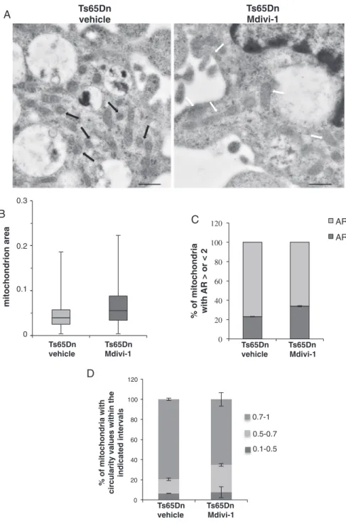
To give more information of Mdivi-1 effect on mitochondrial mor-phology and dimensions, ultrastructural analysis was performed in Mdivi-1-treated Ts65Dn cells. Mitochondrial area and the percentage of mitochondria sections with AR > 2 were notably increased following Mdiv-1 treatment while mitochondria with a circularity value > 0.7 decreased (Fig. 5 andTable 1), which restores a wt-like condition of mitochondrial area and structure (see alsoFig. 2A andTable 1).
Altogether, these results showed that inhibition of mitochondrial fission by Mdivi-1 resulted in an improvement of mitochondrial 0 20 40 60 80 100 120
wt Ts65Dn
% of mitochondria withcircularity values within the
indicated intervals 0 20 40 60 80 100 120
% of mitochondria
with
AR > or < 2
AR<2 AR>2mitochondrion area
B
C
0 0.1 0.2 0.3wt Ts65Dn
wt Ts65Dn
wt
Ts65Dn
A
D
0.7-1 0.5-0.7 0.1-0.5Fig. 2. Ultrastructure analysis reveals mitochondria fragmenta-tion in Ts65Dn-NPCs. (A) Representative images of mitochon-dria visualized by transmission electron microscopy in a wt and Ts65Dn cell. Scale bars correspond to 0.6μm. White arrows indicate representative elongated mitochondria and black ar-rows highlight representative spheroid mitochondria. (B) Box plot shows the distribution of mitochondria section area in Ts65Dn and wt cells; horizontal lines in the box plot indicate the mean values. (C) Histogram depicts the percentage of mi-tochondria with AR values > of 2 (i.e. elongated) and < 2 (i.e. near a roundish shape) in Ts65Dn and control cells. (D) Histogram depicts the percentage of mitochondria with circu-larity values included in the intervals 0.1–0.5; 0.5–0.7; 0.7–1 (a perfect circle shows a circularity value of 1). Data result from the analysis of a minimal number of 100 mitochondria sections from at least 10 individual cells from three independent samples for each NPC genotype (wt and Ts65Dn). For significant differ-ences and statistical analysis please refer toTable 1.
Table 1
Data from the ultrastructural analysis performed on mitochondria from wild-type and Ts65Dn NPCs or on mitochondria from Ts65Dn NPCs treated either with 10μM Mdivi-1 (Ts65Dn NPCs + Mdiv-1) or DMSO (Ts65Dn NPCs + vehicle).
Wild-type NPCs Ts65Dn NPCs Ts65Dn NPCs + vehicle Ts65Dn NPCs + Mdivi-1
Area Mean surface area (square pixels) 0.076 0.050 0.048 0.069
SD between single values for cell type 0.0552 0.043 0.0378 0.0486
SD between mean values of independent samples 0.0005 0.0048 0.0044 0.0122
P value between mean values of single samples from each cell type 0.048 0.05
P value between all recorded values for cell type 1.21E-09 2.30E-09
AR Mean area ratio values 2.06 1.85 1.77 1.95
SD between single values for cell type 0.9246 0.9909 0.8925 0.9216
SD between mean values of independent samples 0.058 0.027 0.003 0.1
P value between mean values of single samples from each cell type 0.06 0.08
P value between all recorded values for cell type 0.01 0.01
Circularity Mean circularity values 0.73 0.79 0.81 0.76
SD between single values for cell type 0.1782 0.1692 0.1490 0.1602
SD between mean values of independent samples 0.0039 0.0019 0.0032 0.0258
P value between mean values of single samples from each cell type 0.01 0.08
P value between all recorded values for cell type 7.92E-05 0.01
Cells were processed as described in the legends ofFigs. 2 and 5. Mitochondrial area (expressed in square pixels), AR (the ratio between major and minor axe) and the circularity (4π × [area/(perimeter)2]) were recorded for each mitochondrion section by using the image J software. Statistical analysis was performed by Student's t-test analysis evaluated as follow: i) the P value between all recorded values for cell type is calculated considering the single values of all mitochondrial section recorded in wt NPCs vs Ts65Dn NPCs or Ts65Dn NPCs + vehicle vs Ts65Dn NPCs + Mdivi-1; ii) the P value between all recorded values for cell type is calculated considering the mean values of the three independent experiments in wt NPCs vs Ts65Dn NPCs or Ts65Dn NPCs + vehicle vs Ts65Dn NPCs + Mdivi-1. Statistically significant values are indicated in bold.
0 20 40 60 80 100 120 140 160 Protein content (% of wt) wt Ts65Dn Drp1 (81 kDa) actin
A
P-Drp1 RCAN1 (28 kDa) P-DRP1/DRP1 wt Ts65Dn Mnf2 (86 kDa) actin wt Ts65Dn wt Ts65Dn 0 0.2 0.4 0.6 0.8 1.0 1.2**
**
**
**
B
C
D
0 20 40 60 80 100 120 Protein content (% of wt) wt Ts65Dn*
E
F
S-Opa1 L-Opa1 wt Ts65Dn actin 0 20 40 60 80 100 120 wt Ts65Dn S-Opa1 L-Opa1 % of ADU Mnf2Drp1
P-Drp1
RCAN1
Fig. 3. Levels of proteins involved in the mitochondrialfission and fusion process.(A) Representative immunoblot and (B) densitomentric analysis of Drp1, Drp1 phosphorylation at S637 (P-DRP1) and RCAN1 protein levels measured in cell extracts (0.05 mg protein) of wt and Ts65Dn NPCs. (C) Histrogram shows the ratio between P-Drp1 and Drp1. (D) Representative immunoblot and densitomentric analysis of Mnf2 protein levels measured in cell extracts (0.05 mg protein) of wt and Ts65Dn NPCs. (E) Representative immunoblot of long (L-Opa1) and short (S-Opa1) forms of Opa1 proteins measured in cell extracts (0.05 mg protein) of wt and Ts65Dn NPCs. (F) The histogram represents the percentage of arbitrary densitometric unit (ADU) of L and S forms of Opa1 in each lane. Data are mean values ( ± SD) of two independent measurements carried out in three independent samples for each NPC genotypes (wt and Ts65Dn) and expressed as percentage of wt. Significant differences, calculated with Student's t-test, are indicated with asterisks (** = P < 0.01; * = P < 0.05).
network organization in Ts65Dn cells.
3.3. Mdivi-1 treatment counteracts mitochondrial dysfunctions, restores cell energy deficit and enhances in vitro neurogenesis of Ts65Dn NPCs
To test whether inhibition of mitochondrialfission by Mdivi-1 could counteract the deficit of OXPHOS capacity found in Ts65Dn NPCs[18], we measured mitochondrial ATP production and ATPase activity in cultured NPCs in the presence of 10μM Mdivi-1 or vehicle (DMSO), for 24 h.
As shown inFig. 6, exposure of Ts65Dn cells to Mdivi-1 prevented both the impairment of mitochondrial ATP production via OXPHOS (Fig. 6A) and the deficit of ATPase activity in Ts65Dn cells (Fig. 6B).
Consequently, measurement of the cellular ATP levels demonstrated that Mdivi-1 treatment significantly prevents the strong reduction of cellular ATP pool (64.8% ± 5 respect to wt, P < 0.01) and confers to Ts65Dn cells the capability to maintain the cellular ATP content
comparable to that of wt cells (Fig. 6C).
Next, we investigated whether improvement of mitochondrial dy-namics and bioenergetics could affect Ts65Dn cell proliferation and differentiation, a measure of neurogenesis in vitro [45]. We first de-termined NPC proliferative capacity in untreated and Mdivi-1-treated Ts65Dn cells by BrdU incorporation experiments. As shown inFig. 6D, Ts65Dn NPCs showed about 40% reduction of proliferation as com-pared to wt cultures. Mdivi-1 treatment significantly promotes pro-liferation capacity in Ts65Dn NPCs, making them able to divide at the same level of wt cells. We next evaluated the Mivi-1 effect on Ts65Dn NPC differentiation. A statistically significant increase in DCX-positive NPCs, a marker for newly generated hippocampal neurons[46], was found in the presence of 10μM Mdivi-1 under differentiation condition (Fig. 6F; 13% ± 5 and 28% ± 7 DCX-positive neurons in untreated and Mdivi-1-treated Ts65Dn cells, respectively; P < 0.01). Overall, these data indicated enhanced proliferation and differentiation of Ts65Dn hippocampal cells following Mdivi-1 treatment.
Ts65Dn
vehicle
Ts65Dn
Mdivi-1
Drp1 Ts65Dn Ts65Dn vehicle Mdivi-1 p-Drp1 actinA
B
10 µm 10 µm 0 20 40 60 80 100 120 140 160 DRP1 P-DRP1 Protein content (% of wt) Ts65Dn, vehicle Ts65Dn, Mdivi-1 0 10 20 30 40 50 60 Ts65Dn f index vehicle Mdivi-1 Ts65Dn T ubularity vehicle Mdiv-1 0.00 0.05 0.10 0.15 0.20 0.25**
**
C
D
Fig. 4. Mdivi-1 has no effect on Drp1 protein expression and phosphorylation but improves mitochondrial network organization. Ts65Dn NPCs were incubated either with 10μM Mdivi-1 (Ts65Dn Mdivi-1) or vehicle (0.05% DMSO) (Ts65Dn vehicle) for 24 h. (A) Representative immunoblot and (B) densitomentric analysis of Drp1 and Drp1 phosphorylation at S637 (P-Drp1) protein levels measured in cell extracts (0.05 mg protein) of NPCs treated and untreated Ts65Dn cells. Data are mean values ( ± SD) of three in-dependent experiments and expressed as per-centage of wt. Differences between Ts65Dn ve-hicle and Ts65Dn NPCs treated with Mdivi-1 are not significant (P > 0.05). (B) Representative images obtained by confocal microscopy of cul-tured Ts65Dn NPCs treated with Mdivi-1 or ve-hicle loaded with 0.5μM MitoTracker®deepRed. The panel inside shows a higher magnification of the image in the white box. (C) Quantification of the fragmentation index (f index) and (D) the tubularity of mitochondrial networks in un-treated and Mdivi-1-un-treated Ts65Dn NPCs. Data are the mean values ( ± SD) of the analyses performed in 20 Ts65Dn cells plus vehicle and 27 Ts65Dn cells plus Mdivi-1. Significant differ-ences, calculated with Student's t-test, are in-dicated with asterisks (** = P < 0.01).
4. Discussion
In this study we provide evidence that an increase of Drp1-depen-dent mitochondriafission occurs during the proliferation of cultured adult hippocampal progenitors from Ts65Dn mouse model of DS. In these cells, mitochondrial fission is associated with an increase in protein level and dephosphorylation of Drp1, a decrease of Mnf2 and a conversion of L-Opa1 into S-Opa1. Interestingly, the treatment of these cells with Mdivi-1, an inhibitor of Drp1, decreases mitochondrial fis-sion, restores mitochondrial network, prevents mitochondrial dysfunc-tions and stimulates NPC proliferation and neural differentiation. Our results provide new insight on the origin of mitochondrial dysfunction in DS, shed light on how mitochondrial dysfunctions could contribute to impaired adult neurogenesis in DS, and pave the way for the use of new therapeutic drugs in managing some energy deficit-associated DS clin-ical manifestations.
Neurons critically depend on mitochondrial structural network
organization and bioenergetics to execute the complex processes of neurogenesis, neurotransmission and synaptic plasticity [4,5,47,48]. Indeed, alteration of mitochondrial dynamics strongly affects neuronal progenitor proliferation and neuronal function, survival and differ-entiation in several models of neurodegenerative diseases (for refs see [49]). Mitochondrial morphology, size and position within cells are maintained through a balance offission and fusion events. Perturbation of the steady state between these opposing processes has been directly implicated in several human disorders[50].
A reduction in fusion process has been recently described in per-ipheral DS fibroblasts [15]. Consistently, we found in NPCs from Ts65Dn mice a reduction in mitochondrial fusion due to the down-regulation of Mnf2 protein level and, increased processing of Opa1 into short Opa1 form, both known to be involved in promoting mitochon-drial fragmentation[39,40]. Given that PGC-1α has been reported to promote the expression of the Mnf2[15,51], the reduced protein levels of Mnf2 in Ts65Dn NPCs may be linked to the PGC-1α down-regulation 0 20 40 60 80 100 120 % of mitochondria with AR > or < 2 AR<2 AR>2 mitochondrion area
A
B
C
Ts65Dn
vehicle
Ts65Dn
Mdivi-1
0 0.1 0.2 0.3 Ts65Dn Ts65Dn vehicle Mdivi-1 Ts65Dn Ts65Dn vehicle Mdivi-1D
0 20 40 60 80 100 120 % of mitochondria withcircularity values within the
indicated intervals 0.7-1 0.5-0.7 0.1-0.5 Ts65Dn Ts65Dn vehicle Mdivi-1
Fig. 5. Mdivi-1 inhibits mitochondrial fragmentation. Ts65Dn NPCs were incubated either with 10μM Mdivi-1 (Ts65Dn Mdivi-1) or vehicle (0.02% DMSO) (Ts65Dn ve-hicle) for 24 h andfixed with glutaraldehyde and ma-nipulated as described inMaterial and Methods(A) Re-presentative images of mitochondria visualized by Trasmission Electron Microscopy. Scale bars correspond to 0.6μm. Black arrows indicate representative spheroid tochondria, white arrows representative elongated tochondria. (B) Box plot shows the distribution of mi-tochondria section area in untreated or Mdiv-1-treated Ts65Dn cells; horizontal lines in the box plot indicate the mean values. (C) Histogram depicts the percentage of mi-tochondria with AR values > of 2 (i.e. elongated) and < 2 (i.e. near a roundish shape) in untreated or Mdiv-1-treated Ts65Dn NPCs. (D) Histogram depicts the percentage of mitochondria with circularity values included in the in-tervals 0.1–0.5; 0.5–0.7; 0.7–1 (a perfect circle shows a circularity value of 1). Data result from the analysis of a minimal number of 100 mitochondria sections from at least 10 individual cells for each sample in three independent experiments. For significant differences and statistical analysis please refer toTable 1.
that we previously demonstrated in these cells[18].
In neuronal progenitor cells from trisomic Ts65Dn mice we also found a shift of the balancefission/fusion toward a higher fission with an up-regulation of Drp1 expression and activity. Imbalance of the only Drp1 protein is sufficient to produce excessive fission that leads to mitochondrial dysfunction[52]. For thefission event, Drp1 translocates from the cytoplasm to mitochondria where it forms an oligomeric structure that induces fragmentation of mitochondrial membranes[53]. We confirmed a high fission process in Ts65Dn NPCs by both ultra-structural evaluation of mitochondrial area and shape, as well as by mitochondrial network visualization by confocal microscopy. Our data are in agreement with recent studies showing that an up-regulation of Drp1-mediated mitochondrialfission is commonly observed in various neurological disorders (for refs see[54]) together with a decrease in calcineurin-dependent dephosphorylation of Drp1 at S637, which reg-ulates Drp1 translocation from cytoplasm to mitochondria[41,42]. Our results, showing an up-regulation of Drp1 protein content and a de-crease of S637 phosphorylation, could account for higher translocation
of Drp1 from cytosol to mitochondria in Ts65Dn NPCs with respect to wt cells, thus providing further explanation for the alteration in shape and network. It is known that PKA-dependent phosphorylation of S637 exerts the opposing effect, i.e. maintains Drp1 in the cytosol and in-hibitsfission[55]. Thus, it could be that calcineurin-mediated depho-sphorylation of Drp1 prevails over PKA-dependent phodepho-sphorylation. This is consistent with thefinding that a down-regulation of PKA ac-tivity and cAMP signalling occurs in hippocampus of Ts65Dn mice[56] and DS cells [9]. Interestingly, we confirmed that, as previously re-ported[28], the chromosome 21 gene RCAN1, the regulator of calci-neurin 1, is overexpressed in Ts65Dn cells. This suggests that RCAN1 overexpression in DS could account for Drp1 up-regulation and the consequent increase in mitochondrialfission.
It has been reported that shifting the balance of mitochondrial morphology toward fission enhances susceptibility to mitochondrial dysfunctions, oxidative stress and cell death; conversely, fused mi-tochondria are energetically more active, preserve cell functions, and can better tolerate oxidative stress [57]. Recently, Khacho and co-0 20 40 60 80 V o (nmol/min x mg protein)
Mitochondria ATP production
0 5 10 15 20 25 V o (nmol/min x mg protein)
ATPase activity
0 2 4 6 8 10 12 A TPcontent (nmol /mg protein)
Cellular ATP
A
B
C
BrdU incorporation (OD 450/595)NPCs proliferation
D
wt Ts65Dn Ts65Dn vehicle Mdivi1 wt Ts65Dn Ts65Dn vehicle Mdivi1 wt Ts65Dn Ts65Dn vehicle Mdivi1 0.0 0.2 0.4 0.6 0.8 1.0 wt Ts65Dn Ts65Dn vehicle Mdivi1"
E
wt Ts65Dn Ts65Dn vehicle Mdivi1 0 5 10 15 20 25 30 35 40 % of DCX-positive cellsNPCs differentiation
**
**
**
**
**
Fig. 6. Mdivi-1 rescues the deficit of mitochondrial bioenergetics and enhances proliferation and neural differentiation of Ts65Dn NPCs. Ts65Dn NPCs were incubated either with 10μM Mdivi-1 (Ts65Dn Mdivi-1) or vehicle (0.05% DMSO) (Ts65Dn vehicle) for 24 h. wt NPCs were also used as control (wt). (A) The rate of mitochondrial ATP production via OXPHOS was measured spectrophotometrically in 0.3 mg of protein in the presence of the respiratory substrate succinate (5 mM), as described inMaterial and Methods. ATP production was expressed as nmol/min × mg protein. (B) The ATPase activity was measured spectrophotometrically in mitochon-drial membrane enriched fractions (0.3 mg protein) as described underMaterials and Methodsand ex-pressed as nmol/min × mg protein. (C) ATP cellular content was measuredfluorimetrically in cell extract as described underMaterials and Methodsand ex-pressed as nmol/min × mg protein. (D) NPC pro-liferation was analyzed by BrdU incorporation. Data are represented as relative levels of BrdU in-corporation in wt and untreated (vehicle) and treated (Mdivi-1) Ts65Dn NPCs. Results are mean values of optic density (OD) at the measure-re-ference wavelengths of 450–595 nm obtained from ELISA assay experiments. (E) Ts65Dn NPCs were treated with Mdivi-1 or vehicle under differentiation conditions (seeMaterials and Methods). Expression of DCX indicates neuronal differentiation. Data in each panel represent the mean value ( ± SD) of at least three independent experiments and sig-nificant differences, calculated with one-way ANOVA and Bonferroni test, are indicated as follow: wt NPCs vs Ts65Dn NPCs, ** = P < 0.01; Ts65Dn NPCs vs Ts65Dn NPCs treated with Mdivi-1, αα = P < 0.01.
workers [58] have also demonstrated that increased mitochondrial fission is associated with defective neural stem cell proliferation and that mitochondrial dynamics is an upstream regulator of essential me-chanisms governing neural stem cell self-renewal and differentiation.
Given the involvement of Drp1 in mitochondrialfission, and the defective hippocampal neurogenesis showed in Ts65Dn NPCs[16,17], we hypothesized that blocking Drp1 and the Drp1-dependent excessive fission, might, by one hand, give new information on the importance of mitochondrial dynamics in the regulation of adult neurogenesis in DS and, by the other, represent a new attractive strategy to correct DS-associated clinical phenotypes linked to energy deficit and defective neurogenesis. The effects of Mdivi-1 on the excessive fragmentation of mitochondria, the bioenergetics impairment and the altered neuro-genesis, have proved, at least in part, our hypothesis. Mdivi-1 is a small molecule derivative of quinazolinone, which acts mainly as a selective inhibitor of the mitochondrial fission protein Drp1 through blocking Drp1 self-assembly and GTP hydrolysis [43]. Indeed, we found that Mdivi-1-dependent restoring of mitochondrial network organization counteracts mitochondrial bioenergetics impairment and, strikingly, promotes both in vitro proliferation and differentiation of hippocampal neural precursor cells. Our results are consistent with several other studies in which it has been demonstrated that mitochondrialfission is required for hippocampal neuronal proliferation and differentiation [58,59]and that Mdivi-1, by blocking the mitochondrialfission, exerts neuroprotective effects in animal models of various neurodegenerative disorders[30,32,60–62].
In conclusion, the present study provides new information on the molecular mechanisms responsible for energy deficit and altered mi-tochondrial bioenergetics and dynamics in DS. Moreover it demon-strates that alteration of mitochondrial network is closely linked to the impaired mitochondrial bioenergetics and in vitro hippocampal neuro-genesis. Our conclusion is consistent with a recent study by Izzo and co-workers[15]showing mitochondrial fragmentation in DS human fetal fibroblasts and the potential of metformin, a drugs able to target mi-tochondria [5]and to promote mouse adult neurogenesis and spatial memory formation [63], in counteracting both mitochondrial frag-mentation and dysfunctions. The specific inhibition of Drp1-dependent mitochondrial fragmentation by Mdivi-1 and the consequent effect on hippocampal neural progenitor cell proliferation and differentiation, provide a further evidence of the biological role of mitochondria in neurogenesis and bring insights on how the promotion of mitochondrial dynamics can contribute to the therapeutic treatment of Down syn-drome.
Transparency document
The http://dx.doi.org/10.1016/j.bbadis.2017.09.014 associated with this article can be found, in online version.
Acknowledgment
This study was partially supported by a grant from Fondation Jerome Lejeune (VACCA/1093-VR2012B). We are particularly grateful to people that very kindly contributed to support in part this research by individual donations, in particular to “Associazione Progetto 21-Onlus” and “A.M.A.R. Down-21-Onlus”. We thank Dr. Andrea Contestabile for the kind gift of the cellular lines used in this study.
Disclosure
None of the authors declaresfinancial interests or potential conflict of interests.
References
[1] S.E. Antonarakis, Down syndrome and the complexity of genome dosage imbalance,
Nat. Rev. Genet. 18 (2017) 147–163.
[2] M. Kazemi, M. Salehi, M. Kheirollahi, Down syndrome: current status, challenges
and future perspectives, Int. J. Mol. Cell. Med. 5 (2016) 125–133.
[3] E. Head, I.T. Lott, D.M. Wilcock, C.A. Lemere, Aging in Down syndrome and the
development of Alzheimer's disease neuropathology, Curr. Alzheimer Res. 13 (2016) 18–29.
[4] D. Valenti, L. de Bari, B. De Filippis, A. Henrion-Caude, R.A. Vacca, Mitochondrial
dysfunction as a central actor in intellectual disability-related diseases: an overview of Down syndrome, autism, Fragile X and Rett syndrome, Neurosci. Biobehav. Rev. 2 (2014) 202–217.
[5] D. Valenti, N. Braidy, D. De Rasmo, A. Signorile, L. Rossi, A.G. Atanasov, M. Volpicella, A. Henrion-Caude, S.M. Nabavi, R.A. Vacca, Mitochondria as phar-macological targets in Down syndrome, Free Radic. Biol. Med. (2017),http://dx. doi.org/10.1016/j.freeradbiomed.2017.08.01.
[6] B. De Filippis, D. Valenti, L. de Bari, D. De Rasmo, M. Musto, A. Fabbri, L. Ricceri,
C. Fiorentini, G. Laviola, R.A. Vacca, Mitochondrial free radical overproduction due to respiratory chain impairment in the brain of a mouse model of Rett syndrome: protective effect of CNF1, Free Radic. Biol. Med. 83 (2015) 167–177.
[7] M. Golpich, E. Amini, Z. Mohamed, R. Azman Ali, N. Mohamed Ibrahim,
A. Ahmadiani, Mitochondrial dysfunction and biogenesis in neurodegenerative diseases: pathogenesis and treatment, CNS Neurosci. Ther. 23 (2017) 5–22.
[8] D. Valenti, A. Tullo, M.F. Caratozzolo, R.S. Merafina, P. Scartezzini, E. Marra,
R.A. Vacca, Impairment of F1F0-ATPase, adenine nucleotide translocator and adenylate kinase causes mitochondrial energy deficit in human skin fibroblasts with chromosome 21 trisomy, Biochem. J. 431 (2010) 299–310.
[9] D. Valenti, G.A. Manente, L. Moro, E. Marra, R.A. Vacca, Deficit of complex I
ac-tivity in human skinfibroblasts with chromosome 21 trisomy and overproduction of
reactive oxygen species by mitochondria: involvement of the cAMP/PKA signalling pathway, Biochem. J. 435 (2011) 679–688.
[10] D. Valenti, De D. Rasmo, A. Signorile, L. Rossi, L. de Bari, I. Scala, B. Granese,
S. Papa, R.A., Vacca, epigallocatechin-3-gallate prevents oxidative phosphorylation deficit and promotes mitochondrial biogenesis in human cells from subjects with Down's syndrome, Biochim. Biophys. Acta 1832 (2013) 542–552.
[11] R.A. Vacca, D. Valenti, Green tea EGCG plusfish oil omega-3 dietary supplements
rescue mitochondrial dysfunctions and are safe in a Down's syndrome child, Clin. Nutr. 34 (2015) 783–784.
[12] C. Piccoli, A. Izzo, R. Scrima, F. Bonfiglio, R. Manco, R. Negri, G. Quarato, O. Cela,
M. Ripoli, M. Prisco, F. Gentile, G. Calì, P. Pinton, A. Conti, L. Nitsch, N. Capitanio, Chronic pro-oxidative state and mitochondrial dysfunctions are more prounounced
infibroblasts from Down syndrome foeti with congenital heart defects, Hum. Mol.
Genet. 22 (2013) 1218–1232.
[13] E.A. Shukkur, A. Shimohata, T. Akagi, W. Yu, M. Yamaguchi, M. Murayama,
D. Chui, T. Takeuchi, K. Amano, K.H. Subramhanya, T. Hashikawa, H. Sago, C.J. Epstein, A. Takashima, K. Yamakawa, Mitochondrial dysfunction and tau hy-perphosphorylation in Ts1Cje, a mouse model for Down syndrome, Hum. Mol. Genet. 15 (2006) 2752–2762.
[14] J. Busciglio, A. Pelsman, C. Wong, G. Pigino, M. Yuan, H. Mori, B.A. Yankner,
Altered metabolism of the amyloid beta precursor protein is associated with mi-tochondrial dysfunction in Down's syndrome, Neuron 33 (2002) 677–688.
[15] A. Izzo, M. Nitti, N. Mollo, S. Paladino, C. Procaccini, D. Faicchia, G. Calì,
R. Genesio, F. Bonfiglio, R. Cicatiello, E. Polishchuk, R. Polishchuk, P. Pinton, G. Matarese, A. Conti, L. Nitsch, Metformin restores the mitochondrial network and reverses mitochondrial dysfunction in Down syndrome cells, Hum. Mol. Genet. 26 (2017) 1056–1069.
[16] R.H. Reeves, N.G. Irving, T.H. Moran, A. Wohn, C. Kitt, S.S. Sisodia, C. Schmidt,
R.T. Bronson, M.T. Davisson, A mouse model for Down syndrome exhibits learning and behavior deficits, Nat. Genet. 11 (1995) 177–184.
[17] A. Contestabile, B. Greco, D. Ghezzi, V. Tucci, F. Benfenati, L. Gasparini, Lithium
rescues synaptic plasticity and memory in Down syndrome mice, J. Clin. Invest. 123 (2013) 348–361.
[18] D. Valenti, L. de Bari, D. de Rasmo, A. Signorile, A. Henrion-Caude, A. Contestabile,
R.A. Vacca, The polyphenols resveratrol and epigallocatechin-3-gallate restore the severe impairment of mitochondria in hippocampal progenitor cells from a Down syndrome mouse model, Biochim. Biophys. Acta 1862 (2016) 1093–1104.
[19] R.A. Vacca, D. Valenti, S. Caccamese, M. Daglia, N. Braidy, S.M. Nabavi, Plant
polyphenols as natural drugs for the management of Down syndrome and related disorders, Neurosci. Biobehav. Rev. 71 (2016) 865–877.
[20] G. Benard, N. Bellance, D. James, P. Parrone, H. Fernandez, T. Letellier,
R. Rossignol, Mitochondrial bioenergetics and structural network organization, J. Cell Sci. 120 (2007) 838–848.
[21] P. Mishra, D.C. Chan, Metabolic regulation of mitochondrial dynamics, J. Cell Biol.
212 (2016) 379–387.
[22] N. Birsa, R. Norkett, N. Higgs, G. Lopez-Domenech, J.T. Kittler, Mitochondrial
trafficking in neurons and the role of the Miro family of GTPase proteins, Biochem. Soc. Trans. 41 (2013) 1525–1531.
[23] S.B. Berman, F.J. Pineda, J.M. Hardwick, Mitochondrialfission and fusion
dy-namics: the long and short of it, Cell Death Differ. 15 (2008) 1147–1152.
[24] H. Lee, Y. Yoon, Mitochondrialfission and fusion, Biochem. Soc. Trans. 44 (2016)
1725–1735.
[25] A.M. Bertholet, T. Delerue, A.M. Millet, M.F. Moulis, C. David, M. Daloyau,
L. Arnauné-Pelloquin, N. Davezac, V. Mils, M.C. Miquel, M. Rojo, P. Belenguer, Mitochondrial fusion/fission dynamics in neurodegeneration and neuronal plasti-city, Neurobiol. Dis. 90 (2016) 3–19.
[26] H. Wong, J. Levenga, P. Cain, B. Rothermel, E. Klann, C. Hoeffer, RCAN1
over-expression promotes age-dependent mitochondrial dysregulation related to neuro-degeneration in Alzheimer's disease, Acta Neuropathol. 130 (2015) 829–843.
[27] A. Patel, N. Yamashita, M. Ascaño, D. Bodmer, E. Boehm, C. Bodkin-Clarke, Y.K. Ryu, R. Kuruvilla, RCAN1 links impaired neurotrophin trafficking to aberrant development of the sympathetic nervous system in Down syndrome, Nat. Commun. 6 (2015) 10119.
[28] M. Rachidi, C. Lopes, Mental retardation and associated neurological dysfunctions
in Down syndrome: a consequence of dysregulation in critical chromosome 21 genes and associated molecular pathways, Eur. J. Paediatr. Neurol. 12 (2008) 168–182.
[29] L.L. Lackner, J. Nunnari, Small molecule inhibitors of mitochondrial division: tools
that translate basic biological research into medicine, Chem. Biol. 17 (2010) 578–583.
[30] Q. Wu, S.X. Xia, Q.Q. Li, Y. Gao, X. Shen, L. Ma, M.Y. Zhang, T. Wang, Y.S. Li,
Z.F. Wang, C.L. Luo, L.Y. Tao, Mitochondrial division inhibitor 1 (Mdivi-1) offers neuroprotection through diminishing cell death and improving functional outcome in a mouse model of traumatic brain injury, Brain Res. 1630 (2016) 134–143.
[31] Y. Li, P. Wang, J. Wei, R. Fan, Y. Zuo, M. Shi, H. Wu, M. Zhou, J. Lin, M. Wu,
X. Fang, Z. Huang, Inhibition of Drp1 by Mdivi-1 attenuates cerebral ischemic in-jury via inhibition of the mitochondria-dependent apoptotic pathway after cardiac arrest, Neuroscience 311 (2015) 67–74.
[32] N. Zhang, S. Wang, Y. Li, L. Che, Q. Zhao, A selective inhibitor of Drp1, mdivi-1,
acts against cerebral ischemia/reperfusion injury via an anti-apoptotic pathway in rats, Neurosci. Lett. 535 (2013) 104–109.
[33] S. Agarwal, A. Yadav, S.K. Tiwari, B. Seth, L.K. Chauhan, P. Khare, R.S. Ray,
R.K. Chaturvedi, Dynamin-related protein 1 inhibition mitigates bisphenol A-mediated alterations in mitochondrial dynamics and neural stem cell proliferation and differentiation, J. Biol. Chem. 291 (2016) 15923–15939.
[34] T.D. Fischer, M.J. Hylin, J. Zhao, A.N. Moore, M.N. Waxham, P.K. Dash, Altered
mitochondrial dynamics and TBI pathophysiology, Front. Syst. Neurosci. 10 (2016) 29.
[35] H. Babu, J.H. Claasen, S. Kannan, A.E. Rünker, T. Palmer, G. Kempermann, A
protocol for isolation and enriched monolayer cultivation of neural precursor cells from mouse dentate gyrus, Front. Neurosci. 5 (2011) 89.
[36] J. Vowinckel, J. Hartl, R. Butler, M. Ralser, MitoLoc: a method for the simultaneous
quantification of mitochondrial network morphology and membrane potential in single cells, Mitochondrion 24 (2015) 77–86.
[37] M.D. Abramoff, P.J. Magelhaes, S.J. Ram, Image processing with ImageJ, Biophot.
11 (2004) 36–42.
[38] N.C. Yang, W.M. Ho, Y.H. Chen, M.L. Hu, A convenient one-step extraction of
cellular ATP using boiling water for the luciferin-luciferase assay of ATP, Anal. Biochem. 306 (2002) 323–327.
[39] T. MacVicar, T. Langer, OPA1 processing in cell death and disease - the long and
short of it, Thomas, J. Cell Sci. 129 (2016) 2297–2306.
[40] A. Signorile, A. Santeramo, G. Tamma, T. Pellegrino, S. D'Oria, P. Lattanzio, D. De
Rasmo, Mitochondrial cAMP prevents apoptosis modulating Sirt3 protein level and OPA1 processing in cardiac myoblast cells, Biochim. Biophys. Acta 1864 (2017) 355–366.
[41] A. Jahani-Asl, R.S. Slack, The phosphorylation state of Drp1 determines cell fate,
EMBO Rep. 8 (2007) 912–913.
[42] G.M. Cereghetti, A. Stangherlin, O. Martins de Brito, C.R. Chang, C. Blackstone,
P. Bernardi, L. Scorrano, Dephosphorylation by calcineurin regulates translocation of Drp1 to mitochondria, Proc. Natl. Acad. Sci. U. S. A. 105 (2008) 1508–15803.
[43] A. Tanaka, R.J. Youle, A chemical inhibitor of DRP1 uncouples mitochondrial
fis-sion and apoptosis, Mol. Cell 29 (2008) 409–410.
[44] J.M. Liu, Z. Yi, S.Z. Liu, J.H. Chang, X.B. Dang, Q.Y. Li, Y.L. Zhang, The
mi-tochondrial division inhibitor mdivi-1 attenuates spinal cord ischemia-reperfusion
injury both in vitro and in vivo: involvement of BK channels, Brain Res. 1619 (2015) 155–165.
[45] R. Lin, L. Iacovitti, Classic and novel stem cell niches in brain homeostasis and
repair, Brain Res. 1628 (2015) 327–342.
[46] M.S. Rao, A.K. Shetty, Efficacy of doublecortin as a marker to analyse the absolute
number and dendritic growth of newly generated neurons in the adult dentate gyrus, Eur. J. Neurosci. 19 (2004) 234–246.
[47] M.P. Mattson, M. Gleichmann, A. Cheng, Mitochondria in neuroplasticity and
neurological disorders, Neuron 60 (2008) 748–766.
[48] A. Cheng, Y. Hou, M.P. Mattson, Mitochondria and neuroplasticity, ASN Neuro 2
(2010) e00045.
[49] K.H. Flippo, S. Strack, Mitochondrial dynamics in neuronal injury, development and
plasticity, J. Cell Sci. 130 (2017) 671–681.
[50] D.C. Chan, Mitochondria: dynamic organelles in disease, aging and development,
Cell 125 (2006) 1241–1252.
[51] M. Liesa, B. Borda-d'Agua, G. Medina-Gómez, C.J. Lelliott, J.C. Paz, M. Rojo,
M. Palacín, A. Vidal-Puig, A. Zorzano, Mitochondrial fusion is increased by the nuclear coactivator PGC-1 beta, PLoS One 3 (2008) e3613.
[52] L. Lin, M. Zhang, R. Yan, H. Shan, J. Diao, J. Wei, Inhibition of Drp1 attenuates
mitochondrial damage and myocardial injury in Coxsackievirus B3 induced myo-carditis, Biochem. Biophys. Res. Commun. 484 (2017) 550–556.
[53] N.H. Fukushima, E. Brisch, B.R. Keegan, W. Bleazard, J.M. Shaw, The GTPase
ef-fector domain sequence of the Dnm1p GTPase regulates self-assembly and controls
a rate-limiting step in mitochondrialfission, Mol. Biol. Cell 12 (2001) 2756–2766.
[54] Q. Wu, C.L. Luo, L.Y. Tao, Dynamin-related protein 1 (Drp1) mediating mitophagy
contributes to the pathophysiology of nervous system diseases and brain injury, Histol. Histopathol. 10 (2016) 11841.
[55] H.W. Hyun, S.J. Min, J.E. Kim, CDK5 inhibitors prevent astroglial apoptosis and
reactive astrogliosis by regulating PKA and DRP1 phosphorylations in the rat hip-pocampus, Neurosci. Res. 119 (2017) 24–37.
[56] M. Dierssen, I.F. Vallina, C. Baamonde, S. García-Calatayud, M.A. Lumbreras,
J. Flórez, Alterations of central noradrenergic transmission in Ts65Dn mouse, a model for Down syndrome, Brain Res. 749 (1997) 238–244.
[57] S.B. Ong, S. Subrayan, S.Y.D.M. Yellon Lim, S.M. Davidson, D.J. Hausenloy,
Inhibiting mitochondrialfission protects the heart against ischemia/reperfusion
injury, Circulation 121 (2010) (2012–2022).
[58] M. Khacho, A. Clark, D.S. Svoboda, J. Azzi, J.G. MacLaurin, C. Meghaizel, H. Sesaki,
D.C. Lagace, M. Germain, M.E. Harper, D.S. Park, R.S. Slack, Mitochondrial dy-namics impacts stem cell identity and fate decisions by regulating a nuclear tran-scriptional program, Cell Stem Cell 19 (2016) 232–247.
[59] K. Steib, I. Schaffner, R. Jagasia, B. Ebert, D.C. Lie, Mitochondria modify
exercise-induced development of stem cell-derived neurons in the adult brain, J. Neurosci. 34 (2014) 6624–6633.
[60] A.A.K. Rosdah, J. Holien, L.M. Delbridge, G.J. Dusting, S.Y. Lim, Mitochondrial
fission - a drug target for cytoprotection or cytodestruction? Pharmacol. Res. Perspect. 4 (2016) e00235.
[61] M. Cui, H. Ding, F. Chen, Y. Zhao, Q. Yang, Q. Dong, Mdivi-1 protects against
ischemic brain injury via elevating extracellular adenosine in a cAMP/CREB-CD39-dependent manner, Mol. Neurobiol. 53 (2016) 240–253.
[62] J.M. Liu, Z. Yi, S.Z. Liu, J.H. Chang, X.B. Dang, Q.Y. Li, Y.L. Zhang, The
mi-tochondrial division inhibitor mdivi-1 attenuates spinal cord ischemia-reperfusion injury both in vitro and in vivo: involvement of BK channels, Brain Res. 1619 (2015) 155–165.
[63] M.B. Potts, D.A. Lim, An old drug for new ideas: metformin promotes adult