©2016 Institute of Parasitology, SAS, Košice DOI 10.1515/helmin-2016-0027
HELMINTHOLOGIA, 53, 3: 276 – 280, 2016
Case Report
Crenosoma vulpis infection in a four-month old puppy
B. MATOS1, V. COLELLA2, A. M. ALHO1*, D. OTRANTO2,R. DOYLE3, L. MADEIRA DE CARVALHO1
1CIISA, Faculty of Veterinary Medicine, ULisboa, Portugal, *E-mail: [email protected]; 2Department of Veterinary Medicine,
University of Bari, Italy; 3The Wylie Veterinary Centre, Upminster, Essex, UK
Article info
Received May 2, 2016 Accepted June 8, 2016
Summary
Here we report the case of a diagnostic and treatment challenge in a four-month old dog from Eng-land, presented with one-month history of unproductive cough. Antigen blood test for Angiostrongy-lus vasorum infection was negative. Thoracic radiographs revealed a generalised bronchointerstitial pattern and bronchoscopy showed moving nematodes in the mucus of the bronchial wall. Additional-ly, Baermann technique revealed a high burden of larvae per gram of faeces. Morphological and mo-lecular analyses confi rmed that they were fi rst stage larvae of Crenosoma vulpis. The infection was fi rstly treated with a spot-on solution containing 10% imidacloprid + 2.5% moxidectin, but the dog was still positive after 13 days. Therefore, a seven-day course of fenbendazole was prescribed. This represents one of the youngest dogs ever reported naturally infected by C. vulpis. The scant number of reported cases of crenosomosis has led practitioners to consider it as a rare parasitic disease in dogs, delaying a correct and targeted on-time diagnosis. Further studies are needed to perceive the real prevalence of this lungworm and to understand if it is a rare parasite or just rarely diagnosed.
Keywords: Crenosoma vulpis; puppy; Baermann technique; morphologic identifi cation; molecular
identifi cation; England
Introduction
Nematodes parasitising the respiratory tract of mammals, com-monly referred as lungworms, are increasingly reported in Europe as a cause of infection in animals and humans (Traversa et al., 2010). Among these nematodes, Crenosoma vulpis, also known as the fox lungworm, is a metastrongylid that affects the bronchi, bronchioles and trachea of wild and domestic canids (Bihr &Con-boy, 1999). This parasite is endemic in the red fox population in Europe (Sréter et al., 2003; Saeed et al., 2006), including Great Britain (Taylor, 2015), and it was reported for the fi rst time in dogs in 1992 in the UK (Cobb & Fisher, 1992). Adult nematodes inhabit the lungs of the defi nitive hosts and release larvated eggs that hatch to fi rst-stage larvae (L1) and are coughed up and swallowed.
L1 are passed in the faeces (Anderson, 1992) and once in the en-vironment, infect gastropod intermediate hosts, developing into in-fective third-stage larvae (L3) in approximately 17 days (Wetzel & Mueller, 1935). In the common garden snail Cornu aspersum (syn Helix aspersa) for example, L3 have been detected after 10 days post-infection (Colella et al., 2016). Caniids acquire the infection via the ingestion of infected terrestrial snails or slugs (Stockdale & Hulland, 1970), however alternative routes of infection for the de-fi nitive and intermediate hosts have also been reported for metas-trongyles infecting cats and dogs (Barçante et al., 2003; Colella et al., 2015; Giannelli et al., 2015).
Canine crenosomosis is typically characterised by bronchitis with a dry, unproductive cough that can be elicited by tracheal palpa-tion, with occasional gagging (Cobb & Fisher, 1992). High parasite
burdens may induce mucoid or mucopurulent discharge from the airways along with a chronic and productive cough, which could detrimentally affect dogs’ quality of life (Conboy, 2009). In most cases, mild to moderate bronchial patterns with a diffuse interstitial component (more evident at the diaphragmatic lobes) is observed on radiographs (Unterer et al., 2002). Indeed, the diagnosis of crenosomosis is challenging as the clinical presentation closely mimics other respiratory diseases, characterised by minor to mild respiratory signs, such as bronchitis with mucopurulent discharge and chronic cough (Conboy, 2009). Occasionally, small animal practitioners fail to consider C. vulpis infection in dogs suffering from chronic cough, misdiagnosing and treating the condition as an allergic respiratory disease (Traversa et al., 2010). In addition, crenosomosis frequently presents with no specifi c radiographic or haematological abnormalities (Traversa et al., 2010), making crit-ical a timely and reliable diagnosis, based on correct procedures and methodologies (Foster et al., 2004). Here we report the clinical presentation and diagnostic approach carried out in a puppy from England.
Material and Methods
A four-month old, entire female, wirehaired Dachshund was re-ferred to The Wylie Veterinary Centre, Upminster, Essex UK, in November 2015 with a one-month history of unproductive cough-ing, refractory to non-steroidal anti-infl ammatory drugs (NSAIDs) and several broad spectrum antibiotic therapies (including
cefo-vecin, metronidazole, amoxicillin and clavulanic acid). The pa-tient lived with another dog in a semi-rural area in Essex (UK). In mid-October, she was core vaccinated against canine distemper, hepatitis, parvovirus, parainfl uenza and leptospirosis and treated with a topical parasiticide containing 10 % imidacloprid + 2.5 % moxidectin.
After a physical examination, blood was collected from the jugular vein for a complete blood count (CBC), serum chemistry profi le and an antigen blood test specifi c for the detection of Angiostron-gylus vasorum infection (IDEXX Angio DetectTM Test).
In order to assess the lungs, heart and chest wall, the dog was premedicated with acepromazine and butorphanol, and general anaesthesia was induced with propofol and maintained with iso-fl urane after tracheal intubation. Lateral and dorso-ventral radio-graphic projections of the thorax were taken at full inspiration. A fl exible bronchoscopy was performed, as well as, a bronchoalve-olar lavage (BAL) with 15 mL of warm sterile saline instilled and aspirated fi ve seconds later. The BAL was stored in sterile tubes for cytological evaluation.
A qualitative fl oatation analysis with a sucrose solution, a sedimen-tation test and a Baermann technique were performed with fresh faecal samples collected through an enema. Larvae were stored in 70 % ethanol and sent to the Parasitology Unit at the Faculty of Veterinary Medicine, ULisboa (Portugal) and to the Department of Veterinary Medicine, University of Bari (Italy) for morphological and molecular parasitological identifi cation. In order to perform molecular characterization, larvae were isolated from the
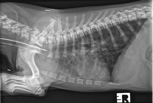
sus-Fig. 1. Right lateral thoracic radiograph of the dog showing thickening of the bronchial walls into the periphery of the lung fi elds and a diffuse increase in pulmonary opacity throughout the lungs
pension using a 10 μl micropipette and stored in phosphate buffer saline (PBS) solution. Genomic DNA was extracted using a com-mercial kit (DNeasy Blood & Tissue Kit, Qiagen, GmbH, Hilden, Germany), in accordance with the manufacturer’s instructions, and partial mitochondrial 12S rRNA (~330 bp) and nuclear 18S rRNA (~1700 bp) genes were amplifi ed as previously described (Latrofa et al., 2015).
Results
On physical examination the dog was bright, alert and responsive with a body condition score of 3/9. Her inspiratory effort was in-creased and an unproductive cough with terminal retch was no-ticed during the consultation, although there was no response on tracheal pinch. On thoracic auscultation, normal respiratory sounds were slightly exaggerated over the entire lung fi eld, al-though there were no adventitious lung sounds. Her heart sounds were normal with synchronous femoral pulses. Her rectal temper-ature was within normal limits and no enlarged lymph nodes were noticeable. CBC revealed eosinophilia, with no other abnormalities and serum chemistry was unremarkable. The antigen blood test specifi c for the detection of A. vasorum (IDEXX Angio DetectTM
Test) revealed a negative result.
Lateral and dorso-ventral radiographic projections of the thorax were taken at full inspiration, revealing a normal cardiac silhou-ette, enhancement and thickening of the bronchial walls spreading into the periphery of the lung fi elds, and a diffuse increase in pul-monary opacity throughout the lungs (Fig. 1). Flexible bronchos-copy was performed and visualization of the trachea and bron-chioles revealed a moderate quantity of mucous and absence of foreign bodies, nodules or masses. After inspection of all primary and secondary bronchioles, a bronchoalveolar lavage (BAL) was performed. During the instillation, a single motile white worm was observed on the mucus of the bronchial wall and cytological evalu-ation of the BAL showed the presence of a non-septic exudate with marked eosinophilic infi ltration.
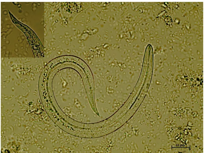
After the visualization of the nematode in the respiratory tract, an enema was performed to collect fresh faecal samples. Qualitative fl oatation analysis and sedimentation test were negative. The sediment collected from the Baermann apparatus revealed the presence of several motile larvae under light microscopy. A quan-titative Baermann was performed, revealing 6066 larvae per gram of faeces (LPG). Several fi liform C-shaped larvae were visualized, 260 – 290 μm long and 12 – 14 μm wide, consistent with C. vulpis L1 (Fig. 2). Regarding the molecular identifi cation, the 12S rDNA sequences obtained from the collected larvae (accession no.
Fig. 2. Crenosoma vulpis fi rst stage larvae detected at the Baermann technique, 40x magnifi cation. Note the fi liform C-shaped larvae with conical tail, consistent with C. vulpis
KR920039) revealed 100 % identity to the nucleotide sequences of C. vulpis available in GenBank®.
The patient was treated with a second administration of 10 % im-idacloprid + 2.5 % moxidectin spot-on. However, at a re-exami-nation 13 days after the spot-on application, L1 of C. vulpis were still present in the dog faeces. Therefore, an alternative approach using fenbendazole (50 mg/kg, PO, q 24 hours) was prescribed for seven days. One month later, thoracic radiographs and Bae-rmann analysis, using faeces from three successive days were performed. Radiographs showed a normal lung pattern, no fi rst stage larvae were detected in faeces and the cough resolved after the fourth day of treatment.
Discussion
Here we report a case of natural infection by C. vulpis in a very young puppy, one of the earliest cases of infection ever reported for this lungworm. Since the fi rst report of this nematode in a do-mestic dog from the UK (Cobb & Fisher, 1992), only sporadic cas-es have been diagnosed in the last decade, including reports from Ireland (Reilly et al., 2000), Switzerland (Unterer et al., 2002), Ger-many (Barutzki & Schaper, 2003), Italy (Rinaldi et al., 2007) and Belgium (Caron et al., 2014). The scant number of reported cases has led practitioners to consider crenosomosis as a rare parasitic disease in dogs. C. vulpis is recognized as the primary cause of pulmonary infections in foxes (Magi et al., 2009), with 10.8 % of foxes scoring positive for this parasite in Great Britain (Taylor et al., 2015). This high prevalence in foxes may suggest that this animal species may act as a reservoir host for the infection of dogs living in the UK and that it may also be prevalent in dogs despite the low evidence in the UK (Cobb & Fisher, 1992; Reilly et al., 2000). Indeed, temperature and the high relative humidity in this country represent optimal conditions for the lifecycle of this lungworm (Fer-dushy and Hasan 2010). In addition, the free-roaming behaviour of foxes has been considered as one of the major drivers for the geographical dispersal of lungworms (Otranto et al., 2015). Further studies are needed to investigate long term effects and po-tential complications when other concomitant infections are pres-ent. Clinicians should be mindful that C. vulpis can cause chronic respiratory signs refractory to several fi rst line treatments, leading to irresponsible use of medicaments and client frustration due to lack of resolution of troublesome symptoms. Besides, towards a negative A. vasorum antigen blood test and the presence of res-piratory signs, other techniques must be performed to exclude oth-er lungworm infections in dogs. In fact, C. vulpis cannot be easily diagnosed with standard faecal fl otation techniques commonly carried out at most veterinary clinics, but by using the Baermann technique, a coprological test considered the gold standard for the diagnosis of lungworm infections (Conboy, 2009). This is a sim-ple, non-invasive, inexpensive diagnostic tool (Koch & Willesen, 2009), which allows lungworm detection and identifi cation based on the morphological characteristics of L1 (McGarry & Morgan,
2009; Colella et al., 2016). Multiple faecal examinations should be performed to improve detection rates and avoid false nega-tives (Foster et al., 2004; Koch & Willesen, 2009). However, this technique is not commonly used in current practice, particularly in patients with respiratory signs, prior to a presumptive diagnosis of allergic respiratory disease and the administration of long-term corticosteroid therapy (Bihr & Conboy, 1999). Considering the time before the patency period (i.e. 18 – 21 days), this dog acquired the infection in a very early age, at approximately three-months old, and the cases available in the literature exclusively report natural infections in dogs older than one year (Reilly et al., 2000; Rinaldi et al., 2007; Caron et al., 2014). Hence, canine crenosomosis should also be taken into account in very young patients suffering from respiratory disease.
Successful treatments in naturally infected dogs have been report-ed with the use of febantel, fenbendazole, ivermectin, milbemycin oxime (Bihr & Conboy, 1999) and moxidectin (Colella et al., 2016). In two studies, milbemycin oxime (Conboy et al., 2013) and mox-idectin (Conboy et al., 2009) showed 98.7 and 100 % effi cacy in the treatment of experimentally infected dogs, respectively. In the case here reported, the treatment with a spot-on solution contain-ing 10 % imidacloprid + 2.5 % moxidectin was unsuccessful in treating C. vulpis infection. However, this dog had a larger larval shedding (i.e. 6066 LPG) than those of experimentally infected dogs (i.e. 0 – 152.5 LPG) (Conboy et al., 2009). This could explain why contrarily to the effi cacy showed 7 days after the adminis-tration of the same spot-on association in experimentally infected animals (Conboy et al., 2009), the dog herein examined was still positive at the re-examination after 13 days. Following the second treatment with fenbendazole on day 13, the dog cured C. vulpis infection on day 43, thought it was not possible to determine which of the two treatments was effi cacious.
Baermann tests should be performed more routinely to perceive the real prevalence of this lungworm and to understand if it is a rare parasite or just rarely diagnosed. Besides, practitioners should include crenosomosis in the differential diagnosis of res-piratory diseases in dogs, particularly in areas where foxes are present, and should foster targeted preventive therapy against this lungworm infection.
Acknowledgement
To The Wylie Veterinary Centre team, Upminster, Essex UK for their invaluable help, to Angela Doyle for their precious help in English review and to CIISA, Faculty of Veterinary Medicine, ULis-boa, Portugal, reference UID/CVT/00276/2013. Ana Margarida Alho holds a PhD fellowship reference SFRH/BD/85427/2012.
References
ANDERSON, R.C. (1992): Nematode parasites of vertebrates, their
BARÇANTE, T.A., BARÇANTE, J.M., DIAS, S.R., LIMA WDOS, S. (2003):
Angiostrongylus vasorum (Baillet, 1866) Kamensky, 1905: emer-gence of third-stage larvae from infected Biomphalaria glabrata snails. Parasitol. Res., 91: 471 – 475
BARUTZKI, D., SCHAPER, R. (2003): Endoparasites in dogs and cats
in Germany 1999 – 2002. Parasitol. Res., 90: 148 – 150
BIHR, T., CONBOY, G.A. (1999): Lungworm (Crenosoma vulpis)
infec-tion in dogs on Prince Edward Island. Can. Vet. J., 40: 555 – 559 CARON, Y., MERVEILLE, A.C., LOSSON, B., BILLEN, F. (2014):
Crenoso-ma vulpis infection in two young dogs in Belgium. Vet. Rec. Case Report, 2: e000098
COBB, M.A., FISHER, M.A. (1992): Crenosoma vulpis infection in a
dog. Vet. Rec., 130: 452
COLELLA, V., GIANNELLI, A., BRIANTI, E., RAMOS, R.A., CANTACESSI, C.,
DANTAS-TORRES, F., OTRANTO, D. (2015): Feline lungworms unlock a
novel mode of parasite transmission. Sci. Rep., 5: 13105
COLELLA, V., MUTAFCHIEV, Y., CAVALERA, M.A., GIANNELLI, A., LIA, R.P.,
DANTAS-TORRES, F., OTRANTO. D. (2016): Development of
Crenoso-ma vulpis in the common garden snail Cornu aspersum: implica-tions for epidemiological studies. Parasit Vectors., 9(1): 208 CONBOY, G. (2009): Helminth parasites of the canine and feline
respiratory tract. Vet. Clin. North. Am. Small Anim. Pract., 39: 1109 – 1126
CONBOY, G., BOURQUE, A., MILLER, L., SEEWALD, W., SCHENKER, R.
(2013): Effi cacy of Milbemax (milbemycin oxime + praziquantel) in the treatment of dogs experimentally infected with Crenosoma vulpis. Vet. Parasitol., 198: 319 – 324
CONBOY, G., HARE, J., CHARLES, S., SETTJE, T,, HEINE, J. (2009). Effi
-cacy of a single topical application of Advantage Multi (=Advocate) Topical Solution (10 % imidocloprid + 2.5 % moxidectin) in the treatment of dogs experimentally infected with Crenosoma vulpis. Parasitol. Res., 105: 49 – 54
FERDUSHY, T., HASAN, M.T. (2010): Angiostrongylus vasorum: the
‘French heartworm’. Parasitol Res., 107: 765 – 771
FOSTER, S.F, MARTIN, P., BRADDOCK, J.A, MALIK, R. (2004): A
retro-spective analysis of feline bronchoalveolar lavage cytology and microbiology (1995 – 2000). J. Fel. Med. Surg., 6: 189 – 198 GIANNELLI, A., COLELLA, V., ABRAMO, F., NASCIMENTO RAMOS, R.A., FAL -SONE, L., BRIANTI, E., VARCASIA, A., DANTAS-TORRES, F., KNAUS, M.,
FOX, M.T., OTRANTO, D. (2015): Release of lungworm larvae from
snails in the environment: potential for alternative transmission pathways. PLoS Negl. Trop. Dis., 9, e0003722
KOCH, J., WILLESEN. J.L. (2009): Canine pulmonary
angiostrongylo-sis: An update. Vet. J., 179: 348 – 359
LATROFA, M.S., LIA, R.P., GIANNELLI, A., COLELLA, V., SANTORO, M.,
D’ALESSIO, N., CAMPBELL, B.E., PARISI, A., DANTAS-TORRES, F., MUTAF
-CHIEV, Y., VENEZIANO, V., OTRANTO, D. (2015): Crenosoma vulpis in
wild and domestic carnivores from Italy: a morphological and mo-lecular study. Parasitol. Res., 114: 3611 – 3617
MAGI, M., MACCHIONI, F., DELL’OMODARME, M., PRATI, M.C., CALDERINI,
P., GABRIELLI, S., IORI, A., CANCRINI, G. (2009): Endoparasites of red
fox (Vulpes vulpes) in central Italy. J. Wildl. Dis., 45: 881 – 885 MCGARRY, J.W., MORGAN, E.R. (2009): Identifi cation of fi rst-stage
larvae of metastrongyles from dogs. Vet Rec., 165: 258 – 261 OTRANTO, D., CANTACESSI, C., DANTAS-TORRES, F., BRIANTI, E,, PFEF -FER, M., GENCHI, C., GUBERTI, V., CAPELLI, G., DEPLAZES, P. (2015):
The role of wild canids and felids in spreading parasites to cats and dogs in Europe. Part II: Helminths and arthropods. Vet. Para-sitol., 213: 24 – 37
REILLY, G.A, MCGARRY, J.W,, MARTIN, M., BELFORD, C. (2000):
Creno-soma vulpis, the fox lungworm, in a dog in Ireland. Vet. Rec., 146: 764 – 765
RINALDI, L., CALABRIA, G., CARBONE, S., CARRELA, A., CRINGOLI, G.
(2007): Crenosoma vulpis in dog: fi rst case report in Italy and use of the FLOTAC technique for copromicroscopic diagnosis. Parasi-tol. Res., 101: 1681 – 1684
SAEED, I., MADDOX-HYTTEL, C., MONRAD, J., KAPEL, C.M. (2006):
Hel-minths of red foxes (Vulpes vulpes) in Denmark. Vet Parasitol., 139: 168 – 179
SRÉTER, T., SZÉLL, Z., MARUCCI, G., POZIO, E., VARGA, I. (2003):
Ex-traintestinal nematode infections of red foxes (Vulpes vulpes) in Hungary. Vet Parasitol., 115: 329 – 334
STOCKDALE, P.H.G., HULLAND, T.J. (1970): The pathogenesis, route
of migration, and development of Crenosoma vulpis in a dog. Pathol Vet., 7, 28 – 42
TAYLOR, C.S., GARCIA GATO, R., LEARMOUNT, J., AZIZ, N.A., MONTGOM -ERY, C., ROSE, H., COULTHWAITE, C.L, MCGARRY, J.W., FORMAN, D.W.,
ALLEN, S., WALL, R., MORGAN, E.R. (2015): Increased prevalence
and geographic spread of the cardiopulmonary nematode Angi-ostrongylus vasorum in fox populations in Great Britain. Parasitol-ogy, 142: 1190 – 1195
TRAVERSA, D., DI CESARE, A., CONBOY, G. (2010): Canine and feline
cardiopulmonary parasitic nematodes in Europe: emerging and underestimated. Parasit. Vectors, 3: 62
UNTERER, S., DEPLAZES, P., ARNOLD, P., FLUCKIGER, M., REUSCH, C.E.,
GLAUS, T.M. (2002): Spontaneous Crenosoma vulpis infection in 10
dogs: laboratory, radiographic and endoscopic fi ndings. Schweiz Arch. Tierheilkd., 144: 174 – 179
WETZEL, Z., MUELLER, R. (1935): The life cycle of Crenosoma vulpis,
the lung worm of foxes, and ways and means of combat. Fur Trade J. Can., 13: 16 – 17