http://ats.ctsnetjournals.org/cgi/content/full/80/6/2008
located on the World Wide Web at:
The online version of this article, along with updated information and services, is
Print ISSN: 0003-4975; eISSN: 1552-6259.
Southern Thoracic Surgical Association. Copyright © 2005 by The Society of Thoracic Surgeons.
is the official journal of The Society of Thoracic Surgeons and the
Late-Onset Occult Pneumothorax After Lung
Volume-Reduction Surgery
Federico Tacconi,
MD,
Eugenio Pompeo,
MD,
and Tommaso C. Mineo,
MD
Division of Thoracic Surgery, Policlinico Tor Vergata University, Rome, Italy
Background. Lung volume-reduction surgery has proved to be a reliable palliative surgical treatment for patients with severe emphysema. Nonetheless, late com-plications can arise after lung volume-reduction surgery although this matter has been poorly investigated by previous studies.
Methods. We report a series of 6 patients undergoing unilateral lung volume-reduction surgery at our institu-tion between October 1995 and December 2004, who were readmitted several months after discharge because of the occurrence of occult pneumothorax mimicking acute re-spiratory failure.
Results. Occult pneumothorax occurred in 3.3% of the 182 patients treated with lung volume-reduction surgery at our institution. Patients were readmitted after a mean of 94 days (range, 20 to 700 days) from the discharge. Chest roentgenography was unable to detect the occur-rence of pneumothorax, which was instead revealed by means of a computed tomographic scan in all patients.
The interval between admission and correct diagnosis averaged 22.4 hours. The number of air collections ranged between two and four. Treatment entailed solely blind chest drainage placement in 2 patients and awake video-assisted thoracoscopic surgery in the others, in-cluding placement of chest tube under direct vision in 1 patient, repair of lung tears by means of cyanoacrylate glue in 2 and bovine pericardium patch plus cyanoacry-late glue apposition in 1 patient.
Conclusions. In conclusion, we believe that occult pneumothorax should be kept in mind as one of the possible late complications of lung volume-reduction surgery and should be suspected whenever sudden wors-ening of dyspnea is noticed even in the presence of an uneventful chest roentgenogram. Awake video-assisted thoracoscopic surgery management can represent an ef-fective option in these instances.
(Ann Thorac Surg 2005;80:2008 –12) © 2005 by The Society of Thoracic Surgeons
L
ung volume-reduction surgery (LVRS) has proved to be a reliable palliative surgical treatment for pa-tients with heterogeneously distributed severe emphy-sema. Nonetheless, several late complications can occur after LVRS including development of giant bullous em-physema [1] and the occurrence of secondary pneumo-thorax. This latter in particular has been poorly investi-gated by the previous literature. More to the point, we have found that loculated air collections can occur after LVRS even several months after discharge, leading to severe impairment of respiratory function. Prompt rec-ognition of LVRS-related complications is crucial to avoid acute respiratory failure, which can be associated with a high mortality rate in these delicate patients. Unfortu-nately, detection of this complication can be a difficult first-line assessment owing to the presence of postoper-ative pleural adhesions resulting in complex summation artifacts on standard chest roentgenogram. Herein, we describe peculiar clinical features and surgical manage-ment of occult loculated pneumothorax occurring in a large single-center consecutive series of patients under-going LVRS.Patients and Methods
We reviewed a series of 6 patients undergoing stapled LVRS at our institution between October 1995 and De-cember 2004, and presenting several months after dis-charge as critical care patients because of the occurrence of secondary pneumothorax.
All patients had advanced smoking-related upper-lobe prevailing emphysema with severe airflow obstruction and marked air trapping (mean forced expiratory volume in 1 second, 33.5%; mean residual volume, 210.8% of predicted). No patient in this series had giant bullae, ho-mogeneous emphysema, or homozygous␣1-antitrypsin
de-ficiency. All patients also met standard inclusion criteria for LVRS including smoking cessation of at least 6 months, poor expectoration, and absence of cor pulmonale and other concomitant diseases affecting the outcome. Addi-tional details regarding our inclusion criteria and preoper-ative assessment are described elsewhere [2]. Five pa-tients underwent unilateral videothoracoscopic LVRS, and 1 underwent staged bilateral operation. The most damaged portions of the lung were identified by visual inspection as the regions retaining inflation after venti-latory exclusion. Both computed tomographic (CT) and scintigraphic scan findings were also taken into the account. The targeted areas were resected by endoscopic staplers (Endopath 45; Ethicon Endosurgery, Pomezia, Italy). In the first 3 patients, suture lines were reinforced Accepted for publication June 7, 2005.
Address correspondence to Dr Pompeo, Cattedra di Chirurgia Toracica, Policlinico Tor Vergata, V.le Oxford 81, 0133, Rome, Italy; e-mail: [email protected].
with the use of bovine pericardial strips (Peristrips; Biovascular Inc, St. Paul, MN). After resection, careful assessment of pneumostasis was performed. If major leaks were identified, they were ablated by additional “no-cut” stapling (Endopath NK45B; Ethicon Endosur-gery). Two chest tubes were inserted at the end of the procedure and placed under water-seal. No patient in this series required additional suction at the end of the procedure. After operation, 4 patients were discharged after complete cessation of air leaks (days 7, 10, 12, and 15). Two patients were discharged with a Heimlich valve owing to small persisting air leaks, and were controlled as outpatients thrice weekly until air leak cessation was ensured (postoperative days 18 and 21). In both groups, criteria for chest drainage removal were radiologically documented complete lung reexpansion and the absence of bubbling in the drainage chamber. After tube removal, patients underwent first follow-up within 7 days entail-ing solely physical examination, and complete reassess-ment within 15 days including respiratory function tests, 6-minute walking test, blood-gas assay, and in-expiratory chest roentgenogram. Four patients underwent postop-erative respiratory rehabilitation performed either on an outpatient or on an inpatient basis, depending on indi-vidual preference.
Results
The study cohort (all men, mean age, 66.4 years) accounts for 3.3% of the 182 patients treated by LVRS at our institution. Patients were readmitted after a mean of 168 days from discharge (range, 20 to 700 days; median, 94
days). In this subgroup, mean air leak duration after LVRS was 13.8 days (range, 7 to 21 days), a number that reaches statistical difference when compared with the average duration observed in our historical cohort (5.2 days; Mann-Whitney U test, p⫽ 0.00009).
Four patients arrived directly at the emergency depart-ment of our institution because of acute respiratory failure. The other 2 patients were referred at our institu-tion after preliminary admission to the emergency de-partment of a peripheral hospital. Both these subjects arrived with a misdiagnosis of exacerbated chronic ob-structive pulmonary disease and deteriorating bullous emphysema, respectively.
In all patients, the acute scenario entailed a marked impairment in subjective dyspnea and severe hypoxia in the absence of fever, cough, sputum, and leukocytosis. Three patients also had cardiovascular symptoms (ie, tachycardia and hypotension). Hypercarbia was present in 2 patients.
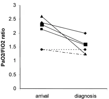
Preliminary chest roentgenography showed signs of unilateral lung hyperinflation with flattening of the dia-phragmatic dome and slight mediastinal shift (2 pa-tients), and a slight hypertranslucent area in the involved hemithorax (3 patients). In 2 patients, chest roentgeno-gram was unremarkable. All patients’ CT scan was per-formed after a mean of 22.4 hours from admission and revealed definitive diagnosis (Figs 1, 2). Figure 3 depicts the fall in arterial oxygenation (expressed as arterial partial pressure of oxygen to fraction of inspired oxygen ratio) per patient in the interval between initial admis-sion and definitive diagnosis.
Three patients underwent initial blind chest drainage
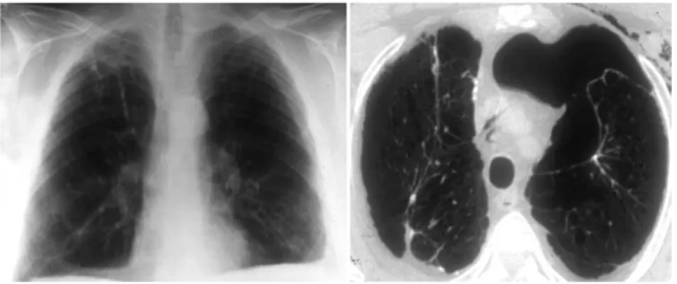
Fig 1. Figure shows roentgenogram (left) and computed tomographic (right) feature of a loculated pneumothorax occupying almost 45% of the left hemithorax. Despite the large size revealed by computed tomography, chest roentgenogram showed solely a minimal hy-perlucency area on the left upper field.
Fig 2. Tomographic feature of a very large right pneumothorax (right), which was mim-icking lung hyperinflation at scout scanogram (left; slight leftward tracheal shift and com-pression of the upper lobe vessels).
GENERAL
placement, which allowed definitive resolution of the pneumothorax in 2 patients. One patient received first blind drainage placement and was subsequently sched-uled for video-assisted thoracoscopic surgery because of persisting air collection. Three patients were immedi-ately scheduled for video-assisted thoracoscopic surgery because of the presence of multiple loculated pneumo-thoraces whose treatment by blind drainage was deemed dangerous. To avoid the risks of general anesthesia and mechanical ventilation, all the procedures were per-formed with the patient awake, under spontaneous ven-tilation. Analgesia entailed intercostal nerve blockade with a mix of lidocaine 2% and ropivacaine 7.5%. Mild sedation was obtained with 2 to 5 mL of intravenous midazolam. No patient required intubation or suffered functional impairment throughout the procedure. Two 15-mm trocar ports were sufficient for the operation in all patients but 1, who required an additional 7-mm port because of particular anatomic complexity. The air leak was easily discovered in 5 patients. In 1 patient the leak was not found, and operative technique entailed only video-assisted placement of a chest drainage. Sealing of a small lung tear lying in the vicinity of the suture line by means of cyanoacrylate glue instillation was carried out in 2 patients. In 1 patient, a gross bronchoalveolar leak lying on the major fissure was repaired by means of a cyanoacrylate layer covered with a bovine pericardium patch. Operative time ranged from 35 to 90 minutes. Air leak cessation was achieved after 1 to 3 days; mean hospital stay was 3.2 days. Follow-up ranged from 6 to 36 months, with no patients lost to follow-up. No patient developed a second pneumothorax during follow-up.
Tables 1 and 2 report major clinical and radiologic findings of our series.
Comment
The most meaningful finding of our study is that a multiloculated pneumothorax may develop late after LVRS, and that this complication may be easily missed at standard radiographic assessment even in the presence of large air collections, thus leading to erroneous misdi-agnosis of respiratory failure caused by exacerbated chronic obstructive pulmonary disease or deteriorating bullous emphysema. The causal relation of late-onset pneumothorax with previous LVRS can be exclusively hypothetical. In some cases, an alveolar, or even bron-choalveolar, leak might develop after several days as an effect of the mechanical stress induced by reexpansion of the lung surface. This mechanism supplies a better ex-planation of cases that present after a period of relative well-being after discharge. Alternatively, we suggest that in some patients, a minimal air leak can persist despite radiologically documented full lung reexpansion and the absence of bubbling on the water seal chamber. In these instances, pneumothorax is likely to occur early after discharge, although the presence of thick pleural adhe-sions might initially limit its enlargement and its radio-logic detection even with a provocative clamping test. In these patients, the presence of a minuscule air collection may explain the lack of subjective improvement at first outpatient visit after surgery in some patients. In these instances, uneventful chest roentgenogram may lead the surgeon to rule out other procedure-related complica-tions. Therefore, we now prefer to include CT scan even in the short-term follow-up of LVRS patients presenting with unsatisfactory outcome, particularly if they had prolonged postoperative air leaks. The basic mechanism of very late onset pneumothorax is more demanding to hypothesize. We acknowledge that these pneumothora-ces might simply develop as a result of a progression of the native disease, without reasonable relationship with the previous surgery. Nonetheless, the regional differ-ences in elastic recoil induced by LVRS might at least exaggerate the disruption of lung tissue and facilitate the rupture of superficial bullae, even in sites distant from the suture lines. A similar mechanism has been proposed to explain the late development of giant bullae after LVRS[1].
In our series, conventional chest roentgenography failed to pinpoint the exact diagnosis even in presence of very large sized pneumothoraces, which were instead
Fig 3. Decline in arterial blood oxygenation (arterial partial pres-sure of oxygen to fraction of inspired oxygen ratio [Pao2/Fio2]) in
the interval time between admission and diagnosis.
Table 1. Patient Data
Patient Pao2/Fio2 Ratio Days From Discharge Diagnostic Delay (hours) 1 2.62 92 32 2 1.45 37 16 3 2.42 106 24 4 1.35 55 32 5 2.38 20 8 6 1.44 700 3
Fio2⫽ inspiratory fraction of oxygen; Pao2⫽ arterial tension of oxygen.
revealed at CT scan. The merits of CT in this setting are not surprising, and have been evidenced in both clinical and experimental studies [3–5]. In particular, Omert and coworkers [6] have stressed that CT scan results in a significant change in clinical management of trauma patients with intrathoracic complications. Nonetheless, physicians involved in primary care are sometimes reluc-tant to include CT in first-line assessment of patients with thoracic complications, on account of cost-saving concerns. This kind of behavior resulted in a remarkable diagnostic delay for our patients that translated in a significant decrease in arterial blood oxygenation (Fig 3), particularly in those who were first referred to peripheral hospitals. On the basis of this observation, we advise LVRS patients to be immediately referred to their spe-cialized center rather than be seen at primary care structures not experienced with this kind of surgery nor with its particular critical care needs.
Several authors have suggested that any pneumotho-rax occurring in critically ills patients should be treated expeditiously by chest tube placement because of the high rate of progression to tension, especially in case of ventilatory support [7, 8]. However, although blind chest tube placement may achieve initial stabilization of the patient or even definitive resolution in certain cases, it may be demanding or even hazardous owing to the risk of lung injury in the presence of loculated pneumothorax. Moreover, chest drainage alone may not resolve the air leaks when these are sustained by a large lung tear, leading to prolonged hospital stay with increased mor-bidity and overall costs. We therefore now prefer video-assisted thoracoscopic surgery management with the goal of sealing pulmonary tears and reducing the risks of triggering further complications. In addition, having fur-ther experience with awake procedures for resection of pulmonary nodules [9] and LVRS [10], we are prone to adopt this option even in this setting, with the aim of reducing surgical risks in these delicate patients. Previ-ous studies have focused on the advantages of this approach to managing air leak problems [11, 12]. Differ-ent video-assisted thoracoscopic surgery techniques can be adopted to seal the air leak, including limited resec-tion, “no-cut” stapling, instillation of biologic glues, or a combination of these [11–13]. In particular, we have found that definitive air leak control can be rapidly achieved with the use of instilled cyanoacrylate glue, which can be also used in combination with a bovine
pericardium patch to seal large lung tears as reported by Horsley and Miller [12]. Using this technique, operative time was short even in the most demanding instances. Despite the finding that more detailed evaluation of cost-to-benefit analysis of minimally invasive surgery versus only chest drainage or interventional radiologic procedures is warranted in future studies, we believe that this approach translates into decreased morbidity, shorter hospital stay, and lower hospital costs.
In conclusion, we believe that occult pneumothorax should be considered as a possible late-onset life-threatening complication of LVRS, especially in patients with a history of prolonged postoperative air leaks. Oc-cult pneumothorax may present even several months after the procedure as sudden, progressively deteriorat-ing respiratory impairment in relatively well-fardeteriorat-ing pa-tients. Nonetheless, in some patients, the presence of a minimal active air leak should already be suspected at short-term follow-up as exacerbating dyspnea is noticed despite uneventful chest roentgenogram. We therefore suggest that CT scan should be routinely included in the early postoperative management of this subgroup. More-over, because immediate recognition and treatment of a late-onset pneumothorax is crucial to avoid the need of mechanical ventilation, we recommend that criti-cally ill LVRS patients should be sent immediately to their referral center for prompt diagnosis and optimal management.
Supported by MURST COFIN grants No. 9906274194 – 06, CNR No. CU0100935 2002, and Centro di Eccellenza 2001. This study was carried out within the Research Fellowship Program Tec-nologie e Terapie Avanzate in Chirurgia awarded by the Tor Vergata University.
References
1. Iqbal M, Rossoff L, McKeon K, Graver M, Scharf SM. Development of a giant bulla after lung volume reduction surgery. Chest 1999;116:1809 –11.
2. Mineo TC, Pompeo E, Mineo D, Rogliani P, Leonardis C, Nofroni I. Results of unilateral lung volume reduction sur-gery in patients with distinct heterogeneity of emphysema between lungs. J Thorac Cardiovasc Surg 2005;129:73–9. 3. Engdhal O, Toft T, Boe J. Chest radiography: a poor method
for determining the size of a pneumothorax. Chest 1993;103: 26 –9.
4. Phillips GD, Trotman-Dickensen B, Hodson ME, Geddes DM. Role of CT in the management of pneumothorax in
Table 2. Radiologic and Operative Data
Patient Site of Air Leak Radiographic Findings Treatment Hospital Stay (days)
1 Staple line None VATS 3
2 Staple line Hyperlucency, mediastinal shift VATS 4
3 Major fissure None DP, VATS 2
4 Lower lobe Mediastinal shift VATS 4
5 Unknown Hyperlucency DP 3
6 Unknown Hyperlucency DP 5
DP⫽ drainage placement; VATS⫽ video-assisted thoracoscopic surgery.
GENERAL
patients with complex cystic lung disease. Chest 1997;112: 275– 8.
5. Wolfman NT, Wendell SM, Glauser SJ, Meredith JW, Chen MY. Validity of CT classification on management of occult pneumothorax: a prospective study. AJR Am J Roentgenol 1998;171:1317–20.
6. Omert L, Yeaney WW, Protetch J. Efficacy of thoracic com-puterized tomography in blunt chest trauma. Am Surg 2001:67:660 – 4.
7. Gobien RP, Reines HD, Schabel SI. Localized tension pneu-mothorax: unrecognized form of barotrauma in adult respi-ratory distress syndrome. Radiology 1982;142:15–9. 8. Streiter RM, Lynch JP. Complications in the ventilated
patients. Clin Chest Med 1988;9:127–39.
9. Pompeo E, Mineo D, Rogliani P, Sabato AF, Mineo TC. Feasibility and results of awake thoracoscopic resection of
solitary pulmonary nodules. Ann Thorac Surg 2004;78: 1761– 8.
10. Mineo TC, Pompeo E, Mineo D, Pampana E, Dauri M, Sabato AF. Awake non-resectional lung volume reduction surgery. Ann Surg. In press.
11. Thistlethwaite PA, Luketich JD, Ferson PF, Keenan RJ, Jamieson SW. Ablation of persistent air-leaks after thoracic procedures with fibrin sealant. Ann Thorac Surg 1999;67: 575–7.
12. Mukaida T, Andou A, Date H, Aoe M, Shimizu N. Thoraco-scopic operation for secondary pneumothorax under local anesthesia in high-risk patients. Ann Thorac Surg 1998;65: 924 – 6.
13. Horsley WS, Miller JI. Management of the uncontrollable air-leak with cyanoacrylate glue. Ann Thorac Surg 1997;63:1492–3.
Online Discussion Forum
Each month, we select an article from the The Annals of Thoracic Surgery for discussion within the Surgeon’s Fo-rum of the CTSNet Discussion FoFo-rum Section. The arti-cles chosen rotate among the six dilemma topics covered under the Surgeon’s Forum, which include: General Thoracic Surgery, Adult Cardiac Surgery, Pediatric Car-diac Surgery, CarCar-diac Transplantation, Lung Transplan-tation, and Aortic and Vascular Surgery.
Once the article selected for discussion is published in the online version of The Annals, we will post a notice on the CTSNet home page (http://www.ctsnet.org) with a FREE LINK to the full-text article. Readers wishing to comment can post their own commentary in the discus-sion forum for that article, which will be informally moderated by The Annals Internet Editor. We encourage all surgeons to participate in this interesting exchange and to avail themselves of the other valuable features of the CTSNet Discussion Forum and Web site.
For December, the article chosen for discussion under the Adult Cardiac Dilemma Section of the Discussion forum is:
Vacuum-Assisted Wound Closure of Deep Sternal Infec-tions in High-Risk Patients After Cardiac Surgery Kyle Northcote Cowan, MD, PhD, Laura Teague, RN, MN, Sammy C. Sue, BS, and James L. Mahoney, MD Tom R. Karl, MD
The Annals Internet Editor UCSF Children’s Hospital Pediatric Cardiac Surgical Unit 505 Parnassus Ave, Room S-549 San Francisco, CA 94143-0118 Phone: (415) 476-3501
Fax: (212) 202-3622
e-mail: [email protected]