37/661 (2), Fort P.O. Trivandrum-695 023 Kerala, India
Recent Advances in Pharmaceutical Sciences VI, 2016: 165-188 ISBN: 978-81-308-0566-5 Editors: Diego Muñoz-Torrero, Àngela Domínguez and Àngels Manresa
10. Advances in the research of new genetic
markers for the detection of
Helicobacter pylori infection
Núria Piqué, Montserrat Palau, Mercedes Berlanga and David Miñana-Galbis Departament de Microbiologia i Parasitologia Sanitàries, Facultat de Farmàcia, Universitat de
Barcelona, Av. Joan XXIII s/n, 08028 Barcelona, Catalonia, Spain
Abstract. Helicobacter pylori is one of the human pathogens with
highest prevalence around the world. Colonizing the human stomach, H. pylori could lead to peptic ulceration, gastric adenocarcinoma and gastric lymphoma. H. pylori is a genetically diverse bacterial species, and variability in virulence factors has a role in bacterial pathogenesis and progression to gastric cancer, although bacterium and host factors of progression are not completely understood. In a recent study, we have demonstrated that six housekeeping genes related to H. pylori pathogenesis were specifically amplified for H. pylori in a total of 52 H. pylori clones isolated from 11 patients. Although most clones isolated from the same patient showed identical gene sequences, events of multiple infection and microevolution were detected. We consider that housekeeping genes could be useful for H. pylori detection and to elucidate the mode of transmission and the relevance of the N. Piqué and M. Palau contributed equally.
Correspondence/Reprint request: Prof. Dr. David Miñana-Galbis, Departament de Microbiologia i Parasitologia Sanitàries, Facultat de Farmàcia, Universitat de Barcelona, Av. Joan XXIII s/n, 08028 Barcelona, Catalonia, Spain. E-mail: [email protected]
multiple infection. Further genetic studies are required to provide powerful tools to face all current unmet challenges of H. pylori infection, such as the elucidation of mode of transmission, the development of new sensitive and specific PCR methods for H. pylori detection, and implication of H. pylori in other diseases.
Introduction
H. pylori is one of the human pathogens with highest prevalence around the world (50% of the world´s population, with proportions as high as 80% in developing countries) [1,2], which was first isolated in culture media by Warren and Marshall in 1982. This Gram negative, spiral-shaped, microaerophilic bacterium, with positive findings for urease, oxidase and catalase, colonizes the human gastric epithelium [2,3], where it can be found in two forms, the bacillary and coccoid forms [4]. H. pylori is difficult to culture in vitro, particularly in liquid media, being its growth demonstrated in several nutrient-rich media at 37ºC [5].
H. pylori is a genetically diverse bacterial species, and variability in
virulence factors has a role in bacterial pathogenesis [6,7]. Accumulating evidences support that H. pylori infection cause a list of diseases, ranging from gastric to extra-gastric diseases, from chronic gastritis to gastric carcinoma, and thus this bacterium is recognized as Class I carcinogenic pathogen in humans [8,9]. Gastric adenocarcinoma is the second highest cause of cancer deaths worldwide. The nearly one million deaths per year are due to a combination of high incidence, aggressive disease course, and lack of effective treatment options. H. pylori causes distal but not proximal gastric adenocarcinoma, distal being the most common form. H. pylori also causes B cell mucosa-associated lymphoid tissue (MALT) lymphoma of the stomach [3,10].
Progression from initial gastritis toward more severe disease such as gastric cancer occurs only in a small proportion of the infected individuals and is likely to depend on host factors, exposure to lifestyle factors, and bacterial factors [1,7,10]. Furthermore, epidemiological and eradication studies have demonstrated a casual relationship between H. pylori infections and endothelial dysfunction, leading to vascular diseases [3,11]. It has also been suggested the possible association of H. pylori infection with several extragastric effects, including hepatobiliary and pancreatic diseases, although further research is needed in this issue [12].
H. pylori colonization usually occurs in childhood, particularly in
developing areas, and usually in the same family for a cohort effect [3], but infection persists lifelong in the absence of treatment. H. pylori persistence is central to pathogenesis; ulcers occur mainly in mid- or late adulthood after
many years of infection and inflammation, and gastric adenocarcinoma occurs in late adulthood after an even longer period of chronic inflammation and epithelial damage [10].
H. pylori strains appear to be spread by person-to-person contact and
DNA fingerprinting has provided evidence of transmission between family members [13]. To date, however, the exact mode of transmission is still uncertain. While this organism is isolated from the human stomach, it has not been consistently isolated from other niches, and thus the mechanism by which it colonizes the human stomach remains largely unknown [2].
Different invasive and non-invasive diagnostic tests are available for the diagnosis of H. pylori in the individual patient. The non-invasive tests obviate the need for endoscopy and can be surely more accepted by the subjects [3]. Therefore, further research is needed to improve non-invasive methods, mainly based on PCR methods.
The goal of H. pylori treatment is the complete elimination of the organism. Once this has been achieved, reinfection rates are low; thus, the benefit of treatment is durable [14]. Curing the infection interferes with the precancerous cascade if accomplished early in the process, and can prevent cancer development [15]. The so-called triple therapies, combinations of one antisecretory agent with two antimicrobial agents for 7 to 14 days, have been extensively evaluated and approved [14]. Failure of antibiotic treatment, often caused by antibiotic resistant H. pylori strains, however, is frequent [14] and appears to be increasing, so susceptibility testing for antibiotics plays an important role in treatment [16].
The recent findings on the bacterial virulence factors, effects of
H. pylori on epithelial cells, genetic polymorphism of both the bacterium and
its host, and the environmental factors can help to understand the role of this bacterium in gastric carcinogenesis [9]. However, more research is needed to globally face this prevalent infection, such as: to screen and treat only strains that are known to cause disease, the elucidation of mode of transmission, implication of H. pylori in other diseases, the role of mixed infections, the development of new sensitive and specific PCR methods for H. pylori detection in gastric samples and other specimens, and the development of a safe and efficient preventive vaccine that could address the wide diversity of strains.
1. Epidemiology and population genetics of H. pylori
H. pylori is one of the most common bacterial infectious agents; it
inhabits the stomachs of more than half of the world’s population. The prevalence of H. pylori infection is decreasing in developed countries as
showed by the lower prevalence in the younger generations. In north European and North American populations, about one-third of adults are still infected, whereas in south and east Europe, South America, Africa, and Asia, the prevalence of H. pylori is often higher than 50%. These prevalences may differ between different ethnic, social or age groups within the same country [2,10,17]. The prevalence of infection seems to mostly depend on the rate of acquisition, but also on the rate of loss of infection and the length of the persistence period between acquisition and loss [2]. Several factors have been associated with H. pylori infection as a low socioeconomic status, living in a rural area, in crowded homes, and having contaminated sources of drinking water [17].
Despite the high prevalence of H. pylori infection in Africa and South Asia, the incidence of gastric cancer in these areas is much lower than in other countries. Furthermore, the incidence of gastric cancer tends to decrease from north to south in East Asia. Such geographic differences in the pathology can be explained, at least in part, by the presence of different types of H. pylori virulence factors determinants, especially cagA, vacA genes, and the right end of the cag pathogenicity island. The genotype of the virulence genes is useful not only for the study of gastroduodenal diseases related to H. pylori but also as a tool to track human migration although with less accuracy than the multilocus sequence typing (MLST) [18].
MLST analysis using seven housekeeping genes (atpA, efp, mutY, ppa,
trpC, ureI, and yphC) is useful to predict the history of human migrations
and may be at least as informative as most human genetic markers [18,19].
H. pylori is ubiquitous and possesses strong phylogeographic structure,
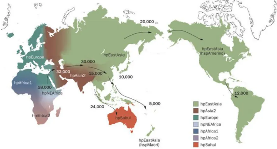
suggesting that bacterial polymorphisms reflect human phylogeography and historical migrations. Population structure analysis based on MLST has revealed seven modern population types of H. pylori (Fig. 1): hpEurope, hpAfrica1 (with two subpopulations, hspSAfrica, and hspWAfrica), hpAfrica2, hpAsia2, hpEastAsia (with three subpopulations, hspAmerind hspEAsia, and hspMaori), hpNEAfrica, and hpSahul [18,20,21,22,23].
The geographic sources of the different strains isolated reflect major events in human settlement history (Fig. 1), such as the colonization of Polynesia and the Americas and the African Bantu migrations. Like humans, simulations indicate that H. pylori seems to have spread from east Africa around 58,000 years ago. Therefore, these data indicate that anatomically modern humans were already infected by H. pylori before their migrations from Africa and demonstrate that H. pylori has remained intimately associated with their human host populations ever since [20,21,22,23].
The existing European population of H. pylori is known to be a hybrid between Asian and African bacteria, although there are hypotheses about
when and where the hybridization took place. In a recent study, a 5300-year-old H. pylori genome from a European Copper age glacier mummy was determined and reconstructed. Results obtained showed that the ―Iceman‖
H. pylori was a nearly representative of the bacterial population of Asian
origin that existed in Europe before hybridization, suggesting that the African population arrived in Europe within the past few thousand years [24].
Figure 1. Distribution of Helicobacter pylori genotypes before Columbus found the
New World and human migration to America and Oceania began. There are seven modern H. pylori population types—hpEurope, hpEastAsia, hpAfrica1, hpAfrica2, hpAsia2, hpNEAfrica and hpSahul. hpEurope includes almost all H. pylori strains isolated from ethnic Europeans, including people from countries colonized by Europeans. Most H. pylori isolates from East Asia belong to hpEastAsia, which includes hspMaori (Polynesians, Melanesians, and native Taiwanese), hspAmerind (American Indians) and hspEAsia (East Asia) subpopulations. hpAsia2 strains are isolated in South, Southeast and Central Asia; hpAfrica1 in west Africa, South Africa and African Americans. hpNEAfrica is predominantly made up of isolates from Northeast Africa. hpAfrica2 is very distinct from any other type and has currently only been isolated in South Africa. hpSahul is a novel group specific to H. pylori
strains isolated from Australian Aborigines and Highlanders of New Guinea.
H. pylori is predicted to have spread from East Africa over the same time period as
anatomically modern humans (~58,000 years ago), and has remained intimately associated with their human hosts ever since. Estimated global patterns of H. pylori migration are indicated by arrows and the numbers show the estimated time since they migrated (years ago). The broken arrow indicates an unconfirmed migration pattern. (reprinted by permission from Macmillan Publishers Ltd: ref. [23], copyright 2010).
Elucidation of the pattern of population subdivision is also of medical interest. Geographically variable results regarding the association of putative virulence factors with disease might well reflect differences in the local prevalence of the individual H. pylori populations. Similarly, the development of diagnostic tests, antibiotics, and vaccines needs to account for global diversity and will be aided by the availability of representative isolates [20].
2. Transmission of H. pylori infection
Currently, the majority of available evidence points to the transmission of H. pylori from human-to-human. The exact route of transmission from person-to-person is still unknown. Ingestion of the bacteria, which is the most likely portal of entry, may occur by one or a combination of three means: oral-oral, gastro-oral, or fecal-oral. However, determination of the dominant route has not been possible to date [2].
H. pylori has been detected in dental plaque and saliva, suggesting that
the oral cavity may be an extra-gastric reservoir and play an important role in both transmission and recurrence [25]. Since the human stomach is the primary niche of H. pylori, it is reasonable to suggest a direct gastro-oral route of transmission mediated by refluxed gastric juice. This hypothesis is supported by studies showing the presence of H. pylori in the gastric juice, as vomitus [2,26,27].
Although H. pylori is sensitive to the bile’s bactericidal effect, different studies have detected viable H. pylori in stools [2,28,29], thus supporting the fecal-oral route of transmission.
H. pylori has also been detected in environmental or animal reservoirs,
although it has not been demonstrated yet if they are natural or primary vehicles of transmission [2].
Presence of H. pylori has been detected in un-washed vegetables, as leek, traditional salad, basil or lettuce [30]. More research is needed to establish different foods at high risk of H. pylori presence and transmission.
Moreover, the demonstration of transmission of viable cells is even more complicated taking into account the difficulties of the bacterium to grow in vitro [5].
3. Role of Helicobacter pylori infection in pathogenesis of gastric
carcinoma
The gastric mucosa is well protected against bacterial infections because of its acidic milieu that acts as a first line of defense against food-borne
microbes. In addition, the reflux of bile acids in the stomach, the thickness of the mucus layer, and the effectiveness of gastric peristalsis might have impeded bacterial colonization of the stomach. Nevertheless, H. pylori is highly adapted to this ecological niche, with a unique array of features that permit entry into the mucus, swimming and spatial orientation in the mucus, attachment to epithelial cells, evasion of the immune response, and, as a result, persistent colonization and transmission [14].
For colonization and survival in the human stomach all Helicobacter isolates require urease and flagella. Urease metabolizes urea to carbon dioxide and ammonia to buffer the gastric acid. Flagella allow the bacterium to swim across the viscous gastric mucus and reach the more neutral pH below the mucus. H. pylori may use at least five different adhesins to attach to gastric epithelial cells. Adhesion by adhesins to epithelial gastric cells is important for the beginning of infection and for the enhanced inflammatory response. Following colonization, H. pylori must acquire nutrients from the gastric mucosa, in which the acquisition of iron from the host is particularly important [3].
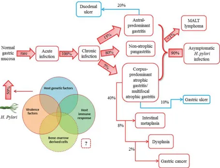
In most infected people, the bacterium acts as a commensal organism inducing chronic asymptomatic gastritis that can last for life. In other cases, however, it is responsible for a high morbidity and mortality as a consequence of peptic ulcers and gastric cancer. Chronic gastritis may progress to intestinal metaplasia, dysplasia and eventually gastric cancer (Fig. 2), a multi-step process known as the Correa pathway [15,31]. H. pylori infection is the strongest known risk factor for gastric cancer, as supported by epidemiological, preclinical and clinical studies [9,14]. In fact, gastric cancer is the fourth cause of death because of a cancer in Europe (GLOBOCAN 2012; http://globocan.iarc.fr/).
However, the mechanism of H. pylori developing gastric carcinoma has not been well defined. Among infected individuals, approximately 10% develop severe gastric lesions such as peptic ulcer disease and 1–3% progresses to gastric cancer. The outcomes of H. pylori infection are determined by bacterial virulence, genetic polymorphism of both hosts and bacteria, as well as environmental factors [9,31]. Environmental factors, such as cigarette smoking, are major risk factors for duodenal ulceration among H. pylori-infected people [3]. Other important factors include stress, childhood living conditions, diet, alcohol and use of non-steroidal anti-inflammatory drugs (NSAIDs) [3,31,32].
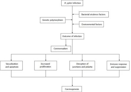
H. pylori infection commonly lasts for decades, provoking a series of
histological changes including destruction of intercellular junctions, apoptosis and proliferation of epithelial cells and malignant transformation (Fig. 3) [9,33,34]. The genotypes of H. pylori strains, host genetic
polymorphisms, environmental factors like high salt diet, smoking habit and certain gastric commensal organisms have been determined to be associated with occurrence of gastric cancer. H. pylori genetic polymorphisms, effects of specific H. pylori products on gastric epithelium and cellular signaling process have been intensively investigated in recent decades [9,35].
Figure 2. Natural history of H. pylori infection. H. pylori is usually acquired in
childhood, whereas acute infection with the bacterium is rarely diagnosed. Instead, chronic gastritis develops in almost all persistently colonized individuals, 90% of whom will remain asymptomatic. The clinical course of H. pylori infection is highly variable depending on bacterial and host (genetic and immune) factors. Recent studies have supported the possible role of bone marrow-derived cells (i.e., gastric stem cells) in tumor progression. Patients with increased acid secretion are more likely to have antral-predominant gastritis, which predisposes to duodenal ulcers. Patients with low acid secretion will more likely develop gastritis in the body of the stomach and are thus more likely to develop gastric ulcer, leading to gastric atrophy, intestinal metaplasia, dysplasia and, finally, in rare cases, gastric carcinoma. This sequence of events is more frequent in people of advanced age. H. pylori infection induces the formation of mucosa-associated lymphoid tissue (MALT) in the gastric mucosa and MALT lymphoma is another rare complication of H. pylori infection. (reprinted by permission from Spandidos Publications: ref. [31], copyright 2013).
Besides its arsenal of virulence factors, persistence of H. pylori is strongly influenced by the ability of the bacterium to evade, overthrow and manipulate the host’s immune system. This bacterium can evade detection by several innate immune receptors through target modification and it can subvert other innate recognition pathways through the suppression of downstream signal transduction, whereas evasion of adaptive immunity is achieved by the modulation of effector T cell functions [36].
Despite the host’s vigorous immune response, H. pylori is capable of persisting for decades in its human host. Interestingly, coccoid forms appear to be the persisting form, allowing H. pylori to spread between human hosts. It has been shown that the morphological transition from bacillary to coccoid forms is accompanied by modifications of the bacterial cell wall peptidoglycan, in which AmiA protein is essential. Cell wall modifications and morphological transition allow the coccoid forms to escape detection by
the immune system and therefore could participate in the persistence of
H. pylori infection during the lifetime of its human host [4].
Figure 3. The pathogenesis of Helicobacter pylori-associated gastric cancer is a
multi-factorial process, its development depends on a combination of host, bacterial and environmental factors, and the pathological changes might progress in steps. (reprinted by permission from Baishideng Publishing Group Inc.: ref. [9], copyright 2016).
4. H. pylori virulence factors
Crucial bacterial factors associated with pathogenicity comprise flagella, urease, peptidoglycan, lipopolysaccharide (LPS), Hsp60 (= Cpn60) chaperone, a type IV secretion system encoded by the cag pathogenicity island containing the effector protein CagA, the vacuolating cytotoxin (VacA), and others [37].
The high number of these factors and allelic variation of the involved genes generates a highly complex scenario and reveals the difficulties in testing the contribution of each individual factor. Much effort has been put into identifying the molecular mechanisms associated with H. pylori-associated pathogenesis using human primary tissues, Mongolian gerbils, transgenic, knockout and other mice, as well as in vitro cell model systems [37].
Studies on H. pylori heterogeneity have proved that the strongest virulence factors were amongst the genes within the cag pathogenicity island (PAI). The mechanisms underlying H. pylori-related gastric carcinogenesis, however, remain unclear [9], with the virulence factors CagA and VacA having a prominent role.
Development of gastric and hematological carcinoma has been observed in the mice that were genetically modified to express CagA [9,38]. Studies revealed that cagA+/vacAs1+/vacAm1+ H. pylori strains promoted pathogenesis of intestinal metaplasia and gastric carcinoma [9,39].
4.1. Flagella
H. pylori possess two to six sheathed unipolar flagella, which confer
motility and constitute a virulence factor shown to be absolutely essential for colonization [40]. Non-motile mutants lacking flagella are unable to establish persistent infection in animal models [40,41,42], although it has also been shown that the presence of flagella alone is not sufficient for colonization [40,43]
4.2. Urease
Urease can play a broad role in the pathogenesis associated with
H. pylori infection. Although urease is known for its enzymatic activity
(converts urea into ammonia and bicarbonate to counteract the low acidity of the stomach), it is also correlated with the dual function of adhesivity and immunogenicity [3,44].
4.3. Peptidoglycan
The mechanism by which H. pylori generates helical shape is unknown. A novel study identified four genes being involved: peptidoglycan endopeptidases (csd1-3) and ccmA. The findings suggest that the coordinated action of multiple proteins relaxes peptidoglycan crosslinking, enabling helical cell curvature and twist, which is required for robust bacterial colonization in the stomach [35,45].
4.4. Lipopolysaccharide
O polysaccharides of the gastric pathogen H. pylori contain Lewis antigens, mimicking glycan structures produced by human cells. The interaction of Lewis antigens with human dendritic cells induces a modulation of the immune response, contributing to the H. pylori virulence.
The amount and position of Lewis antigens in the LPS varies among
H. pylori isolates, indicating an adaptation to the host [46].
Two new enzymes typically involved in LPS biosynthesis were discovered in H. pylori, glycosyltransferase WecA and O-antigen ligase WaaL, but a flippase enzyme, normally involved in O-antigen synthesis, could not be detected. Instead, H. pylori uses a translocase named Wzk in a novel LPS biosynthetic pathway, evolutionarily connected to protein N-glycosylation [35,46]. In addition, 3-deoxy-d-manno-octulosonic acid (Kdo) hydrolase genes (HP0579 and HP0580) were shown to affect the expression of O-antigen Lewis epitopes [47]. Finally, an ADP-l-glycero-d-manno-heptose-6-epimerase ortholog (HP0859) was characterized. DHP0859 mutants exhibited severe truncation of LPS, decreased growth rate, higher susceptibility to novobiocin, and failed to induce the AGS elongation phenotype in AGS cells. Genetic complementation restored these phenotypes, revealing HP0859 essential for LPS biosynthesis and virulence [35,48].
4.5. Hsp60 superficial protein
H. pylori produces two heat shock proteins (Hsps): a groEs-like HspsA
(size 13 kD) and groEL-like HspsB (size 54–60 kD, Hsps60). Hsps60 is in particular shared by H. pylori and eukaryotic cells [3,49]. The homologies between bacteria and human Hsp60 antigens leads to an antibody response directed to both the bacteria and to human tissues that express Hsps, including vascular endothelial cells (autoimmune response) [3].
4.6. CagA
CagA, a highly immunogenic protein, is encoded at one end of the cag PAI, which encode the components to form the type IV secretion system (T4SS) [7,9,50]. As a component of T4SS, CagL protein binds to and activates the integrin α5β1 receptor on gastric epithelial cells and triggers CagA delivery into the target cells [9,51].
H. pylori strains harboring the cag PAI or producing CagA are related
to enhanced inflammation and risk of ulcers and carcinoma [9,52,53]. Moreover, the C-terminal EPIYA-containing region of CagA is polymorphic and isolates from gastric cancer patients frequently have a high number of EPIYA motifs [54]. CagA contributes to myriad signaling alterations, which profoundly affects physiology of host epithelial cells [9]. Once inside host cells, CagA is tyrosine phosphorylated, triggering the cellular signaling pathways leading to expression of proinflammatory cytokines and chemokines, and deregulates the signaling pathways that control host cell shape, adhesion and transformation [9,35,55].
Unphosphorylated CagA interacts with certain intracellular proteins, up-regulate production of proinflammatory cytokines, provoke mitogenic responses and disrupt intercellular junctions and epithelial cell polarit y [9,55,56]. Additionally, CagA intoxicates dendritic cells leading to impaired activation, decreased inflammatory cytokine production and Th1 immune response [9,57]. Recently, it was confirmed that H. pylori infection resulted in rapid association of the virulence factor CagA with the c-Met receptor, activation of signaling and epithelial proliferation [9,58].
4.7. Vacuolating cytotoxin A
Vacuolating cytotoxin A (VacA) is a bacterial toxin with multiple activities, contributing to multiple structural and functional alterations of epithelial cells [7,9]. After secretion by the bacterium through the type V secretion system, VacA binds to host cells interfering with endosomal maturation and leading to vacuolation, enhances leakage of nutrients by destruction of barrier function at tight junctions of epithelial cells,
provokes mitochondrial damage and cell apoptosis, which improves
H. pylori growth [9,59,60,61]. Recent studies proved that VacA could
disrupt phagocytosis, interfere with antigen presentation, restrain T cell activation in vitro and inhibit T cell proliferation independing of NFAT
(nuclear factor of activated T cells) activation or IL-2 expression [9,62,63,64]. These effects of VacA on the immune system may explain how H. pylori evades adaptive immune responses to establish persistent infection [9].
The secreted mature form of VacA is a 88 kDa monomer comprising two domains designated p33 (residues 1–311) and p55 (residues 312–821) [65]. VacA harbours different allelic variants that modulate its activity. The amino-terminal end of VacA (residues -33–1), designated as the signal peptide or ―s-region‖, carrying the s1 or s2 alleles; the intermediate region or ―i-region‖ that encodes part of the p33 (residues 190–223) with the allelic variants i1 or i2; and the mid region or ―m-region‖, existing as an m1 or m2 allele [7,65,66]. Mosaic combinations of the s-, i- and m-regions exist and relate to the in vitro cytotoxin activity. Strains with alleles s1/i1/m1 have been associated with peptic ulcer disease and gastric cancer [67]. González et al. (2011) [7] showed that patients infected with high virulence strains (cagA+, vacA s1i1m1) have a higher risk of progression of the preneoplastic lesions to gastric cancer in relation to patients infected with low virulence strains.
4.8. Other virulence factors
Adherence to epithelial cells is essential for H. pylori colonization and delivery of virulence factors to host cells. Some H. pylori adhesins have been described, including the sialic acid-binding adhesins BabA and SabA [9,68,69], and OipA, an inflammation-related outer membrane protein [9,70]. Other outer membrane proteins (OMPs) as HomA and HomB have been associated with peptic ulcer disease (PUD) [3,71,72]. The development of duodenal ulcer has also been correlated to the presence of the duodenal ulcer promoting gene A (dupA), but its role in gastric cancer is controversial [70].
Other H. pylori virulence factors described are HrgA, GGT, HP-NAP, HopQ, etc. Restriction endonuclease-replacing gene A (hrgA) is considered as an important virulence determinant in H. pylori-associated gastric diseases such as carcinoma [3,73,74]. GGT is a gamma-glutamyl transpeptidase related to increased levels of hydrogen peroxide and IL-8 in epithelial cells and H. pylori-associated diseases [9,75,76,77]. The H. pylori neutrophil-activating protein (HP-NAP) is another virulence determinant that stimulates neutrophil high production of oxygen radicals and adhesion to endothelial cells [3,78]. It has also been shown that the outer membrane
protein HopQ is involved in leukocyte migration inhibition, together with CagA, suggesting an unprecedented role of the HopQ-CagA axis in immune modulation [79].
5. Currently used diagnosis methods
In patients with H. pylori-related diseases, a reliable diagnosis of infection with this bacterium is crucial, but no single test can be considered the gold standard [80,81]. Testing for H. pylori infection is indicated in patients with active peptic ulcer disease, a past history of documented peptic ulcer, or gastric MALT lymphoma [82].
Different invasive and non-invasive diagnostic tests are available for the diagnosis of H. pylori in the individual patient. The non-invasive tests obviate the need for endoscopy and can be surely more accepted by the subjects [3].
Histology is usually considered to be the gold standard in the direct detection of H. pylori infection and is also the first method used for the detection of H. pylori. Several factors, however, influence the diagnostic accuracy of histology, such as site, size and number of biopsies, staining methods, proton pump inhibitor, antibiotics and experience of the examining pathologist [81]. The rapid urease test (RUT) is the most commonly used biopsy-based method to diagnose H. pylori infection because it is simple, rapid and accurate. However, it requires a high density of bacteria, and anything that reduces the bacterial load may produce false-negative tests [80,83]. Culturing of H. pylori from gastric biopsy specimen is a highly specific but less sensitive method. In general, culturing has almost 100% specificity, but the sensitivity of culture shows significant variation, between 85-95% [81].
The non-invasive tests as diagnostic tool in H. pylori infections of patients with various gastrointestinal disorders are strongly important because they make the endoscopy unnecessary in different situations [3]. Urea breath test (UBT) has been used for almost 30 years and is still the most popular and accurate noninvasive test for diagnosis of H. pylori infection. By the urease activity of H. pylori, the 13C- or 14C-labeled urea ingested by the patient is hydrolyzed to labeled CO2 in stomach, then labeled CO2 is absorbed in the blood and exhaled by breathing in which labeled CO2 is measured [81]. Stools antigen test (SAT) is the other noninvasive method with good sensitivity and specificity (94% and 97%, respectively) in the diagnosis of H. pylori infection [81].
The pre-endoscopy screening may be performed principally through serological markers (detection of different kinds of immunoglobulins). For CagA detection, serology has proved to be useful, being CagA protein a factor with good antigenic properties, easy and reliable to perform and prone to reveal the presence of cag pathogenicity island [3,84]. Hsp60 is also a good antigen so that its detection can be performed through the appearance of specific antibodies against it [3,85].
The rapid and easy detection of virulent strains using novel PCR methods has been suggested as a mean to avoid the consequences of a long-lasting untreated infection [3,80]. Real-time PCR is a sensitive, accurate method of diagnosing H. pylori infection. Compared to other available methods for diagnosing H. pylori for clinical and research purposes, PCR yields high sensitivity and specificity for H. pylori in frozen samples from patients with nonbleeding and bleeding peptic ulcer [86].
A useful approach is to use the new and advanced multiplex PCR methods in gastric tissue samples, which could contribute to gain insights into the genotypic variability exhibited by this pathogen [3,80,87]. Novel ultra-sensitive methods as digital PCR could also contribute to detect cases of ―occult infection‖, which were traditionally considered as false-positive results in the 13C-urea breath test [88].
6. Bacterial genetic diversity and role of mixed infections
The H. pylori genome (1.65 Mbp) codes for about 1,500 proteins [14].
H. pylori has been reported to be genetically extremely variable and this
heterogeneity is proposed to be involved in the ability of H. pylori to cause different diseases [3,89,90], detrimental and non-detrimental chronic infections [3,7,91].
The remarkable adaptive capacity of H. pylori can be partly attributed to the enormous plasticity of its genome, caused by high mutation and recombination rates, and by the ability for aberrant genomic rearrangements and incorporation of non-homologous DNA. This results in an exceptionally high sequence diversity among strains that were isolated from unrelated human hosts and in an H. pylori core genome that consists of only approximately 1,100 genes, while the other approximately 400–500 genes per strain are strain-specific and variably present [92].
Strains of H. pylori exhibit considerable genetic diversity following a panmictic (non-clonal) population structure due to horizontal gene transfer and frequent recombination [21,22,93]. The estimated mutation rate reveals a mutation burst during the acute infection phase that is over 10 times faster
than the mutation rate during chronic infection, and orders of magnitude faster than mutation rates in any other bacteria. The elevated frequency of mutations in outer membrane protein genes suggests that the mutation burst facilitates rapid host adaptation of the bacteria [92].
In a recent study, it has been suggested that prolonged in vivo exposure of H. pylori to the gastric environmental conditions associated with a high-salt diet can lead to the emergence of strains adapted to these conditions. It has been shown that the production of proteins involved in iron acquisition and oxidative-stress resistance in H. pylori strains cultured from animals on high-salt diet differs from that of the input strain and strains isolated from animals on a regular diet [94].
It seems that the number of H. pylori clones isolated from a single host varies depending on the geographic region and other factors as age, socioeconomic and hygienic status [16]. It has also been demonstrated the importance of mixed infections for the genetic diversification of H. pylori through recombination [95]. H. pylori mixed infections with facilitate interstrain gene transfer and the maintenance of genetic diversity for adaptation to the gastric environment, but whether mixed infections with histological significance and tissue tropism occur in the human stomach is still unclear [96].
Recent data suggest that around 23% of H. pylori-infected patients have mixed infections, with different dominant strains isolated from the antrum and the corpus specimens, and being mixed infection significantly related to the appearance of intestinal metaplasia in the antrum [96]. The role of mixed infections in the development of gastric cancer, however, deserves further investigation.
Mixed infection has also been described in the pediatric population [52]. It has been shown that the panmictic population structure of H. pylori results from very frequent recombination during mixed colonization by unrelated strains [97].
Results of genetic studies comparing sequences of housekeeping genes have shown natural mixed infection in family members. Identical alleles found in some strains isolated from the children and parents, but not from unrelated patients, demonstrated that strains had circulated within the family. Several mechanisms, such as point mutations, intragenic recombination, and introduction of foreign alleles, were shown to enhance strain diversity within the family [13].
Using enzyme-linked immunosorbent assay and line probe assay (LiPA) techniques, the prevalence of mixed H. pylori colonization was analyzed in 127 subjects from Venezuela, a country of high H. pylori prevalence, from three regions representing different population groups: the Andes (Merida),
where Caucasian mestizos predominate, a major city near the coast (Caracas), where Amerindian-Caucasian-African mestizos predominate, and an Amazonian community (Puerto Ayacucho), where Amerindians predominate and mestizos reflect Amerindian and Caucasian ancestry. Among 121 H. pylori-positive persons, the prevalence of cagA-positive strains varied from 50% (Merida) to 86% (Puerto Ayacucho) by LiPA. Rates of mixed colonization also varied, as assessed by LiPA of the vacA s (mean, 49%) and m (mean, 26%) regions. In total, 55% of the individuals had genotypic evidence of mixed colonization. vacA s1c, a marker of Amerindian (East Asian) origin, was present in all three populations, especially from Puerto Ayacucho (86%). These results demonstrate the high prevalence of mixed colonization and indicate that the H. pylori East Asian
vacA genotype has survived in all three populations tested [1].
7. Research on new markers of H. pylori infection and
progression
The low incidence of severe disease associated with infection has suggested that there may be ―beneficial‖ H. pylori organisms in addition to
those that cause disease. In fact, since humans have co-evolved with
H. pylori, they might derive some benefits from them [3,98]. Certainly, man
adapts physiologically to infection during his lifetime and treatment can lead to problems such as reflux oesophagitis. In view of these real problems, it would seem preferable to screen for and treat only strains that are known to cause disease. In H. pylori a lot of virulence determinants have been detected affecting the infection course [3]. To date, despite intensive research, not enough knowledge exit on clear risk or disease-specific markers [99].
Recent studies have analysed miRNA expression in human H. pylori-infected patients with or without duodenal ulcer disease. Results obtained have shown that a set of miRNA are deregulated during chronic gastric inflammation and could be useful as a surrogate marker for determining the presence of H. pylori [99].
In our research group, we have amplified and sequenced six housekeeping genes (amiA, cgt, cpn60, cpn70, dnaJ, and luxS) related to H.
pylori pathogenesis to evaluate their usefulness for the specific detection of H. pylori, the genetic discrimination at strain level and the detection of
multiple infection. A total of 52 H. pylori clones, isolated from 14 gastric biopsies from 11 patients, were analyzed for this purpose [93].
All genes were specifically amplified for H. pylori and all clones isolated from different patients were discriminated, with gene distances ranged from 0.9 to 7.8%. Although most clones isolated from the same
patient showed identical gene sequences, an event of multiple infection was detected in all the genes and microevolution events were showed for amiA and cpn60 genes [93].
These results suggest that housekeeping genes could be useful for
H. pylori detection and to elucidate the mode of transmission and the
relevance of the multiple infection [93]. These genes are potential candidates to detect H. pylori infection in gastric biopsies and other specimens (as gastric juice, stool, saliva, dental plaque, water, and food samples), together with others, mainly used for gastric biopsies, as ureA, vacA, 16S rRNA, and 23S rRNA genes, previously described [81,86,93].
In our study, two different strains isolated from the antral biopsy B657A were clearly detected by all genes with high distance values (1.6–7.3%). One strain was represented by the identical clones B657A-1, -2 and -3, and the other strain by the clone B657A-4, which was identical to clones isolated from B657C (corpus biopsy from the same patient), except in the case of
amiA. This high divergence observed between both strains indicates an event
of multiple infection in the antrum of the stomach [93]. Since the role of multiple infections on disease outcome and progression is still unclear [1,13,93,96], we consider that the genes sequenced in our study could be used in further research in this issue.
Of note, concatenation of amiA, cgt, cpn60, cpn70, dnaJ, and luxS partial sequences allowed us to conduct a multilocus sequence analyses (MLSA) approach, which is useful to elucidate intra- and interspecies phylogenetic relationships [93,100,101] and, in the case of H. pylori, the phylogeographic differentiation of bacterial populations associated to the migration of human populations [18,21,93]. Most of these genes had never been used for these purposes before, except dnaJ and, especially, cpn60, a gene useful for microbial phylogeny, detection and identification, ecology, and evolution through the analysis of the 555 bp region known as universal target (UT) analyzed in our study [93,102,103].
In conclusion, our results provide new tools for the detection of
H. pylori, with potential applications for H. pylori detection in different
specimens and to assess the mode of transmission, the role associated to virulence or the phylogeographic differentiation of H. pylori populations [93].
8. Conclusions
Stomach cancer is one of the leading causes of cancer death worldwide, despite its incidence and mortality falling in many places. The discovery in 1984 that a bacterial infection with H. pylori could cause stomach and
duodenal ulcers prompted work in its role in causing gastritis, and led to the first prospective study in 1991 by Forman et al., showing that infection with
H. pylori increased the risk of stomach cancer in those infected by almost
three-fold [104].
Efforts to reduce the incidence of gastric cancer by means of eradicating
H. pylori have been variable (because of re-infections) and there is still the
debate around population screening and opportunistic ―seek and treat‖ of
H. pylori in the absence of symptoms [104]. Instead, it would seem
preferable to screen for and treat only strains that are known to cause disease, as a promise targeted intervention [3,104].
Research on the bacterial virulence factors, effects of H. pylori on epithelial cells, genetic polymorphism of both the bacterium and its host, and the environmental factors can help to understand the role of this bacterium in gastric carcinogenesis [9].
Other challenging issues, needing further research, are the elucidation of transmission, implication of H. pylori in other diseases, the role of mixed infections, the development of new sensitive and specific PCR methods for
H. pylori detection in gastric samples and other specimens, and the
development of a safe and efficient preventive vaccine that could address the wide diversity of strains.
In this context, results of our genetic study from H. pylori clones isolated from gastric biopsies can contribute to the advances in all these fields. Our results suggest that housekeeping genes could be useful for
H. pylori detection and to elucidate the mode of transmission and the
relevance of multiple infection [93]. We consider that further genetic studies, including sequencing of complete genomes of virulent strains and metagenomics, will provide powerful tools to face all current unmet challenges of H. pylori infection.
Acknowledgements
This work has been supported by a project from the Fundació La Marató de TV3 (1007/C/2013), Catalonia.
References
1. Ghose, C., Pérez-Pérez, G. I., van Doorn, L. J., Domínguez-Bello, M. G., Blaser, M. J. 2005, J. Clin. Microbiol., 43, 2635.
2. Khalifa, M. M., Sharaf, R. R., Aziz, R. K. 2010, Gut Pathog., 2, 2. 3. Mascellino, M. T., Margani, M., Oliva, A. 2009, Dis. Markers., 27, 137.
4. Chaput, C., Ecobichon, C., Cayet, N., Girardin, S. E., Werts, C., Guadagnini, S., Prévost, M. C., Mengin-Lecreulx, D., Labigne, A., Boneca, I. G. 2006, PLoS
Pathog., 2, e97.
5. Vega, A. E., Cortiñas, T. I., Mattana, C. M., Silva, H. J., Puig de Centorbi, O. 2003, J. Clin. Microbiol., 41, 5384.
6. Correa, P., Schneider, B. G. 2005, Cancer Epidemiol. Biomarkers Prev., 14, 1865.
7. González, C. A., Figueiredo, C., Lic, C. B., Ferreira, R. M., Pardo, M. L., Ruiz Liso, J. M., Alonso, P., Sala, N., Capella, G., Sanz-Anquela, J. M. 2011, Am. J.
Gastroenterol., 106, 867.
8. Mishra, S. 2013, Eur. J. Clin. Microbiol. Infect. Dis., 32, 301.
9. Zhang, R. G., Duan, G. C., Fan, Q. T., Chen, S. Y. 2016, World J. Gastrointest.
Pathophysiol., 7, 97.
10. Atherton, J. C. 2006, Annu. Rev. Pathol., 1, 63.
11. Ando, T., Minami, M., Ishiguro, K., Maeda, O., Watanabe, O., Mizuno, T., Fujita, T., Takahashi, H., Noshiro, M., Goto, H. 2006, Aliment Pharmacol.
Ther., 24, 58.
12. Rabelo-Gonçalves, E. M., Roesler, B. M., Zeitune, J. M. 2015, World J.
Hepatol., 7, 2968.
13. Raymond, J., Thiberg, J. M., Chevalier, C., Kalach, N., Bergeret, M., Labigne, A., Dauga, C. 2004, Emerg. Infect. Dis., 10, 1816.
14. Suerbaum, S., Michetti, P. 2002, N. Engl. J. Med., 347, 1175. 15. Correa, P., Houghton, J. 2007, Gastroenterology, 133, 659.
16. Toita, N., Yokota, S., Fujii, N., Konno, M. 2013, Gastroenterol. Res. Pract., 2013, 721306.
17. Eusebi, L. H., Zagari, R. M., Bazzoli, F. 2014, Helicobacter, 19, 1. 18. Suzuki, R., Shiota, S., Yamaoka, Y. 2012, Infect. Genet. Evol., 12, 203.
19. Wirth, T., Wang, X., Linz, B., Novick, R. P., Lum, J. K., Blaser, M., Morelli, G., Falush, D., Achtman, M. 2004, Proc. Natl. Acad. Sci. USA, 101, 4746.
20. Falush, D., Wirth, T., Linz, B., Pritchard, J. K., Stephens, M., Kidd, M., Blaser, M. J., Graham, D. Y., Vacher, S., Perez-Perez, G. I., Yamaoka, Y., Mégraud, F., Otto, K., Reichard, U., Katzowitsch, E., Wang, X., Achtman, M., Suerbaum, S. 2003, Science, 299, 1582.
21. Linz, B., Balloux, F., Moodley, Y., Manica, A., Liu, H., Roumagnac, P., Falush, D., Stamer, C., Prugnolle, F., van der Merwe, S. W., Yamaoka, Y., Graham, D. Y., Perez-Trallero, E., Wadstrom, T., Suerbaum, S., Achtman, M. 2007, Nature, 445, 915.
22. Moodley, Y., Linz, B., Yamaoka, Y., Windsor, H. M., Breurec, S., Wu, J. Y., Maady, A., Bernhöft, S., Thiberge, J. M., Phuanukoonnon, S., Jobb, G., Siba, P., Graham, D. Y., Marshall, B. J., Achtman, M. 2009, Science, 323, 527. 23. Yamaoka, Y. 2010, Nat. Rev. Gastroenterol. Hepatol., 7, 629.
24. Maixner, F., Krause-Kyora, B., Turaev, D., Herbig, A., Hoopmann, M. R., Hallows, J. L., Kusebauch, U., Vigl, E. E., Malfertheiner, P., Megraud, F., O'Sullivan, N., Cipollini, G., Coia, V., Samadelli, M., Engstrand, L., Linz, B.,
Moritz, R. L., Grimm, R., Krause, J., Nebel, A., Moodley, Y., Rattei, T., Zink, A. 2016, Science, 351, 162.
25. Anand, P. S., Kamath, K. P., Anil, S. 2014, World J. Gastroenterol., 20, 5639. 26. Parsonnet, J., Shmuely, H., Haggerty, T. 1999, JAMA, 282, 2240.
27. Luzza, F., Mancuso, M., Imeneo, M., Contaldo, A., Giancotti, L., Pensabene, L., Doldo, P., Liberto, M. C., Strisciuglio, P., Focà, A., Guandalini, S., Pallone, F. 2000, Eur. J. Gastroenterol. Hepatol., 12, 623.
28. Thomas, J. E., Gibson, G. R., Darboe, M. K., Dale, A., Weaver, L. T. 1992,
Lancet, 340, 1194.
29. Kelly, S. M., Pitcher, M. C., Farmery, S. M., Gibson, G. R. 1994,
Gastroenterology, 107, 1671.
30. Atapoor S., Safarpoor Dehkordi, F., Rahimi, E. 2014, Jundishapur J.
Microbiol., 7, e10013.
31. Conteduca, V., Sansonno, D., Lauletta, G., Russi, S., Ingravallo, G., Dammacco, F. 2013, Int. J. Oncol., 42, 5.
32. Piqué, N., Ponce, M., Garrigues, V., Rodrigo, L., Calvo, F., de Argila, C. M., Borda, F., Naranjo, A., Alcedo, J., Soria, M. J., Rey, E., Bujanda, L., Gisbert, J. P., Suárez, D., Calvet, X., Ponce, J. 2016, United European Gastroenterol. J., 4, 229.
33. Watari, J., Chen, N., Amenta, P. S., Fukui, H., Oshima, T., Tomita, T., Miwa, H., Lim, K. J., Das, K. M. 2014, World J. Gastroenterol., 20, 5461. 34. Xia, H. H., Talley, N. J. 2001, Am. J. Gastroenterol., 96, 16.
35. Wen, S., Moss, S. F. 2009, Cancer Lett., 282, 1.
36. Salama, N. R., Hartung, M. L., Müller, A. 2013, Nat. Rev. Microbiol., 11, 385. 37. Backert, S., Clyne, M. 2011, Helicobacter, 16, 19.
38. Ohnishi, N., Yuasa, H., Tanaka, S., Sawa, H., Miura, M., Matsui, A., Higashi, H., Musashi, M., Iwabuchi, K., Suzuki, M., Yamada, G., Azuma, T., Hatakeyama, M. 2008, Proc. Natl. Acad. Sci. USA, 105, 1003. 39. Wang, F., Wu, X., Liu, Z., Bu, G., Li, X., Qu, N., Peng, J., Xu, C., Shen, S.,
Yuan, Y. 2015, Gastroenterol. Res. Pract., 2015, 648479.
40. Dunne, C., Dolan, B., Clyne, M. 2014, World J. Gastroenterol., 20, 5610.
41. Eaton, K. A., Morgan, D. R., Krakowka, S. 1992, J. Med. Microbiol., 37, 123. 42. Eaton, K. A., Suerbaum, S., Josenhans, C., Krakowka, S. 1996, Infect.
Immun., 64, 2445.
43. Ottemann, K. M., Lowenthal, A. C. 2002, Infect. Immun., 70, 1984.
44. Beswick, E. J., Pinchuk, I. V., Minch, K., Suárez, G., Sierra, J. C., Yamaoka, Y., Reyes, V. E. 2006, Infect. Immun., 74, 1148.
45. Sycuro, L. K., Pincus, Z., Gutierrez, K. D., Biboy, J., Stern, C. A., Vollmer, W., Salama, N. R. 2010, Cell, 141, 822.
46. Hug, I., Couturier, M. R., Rooker, M. M., Taylor, D. E., Stein, M., Feldman, M. F. 2010, PLoS Pathog., 6, e1000819.
47. Stead, C. M., Zhao, J., Raetz, C. R., Trent, M. S. 2010, Mol. Microbiol., 78, 837. 48. Chang, P. C., Wang, C. J., You, C. K., Kao, M. C. 2011, Biochem. Biophys. Res.
49. Lenzi, C., Palazzuoli, A., Giordano, N., Alegente, G., Gonnelli, C., Campagna, M. S., Santucci, A., Sozzi, M., Papakostas, P., Rollo, F., Nuti, R., Figura, N. 2006, World J. Gastroenterol., 12, 7815.
50. Tegtmeyer, N., Wessler, S., Backert, S. 2011, FEBS J., 278, 1190.
51. Murata-Kamiya, N., Kikuchi, K., Hayashi, T., Higashi, H., Hatakeyama, M. 2010, Cell Host Microbe, 7, 399.
52. Talarico, S., Gold, B. D., Fero, J., Thompson, D. T., Guarner, J., Czinn, S., Salama, N. R. 2009, J. Clin. Microbiol., 47, 1680.
53. Kim, S. S., Ruiz, V. E., Carroll, J. D., Moss, S. F. 2011, Cancer Lett., 305, 228. 54. Argent, R. H., Kidd, M., Owen, R. J., Thomas, R. J., Limb, M. C., Atherton, J.
C. 2004, Gastroenterology, 127, 514.
55. Alzahrani, S., Lina, T. T., Gonzalez, J., Pinchuk, I. V., Beswick, E. J., Reyes, V. E. 2014, World J. Gastroenterol., 20, 12767.
56. Amieva, M. R., Vogelmann, R., Covacci, A., Tompkins, L. S., Nelson, W. J., Falkow, S. 2003, Science, 300, 1430.
57. Tanaka, H., Yoshida, M., Nishiumi, S., Ohnishi, N., Kobayashi, K., Yamamoto, K., Fujita, T., Hatakeyama, M., Azuma, T. 2010, Arch. Biochem. Biophys., 498, 35.
58. McCracken, K. W., Catá, E. M., Crawford, C. M., Sinagoga, K. L., Schumacher, M., Rockich, B. E., Tsai, Y. H., Mayhew, C. N., Spence, J. R., Zavros, Y., Wells, J. M. 2014, Nature, 516, 400.
59. Palframan, S. L., Kwok, T., Gabriel, K. 2012, Front. Cell Infect. Microbiol. 2, 92.
60. Manente, L., Perna, A., Buommino, E., Altucci, L., Lucariello, A., Citro, G., Baldi, A., Iaquinto, G., Tufano, M. A., De Luca, A. 2008, J. Cell Physiol., 214, 582.
61. Papini, E., Satin, B., Norais, N., de Bernard, M., Telford, J. L., Rappuoli, R., Montecucco, C. 1998, J. Clin. Invest. 102, 813.
62. Amieva, M. R., El-Omar, E. M. 2008, Gastroenterology, 134, 306. 63. Allen, L. A., Schlesinger, L. S., Kang, B. 2000, J. Exp. Med., 191, 115.
64. Torres, V. J., VanCompernolle, S. E., Sundrud, M. S., Unutmaz, D., Cover, T. L. 2007, J. Immunol., 179, 5433.
65. Atherton, J. C., Cao, P., Peek, R. M. Jr, Tummuru, M. K., Blaser, M. J., Cover, T. L. 1995, J. Biol. Chem., 28, 270.
66. Kim, I. J., Blanke, S. R. 2012, Front. Cell. Infect. Microbiol., 2, 37.
67. Basso, D., Zambon, C. F., Letley, D. P., Stranges, A., Marchet, A., Rhead, J. L., Schiavon, S., Guariso, G., Ceroti, M., Nitti, D., Rugge, M., Plebani, M., Atherton, J. C. 2008, Gastroenterology, 135, 91.
68. Mahdavi, J., Sondén, B., Hurtig, M., Olfat, F. O., Forsberg, L., Roche, N., Angstrom, J., Larsson, T., Teneberg, S., Karlsson, K. A., Altraja, S., Wadström, T., Kersulyte, D., Berg, D. E., Dubois, A., Petersson, C., Magnusson, K. E., Norberg, T., Lindh, F., Lundskog, B. B., Arnqvist, A., Hammarström, L., Borén, T. 2002, Science, 297, 573.
69. Ishijima, N., Suzuki, M., Ashida, H., Ichikawa, Y., Kanegae, Y., Saito, I., Borén, T., Haas, R., Sasakawa, C., Mimuro, H. 2011, J. Biol. Chem., 286, 25256.
70. Yamaoka, Y., Kwon, D. H., Graham, D. Y. 2000, Proc. Natl. Acad. Sci. USA, 97, 7533.
71. Ogura, K., Maeda, S., Nakao, M., Watanabe, T., Tada, M., Kyutoku, T., Yoshida, H., Shiratori, Y., Omata, M. 2000, J. Exp. Med., 192, 1601.
72. Oleastro, M., Cordeiro, R., Ferrand, J., Nunes, B., Lehours, P., Carvalho-Oliveira, I., Mendes, A. I., Penque, D., Monteiro, L., Mégraud, F., Ménard, A. 2008, J. Infect. Dis., 198, 1379.
73. Ando, T., Wassenaar, M., Peek, R. M., Aras, R. A., Tschumi, A. I., van Doorn, L. J., Kusugami, K., Blaser, M. J. 2002, Cancer Res., 62, 2385.
74. Ando, T., Aras, R. A., Kusugami, K., Blaser, M. S., Wassenaar, T. M. 2003, J.
Bacteriol., 185, 295.
75. Gong, M., Ling, S. S., Lui, S. Y., Yeoh, K. G., Ho, B. 2010, Gastroenterology, 139, 564.
76. Olofsson, A., Vallström, A., Petzold, K., Tegtmeyer, N., Schleucher, J., Carlsson, S., Haas, R., Backert, S., Wai, S. N., Gröbner, G., Arnqvist, A. 2010,
Mol. Microbiol., 77, 1539.
77. Rimbara, E., Mori, S., Kim, H., Shibayama, K. 2013, Microbiol. Immunol., 57, 665.
78. Amedei, A., Cappon, A., Codolo, G., Cabrelle, A., Polenghi, A., Benagiano, M., Tasca, E., Azzurri, A., D'Elios, M. M., Del Prete, G., de Bernard, M. J. 2006,
Clin. Invest., 116, 1092.
79. Busch, B., Weimer, R., Woischke, C., Fischer, W., Haas, R. 2015, Int. J. Med.
Microbiol., 305, 355.
80. Chung, W. C., Jung, S. H., Oh, J. H., Kim, T. H., Cheung, D. Y., Kim, B. W., Kim, S. S., Kim, J. I., Sin, E. Y. 2014, World J. Gastroenterol., 20, 6547. 81. Wang, Y. K., Kuo, F. C., Liu, C. J., Wu, M. C., Shih, H. Y., Wang, S. S., Wu, J.
Y., Kuo, C. H., Huang, Y. K., Wu, D. C. 2015, World J. Gastroenterol., 21, 11221.
82. Chey, W. D., Wong, B. C. 2007, Am. J. Gastroenterol., 102, 1808.
83. Yakoob, J., Jafri, W., Abid, S., Jafri, N., Abbas, Z., Hamid, S., Islam, M., Anis, K., Shah, H. A., Shaikh, H. 2005, BMC Gastroenterol., 5, 38.
84. Bodger, K., Wyatt, J. I., Heatley, R. V. 1999, Scand. J. Gastroenterol., 34, 856. 85. Kusters, J. G., Van Vliet, A. H. M., Kuipers, E. J. 2006, Clin. Microbiol. Rev.
19, 449.
86. Ramírez-Lázaro, M. J., Lario, S., Casalots, A., Sanfeliu, E., Boix, L., García-Iglesias, P., Sánchez-Delgado, J., Montserrat, A., Bella-Cueto, M. R., Gallach, M., Sanfeliu, I., Segura, F., Calvet, X. 2011, PLoS One., 6, e20009.
87. Tiwari, S. K., Khan, A. A., Manoj, G., Ahmed, S., Abid, Z., Habeeb, A., Habibullah, C. M. 2007, J. Appl. Microbiol., 103, 2353.
88. Ramírez-Lázaro, M. J., Lario, S., Calvet, X., Sánchez-Delgado, J., Montserrat, A., Quílez, E. M., Casalots, A., Suárez, D., Campo, R., Brullet, E., Junquera, F., Sanfeliu, I., Segura, F. 2015, United European Gastroenterol. J. 3, 437.
89. Dubois, A., Berg, D. E., Incecik, E. T., Fiala, N., Heman-Ackah, L. M., Perez-Perez, G. I., Blaser, M. J. 1996, Infect. Immun., 64, 2885.
90. Atherton, J. C., Peek, R. M. Jr, Tham, K. T., Cover, T. L., Blaser, M. J. 1997,
Gastroenterology, 112, 92.
91. Logan, R. P. H., Berg, D. E. 1996, Lancet, 348, 1462.
92. Linz, B., Windsor, H. M., McGraw, J. J., Hansen, L. M., Gajewski, J. P., Tomsho, L. P., Hake, C. M., Solnick, J. V., Schuster, S. C., Marshall, B. J. 2014,
Nat. Commun., 5, 4165.
93. Palau, M., Kulmann, M., Ramírez-Lázaro, M. J., Lario, S., Quilez, M. E., Piqué, N., Calvet, X., Miñana-Galbis, D. 2016, Helicobacter, doi: 10.1111/hel.12304. 94. Loh, J. T., Gaddy, J. A., Algood, H. M., Gaudieri, S., Mallal, S., Cover, T. L.
2015, Infect. Immun., 83, 4871.
95. Kennemann, L., Didelot, X., Aebischer, T., Kuhn, S., Drescher, B., Droege, M., Reinhardt, R., Correa, P., Meyer, T. F., Josenhans, C., Falush, D., Suerbaum, S. 2011, Proc. Natl. Acad. Sci. USA, 108, 5033.
96. Sheu, S. M., Sheu, B. S., Lu, C. C., Yang, H. B., Wu, J. J. 2009, Clin. Microbiol.
Infect., 15, 253.
97. Falush, D., Kraft, C., Taylor, N. S., Correa, P., Fox, J. G., Achtman, M., Suerbaum, S. 2001, Proc. Natl. Acad. Sci. USA, 98, 15056.
98. Atherton, J. C. 1998, Br. Med. Bull., 54, 105.
99. Lario, S., Ramírez-Lázaro, M. J., Aransay, A. M., Lozano, J. J., Montserrat, A., Casalots, Á., Junquera, F., Álvarez, J., Segura, F., Campo, R., Calvet, X. 2012,
Clin. Microbiol. Infect., 18, E273.
100.Gevers, D., Cohan, F. M., Lawrence, J. G., Spratt, B. G., Coenye, T., Feil, E. J., Stackebrandt, E., Van de Peer, Y., Vandamme, P., Thompson, F. L., Swings, J. 2003, Nat. Rev. Microbiol., 3, 733.
101.Tindall, B. J., Rosselló-Móra, R., Busse, H. J., Ludwig, W., Kämpfer, P. 2010,
Int. J. Syst. Evol. Microbiol., 60, 249.
102.Hill, J. E., Paccagnella, A., Law, K., Melito, P. L., Woodward, D. L., Price, L., Leung, A. H., Ng, L. K., Hemmingsen, S. M., Goh, S. H. 2006, J. Med.
Microbiol., 55, 393.
103.Miñana-Galbis, D., Farfán, M., Lorén, J. G., Fusté, M. C. 2010, Int. J. Syst. Evol.
Microbiol., 60, 715.