FACULTY OF MEDICINE AND SURGERY
PhD Course in Experimental and Regenerative Medicine
XXX Cicle
Mutational analysis of Kabuki Syndrome patients and
functional dissection of KMT2D mutations
Tutor PhD Student
Prof. V.M Fazio Dario Cocciadiferro Supervisor
Dr. Giuseppe Merla
I
INDEX
ABSTRACT...1
INTRODUCTION KABUKI SYNDROME: CLINICAL CHARACTERISTICS...3
PREVALENCE AND PROGNOSIS...4
GENETICS OF KABUKI SYNDROME: KMT2D...5
GENETICS OF KABUKI SYNDROME: KDM6A...8
DIFFERENTIAL DIAGNOSIS...10
GENE-SPECIFIC DNA METHYLATION SIGNATURES IN DISEASES...12
HISTONE METHYLATION ...13
KABUKI SYNDROME AS DISORDER OF THE EPIGENETIC MACHINERY...15
MOSAICISM AND KABUKI SYNDROME...17
READTHROUGH STRATEGIES FOR SUPPRESSION OF NONSENSE VARIANTS...19
NONSENSE MEDIATED mRNA DECAY IN MAMMALS...20
AIM OF THE THESIS...22
MATERIAL AND METHODS PATIENTS AND SAMPLES PREPARATION...23
KMT2D AND KDM6A SEQUENCE MUTATION, MLPA AND PYROSEQUENCING...23
IN SILICO ANALYSIS OF KMT2D AND KDM6A VARIANTS...24
IN SILICO ANALYSIS OF KMT2D PROTEIN DOMAINS...24
CELL LINES, PLASMIDS AND TRANSFECTION ASSAYS...25
ESTABLISHMENT OF LYMPHOCYTE CELL LINES AND NMD ASSAY...25
QUANTIFICATION OF READTHROUGH LEVELS IN HEK293...26
SITE-DIRECTED MUTAGENSIS...26
TRANSFORMATION...27
SCREENING FOR RECOMBINANTS...27
PLASMID DNA EXTRACTION...28
TOTAL RNA ISOLATION AND REVERSE TRASCRIPTION (RT-PCR)...29
REAL-TIME POLYMERASE CHAIN REACTION (qPCR)...30
IN VITRO HISTONE METHYLTRANSFERASE (HMT) ASSAY AND EPIGENETIC REPORTER ALLELE...31
CO-IMMUNOPRECIPITATION ASSAY AND WESTERN BLOTTING ANALYSIS...31
II
RESULTS
MUTATION SCREENING OF KMT2D AND KDM6A...33
KMT2D NONSENSE AND FRAMESHIFT VARIANTS...37
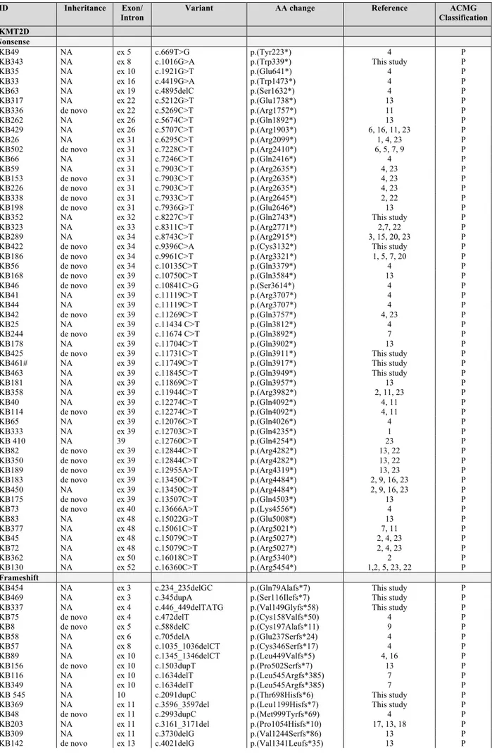
IDENTIFICATION OF KMT2D NONSENSE VARIANTS RESPONSIVE TO GENTAMICIN TREATMENT...38
KMT2D SPLICE SITE VARIANTS...40
KMT2D MOSAIC VARIANTS...43
MISSENSE VARIANTS PATHOGENIC ASSESSMENT BY BIOINFORMATIC APPROACHES...44
MISSENSE VARIANTS IMPAIR KMT2D METHYLTRANSFERASE ACTIVITY...47
KMT2D-COMPLEX PROTEIN INTERACTION...50
KMT2D MEDIATES NEURONAL DIFFERENTIATION OF NT2D1 CELLS BY ACTIVATING DIFFERENTIATION SPECIFIC GENES...51
DISCUSSION...53
CONCLUSIONS...56
1
ABSTRACT
The discovery of histone methyltransferase KMT2D and demethylase KDM6A genetic alterations in Kabuki Syndrome (KS) expanded and highlighted the role of histone modifiers in causing congenital anomalies and intellectual disability syndromes. KS is a rare autosomal dominant condition characterized by facial features, various organ malformations, postnatal growth deficiency, and intellectual disability. Since 2011 we performed a mutational screening of our KS cohort, that includes now 505 KS patients, by Sanger sequencing and MLPA of KMT2D, followed by KDM6A analysis in those patients resulted as KMT2D-negative. Of these 505 patients, we identified 196/505 (39%) patients with KMT2D variants and 208 different KMT2D variations; of them 37/208 (18%) never described before. The majority of KS patients carry nonsense and splicesite variants, suggesting the loss of function, and therefore haploinsufficiency, as the likely mechanism for the KS phenotype. RT-PCR and direct sequencing on cDNA from Kabuki patients carrying KMT2D splice site variants demonstrated that these cause aberrant splicing of the corresponding transcript, resulting in a truncating and not functional translated protein.
Molecular assays also showed that KMT2D mRNAs bearing premature stop codon are degraded by the nonsense mediated mRNA decay, contributing to KMT2D protein haploinsufficiency. We hypothesized that KS patients may benefit from a readthrough therapy that mediates translational suppression of nonsense variants, restoring the physiologically levels of endogenous KMT2D protein. Fourteen KMT2D nonsense variants were tested for their response to readthrough treatment through an in vitro dual reporter luciferase vector system, identifying 11/14 variants that displayed high levels of readthrough in response to gentamicin treatment.
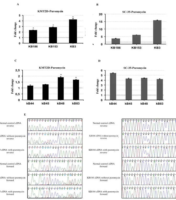
Among our cohort we identified three new cases with a mosaic variants in KMT2D gene, consisting in single nucleotide change resulting in two already reported nonsense variants, the c.13450C=/>T (p.R4484X) and the c.15061C=/>T (p.R5021X) and in a new frameshift variant, the c.3596_3597=/del (p.L1199HfsX7) KMT2D, respectively.
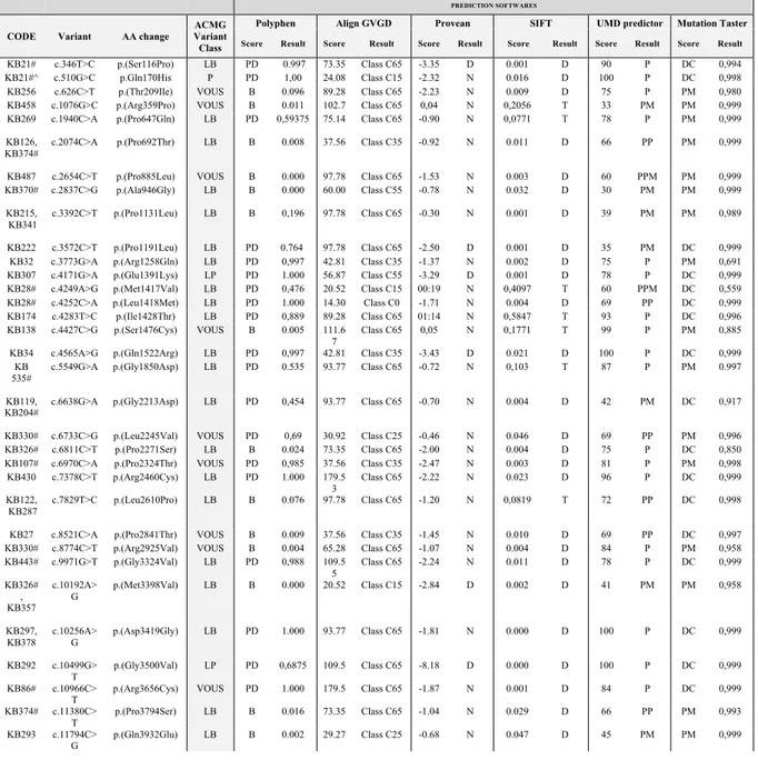
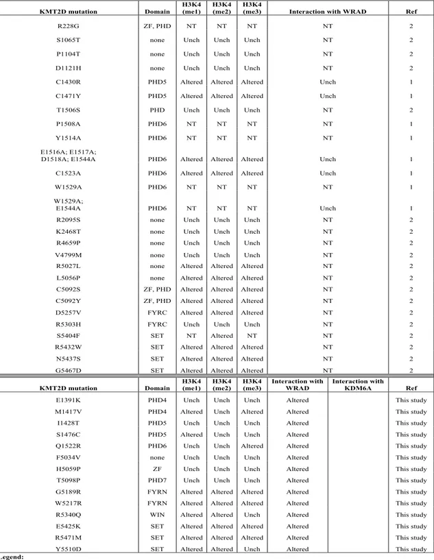
Moreover, relevant for diagnostic and counselling purposes, we implemented a number of bioinformatics tools to assess the pathogenicity of 69 KMT2D missense variants, found overall in our cohort of 505 KS patients, and for 14 of them we adopted a combination of biochemical and cellular approaches to investigate their role and characterize their functional impact in the pathogenesis of the disease. We found 9/14 missense variants showing altered H3K4 methylation activity. We additionally assessed the impact on complex formation with
2
WRAD protein complex, and we found that the reduced methyltransferase activity could be a consequence of lack of interaction.
Overall this study:
- Expands the number of KMT2D and KDM6A variants that cause KS, as a result of a 505 KS patients cohort.
- Enlarges the number of KS patients with mosaic mutations by reporting three additional cases with a single nucleotide change in KMT2D.
- Adds some insight to the functional mechanisms that cause the disease.
- Provides a preliminary proof-of-concept that naturally occurring nonsense mutations in KMT2D can be effectively suppressed by treatment with readthrough inducers, moving us closer to personalized medicine for KS.
- Affords a strategy to estimate the real deleterious effect of KMT2D missense variants in KS.
4
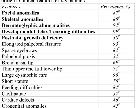
infection leading to recurrent infections of the upper airway tract and the middle ear is also a known and a common feature of KS [9]. KS patients have particular delays in speech and language acquisition and autism or autistic-like behavior, with difficulties in both communication and peer interactions [10, 11]. Consensus clinical diagnostic criteria for Kabuki Syndrome have not been established yet, although individuals with this condition usually have five cardinal manifestations according to Niikawa et al. 1988 [12], including: typical facial features, skeletal anomalies, dermatoglyphic abnormalities (persistence of fetal fingertip pads), mild to moderate intellectual disability, and postnatal growth deficiency (Table1).
Table 1: Clinical features of KS patients
Features Prevalence %
Facial anomalies 87#
Skeletal anomalies 80#
Dermatoglyphic abnormalities 92#
Developmental delay/Learning difficulties 99#
Postnatal growth deficiency 58#
Elongated palpebral fissures 95*
Sparse eyebrows 82*
Palpebral ptosis 52*
Broad nasal tip 69*
Thin upper and full lower lip 71*
Large dysmorhic ears 90*
Short stature 70#
Feeding difficulties 82#
Cleft palate 37#
Cardiac defects 46#
Urogenital anomalies 42#
Legend: KS, Kabuki Syndrome; * Micale et al. 2011 [4], # Bogershausen, et al. 2016 [18].
Prevalence and Prognosis
KS was initially thought to be specific to Japanese individuals, with an estimated prevalence in Japan of 1/32,0005 [12]. Even if the global incidence of KS is still not known, there are now reports of a variety of ethnic groups, including European, Brazilian, Vietnamese, Filipino, East Indian, Arabic, Chinese, Mexican, and African [3, 13] that presumably approximate the global prevalence to the Japanese one (1/32,0005). Because KS is not classically associated with severe medical complications, the adulthood prognosis is good, particularly if congenital anomalies and infections are properly managed.
5
Genetics of Kabuki Syndrome: KMT2D
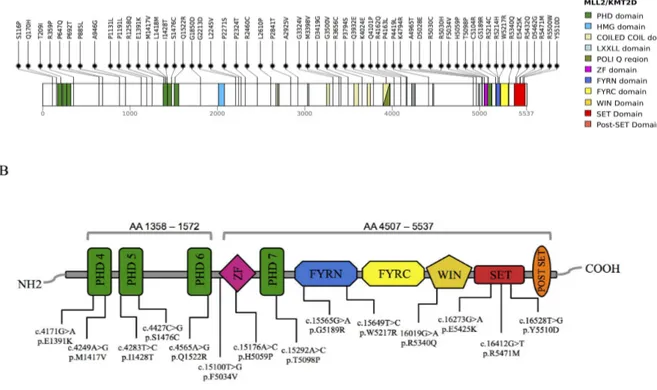
In 2010, whole-exome sequencing successfully identified heterozygous loss of function variants in the KMT2D (NM_003482.3; MIM #602113, also known as MLL2 and MLL4) gene as the major cause of KS [14]. KMT2D encodes a conserved member of the SET1 family of histone lysine methyltransferases (KMTs), which catalyzes the methylation of lysine 4 on histone H3 (H3K4), a modification associated with active transcription [15-17]. The enzymatic function of KMT2D depends on a cluster of conserved C-terminal domains, including plant homeodomains (PHD), two phenylalanine and tyrosine (FY)-rich motifs (FY-rich, N-terminal (FYRN) and FY-(FY-rich, C-terminal (FYRC) and a catalytic Su(var)3-9, Enhancer-of-zeste, Trithorax (SET) domain. Six different KMTs have been identified in higher eukaryotes, which fall into three subgroups on the basis of homologies in protein sequence and subunit composition: SET1A-SET1B, MLL1-MLL4 (KMT2A-KMT2B) and MLL3-MLL2 (KMT2C-KMT2D) [15-18]. KMT2C-KMT2D subgroup localizes and functions as major histone H3K4 mono, di and trimethyltransferases in a tissue specific manner at enhancers and promoters in mammalian cells [19-24] playing an important role in the epigenetic control of chromatin states [25] acting as transcriptional coactivator involved in the expression control of genes essential for embryogenesis and development, such as the HOX genes [26, 27]. The SET1 protein family, including KMT2D, is active in the context of a multisubunit complex, sharing four common highly conserved components, namely, WDR5, RbBP5, Ash2L and Dpy30 (a mini complex referred to as WRAD) that are related to yeast Set1 COMPASS complex [15], with the minimal four-component complex, including WDR5, RbBP5 and Ash2L along with the KMT2D SET-domain subunit, that can reconstitute most of the H3K4-specific histone methyltransferase activity (Figure 2) [28-31].
In addition, KMT2D also interacts with the Histone H3 lysine-27 demethylase KDM6A, who plays an important role for general chromatin remodelling and regulation of homeobox (HOX) genes, contributing to the correct reprogramming and tissue-specific differentiation during development [32-34]. During adipogenesis and myogenesis, KMT2D exhibits cell type and differentiation-stage-specific binding, co-localizing with lineage determining transcription factors on active enhancers [22]. The localization of KMT2D on active enhancers is also required for the enhancer-binding and enhancer activation of H3K27 acetyltransferase p300, one of the major enhancer epigenomic writers with KMT2D, both drivers of the enhancer epigenome and transcriptome processes during brown adipogenesis and ESC differentiation [35, 36]. The inactivation of the zebrafish kmt2d orthologue by morpholino is associated with significant craniofacial defects with severe hypoplasia of the viscerocranium, including loss of branchial arches, Meckel’s cartilage and the ceratohyal
7
oligomers. KMT2D contains five LXXLL motifs that participate in protein–protein interactions associated with different aspects of transcriptional regulation [41]. LXXLL motifs were originally observed in cofactor proteins that interact with hormone-activated nuclear receptors. Mo et al. (2006) [42] demonstrated that KMT2D physically interacts with ERα, through two LXXLL motifs after stimulation with the steroid estrogen hormone. As matter of fact, KMT2D suppression by its specific siRNA, decreased estrogen-induced expression of ERα target genes, such as cathepsin D and pS2 [42]. KMT2D may also interact with ERα via different KMT2D interacting proteins (NR-box containing), such as Menin, ASC2 and INI1 that contain multiple LXXLL domains. The tumour suppressor Menin interacts with ERα, recruits KMT2D complex into the promoter of estrogen-responsive genes and regulates their expression in a ligand-dependent manner [43], working as a critical link between activated ERα and the KMT2D-coactivator complex. A similar interaction involving Menin and KMT2D was observed at the Hox loci; in absence of the Menin protein, H3K4 trimethylation of the entire Hox cluster is abolished [44, 45].
3. FYRN and FYRC domains
These motifs are two poorly characterized phenylalanine/tyrosine-rich regions of approximately 50–100 aminoacids, respectively, that are found in a variety of chromatin-associated proteins [46]. The FYRN and FYRC motifs are closely juxtaposed and are involved in heterodimerisation between the N-ter and C-ter fragments of the protein.
4. SET domain
KMT2D is characterized by an evolutionarily conserved SET domain that is found in a number of chromatin-associated proteins with diverse transcriptional activities [47]. The SET domain, is a histone methyltransferase motif named for its presence in three Drosophila chromatin regulators: a modifier of position-effect variegation, Suppressor of variegation 3–9 (Su(var)3–9) [48], the Polycomb-group chromatin regulator Enhancer of zeste (E(z)) [49] and the Trithorax-group chromatin regulator trithorax (Trx) [50]. The role of SET-domain is to transfer a methyl group from the donor S-adenosyl-L-methionine (AdoMet) to the amino-group of a lysine residue on the histone or other proteins, leaving a methylated lysine residue and the cofactor by product S-adenosyl-L-homocysteine (AdoHcy). SET domain proteins can be classified into several families that differ because of the substrate specificity and the presence of associated domains [51]. KMT2D belongs to the SET1 family, which is found in conserved multisubunit complexes that regulate cellular H3K4 methylation levels [52]. The KMT2D SET domain is flanked on the C-terminus by a 22-aminoacid post-SET domain,
10
defining T-box transcription factor family recruit KDM6A to activate specific target gene profile in their natural developmental context like mesoderm, heart and vertebrae, emphasising the role of KDM6A in the developmental processes [63]. Interestingly, most of the pathogenic variants reported for T-box genes in human diseases are located in the KDM6A-interacting T-box domain.
Differential Diagnosis
Some disorders share overlapping features with KS. Evaluation of the patient via clinical assessment as well as molecular analysis is often performed to investigate the causes of clinical findings that are characteristic of KS but may also appear in other genetic syndromes. Disorders with the most overlapping features with Kabuki Syndrome (KS) include the following:
1) Turner syndrome (TS): is a neurogenetic disorder characterized by partial or complete monosomy-X associated with estrogen deficiency, short stature and increased risk for several diseases with cardiac conditions. Genetic analyses identified the Short stature HOmeoboX (SHOX) gene as the candidate gene for short stature and other skeletal abnormalities associated with TS but currently the gene or genes associated with cognitive impairments remain unknown [64]. Overlapping features with KS include heart defects, short stature, scoliosis, and similar facial features [65]. Constitutional KDM6A mutations cause Kabuki Syndrome with variable degrees in heterozygous females because of low expression levels from the normal wild type allele. Most of 45, X Turner females do not have a complete Kabuki Syndrome probably because of the dosage compensation of a single X chromosome in 45, X patients that migh be greater than the one in individuals with two sex chromosomes or because of a differential X chromomose inactivation. As patients with Turner syndrome only share some overlapping features with Kabuki Syndrome, monoallelic KDM6A expression is apparently sufficient to prevent this phenotype [66, 67].
2) CHARGE syndrome: (Coloboma of the eye, Heart defects, Atresia of the choanae, Retardation of growth and/or development, Genital and/or urinary abnormalities, and Ear abnormalities) is a genetic disorder characterized by a specific and a recognizable pattern of anomalies. De novo variants in the gene encoding chromodomain helicase DNA binding protein 7 (CHD7) are the major cause of CHARGE syndrome [68].
11
Overlapping clinical features with KS include: postnatal growth retardation, cleft lip/palate, hearing loss, congenital heart defects, urogenital malformations, developmental delay, and intellectual disability. As well, ocular coloboma, which is a major diagnostic criterion for CHARGE syndrome, has occasionally been reported in individuals with Kabuki Syndrome [69].Typical facial features and prominent fingertip pads in KS are distinct from those in CHARGE syndrome. Pathogenic variants in CHD7 are causative; inheritance is autosomal dominant [70].
3) 22q11.2 deletion syndrome (22q11.2DS): is the most common chromosomal microdeletion disorder, estimated to result mainly from de novo non-homologous meiotic recombination events occurring in approximately 1 in every 1,000 fetuses [71]. The syndrome is now known to have a heterogeneous presentation that includes multiple additional congenital anomalies and later-onset conditions, such as palatal, gastrointestinal and renal abnormalities, autoimmune disease, variable cognitive delays, behavioural phenotypes and psychiatric illness — all far extending the original description of DiGeorge syndrome [71]. The overlapping clinical features with KS comprise the cleft palate, congenital heart defects, and urinary tract anomalies. However, the different characteristic facial features seen in the two conditions should distinguish them [72].
4) IRF6-related disorders: is a group of orofacial clefting disorders including Van der Woude syndrome (VWS) and popliteal pterygium syndrome (PPS) caused by mutations in the interferon regulatory factor 6 (IRF6) gene [73]. The overlapping clinical features with KS are mainly the cleft lip/palate and lip pits although individuals with IRF6-related disorders do not have atypical growth and development, cardiac malformations, or the typical Kabuki Syndrome facies [70].
5) Branchio-oto-renal (BOR) syndrome: is a rare autosomal dominant disorder characterized by pits or ear tags in front of the outer ear, abnormal passages from the throat to the outside surface of the neck (branchial fistulas), branchial cysts, hearing loss and/or kidney abnormalities [74]. Ear pits, cupped ears, hearing loss, and renal anomalies are overlapping features with KS. However, individuals with BOR syndrome have otherwise normal craniofacies, normal growth, and normal development. Branchial cleft cysts may be present in BOR but have not been reported in KS [70].
6) Hardikar syndrome: is a disorder of multiple anomalies predominantly characterized by cleft lip/palate, liver and biliary tract disease, intestinal malrotation, obstructive uropathy, and
12
retinopathy [75]. Overlapping features with KS include prolonged hyperbilirubinemia with cleft lip and palate. However, individuals with KS do not typically develop pigmentary retinopathy or sclerosing cholangitis, as seen in Hardikar syndrome [70].
7) Wiedemann Steiner syndrome (WSS): is an autosomal dominant congenital anomaly syndrome characterized by hairy elbows, dysmorphic facial appearances (hypertelorism, thick eyebrows, downslanted and vertically narrow palpebral fissures), pre- and post-natal growth deficiency, and psychomotor delay. WSS is caused by heterozygous mutations in KMT2A (also known as MLL), a gene encoding a histone methyltransferase. Overlapping clinical features with KS are postnatal growth restriction, hypertrichosis, and intellectual disability [76, 77].
Gene-specific DNA methylation signatures in diseases
Epigenetics was originally defined as the genes-environment interaction process that leads to manifestations of various phenotypes during development [78]. In the recent years, this conception has evolved into the study of heritable changes in gene expression that occur without a change in DNA sequence. To date, the best understood epigenetic mechanisms are CpG DNA methylation and histone modifications. DNA methylation in particular has been the subject of intense interest because of its recently recognized role in disease and cancer as well as in the development and normal function of organisms. Many diseases are a consequence of alterations in gene expression patterns leading to deranged biological functions in cells [79]. Genes engaged in epigenetic regulation (epigenes), including those involved in chromatin remodeling and histone modifications, are increasingly being identified in the etiology of a variety of neurodevelopmental disorders, causing specific patterns of DNA methylation (DNAm) alterations that constitute unique signatures [80-82]. As a matter of fact, unique DNAm signatures are observed in individuals harboring variants in lysine-specific demethylase 5C (KDM5C [MIM: 314690]), which encodes an H3K4 demethylase and causes non-syndromic intellectual disability [MIM: 300534], DNA methyltransferase 1 (DNMT1 [MIM: 126375]), which cause autosomal dominant cerebellar ataxia with deafness and narcolepsy (ADCA- DN [MIM: 604121]), and nuclear receptor binding SET domain protein 1(NSD1 [MIM: 606681]) which encodes for a histone methyltransferase and cause Sotos syndrome [MIM:117550] [80-82]. Moreover, for NSD1 has been shown that genes encoding proteins in growth and neurodevelopmental pathways are highly represented in the DNAm signature reflecting the pathophysiology of the disorder [80].
13
Unique DNAm signatures have also been identified in Kabuki Syndrome patients carrying KMT2D variants, determining a characteristic global DNA methylation profile that distinguishes patients with KMT2D loss of function, missense or splice site variants from normal controls [69, 83]. Interestingly, the DNA methylation changes found in Kabuki Syndrome, showed similar magnitude and some overlapping target genes with Wiedermann-Steiner and Charge syndrome, all sharing overlapping features with Kabuki Syndrome. For instance, the DNA methylation profile of Kabuki Syndrome patients with KMT2D missense variants, clustered with the same of KMT2A missense and splice site variants, the main gene associated to Wiedermann-Steiner syndrome [83], while genome-wide DNAm profiles in patients with CHARGE and Kabuki Syndrome with CHD7-LOF or KMT2D-LOF variants, share 14CpG sites, 11 corresponding to HOXA5 and 3 to SLITRK5, which could account for some of the clinical overlap in CHARGE and Kabuki Syndrome [69].
Diagnosis of CHARGE, Wiedermann-Steiner, Kabuki Syndrome, and a variety of neurodevelopmental disorders in the clinical and molecular setting can be challenging. These data provide new insights into the genotype-epigenotype and phenotype relationship, indicating a cross-talk between histone and DNA methylation machineries exposed by inborn errors of the epigenetic apparatus. DNAm classification signature could be used as a functional molecular test to aid in the interpretation of the pathogenicity of the sequence variants, providing a valuable tool to facilitate in the diagnosis of the syndrome [69].
Histone methylation
The epigenetic control of developmental processes is a mechanism by which spatial and temporal expression of distinct genes and pathways are regulated. Alterations of this epigenetic mechanism, including histone modifications (acetylation, methylation and phosphorylation), have been mainly associated with the pathogenesis of cancer and with a variety of congenital malformation syndromes [84, 85]. In the extended form, chromatin appears as an array of nucleosomes, but in the nucleus the chromatin fibres that form chromosomes undergo several levels of folding, resulting in increasing degrees of condensation [86]. It is known that the histone tails have an important role in this folding process, and the histone tails methylation is important to regulate chromatin structure directly by affecting the higher-order folding of the chromatin fibre [87]. This have important implications for chromatin-templated processes such as transcription and DNA repair, altering the accessibility of DNA to the proteins that mediate these processes [88].
14
Histone lysine methylation marks, transcriptionally activate and inactivate chromatin domains in eukaryotic genomes, playing a key role in the regulation of transcription, gene silencing, and heterochromatin formation. This process is dynamically regulated by different site-specific histone methyltransferases (HMTs), including KMT2D (Figure 5). HMTs are key enzymes that introduce methyl groups into the lysine side chain of histone proteins and regulate both gene activation and silencing depending on the specific lysine residue that becomes methylated and the level of methylation (mono-, di-, or trimethylation). KMT2D mono- and trimethylation of histone H3 at K4 (H3K4) are highly associated with gene activation; H3K4 (me1) is enriched on enhancer regions and is required for enhancer activation and super-enhancer formation during cell differentiation of brown adipose tissue and skeletal muscle development [22, 89], whereas H3K4 (me3) is increased on active promoters/transcription start sites (TSS) and is important for actively transcribed gene regulation [90, 91]. The main sites of lysine methylation that have been associated with gene activity include K4, K36 and K79 of histone H3. Interestingly, the methylation of all three sites seems to be directly coupled to the transcription process. In the case of K4 and H3-K36 methylation, the enzymes responsible for both modifications have been shown to physically associate with RNA polymerase II (RNAPII) during elongation, resulting in histone methylation in the coding regions [88, 92-95] At least six different HMTs have been identified in higher eukaryotes, which fall into three subgroups on the basis of homologies in protein sequence and subunit composition: SET1A-SET1B, MLL1-MLL4 (KMT2A-KMT2B) and MLL3-MLL2 (KMT2C-KMT2D) [15-18].
On the contrary, H3-K9 and H3-K27 histone methylation has been linked to several silencing phenomena including homeotic-gene silencing, X inactivation and genomic imprinting [96]. The core of this silencing system is the POLYCOMB GROUP (PcG) of proteins [97], a complex that varies in different species, although the SET-domain-containing protein enhancer of zeste (E(Z); or its human homologue EZH2), extra sex combs (ESC; or its human homologue embryonic ectoderm development (EED) and suppressor of zeste-12 (SUZ12) proteins are present in all complexes that have been isolated so far [88]. Whereas all forms of H3-K27 methylation require EED79, SUZ12 seems to be dispensable for the monomethylation of H3-K27 in vivo, although it is indispensable for the enzymatic activity in vitro [98, 99]. Dynamic changes in the methylation pattern during gene expression indicated the existence of histone demethylase enzymes. The first histone demethylase identified was LSD1 [100]. To date, many histone demethylases have been identified and are classified into two families, the LSD1 demethylase family and the Jumonji (Jmj) domain-containing demethylase (JHDMs) family, depending on their domain structures and reaction mechanisms
16
1. Writers, that are involved in enzymatic addition of side groups (DNA methylation, RNA methylation and histone modification);
2. Erasers, are enzymes that remove the side groups;
3. Chromatin Remodelers of the DEAD/H ATPase helicase family, which are involved in the regulation of nucleosome positioning;
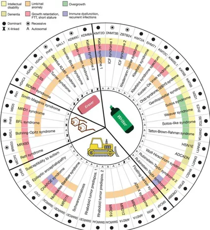
4. Other readers and chromatin remodelers, containing proteins that recognize and bind to their cognate chromatin marks, as well as proteins that are found in transcription repressor and activator complexes and other complexes that regulate DNA accessibility by the basic transcription machinery [103, 107] (Figure 6).
Figure 6. Features of the Mendelian disorders of the epigenetic machinery. The most common phenotype is
intellectual disability (yellow). Other features include growth retardation (red), overgrowth (green), immune dysfunction (purple), and various limb abnormalities (orange). The components of the epigenetic machinery (horizontal labels) and genetic syndromes (vertical labels) are divided into four categories (writer, eraser, reader, or remodeler). The majority of these genes demonstrate dosage sensitivity (filled circle). Bjornsson et al. 2014;[103].
As part of the writer epigenetic machinery, the histone acetyltransferases (HATs) acetylate a variety of lysine residues of histone and non-histone proteins [107, 108]. De novo mutations in HATs cause human genetic disorders as the RubinsteineTaybi syndrome, characterized by intellectual disability, postnatal growth deficiency, microcephaly, broad thumbs and halluces,
17
and characteristic facial appearance and caused by mutations in two HAT genes, CREBBP and EP300 [109, 110]. In contrast to acteyltransferases, Histone Methyltransferases (HMTs) have a higher degree of specificity for catalyzing the modification of specific lysine residues. This reflects the large number of histone methyltransferases (at least 27 according to the HUGO Gene Nomenclature Committee; http:// www.genenames.org/genefamilies/KDM-KAT-KMT) that are encoded in the mammalian genome. One of the most well characterized syndrome associated with HMT defects and intellectual disability is Kabuki Syndrome (KS), which is caused by mutations in two interacting chromatin modifiers: the writer KMT2D and the eraser KDM6A [1, 12, 103].
Mosaicism and Kabuki Syndrome
Genetic disorders are caused by changes in DNA sequence, copy number, or genomic locations (structural changes), which leads to alterations in gene expression and/or protein function. The DNA changes can be inherited; can occur newly during meiosis as the germ cells are being formed, after fertilization, either during development or in a differentiated cell. [111]. When genetic changes occur somatically, the individual is composed of cells with at least two different genotypes, and this state is known as mosaicism. From the human geneticist’s point of view, there are three main types of mosaicism; somatic (occurring only in the cells of the body, but not including the germline), germline (occurring only in the germ cells or their precursors but not found elsewhere in the body) and mixed gonadal and somatic, occurring in the both the cells of the body and the germline (Figure 7) [111]. Can be very difficult to determine the type of mosaicism, since differentiation of germline from somatic mosaicism could require examination of many types of cells, which is not usually possible.
19
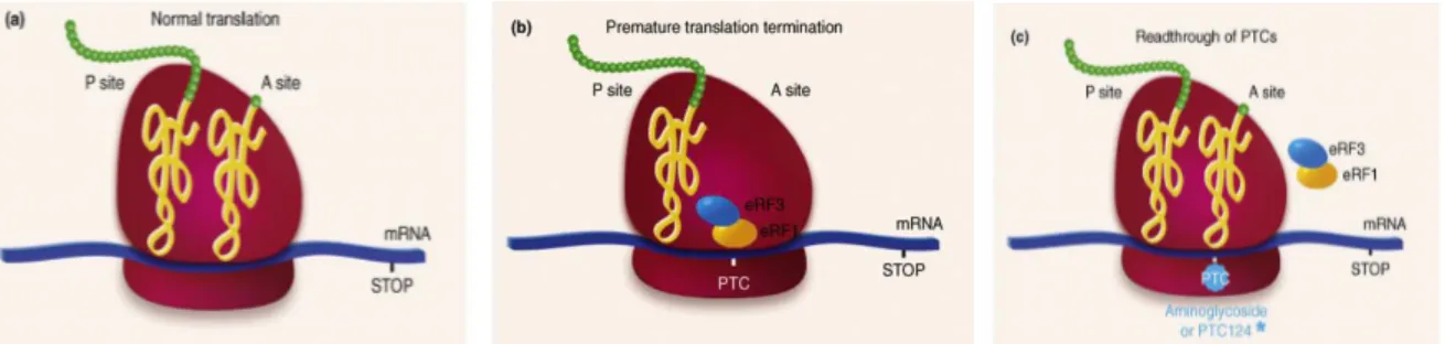
Readthrough strategies for suppression of nonsense variants
An important subset of inherited genetic disorders is caused by frameshifts or nonsense variants, which generate premature termination codons (PTCs) and result in the production of truncated proteins [114, 115]. In the past years, there was an attempt to develop mutation specific pharmacological approaches aimed at achieving sufficient levels of functional proteins. One approach that has been gaining prominence is the one using pharmacological agents to promote nonsense suppression or readthrough of PTCs, thus enabling re-expression of full-length functional proteins (Figure 8) [116]. One such class of these pharmacological agents are aminoglycosides such as gentamicin, G418 and amikacin, which promote in vivo readthrough of nonsense variants leading to the recovery of the biological activity of full-length restored proteins. More recently, novel synthetic readthrough nonaminoglycosides agents with improved biocompatibility, such PTC124 and RTC13, have been developed [117-119]. Examples of studied PTC-mediated diseases are cystic fibrosis, Duchenne muscular dystrophy, ataxia telangiectasia, Rett syndrome, Usher syndrome type I, Hurler syndrome, hemophilia, methylmalonic aciduria, obesity, poor drug metabolism, spinal muscular atrophy, peroxisome biogenesis disorder, and cancer linked to the presence of PTC in p-53 tumor suppressor gene [120].
Figure 8. The effect of readthrough drugs on protein translation (a) Normal protein synthesis (b) Premature
termination of protein synthesis owing to a PTC (c) Readthrough of PTC by aminoglycosides or PTC124. Modified from Lind and Kerem, 2008 [121].
Aminoglycosides are widely used in clinical practice as bactericidal antibiotics with established effects on translational accuracy or efficiency. Their utility as antibacterial agents arise from binding to the decoding site of the 16S or 18S ribosomal RNA (rRNA) interfering with protein synthesis promoting an accurate codon–anticodon pairing. In the presence of aminoglycoside, the conformation of rRNA becomes altered, inducing codon misreading that causes either incorporation of an erroneous amino acid at a sense codon or failure of recognition of the stop codon, leading to translational readthrough rather than chain
20
termination [122]. Although aminoglycosides target a conserved region of the rRNA sequence, these agents are highly active against bacterial and mitochondrial ribosomes but have limited interaction with human ribosomes. The specificity is related to the high affinity binding of aminoglycosides to prokaryotic rRNA, which has an adenine at position 1408 of the 16S rRNA (numbered according to the Escherichia coli sequence). In contrast, eukaryotic ribosomes have a guanine at the corresponding position, causing a low affinity towards aminoglycosides [122]. Consistently, the effects of aminoglycosides on protein translation in eucaryotic cells have been demonstrated at concentrations 10 to 15 times higher than the typical therapeutic antibacterial concentrations. The potential for aminoglycosides to induce misreading was observed in various eukaryotic cell-free systems such as yeast, plants, and human cells.
Readthrough of in-frame PTCs enables the protein synthesis to continue to the end of the transcript, thus, generating a full-length protein with either the correct or an abnormal aminoacid (bound to a tRNA that is a near-cognate of the stop codon) at the PTC. Because the binding of aminoglycosides is inefficient, both full length and truncated proteins will be synthesized. Aminoglycosides have minimal effects on normal translation termination because the normal stop codons of eukaryote genes are surrounded by upstream and downstream sequences, which enhance the efficiency of translation termination, whereas nonsense variants are usually not surrounded by these sequences [123].
Nonsense mediated mRNA decay in mammals
The nonsense-mediated decay (NMD) is an evolutionarily conserved mRNA surveillance mechanism that controls the quality of mRNAs by eliminating aberrant transcripts that prematurely terminate translation, regulating many physiological transcripts that harbor an NMD-eliciting element (uORF) or an intron within the 3′ untranslated region (UTR) [124, 125]. In mammalian cells, NMD generally degrades mRNAs that terminate translation more than 50-55 nucleotides upstream of a splicing-generated exon-exon junction [126]. Although NMD is important to eliminate PTC-bearing mRNAs to minimize the production of mutant proteins, this process is not 100% efficient, often resulting in generation low levels of truncated proteins produced from residual aberrant transcripts [127]. A key issue is how the NMD machinery discriminates between an authentic termination codon and a PTC. Two different mechanistic NMD models have been proposed, (i) the exon junction complex (EJC)-dependent model and (ii) the faux 3′ UTR (untranslated region) model [128]. Both mechanisms imply that a signal downstream of the stop codon determines whether a stop
21
codon is premature or not. In the EJC-dependent model, the multiprotein EJCs are deposited as a consequence of splicing, ~20-24 nucleotides upstream of each exon-exon junction [129]. EJCs components include the conserved NMD factors, UPF1 (up-frameshift) UPF2 and UPF3, as well as other factors that are essential for NMD. If the ribosome runs into a stop codon that is located at least 50–55 nucleotides up-stream of an exon-exon junction, it is recognized as a PTC and the SURF complex, (consisting of the PI3K, SMG-1, UPF1, eRF1 and eRF3), is recruited to the premature termination site [130]. The binding of UPF2 to both UPF1 and SMG-1 bridges the connection between EJC and the SURF complex, which triggers the phosphorylation of UPF1 and the dissociation of eRF1 and eRF3 [130]. Phospho-UPF1 then recruits SMG-5/SMG-7 and SMG-6 proteins, resulting in mRNA decay [131, 132]. The other model, the ‘faux 3′-UTR’ model, proposes that the mechanism of premature termination is intrinsically different from translation termination at a natural stop codon. Accordingly, during natural termination the ribosome is able to interact with 3′-UTR-bound proteins, while premature termination would impair or delay this interaction [127]. Hence, the position of the stop codon relative to the poly(A)-binding protein (PABP) is critical in discriminating premature from natural stop codons. Even if the exact mechanism used to identify a PTC it’s not known, it is clear that the cell can distinguish between a PTC and a natural termination codon. This discrimination suggests that should be possible to identify small molecules that can promote readthrough at a PTC without affecting normal translational termination. NMD mechanism can affect drug-induced readthrough interfearing with the mutated mRNAs, therefore NMD inhibition may increase PTC readthrough [121]. Because NMD occurs upstream bulk of translation, its inhibition may represent an attractive way to treat nonsense-mutation mediated genetic diseases [133].
22
AIM OF THE THESIS
Kabuki Syndrome is a rare autosomal, multiple malformation disorder with an estimated prevalence of 1 in 32000. Mutations in two interacting chromatin modifiers belonging to the ASCOM protein complex, KMT2D/MLL2 and less frequently KDM6A/UTX have been identified as the major cause of KS, although mutations in the RAP1A and RAP1B genes have been recently proposed as additional causatives events. The large prevalence of KMT2D variants is predicted to lead a truncated protein, suggesting a loss of function event, and therefore haploinsufficiency as the likely mechanism for the KS phenotype. However, ~30% of KMT2D variants in our data set and the 16.1% from recent published mutational analysis review [18] harbour missense variants whose classification and interpretation is a challenge in molecular diagnostics and genetic counselling.
Overall this thesis aimed to: i) expand the mutation spectrum of KMT2D and KDM6A in Kabuki Syndrome ii) add some insight to the functional mechanisms that cause the disease iii) investigate the potential drug-mediated readthrough to restore KMT2D function as therapeutical approach and iv) afford a strategy to estimate the real deleterious effect of KMT2D missense variants in KS.
23
MATERIAL AND METHODS
Patients and samples preparation
Our cohort comprised 505 index patients clinically diagnosed as affected by Kabuki Syndrome (Table 3) recruited from 2011 to 2017. Patients were enrolled after obtaining appropriate informed consent by the physicians in charge and approval by the local ethics committees. Genomic DNAs were extracted from fresh and/or frozen peripheral blood leukocytes from patients and their available family members using an automated DNA extractor and commercial DNA extraction Kits (EZ1, Qiagen, Hilden, Germany). Total RNA was extracted from peripheral blood leukocytes using TRIZOL reagent (Life Technology) and reverse transcribed using the Quantitect Transcription kit (Qiagen), according to the manufacturer’s instructions.
KMT2D and KDM6A sequence mutation, MLPA and pyrosequencing analysis
Mutation screening was performed for all 54 coding exons of the KMT2D (MIM #602113, NM_003482.3, also known as MLL2 and MLL4) gene and 29 coding exons of KDM6A (MIM #300128, NM_021140.3). PCR amplifications were carried out in a final volume of 50 µl consisting of:
• 50 ng genomic DNA
• 2.5 mM deoxyribonucleotides
• 15 pmol/µl of sense and antisense primers
• 1X Reaction Buffer (200mM Tris-HCl/ pH 8.8 at 25°), 100 mM KCl, 100mM (NH4)2SO4, 1.0% Triton X-100 and 21 mg/ml nuclease-free BSA) and
• 1 U of Taq DNA polymerase.
Denaturation was carried out at 95° for 30 s, annealing at 60°C for 30 s, extension at 72° for 2 minutes/kb, for 35 cycles.
Primers were designed using the Primer 3 Output program (http://frodo.wi.mit.edu/primer3/) to amplify the 54 coding exons of KMT2D and 29 coding exons of KDM6A gene including the intronic flanking sequences. Amplicons and primers were checked both by BLAST and BLAT against the human genome to ensure specificity. The amplified products were subsequently purified (Exo Sap) and sequenced with a ready reaction kit (BigDyeTerminator v1.1 Cycle, Applied Biosystems). The fragments obtained were purified using DyeEx plates (Qiagen) and resolved on an automated sequencer (3130xl Genetyc analyzer DNA Analyzer, ABI Prism). Sequences were analyzed using the Sequencer software (Gene Codes, Ann
24
Arbor, Michigan). Whenever possible the mutations identified were confirmed on a second independent blood sample. All existing and new mutations were described following the
recommendations of the Human Genome Variation Society
(http://www.hgvs.org/mutnomen). MLPA analysis was performed as reported in Priolo et al. (2012) [134]using probe mixture (SALSA MLPA KIT P389-A1 KMT2D; SALSA MLPA KIT P445-A1 KDM6A MRC-Holland, Amsterdam, The Netherlands) that contains 27 probes targeting exons across the KMT2D gene and 32 probes targeting exons across the KDM6A gene [134]. For Pyrosequencing analysis, PCR reaction was performed three times independently and after checking for amplification on a 2% agarose gel, and 10 µl of each PCR product was pyrosequenced twice on a PyroMark Q24 (Qiagen, Germany) using Pyromark PCR Kit according to the manufacturer’s instructions (Qiagen, Germany). Primers were designed using PyroMark Assay Design 2.0 (Qiagen, Germany) and are available upon request.
In silico analysis of KMT2D and KDM6A variants
The putative causal and functional effect of each identified nucleotide variant was estimated by using the following in silico prediction tools: Polyphen-2 version 2.2.2
(http://genetics.bwh.harvard.edu/pph)[135], Align GVGD
(http://agvgd.iarc.fr/agvgd_input.php) [136], PROVEAN v1.1 (http://provean.jcvi.org/index.php) [137], SIFT v1.03 (http://sift.jcvi.org/) [138], UMD-predictor (http://www.umd.be/) [139], and Mutation Taster (http://www.mutationtaster.org/) [140] using default parameters.Splice-site variants were evaluated for putative alteration of regulatory process at the transcriptional or splicing level with Human Splice Finder (http://www.umd.be/HSF3/)[141], NNSPLICE (http://www.fruitfly.org/seq_tools/splice.html) [142], and NetGene2 (http://www.cbs.dtu.dk/services/NetGene2) [143].
In silico analysis of KMT2D protein domains
We implemented a number of algorithms to analyze the different KMT2D protein domains. Initial models were generated by threading approach using five web-servers: I-Tasser (zhanglab.ccmb.med.umich.edu/I-TASSER/) {Yang, 2015 #10853, Phyre2 (www.sbg.bio.ic.ac.uk/phyre2/) {Kelley, 2015 #10854}, Genesilico (genesilico.pl/meta/) [144], RaptorX (raptorx.uchicago.edu/) [145], Multicom (http://sysbio.rnet.missouri.edu/multicom_toolbox/) [146]. Final model was obtained using
25
Modeller (version 9.9; salilab.org/modeller/) [147] with the five server-generated models as templates. Energy minimization of models was carried out using KoBaMIN web server (http://csb.stanford.edu/kobamin/) [148]. Quality of the resulting models was assessed by Qmean server (swissmodel.expasy.org/qmean/) [149]. Effects of mutations on domain structure and stability were predicted by Rosetta Backrub server (kortemmelab.ucsf.edu/backrub/) [150], FoldX software (foldx.embl.de/) [151].
Cell lines, plasmids, and transfection assays
The pFlag-CMV2 FUSION-KMT2D vector (cDNA spanning the PHD4-5-6 domains (amino acids 1358–1572) and ZF-PHD7- FYRN-FYRC- WIN-SET-post SET domains-(amino acids 4507–5537) was a gift of Professor Min Gyu Lee, Department of Molecular and Cellular Oncology, The University of Texas [152]. We sub-cloned this partial KMT2D clone into the p3XFlag-CMV14 vector (Sigma) using standard procedures. The expression plasmids harbouring patients KMT2D missense mutations were generated by site-directed mutagenesis according to the manufacturer’s instructions (Agilent). The KMT2D C-terminal ORF was assembled into the pcDNA3-Myc-EGFP vector (Reymond et al. 2001) by PCR site directed amplification using human cDNA and pFlag-CMV2 FUSION-KMT2D vector as templates respectively. The WDR5 full length ORF was assembled into the p3XFlag-CMV14 vector (Sigma) by PCR amplification reactions using as template cDNA from HEK 293T cells. HEK 293, 293T and NT2D1 cells were cultured in Dulbecco’s Modified Eagle Medium (Life Technologies) with 10% FBS, penicillin (100 U/ml) and streptomycin (100 µg/ml). Patients’ Fibroblasts were cultured in Dulbecco’s Modified Eagle Medium F12 with 10% FBS, penicillin (100 U/ml) and streptomycin (100 µg/ml). HEK 293 and HEK293T cells were transiently transfectedusing the polyethylenimine method, following published protocols [153]. Cells were harvested 48 h after transfection and used for protein extraction and all the assays.
Establishment of Lymphocyte cell lines and NMD assay
Approximately 10 ml of blood in sodium heparin was processed within 24 hours from taking. Lymphocytes were isolated from whole blood of Kabuki patient by centrifugation using Lymphoprep or similar reagents, transformed by infection with EBV and maintained in RPMI medium (Gibco) containing 20% fetal bovine serum (FBS) and 1% penicillin/streptomycin, in a humidified atmosphere containing 5% carbon dioxide (CO2) at 37°C. NMD was assayed by
26
treating patient lymphoblast cell lines with puromycin at a concentration of 200 ug/ml. After 8h of incubation, total RNA was obtained from lymphoblast cell lines, using the RNasy mini Kit (Qiagen) according to manufacturer instructions and a Quantitect Reverse Transcription kit (Qiagen) was used for cDNA synthesis.
Quantification of readthrough levels in HEK293 cells
A dual gene reporter pCRFL (gently provided by Prof. J-P Rousset) was used to quantify the effect of gentamicin on stop mutation readthrough in culture cells. Sequence to be analyzed spanning 27 nucleotides centered on the different stop mutations were inserted in-frame between the Renilla (LucR) and Firefly Luciferase (LucF) coding sequences. Readthrough efficiency was estimated by calculating the ratio of Renilla and Firelfly activity. Both reporter proteins are expressed from the same message, eliminating in particular potential variation in mRNA stability between the different targets analyzed. A 100% activity control was provided by a construct (TQ) with no stop codon between the coding sequences of the two reporters [154]. A pCRFL reporter vector harboring the 319d Duchenne muscular dystrophy mutation (pCRFL319) exhibiting the highest gentamicin-inducted readthrough efficiency and the highest induction factor in NIH3T3 cultured cells assays [155] was used as positive control.
Site-directed mutagenesis
In vitro approaches to site-directed mutagenesis can be grouped generally into three categories:
i) methods that restructure fragments of DNA, such as cassette mutagenesis; ii) localized random mutagenesis;
iii) oligonucleotide-directed mutagenesis;
All oligonucleotide-directed mutagenesis is based on the same concept: an oligonucleotide pair encoding the desired mutation(s), is annealed to the DNA of interest and serves as a primer for initiation of DNA synthesis. The p3XFlag-KMT2D-FUSION-CMV14 was mutated to insert in-frame missense variant/s corresponding to the variant/s found in our patients with Kabuki Syndrome. All site-directed mutagenesis reactions were performed by using Pfu plymerase from Promega.
50 ng of dsDNA template was added to 10X reaction buffer, 2.5mM dNTP mix ,125 ng of each oligonucleotide primer and 2.5 U/µl Pfu DNA polymerase.
27 • 30 seconds at 95°C followed by
• 1 min at 55°C and
• 2 minutes/kb of plasmid length at 72°C (12 cycles).
Then the parental, supercoiled double-stranded DNA was digested with 10U of Dpn I restriction enzyme at 37°C for 1 h before being transformed.
Transformation
Bacterial transformation is the process by which electrocompetent or chemically competent bacterial cells take up naked molecules. If the foreign DNA has an origin of replication recognize by the host cell DNA polymerases, the bacteria will replicate the foreign DNA along with their own DNA. When transformation is coupled with antibiotic selection techniques, bacteria can be induced to uptake certain DNA molecules, and those bacteria can be selected for that incorporation. All constructs were transformed in DH5 alpha Competent Cells (Invitrogen). 10 µl of the mutagenesis reaction mixture was added to a 120 µl of cells, and the tubes were incubated for 30’ on ice. Cells were heat-shocked in a 42° water bath for 45 seconds, then incubated on ice for 2 minutes, before being additionated with 250 µl LB (10g Bacto-Tryptone, 5g Bacto-yeast extract, 10g NaCl, dd H2O to 1 litre) medium and incubated at 37°C for 1 hour with shaking at 225-250 rpm. 150 µl of the transformation mixture were plated on LB agar plates containing the appropriate antibiotic (Ampicillin, 100 µg/ml).
Screening for recombinants
Screening for recombinants was performed by colony PCR or plasmid miniprep followed by restriction digestion.
i) Colony PCR: this method is designed to quickly screen for plasmid inserts directly from E.coli colonies. Typical colony PCR reaction:
Mix together the following on ice; always adding enzyme last. For multiple samples make a large master mix and aliquot 50 ul in each PCR tube:
-38 µl sterile distilled water -5 µl 10X PCR buffer -3 µl 25mM MgCl2 -1 ul 10mM dNTPs
28 -1 µl 20µM reverse primer
-0.2 µl Taq polymerase
To each cold PCR tube containing the PCR reaction, a small amount of colony was added. ii) plasmid miniprep followed by restriction digestion: well-isolated colonies were picked from a plate and cultured for 8 hours at 37°C in LB culture medium containing the appropriate antibiotic for selection. Miniprep were performed by using GenElute Plasmid Miniprep Kit (Sigma-Aldrich).
Plasmid DNA extraction
Plasmid purification protocols are based on a modified alkaline lysis procedure followed by binding of plasmid DNA to anion-exchange resin under appropriate low-salt and pH conditions. RNA, proteins, dyes, and low-molecular-weight impurities are removed by a medium-salt wash. Plasmid DNA is eluted in a high-salt buffer and then concentrated and desalted by isopropanol precipitation. Starter culture was prepared by inoculating positive colony into 2-5ml of selective LB medium and incubated for 8 hours at 37°C with vigorous shaking (approximately 300 rpm). 1ml of starter culture was diluted in 100 ml of selective LB medium, and grown for 12-16 hours at 37°C, then bacterial cells were harvested by centrifugation at 3500g for 30 minutes at 4°C. Plasmid purification was performed by using QIAGEN Plasmid Midi Kit. Bacterial pellet was resuspended in 4ml of P1 buffer (Resuspension Solution) which contains Rnase A (100 µg/ml). Lysis was performed by incubating for 5 minutes the resuspended pellet with 4 ml of P2 buffer (Lysis buffer) after inverting the tube for mixing, and then reaction was stopped by the addition of 4ml of P3 buffer (Neutralization Buffer). Tubes were incubated on ice for 15 minutes, then cell debris, proteins and genomic DNA were precipitated by centrifugation at 15000g for 30 minutes at 4°C. Column resin was equilibrated applying 4ml of QBT buffer (Equilibration buffer), allowing the column to empty by gravity flow. After centrifugation, supernatant containing plasmid DNA was promptly removed and loaded into the column, allow the lysate to enter the resin by gravity flow. Column resin was washed twice with 10 ml of QC buffer (Wash buffer) to remove all contaminants in the DNA plasmid preparation. Plasmid DNA was eluted by adding 5 ml of QF Eluition buffer in a new tube, then precipitated with 3,5 ml of room-temperature isopropanol, mixed and centrifuged at 15000g for 30 minutes at 4°C. DNA pellet was washed with 2 ml of room-temperature 70% ethanol, centrifuged at 15000 g for 10 minutes. After removal of supernatant, pellet was air-dried, then resuspended in 100-200 µl of H2O.
29
Total RNA Isolation and Reverse Transcription (RT-PCR)
Total RNA was isolated using the TRIzol® protocol (Gibco) from lymphoblast cell lines. RNA extraction with TRIzol is a common method for total RNA extraction from cells. The correct name of the method is Guanidinium thiocyanate-phenol-chlorofrom extraction and uses guanidinium isothiocyanate, a powerful chaotrope used as a protein denaturant, which causes the inactivation of RNases, and acidic phenol/chloroform, which lead to the partitioning of RNA into aqueous supernatant for separation. Cells were homogenized in 1 ml Trizol and incubated for 5 min at RT. 200 µl of chloroform was added, then shaked vigoriously by hand for 15”, incubated for 2-3 minutes at RT and centrifuge at 12,000 rpm for 15 minutes at 4°C. Aqueous (top) phase was transferred to a fresh sterile 1,5 ml microcentrifuge tube. 500 µl isopropyl alcohol was added, then samples were incubated for 10’ RT and centrifuged at 12,000 rpm for 5 minutes at 4°C. Pellet was washed in 1 ml 75% EtOH, RNA pellet was briefly air dry, and dissolved in 50 µl RNase free water. 1 µg RNA was reverse transcribed using the Quantitect Transcription kit (Qiagen). RT-PCR produces DNA copies (complementary DNA, or cDNA) of a RNA template, by using the enzyme reverse transcriptase, and the resulting single-stranded cDNA can be amplified using traditional or real-time PCR. Reverse transcriptase enzyme, in general, has 3 distinct enzymatic acitivities: an RNA-dependent DNA polymerase, a hybrid-dependent exoribonuclease (Rnase H), and a DNA-dependent DNA polymerase. For reverse transcription in vitro, the first 2 activities are utilized to produce single-stranded cDNA: RNA-dependent DNA-polymerase activity (reverse transcription) transcribes cDNA from an RNA template, and RNase H activity specifically degrades only the RNA in RNA: DNA hybrids. The purified RNA samples were incubated in 1X gDNA Wipeout Buffer at 42°C for 2 minutes to remove contaminating genomic DNA. After genomic DNA elimination, the RNA samples were reverse transcribed using a master mix prepared from Reverse Transcriptase (1U), 1X RT Buffer, and RT Primer Mix, a optimized blend of oligo-dT and random primers. The entire reaction was performed at 42°C for 30 minutes and then inactivated at 95°. Each cDNA sample was measured by using a Nanodrop spectrophotometer (NanoDrop Technologies, Willmington, Delaware, USA) and used in qPCR for NMD-assay and in PCR for studying splicing mutations.
30
Real-time polymerase chain reaction (qPCR)
qPCR is a quantitative PCR method which enables both detection and quantification (as absolute number of copies or relative amount when normalized to DNA input or additional normalizing genes) of one or more specific sequences in a DNA sample.
Based on the molecule used for the detection, the real time PCR techniques can be categorically placed under two heads:
• non-specific fluorescent dyes that intercalate with double-stranded DNA, such as SYBR Green, which binds to the minor groove of the DNA double helix, and is the most widely used double-strand DNA-specific dye reported for real time PCR
• sequence-specific DNA probes consisting of oligonucleotides that are labeled with a fluorescent reporter, which permits detection only after hybridization of the probe with its complementary DNA target, such as Molecular Beacons, TaqMan Probes, FRET Hybridization Probes, Scorpion Primers. Oligos for qPCR were designed using the Primer3 program6 with default parameters. Amplicons and primer pairs were checked both by Blast and Blat against the human genome to ensure specificity. Target genes expression was examined by amplification with the primer sets described in the table below (Table 2). EEF1A1 and GAPDH were used as housekeeping genes. The reactions were run in triplicate in 10 µl of final volume with 10 ng of sample cDNA, 0.3 mM of each primer, and 1X Power SYBR Green PCR Master Mix (Applied Biosystems). Reactions were set up in a 384-well plate format with a Biomeck 2000 (Beckmann Coulter, Milan, Italy) and run in an ABI Prism7900HT (Applied Biosystems) with default amplification conditions. Raw Ct values were obtained using SDS 2.3 (Applied Biosystems). Calculations were carried out by the comparative Ct method.
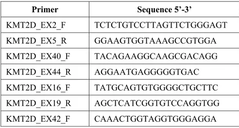
Table 2: Oligos used for splicing mutations analysis
Primer Sequence 5’-3’ KMT2D_EX2_F TCTCTGTCCTTAGTTCTGGGAGT KMT2D_EX5_R GGAAGTGGTAAAGCCGTGGA KMT2D_EX40_F TACAGAAGGCAAGCGACAGG KMT2D_EX44_R AGGAATGAGGGGGTGAC KMT2D_EX16_F TATGCAGTGTGGGGCTGCTTC KMT2D_EX19_R AGCTCATCGGTGTCCAGGTGG KMT2D_EX42_F CAAACTGGTAGGTGGGAGGA
31 KMT2D_EX42_R GGCCCCATAAGGTTTGGTAT KMT2D_EX43+44_F TTCTCCTGACAGCATTGTGC KMT2D_EX45_R TCTAGCCCAGGCTTTCACAT KMT2D_EX45_F TTCCCAGATACCAAACCTTATG KMT2D_EX48_R GCACTCCTTTCCATTTCTTGAG
In vitro histone methyltransferase (HMT) assay and epigenetic reporter allele
Partially purified FLAG-KMT2D wild-type and mutant derivative proteins were obtained from transfected HEK 293T cells by lysis in co-IP buffer (50 mM Tris, pH 7.5, 250 mM NaCl, 1% TritonX-100, 1 mM EDTA), followed by overnight incubation with EZview™ Red Anti-FLAG Affinity Gel (Sigma-Aldrich) at 4 °C and final elution in BC100 buffer (20 mM Tris pH 7.5, 10% Glycerol, 0.2 mM EDTA, 1% TritonX-100, 100 mM NaCl) containing FLAG peptide (Sigma-Aldrich). KMT2D protein amounts were quantified by Coomassie staining and immunoblot analysis using mouse monoclonal FLAG antibodies. Enzymatic activity against native nucleosomes was measured following a published method [152]. Briefly, equal amounts of wild-type or mutant FLAG-KMT2D proteins were incubated at 37 °C for 4 h with HeLa nucleosomes (Reaction Biology) in KMT buffer (50 mM Tris pH 8.5, 100 mM KCl, 5 mM MgCl2, 10% glycerol, 4 mM DTT) supplemented with S-adenosyl methionine (New England BioLabs). Reactions were stopped by adding equal volumes of 2× Laemmli buffer and heated at 100 °C for 5 min before loading onto Tris-glycine 4–20% gradient gels. All assays were performed at least twice independently. Epigenetic reporter allele, a kind gift from Dr Hans T. Bjornsson (McKusick-Nathans Institute of Genetic Medicine, Johns Hopkins University, Baltimore) has been used as described in [103].
Co-immunoprecipitation assay and Western Blotting analysis
Co-immunoprecipitations were performed using Dynabeads magnetic beads (Thermo Fisher Scientific) following manufacturer's instructions. Complexes were analyzed by western blot using the indicated antibodies. Protein extracts were resolved on NuPAGE Tris-acetate 3–8% gels (for KMT2D) or Tris-glycine 4–20% gels (for histone H3) (Life Technologies) and transferred to nitrocellulose membranes (GE Healthcare) according to the manufacturer’s instructions. Antibodies used were: mouse monoclonal antibody to α-tubulin (clone DM1A, Sigma-Aldrich), rabbit polyclonal anti-H3K4me1 (Abcam), anti-H3K4me2 (Active Motif), anti-H3K4me3 (Abcam), rabbit monoclonal anti-Histone H3 (clone D1H2, Cell Signaling
32
Technology), mouse monoclonal anti-Flag (Sigma cat# F3165), rabbit monoclonal anti GFP (Santa Cruz), rat monoclonal anti-HA (Roche), and mouse monoclonal anti Myc (Roche), rabbit polyclonal anti Ash2 (Bethyl) and rabbit polyclonal anti RbBP5 (Bethyl). Horseradish peroxidase conjugated anti-mouse (Santa Cruz), anti-rabbit (Santa Cruz) antibodies, and the ECL chemiluminescence system (GE Healthcare) was used for detection. Quantitation of band signal intensity was obtained by the ImageJ software (http://imagej.nih.gov/ij/). Values are expressed as fold differences relative to the wild-type protein sample, set at 1, after normalization for the loading control.
RNAi and NT2D1 cell differentiation
HEK293 cells were transfected with shKMT2D #1 (SHCLNV-NM_003482; TRCN0000235745), shKMT2D #2 (SHCLNV-NM_003482; TRCN0000013140), shKMT2D #1 + shKMT2D #2, or control shRNA (shLuc) that were all purchased from Sigma (Mission shRNA Lentiviral transduction particles). These shRNA plasmids, which contain a puromycin-resistant marker, were cotransfected along with a packing plasmid (deltaR8) and an envelope plasmid (VSV-G) into HEK293 cells using a calcium phosphate method. Thirteen hours later, the medium was replaced with DMEM supplemented with 10% FBS. Virus particles containing shKMT2D or shLuc were generated for 2 days and used to infect NT2D1 mammalian cells. NT2D1 cells were infected by virus-containing medium, and the infected cells were selected in medium containing 2.5 mg/ml puromycin for 2 days. Knockdown efficiency was examined by Western blot analysis and qRT–PCR. Knockdown cells were treated with 10 mM RA for 0–6 days, monitored for cell differentiation patterns, and harvested for further analysis.
34
Table 3: KMT2D and KDM6A variants identified in our cohort.
ID Inheritance Exon/
Intron
Variant AA change Reference ACMG
Classification KMT2D
N Nonsense
KB49 NA ex 5 c.669T>G p.(Tyr223*) 4 P
KB343 NA ex 8 c.1016G>A p.(Trp339*) This study P
KB35 NA ex 10 c.1921G>T p.(Glu641*) 4 P KB33 NA ex 16 c.4419G>A p.(Trp1473*) 4 P KB63 NA ex 19 c.4895delC p.(Ser1632*) 4 P KB317 NA ex 22 c.5212G>T p.(Glu1738*) 13 P KB336 de novo ex 22 c.5269C>T p.(Arg1757*) 11 P KB262 NA ex 26 c.5674C>T p.(Gln1892*) 13 P KB429 NA ex 26 c.5707C>T p.(Arg1903*) 6, 16, 11, 23 P KB26 NA ex 31 c.6295C>T p.(Arg2099*) 1, 4, 23 P KB502 de novo ex 31 c.7228C>T p.(Arg2410*) 6, 5, 7, 9 P KB66 NA ex 31 c.7246C>T p.(Gln2416*) 4 P KB59 NA ex 31 c.7903C>T p.(Arg2635*) 4, 23 P KB153 de novo ex 31 c.7903C>T p.(Arg2635*) 4, 23 P KB226 de novo ex 31 c.7903C>T p.(Arg2635*) 4, 23 P KB338 de novo ex 31 c.7933C>T p.(Arg2645*) 2, 22 P KB198 de novo ex 31 c.7936G>T p.(Glu2646*) 13 P KB352 NA ex 32 c.8227C>T p.(Gln2743*) This study P KB323 NA ex 33 c.8311C>T p.(Arg2771*) 2,7, 22 P KB289 NA ex 34 c.8743C>T p.(Arg2915*) 3, 15, 20, 23 P KB422 de novo ex 34 c.9396C>A p.(Cys3132*) This study P KB186 de novo ex 34 c.9961C>T p.(Arg3321*) 1, 5, 7, 20 P KB56 de novo ex 34 c.10135C>T p.(Gln3379*) 4 P KB168 de novo ex 39 c.10750C>T p.(Gln3584*) 13 P KB46 de novo ex 39 c.10841C>G p.(Ser3614*) 4 P KB41 NA ex 39 c.11119C>T p.(Arg3707*) 4 P KB44 NA ex 39 c.11119C>T p.(Arg3707*) 4 P KB42 de novo ex 39 c.11269C>T p.(Gln3757*) 4, 23 P KB25 NA ex 39 c.11434 C>T p.(Gln3812*) 4 P KB244 de novo ex 39 c.11674 C>T p.(Gln3892*) 7 P KB178 NA ex 39 c.11704C>T p.(Gln3902*) 13 P
KB425 de novo ex 39 c.11731C>T p.(Gln3911*) This study P KB461# NA ex 39 c.11749C>T p.(Gln3917*) This study P KB463 NA ex 39 c.11845C>T p.(Gln3949*) This study P KB181 NA ex 39 c.11869C>T p.(Gln3957*) 13 P KB358 NA ex 39 c.11944C>T p.(Arg3982*) 2, 11, 23 P KB40 NA ex 39 c.12274C>T p.(Gln4092*) 4, 11 P KB114 de novo ex 39 c.12274C>T p.(Gln4092*) 4, 11 P KB65 NA ex 39 c.12076C>T p.(Gln4026*) 4 P KB333 NA ex 39 c.12703C>T p.(Gln4235*) 1 P KB 410 NA 39 c.12760C>T p.(Gln4254*) 23 P KB82 de novo ex 39 c.12844C>T p.(Arg4282*) 13, 22 P KB350 de novo ex 39 c.12844C>T p.(Arg4282*) 13, 22 P KB189 de novo ex 39 c.12955A>T p.(Arg4319*) 13, 23 P KB183 de novo ex 39 c.13450C>T p.(Arg4484*) 2, 9, 16, 23 P KB450 NA ex 39 c.13450C>T p.(Arg4484*) 2, 9, 16, 23 P KB175 de novo ex 39 c.13507C>T p.(Gln4503*) 13 P KB73 de novo ex 40 c.13666A>T p.(Lys4556*) 4 P
KB83 NA ex 48 c.15022G>T p.(Glu5008*) 13 P KB377 NA ex 48 c.15061C>T p.(Arg5021*) 7, 11 P KB45 NA ex 48 c.15079C>T p.(Arg5027*) 2, 4, 23 P KB72 NA ex 48 c.15079C>T p.(Arg5027*) 2, 4, 23 P KB362 NA ex 50 c.16018C>T p.(Arg5340*) 2 P KB130 NA ex 52 c.16360C>T p.(Arg5454*) 1,2, 5, 23, 22 P Frameshift
KB454 NA ex 3 c.234_235delGC p.(Gln79Alafs*7) This study P KB469 NA ex 3 c.345dupA p.(Ser116Ilefs*7) This study P KB337 NA ex 4 c.446_449delTATG p.(Val149Glyfs*58) This study P KB75 de novo ex 4 c.472delT p.(Cys158Valfs*50) 4 P KB8 de novo ex 5 c.588delC p.(Cys197Alafs*11) 9 P
KB58 NA ex 6 c.705delA p.(Glu237Serfs*24) 4 P
KB57 NA ex 8 c.1035_1036delCT p.(Cys346Serfs*17) 4 P KB89 NA ex 10 c.1345_1346delCT p.(Leu449Valfs*5) 4, 16 P KB156 de novo ex 10 c.1503dupT p.(Pro502Serfs*7) 13 P KB116 NA ex 10 c.1634delT p.(Leu545Argfs*385) 7 P KB349 NA ex 10 c.1634delT p.(Leu545Argfs*385) 7 P KB 545 NA 10 c.2091dupC p.(Thr698Hisfs*6) This study P KB369 NA ex 11 c.3596_3597del p.(Leu1199Hisfs*7) This study P KB48 de novo ex 11 c.2993dupC p.(Met999Tyrfs*69) 4 P KB203 NA ex 11 c.3161_3171del p.(Pro1054Hisfs*10) 17, 13, 18 P KB309 NA ex 11 c.3730delG p.(Val1244Serfs*86) 13 P KB142 de novo ex 13 c.4021delG p.(Val1341Leufs*35) 13 P