Chimeric symbionts expressing a Wolbachia protein
stimulate mosquito immunity and inhibit
filarial
parasite development
Sara Epis
1,2,10
, Ilaria Varotto-Boccazzi
1,2,10
, Elena Crotti
3
, Claudia Damiani
2,4
, Laura Giovati
5
,
Mauro Mandrioli
6
, Marco Biggiogera
7
, Paolo Gabrieli
1,2
, Marco Genchi
8
, Luciano Polonelli
5
,
Daniele Daffonchio
9
, Guido Favia
2,4
& Claudio Bandi
1,2
✉
Wolbachia can reduce the capability of mosquitoes to transmit infectious diseases to humans
and is currently exploited in campaigns for the control of arboviruses, like dengue and Zika.
Under the assumption that Wolbachia-mediated activation of insect immunity plays a role in
the reduction of mosquito vectorial capacity, we focused our attention on the Wolbachia
surface protein (WSP), a potential inductor of innate immunity. We hypothesized that the
heterologous expression of this protein in gut- and tissue-associated symbionts may reduce
parasite transmission. We thus engineered the mosquito bacterial symbiont Asaia to express
WSP (Asaia
WSP). Asaia
WSPinduced activation of the host immune response in Aedes aegypti
and Anopheles stephensi mosquitoes, and inhibited the development of the heartworm parasite
Dirofilaria immitis in Ae. aegypti. These results consolidate previous evidence on the
immune-stimulating property of WSP and make Asaia
WSPworth of further investigations as a potential
tool for the control of mosquito-borne diseases.
https://doi.org/10.1038/s42003-020-0835-2
OPEN
1Department of Biosciences and Pediatric Clinical Research Center“Romeo and Enrica Invernizzi”, University of Milan, Milan, Italy.2Centro Interuniversitario di
Ricerca sulla Malaria/Italian Malaria Network, Milan, Italy.3Department of Food, Environmental and Nutritional Sciences, University of Milan, Milan, Italy.
4School of Biosciences and Veterinary Medicine, University of Camerino, Camerino, Italy.5Department of Medicine and Surgery, University of Parma,
Parma, Italy.6Department of Life Sciences, University of Modena and Reggio Emilia, Modena, Italy.7Department of Biology and Biotechnology“L. Spallanzani”,
University of Pavia, Pavia, Italy.8Department of Veterinary Sciences, University of Parma, Parma, Italy.9King Abdullah University of Science and Technology,
Red Sea Research Center, Thuwal, Saudi Arabia.10These authors contributed equally: Sara Epis, Ilaria Varotto-Boccazzi. ✉email:[email protected]
123456789
T
he microbial communities of insects and mites of medical
relevance, such as mosquitoes, sandflies and ticks, have
attracted a great deal of attention, and it is now well
established that arthropod-associated microbes influence the
fit-ness of the arthropod hosts, as well as their capability to transmit
pathogens to humans and animals
1. Mosquitoes have been in the
focus of this research area, with over 150 papers published in the
last 5 years, on their microbiota and, accordingly, on their
sym-bionts. Two symbiotic bacteria found in mosquitoes have
emerged for their prominent biological role in these insects, as
well for their potential utility for the control of mosquito-borne
diseases: Asaia spp. and Wolbachia pipientis. Representatives of
the genus Asaia have been detected in different mosquito species;
more in general, they have been observed in several insects
2,3.
Asaia spp. are extracellular acetic acid bacteria, which can easily
be cultured in cell-free media and have already been engineered at
both the plasmid and chromosomal level, also for the expression
of molecules interfering with the development of malaria
parasites
2,4–6. These bacteria colonize the gut, salivary glands and
reproductive organs of both male and female mosquitoes. From
the reproductive organs, Asaia can be transmitted venereally
form males to females and vertically from mother to offspring,
via egg-smearing
7. From the salivary glands, Asaia can be
transmitted horizontally among adults through cofeeding
4,7,8.
The actual capability of Asaia to spread into mosquito
popula-tions has recently been demonstrated in semi-field condipopula-tions
9.
Based on the above characteristics, Asaia bacteria have been
defined as very promising mosquito symbionts, suitable for the
control of vector-borne diseases through paratransgenesis
6. In
vector-borne disease control, paratransgenesis is the use of
microbial symbionts manipulated for the expression of molecules
that determine, either directly or indirectly, the reduction of
pathogen transmission
10,11.
The intracellular bacterium Wolbachia is probably the most
widespread intracellular symbiont in arthropods
12, found also in
filarial nematodes
13, and already used in the
field for the control
of mosquito-borne viruses
14. Indeed, through alteration of fatty
acid intracellular trafficking, competition for cholesterol,
manip-ulation of miRNAs expression and/or upregmanip-ulation of innate
immunity responses, Wolbachia strains have been shown to
interfere with the transmission of human pathogens by
mosqui-toes (e.g. dengue and Zika viruses, malaria parasites and
filarial
worms
15–20). However, the biological effects of Wolbachia
infection on the insect host and its vector competence are not
predictable; for example, Dodson and co-workers reported that
Wolbachia enhances West Nile viral infection in the mosquito
Culex tarsalis
21. Field applications for the control of dengue
virus transmission through the release of Wolbachia-infected
Aedes aegypti mosquitoes have been established since 2011, with
very effective results
22,23. The exploitation of Wolbachia in
paratransgenesis is however impaired by the characteristics of
this bacterium: it is an obligate intracellular symbiont and it
is not culturable in cell-free media, and thus not easy to be
engineered
24.
An alternative approach to exploit Wolbachia could be the
identification of molecules from this bacterium able to stimulate
the immune system of the mosquito, thus potentially interfering
with the insect vectorial capacity. The major surface protein
(WSP) of the Wolbachia hosted by the nematode Dirofilaria
immitis has been shown to induce an upregulation of immune
gene transcription in cells from the mosquito Anopheles
gam-biae
25, which is normally not infected by Wolbachia (except for
some local populations
26). WSP has also been shown to activate
innate immune responses in mammalian models, supporting the
activity of this protein as a general trigger of innate immune
activation both in insects and in mammals
27.
According to the above evidence and assumptions, we aimed to
combine properties of Asaia and Wolbachia symbionts, in order
to confer an increased immune-activating capability, derived
from Wolbachia, to the culturable Asaia of mosquitoes. To
accomplish this aim, we engineered Asaia SF2.1 strain
4for the
expression of WSP from the Wolbachia infecting the nematode D.
immitis
25,27. We then tested the capability of the modified
bac-terium to colonize mosquito organs, to stimulate the immune
system, to induce phagocytosis and to interfere with the
devel-opment of
filarial parasites.
Results
WSP expression by Asaia SF2.1 and
fitness of the bacteria. A
schematic presentation of the Asaia-pHM4-WSP (hereafter
Asaia
WSP) construct is shown in Supplementary Fig. 1a, b.
Plasmid pHM4-WSP was constructed by inserting the wsp gene
cassette
flanked by NotI sites in the plasmid pHM4. An E-tag
epitope was included for immunodetection purposes; the
pro-duction of WSP protein by Escherichia coli and Asaia sp. was
evaluated by Western-blot and immunofluorescence assays, with
anti-E-tag antibodies. As shown in Supplementary Fig. 1c,
Asaia
WSPis able to express the protein (26 kDa), while, as
expected, Asaia-pHM4 (hereafter Asaia
pHM4) does not produce
the WSP protein (the same results were observed for E. coli). The
expression of the wsp gene was also verified by RT-qPCR using
bacteria grown at different optical densities (ODs)
(Supplemen-tary Fig. 1d): no expression was observed for Asaia
pHM4, while
Asaia
WSPexpressed the wsp gene, with a substantial increase of
the expression from OD 0.5 (6.253 ± 0.385) to OD 1 (9.970 ±
0.391). Based on these results, we decided to use OD 1 for other
analyses. In addition to Western blot analysis (see above and
Supplementary Fig. 1c), the expression/production of WSP
pro-tein was also verified by immunodetection: both
immuno-fluorescence (Supplementary Fig. 2a–d) and immunogold
staining (Supplementary Fig. 1e–g) confirmed the production of
the protein by Asaia
WSPbacteria, while no staining (or a very
faint background) was observed in Asaia
pHM4control bacteria.
The anti-Etag immunogold staining on Asaia
WSPrevealed a
pattern of colloidal gold deposits associated with the bacterial
cells (Supplementary Fig. 1e,f). To verify if the production of the
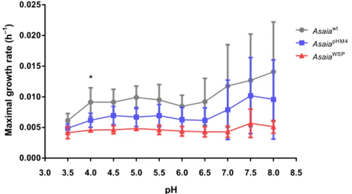
heterologous WSP had negative effects on Asaia growth, we
analyzed growth curves of the bacteria at different pH conditions
along a 24-h period. This test was also performed to reproduce
the different pH condition in mosquito organs and to test the
capability of Asaia to survive and grow. In this
fitness assay, we
compared the strain Asaia
wtwith the engineered strains carrying
the plasmids pHM4 (Asaia
pHM4) or Asaia
WSP: the mean maximal
growth rates (MGRs) of wild type and the two transformed
strains were not significantly different in almost all the tested
growth conditions, with the exception of the MGRs of Asaia
wtand Asaia
WSPat pH 4 (p
= 0.038) (Fig.
1
). In conclusion, WSP
expression does not significantly affect the fitness of Asaia
wtin
most of the tested pH conditions.
In vitro phagocytosis test and immune-related gene expression.
Phagocytosis tests on haemocytes from Ae. aegypti and An.
ste-phensi revealed significant differences, after the stimulation with
Asaia
pHM4or Asaia
WSPfor 1 (p < 0.0001 and p
= 0.0089,
respectively) and 2 h (p
= 0.0001 and p < 0.0001) (Fig.
2
a,b). The
expression of the two selected antimicrobial peptides, defensin
and cecropin, and the nitric oxide synthase (NOS) was
investi-gated on haemocytes from An. stephensi and Ae. aegypti, after an
in vitro stimulation with the two engineered bacteria. Stimulation
of mosquito haemocytes with Asaia
WSPinduced expression of
three time points (6, 9, 12 h), and at only one time point in An.
stephensi (12 h) (Supplementary Fig. 3a, b). In both An. stephensi
and Ae. aegypti haemocytes we detected production of NOS
transcripts after 9, 12 and 24 h of stimulation with Asaia
WSP(Supplementary Fig. 3a, b). Finally, in both An. stephensi and Ae.
aegypti none of the two bacteria determined a significant
upre-gulation of defensin gene expression by the haemocytes,
con-sidering all the time points.
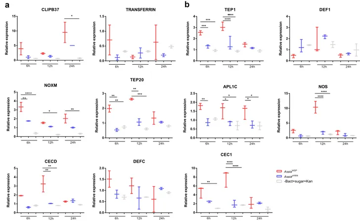
In vivo immune gene expression. Quantitative real-time PCR
assays were used to investigate the capability of Asaia
WSPto
stimulate innate immune responses in An. stephensi and Ae.
aegypti mosquitoes, after a sugar meal containing the engineered
bacteria. To determine the dynamics of this immune response,
the transcription level of immunity genes was monitored at 6, 12
and 24 h post
“bacterial meal”. Only female mosquitoes with
fully- or partially fully-engorged abdomens were selected for these
analyses. As reported in Fig.
3
(Supplementary Table 1 and
Supplementary Data 1), for Aedes mosquitoes, four of the six
analyzed genes were activated after the Asaia
WSPbacteria meal
(Fig.
3
a). On details, cecropin D gene (CECD) showed an
increased expression after 12 h compared to pHM4 and sugar
control; CLIP-domain serine protease gene (CLIPB37) resulted
activated after 24 h compared to the sugar control; thio-ester
containing protein 20 gene (TEP20) showed an upregulation on
the
first two time points compared to Asaia
pHM4and sugar
control;
finally, as for NADPH-oxidases gene (NOXM), the gene
was upregulated at all the time points, especially after 6 h. For
Aedes mosquitoes the expression of the Transferrin gene was also
investigated; after feeding with Asaia
WSPa trend in the
over-expression of the gene was observed, even though the differences
were not significant (Fig.
3
a). This agrees with results obtained on
Ae. aegypti mosquitoes transinfected with Wolbachia, where the
expression of this gene, involved in iron metabolism, immunity
and development, is observed
15.
Anopheles mosquitoes that received Asaia
WSPbacteria showed
upregulation of TEP1, leucine-rich repeat protein 1 (APL1C), NO
synthase (NOS) and cecropin 1 (CEC1)genes, compared to the
controls (Fig.
3
b, Supplementary Data 1). The degree and the
time points of upregulation were different for the different genes:
TEP1 gene for example was upregulated after 6 and 12 h,
compared to the two controls; the expression of CEC1 gene was
enhanced after 12 h; APL1C showed an upregulation after all the
three analyzed time points, while the expression of NOS gene was
very high after 12 h post bacterial meal (Fig.
3
b). Conversely, no
significant expression was detected for defensin gene in both
mosquito species, in coherence with the results obtained in vitro
on haemocytes.
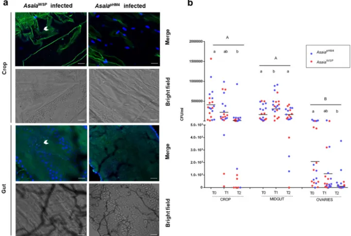
Mosquito colonization by engineered Asaia. Asaia bacteria are
an important and stable component of the microbiota of An.
stephensi and Ae. aegypti. Here, we investigated if the transgenic
bacteria were able to efficiently colonize adult Ae. aegypti female
mosquitoes, performing an immunofluorescence assays on a total
of thirty insects for each of the two different mosquito
popula-tions fed with the two engineered strains of Asaia. Analyses using
a
fluorescent confocal microscopy, after secondary staining on
anti-E-tag antibodies, showed
fluorescence signals inside the crop
and the gut of females, indicating that the bacterium efficiently
colonized these body organs. Most of the individuals showed
fluorescent cells either isolated, aggregated or in microcolonies.
Fluorescent cells and microcolonies were detected in the
mos-quito crop and gut at both 24 and 48 h after the
bacterial-containing meal; colonization of the reproductive system was
observed only 48 h after the meal, with very few bacteria. No
immunofluorescence staining was detected in organs after the
administration of Asaia
pHM4strain (Figs.
4
a), sugar or sugar plus
kanamycin (Supplementary Fig. 4a–d and 5a, b).
3.0 3.5 4.0 4.5 5.0 5.5 6.0 6.5 7.0 7.5 8.0 8.5 0.000 0.005 0.010 0.015 0.020 0.025 Asaiawt AsaiapHM4 AsaiaWSP pH
Maximal growth rate (h
–1)
*
Fig. 1 Growth rates (MGRs) of Asaiawtand recombinant strains
(AsaiaWSPand AsaiapHM4). MGRs were estimated as the slope of the best
regression line whichfitted to the 24 h growth curves calculated for either of the strains by measuring OD620at ten different pH values in GLY
medium. N= three independent experiments were conducted. Bars represent standard deviations. Statistical analysis was carried out by Welch test with GraphPad Prism 5 software; the 24-h growth curves of wild type and both transformed strains showed differences between the mean MGRs in almost all the tested growth conditions, but these differences were not significant, with the exception of the growth at pH 4 (*p < 0.05, AsaiaWSP
vs Asaiawt). Ae. aegypti P hago c yt o s isi n d e x 1h 2h 14 16 18 20 22 24 **** *** An. stephensi P ha g oc y tos is in d e x 1h 2h 14 16 18 20 22 24 ** **** AsaiapHM4 AsaiaWSP
a
b
Fig. 2 Phagocytosis tests. Phagocytic activity was evaluated in vitro using cultured (a) Ae. aegypti and (b) An. stephensi haemocytes exposed to bacterial cells from strains AsaiapHM4and AsaiaWSPincubated with FITC-fluorescent beads suspension. The percentage of haemocytes showing fluorescent
phagocytised bacteria was evaluated after 1 and 2 h. Values are expressed as median±max and min of n= 3 replicates. N = three independent experiments were conducted. Statistical significance for each experiment was determined using the two-way analysis of variance (ANOVA) followed by Sidak’s multiple comparisons test where significance is represented by **p < 0.01, ***p < 0.001 and ****p < 0.0001.
To quantify Asaia colonization, persistence and dynamics in
Ae. aegypti (in view of the successive challenge with D. immitis—
see below), bacteria colony-forming units of Asaia were assessed
at different times after blood feeding, in females previously
infected by the bacteria through sugar meal. In general, no
statistical difference in the colonization by the two Asaia strains
was detected (Fig.
4
b). As for the pattern of organ colonization, in
the midguts bacteria numbers significantly increased 24 h after
the blood meal (T1, p
= 0.0068, Fig.
4
b). In the crops, the
numbers decreased with time, in particular after the blood feeding
(T2, p
= 0.0018). As previously reported for Asaia-GFP
28, the
presence of bacteria was also detected in ovaries, in coherence
with the possibility of a transmission to progeny. Indeed,
Asaia-GFP bacteria have been shown to be transmitted to progeny
through and egg-smearing mechanism
4.
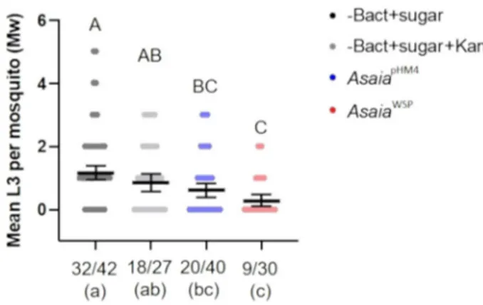
Inhibition of D. immitis infection by Asaia
WSPin Ae. aegypti.
Recombinant Asaia were administered to Ae. aegypti mosquitoes,
Liverpool strain, through a sugar meal, 32 h before mosquitoes
were fed on a D. immitis–infected blood meal. Three days after
the blood meal we recorded an average survival rate of 35% of the
mosquitoes (see Methods and Fig.
5
). Figure
5
shows the results
of the assay. For each group we determined two parameters: the
larval abundance, i.e. the average number of L3 detected in the
dissected mosquitoes; the larval prevalence, i.e. the proportion of
mosquitoes that contained at least one larva at the third stage
(L3), versus the total number of dissected mosquitoes. Asaia
WSP,
in comparison with mosquitoes fed with sugar or sugar plus
kanamycin, determined a significant decrease in L3 abundance,
with a reduction of 75.7% (p < 0.0001) and 66.8% (p
= 0.0083),
respectively. In the comparisons of mosquitoes fed on Asaia
WSPwith those fed on Asaia
pHM4, we observed reduction of 53.8%,
that was however not significant (p = 0.17). Moreover, the
feed-ing on Asaia
WSPdetermined a decrease in the prevalence of L3,
that was significant in the comparisons with the mosquitoes fed
on sugar (p
= 0.0006) or sugar plus kanamycin (p = 0.0243).
Discussion
Evidence has already been reported on the capability of WSP
from the
filarial nematode D. immitis to determine innate
immune responses in both mosquitoes and mammals
25,27,29.
Considering the conservation of the stimuli that induce innate
immunity activation across the animal phyla (e.g.
30) and the
abundance of WSP at the surface of Wolbachia cells
31, it is likely
that this protein represents an important modulator in the
interaction between the symbionts and the host in both insects
and nematodes, as well as in the tripartite system
Wolbachia-filaria-mammalian host
32. For example, it might be a major
player of the immune activation determined by Wolbachia in
mosquitoes, as recently described
33. Based on the above evidence
and considerations, we decided to engineer a mosquito symbiont
of the genus Asaia for the heterologous expression of WSP, in
order to generate a chimeric bacterium capable of inducing
CLIPB37 R el at ive e x pr e s s io n 0 5 10 15 6h 12h 24h * TRANSFERRIN Re la ti ve ex p re ss ion 0.0 0.5 1.0 1.5 6h 12h 24h TEP1 Re la ti ve e x pr ess ion 0 1 2 3 4 6h 12h 24h *** ** *** ******** DEF1 R e la ti ve e xpr e ss io n 0 1 2 3 4 6h 12h 24h NOXM Re la ti v e e xp re s si o n 0 1 2 3 4 5 6h 12h 24h *** * ** * ** Re la tiv e e x p re ss io n 0 1 2 3 6h 12h 24h ** ** TEP20 ** ** * APL1C R e la tiv ee xpr ess ion 0.0 0.5 1.0 1.5 2.0 2.5 6h 12h 24h * * ** ** NOS R e la ti v ee x p re s s ion 0 5 10 15 6h 12h 24h ******** CECD Re la tive ex pre s si on 0 1 2 3 4 5 6h 12h 24h ** ** DEFC R el ati ve e xp ress io n 0.0 0.5 1.0 1.5 2.0 6h 12h 24h R e la tiv e ex pre ss io n 0 2 4 6 8 10 6h 12h 24h ** ** ** CEC1 AsaiaWSP AsaiapHM4 -Bact+sugar+Kan ** **a
b
Fig. 3 qRT-PCR analyses of differentially regulated genes. a The differential regulation of transcript levels in AsaiaWSPinfected Ae. aegypti versus
AsaiapHM4infected Ae. aegypti or versus mosquito fed only with sugar plus kanamycin (100μg ml−1) was examined for six selected genes: cecropin
(CECD), transferrin (TRANSFERRIN), CLIP-domain serine proteases (CLIPB37), thioester-containing protein (TEP20), NADPH oxidase (NOXM) and defensin (DEFC).b The differential regulation of transcript levels in AsaiaWSPinfected An. stephensi versus AsaiapHM4infected An. stephensi or versus mosquito fed
only with sugar plus kanamycin (100μg ml−1) was examined forfive selected genes: cecropin (CEC1), Anopheles Plasmodium-responsive LRR protein-1C (APL1C), nitric oxide synthase (NOS), thioester-containing protein (TEP1) and defensin (DEF1). The values shown are median±max and min of at least two different qRT-PCR experiments with independent samples. Statistical analysis has been performed utilizing the analysis of variance (ANOVA) followed by Bonferroni’s multiple comparisons test where significance is represented by *p < 0.05, **p < 0.01, ***p < 0.001 and ****p < 0.0001.
immune activation in mosquito hosts and thus potentially
interfering with pathogen transmission by the insect.
The
first phase of the study consisted in the engineering of
Asaia strain SF2.1 for the expression of WSP, to determine the
production of the protein and to investigate the
fitness of the
transformed bacteria. The DNA fragment inserted into the
plasmid was synthetized optimizing the codon usage and
including the signal peptide, in order to allow the delivery of the
recombinant protein at the surface of the bacterial cells. Western
blotting and immunofluorence tests proved the expression of the
protein, and the pattern of immunogold staining was coherent
with a localization of the protein at the surface of the bacteria.
Genetically modified microorganisms are considered as poor
competitors and therefore unable to persist in the environment
due to energetic inefficiency. Indeed, several studies support the
idea that engineered bacteria are less
fit than their native strains,
but there are also examples of genetically modified organisms that
display an increased
fitness
34. Therefore, the capability of
Asaia
WSPto grow at different pH conditions for 24 h, under
continuous observation, was tested. As expected, growth rate of
Asaia
WSPdid not surpass either those of Asaia
pHM4or of the wild
type strain. In fact, the growth rate of Asaia
WSPwas slightly
slower, but the differences were not significant in all the tested
conditions, but one. Thus, WSP expression does not appear to
determine a significant reduction of the fitness of Asaia, hence a
significant energetic load.
The studies published so far on the immune-modulating
prop-erties of WSP in humans, dogs, rodents and mosquitoes
25,27,29,35–37have been conducted using a recombinant protein produced in E.
coli, i.e. using a system that implies a possible contamination by
LPS, even after highly accurate purification procedures. In the
current study, immunological assays, carried out in vitro and in vivo
in mosquitoes, prove to be an experimental system in which the
control is very sound. The capability of Asaia
WSPto induce
pha-gocytosis and immune gene activation was higher than that of
Asaia
pHM4, in in vitro assays. Similarly, after in vivo tests in
mos-quitoes, Asaia
WSPincreased the production of antimicrobial
pep-tides and other immune modulators, compared to Asaia
pHM4(see
“Discussion” below). These results rule out the possibility that the
observed higher activation of the immune response is due to
con-tamination by LPS or other molecules, since cells and mosquitoes
were stimulated with two strains of Asaia bacteria, with their load of
LPS and other immune-modulating molecules, differing only for
WSP expression. In summary, our results provide a further
Fig. 4 Asaia bacteria colonize mosquito organs and rapidly proliferate after a blood meal. a Immunofluorescence on AsaiaWSPin Ae. aegypti mosquitoes.
AsaiaWSPor AsaiapHM4were introduced to 2–3-day-old Ae. aegypti females via sugar meal plus kanamycin (100 μg ml−1) for 24 h; 24 h after the bacterial
meal, fed mosquitoes were selected and their organs dissected and probed with anti-E tag antibody, followed by incubation with a FITC-anti-goat IgG secondary antibody. Panels show the bright-field of the organs and the staining of AsaiaWSP(white arrows indicate group of bacteria) or AsaiapHM4. Bars:
100µm. b Population dynamics of AsaiapHM4and AsaiaWSP. AsaiapHM4and AsaiaWSPwere fed to 2–3-day-old Ae. aegypti mosquitoes in a sugar meal for
24 h (T0), then mosquitoes were allowed to feed on a blood meal and collected after 24 (T1) and 48 h (T2). Bacteria colony-forming units were determined by plating serially diluted homogenates of organs on GLY plates containing 100μg ml−1of kanamycin. The maximum bacteria number is reached when microfilariae would be invading the midgut if the blood was infected with the parasite (T1). Different capital letters represent statistically significant differences between examined organs (p < 0.05). Different lowercase letters represent statistically significant differences between time points in each organ (p < 0.05). Bars indicate the means.
evidence of the capability of WSP to induce innate immune
responses in mosquitoes.
It has been proposed that when Wolbachia is forced to create a
new symbiosis with a mosquito that is naturally non infected by
this bacterium, the basal immune response of the insect is
enhanced, with negative effects on the mosquito’s ability to
transmit pathogens
33. Mosquitoes that normally do not harbour
Wolbachia, such as An. stephensi and Ae. aegypti, can therefore be
regarded as good candidates to verify the immune-modulatory
effects of Asaia
WSPand, accordingly, its anti-parasite effects. In
Ae. aegypti both immune deficiency- and Toll-pathways are
activated by Wolbachia upon its introduction into the insect
33,38.
As for Anopheles mosquitoes, no Wolbachia had been detected in
the 38 surveyed species (including An. stephensi)
39,40, till recent
reports that identified Wolbachia in An. gambiae and Anopheles
arabiensis
26,41. In these local mosquito populations, naturally
infected by Wolbachia, the presence of the bacterium negatively
impacts Plasmodium sporozoite development
42. In addition,
studies on An. gambiae transinfected by Wolbachia suggest that
Wolbachia can confer protection to mosquitoes against the
pathogen Plasmodium, with an early activation of the immune
response
43.
The results of our in vivo studies on An. stephensi and Ae.
aegypti proved that Asaia
WSPis able to induce an immune
activation when ingested by mosquitoes. A diverse repertoire of
genes coding for immune effector molecules, such as cecropin,
thio-ester containing proteins, leucine-rich repeat protein and
CLIP-domain serine protease, plus NADPH-oxidases and NO
synthase, were upregulated in the presence of Asaia
WSP; whereas
defensin levels remained unchanged. In both An. stephensi and
Ae. aegypti, TEPs were among the most upregulated genes, after
the bacterial meal with Asaia
WSP. We emphasize that, in
Anopheles mosquitoes, TEP induced by Plasmodium berghei
binds and kills invading Plasmodium ookinetes
44. In transgenic
mosquitoes overexpressing TEP1, a reduced number of
Plas-modium parasites has also been observed
45. In Drosophila
mel-anogaster TEPs are required for efficient phagocytosis of both
Gram-positive and Gram-negative bacteria
46.
Antimicrobial peptides, that displayed upregulation in both
An. stephensi and Ae. aegypti mosquitoes after feeding Asaia
WSP,
are cecropins, at 6 and 12 h. Similar results (i.e. the activation of
cecropin expression after stimulation with the engineered
bac-teria) were obtained in vitro on haemocytes from both species. It
is interesting that cecropins have been shown to inhibit
Plasmo-dium development, and also to display antiviral effects, e.g., on
HIV-1
15. In An. stephensi, the APL1C gene showed an
upregu-lation after all the three analyzed time points; it is reported that
APL1C protein is needed for protection against the rodent
malaria parasites P. berghei and Plasmodium yoelli
47. Another
gene that displayed a strong upregulation (at 24 h), after the
Asaia
WSPstimulus, was CLIPB37; this result is consistent with the
overexpression recorded for this gene after Wolbachia infection in
Ae. aegypti and An. gambiae mosquitoes, which determined an
inhibition of pathogen transmission
48,49. The NOS gene
expres-sion has been determined both in haemocytes and in mosquitoes:
our results recorded expression of NOS gene after the induction
by Asaia
WSP. Mosquito NOS gene, highly homologous to the NOS
genes of vertebrates, is known to be expressed during the malaria
parasite invasion. In An. stephensi mosquitoes, for example, NO
production has been shown to limit the development of
Plas-modium parasites, in particular reducing the release of
spor-ozoites into the hemolymph
50. In summary, our results show that
Asaia
WSPdetermines an upregulation of four genes in An.
ste-phensi, that have been recorded, in previous studies, to be
involved in mosquito defence against Plasmodium spp.
A notable characteristic of Asaia bacteria is their capability to
colonize mosquitoes feeding on sugar meals containing the
bac-teria, offering a potential tool for their introduction in the
field.
Our results show that Asaia
WSPstill possess the ability to colonize
mosquito organs, with an increase in its abundance after the
blood meal (Fig.
4
). We emphasize that, in laboratory conditions,
cotton pads containing Asaia bacteria, either wild type or
genetically modified for WSP expression, resulted more attractive
to mosquitoes than the sterile ones. Moreover, recent laboratory
and
field investigations, using sugar feeding as a mean for
introducing bacteria different from Asaia into adult mosquitoes,
highlighted that these insects are attracted by both sterile sugar
solutions as well as by solutions containing the bacteria
51.
The evidence that Asaia
WSPcolonizes mosquitoes, and primes
the immune response, encouraged us to test the capability of this
strain to interfere with the transmission of a parasite by the
mosquito themselves. As proof of principle, we focused our
attention on a
filarial parasite, also considering that
para-transgenesis has not yet been applied in the control of
insect-borne pathogenic nematodes. The model organisms, that we
selected for this test, were the
filarial parasite D. immitis and the
mosquito vector Ae. aegypti. Our results show that Asaia
WSPindeed interferes with D. immitis infection in mosquitoes, with
differences in terms of developed L3
filarial larvae, in comparison
with insects fed on sugar meals. We also recorded a difference in
L3 numbers in mosquitoes fed on Asaia
WSPin comparison with
those fed on Asaia
pHM4, but this difference, although evident, was
not significant. However, taken together the results on the
immune priming by Asaia
WSPin mosquitoes and the coherence
in the results determined by this bacterium on
filaria
develop-ment in Ae. aegypti (in terms of both larval abundance and
prevalence, Fig.
5
), are highly encouraging. In summary, Asaia
engineered for the expression of WSP can be regarded as an
Fig. 5 Ae. aegypti infection with transgenic bacteria and D.
immitis microfilariae. Scatter dot plots show the number of L3 larvae per mosquito. The mean numbers (±SEM) of infective L3 stage of D. immitis were determined 14 days post microfilarial challenge in Ae. aegypti by microscopical observation. Four treatments have been tested: mosquitoes fed with AsaiaWSP, AsaiapHM4, sugar solution with or without kanamycin,
before the infectious blood meal. The graph reports the average number of L3 detected in the dissected mosquitoes (abundance; y axis). The prevalence, i.e. the proportion of mosquitoes that contained at least one L3 larva versus the total number of survived and dissected mosquitoes (starting from n= 100 individuals per treatment), is shown on x axis. The geometrical mean of L3 larvae per mosquito (William’s mean) was tested using a ONE-way ANOVA followed by Tukey’s post-hoc test. Different capital letters, on the top of the graph, represent statistically significant differences (p < 0.05). The mean number of mosquitoes presenting L3 larvae was tested using contingency analysis and thefinal p values were adjusted using FDR. Different lowercase letters represent statistically significant differences (p < 0.05).
inductor of innate-immune responses in mosquitoes, worth of
further investigations for its potential effects on
filarial parasite
development.
Asaia bacteria have already been investigated for their capacity
to interfere with pathogen transmission by mosquitoes. In a study
conducted on the murine malaria model P. berghei, Asaia
engi-neered for the expression of the scorpine antimicrobial peptide
determined the inhibition of parasite infection in mosquitoes
5.
More recently, a native Asaia strain (SF2.1) has been shown to
activate mosquito immunity, with reduction of P. berghei
devel-opment in An. stephensi
52. Our current study shows that the
immune stimulating capability of Asaia can be boosted through
the expression of a protein form Wolbachia. The obtained results
highlight the plasticity of the Asaia system: the engineered
bac-teria, while expressing a heterologous protein at the surface,
preserved their ability to colonize the insect, determining
over-expression for most of the tested mosquito immune effectors. The
evidence of the immune-activating capability of Asaia, either
native
52or genetically modified, as in the current study, requires
to be validated with assays on wild-collected mosquitoes;
more-over, future investigations should address the potential of Asaia
and Asaia
WSPto interfere with the transmission of arboviruses,
such as dengue and Zika. Indeed, a generalized activation of
mosquito immunity might imply a generalized protection of the
mosquito toward infectious agents, not only
filarial nematodes.
Finally, safety issues should properly be addressed before
pro-posing any strain of Asaia for
field release. In this context,
interesting investigations have been conducted on Wolbachia for
the control of dengue virus
53, including a study on the potential
transmission of this bacterium to humans
54. We emphasize that
there is strong evidence that Asaia colonizes the salivary glands of
mosquitoes; mosquitoes might thus inoculate Asaia into
mam-mals, including humans. The sole epidemiological investigation,
performed so far, has not revealed any evidence for Asaia
infection in humans, either in serological or PCR-based
ana-lyses
55. However, we can hypothesize that Asaia
WSPpossesses
increased immune-stimulating properties also toward humans,
hence an increased pathogenic potential (e.g. proinflammatory
properties
27,56). Therefore, the issue of the potential transmission
of Asaia to mammals would require further consideration.
Methods
Bacterial strains and media. Asaia SF2.1 strain (Asaiawt), originally isolated from an An. stephensi mosquito4, was grown at 30 °C in GLY medium (25 g L−1glycerol, 10 g L−1yeast extract, pH 5; eventually, GLY medium was solidified adding 20 g L−1 agar). E. coli XL1Blue (Stratagene), used as the host for construction of plasmids, was grown at 37 °C in Luria Broth (LB; LB medium was solidified adding 15 g L−1 agar if necessary). If needed, 100μg/mL kanamycin was added to the media. Plasmid construction. Plasmid pHM4 (≈5.5 kbp) was obtained by digesting pHM257with the restriction enzyme SacI (Life technologies Italia). Plasmid pHM4-WSP was then constructed by inserting the WSP cassetteflanked by NotI sites in plasmid pHM4. WSP cassette was synthesized by Eurofins Genomics (Milan) in plasmid pUC57, obtaining the plasmid pUC57-WSP. WSP cassette contains the neomycin phosphotranferase promoter PnptII, the coding DNA sequence of WSP from Dirofilaria immitis including the signal peptide of the gene37, the E-TAG epitope (GAPVPYPDPLEPR11,) and the transcription termi-nator Trrn. E-TAG epitope was inserted in the 4th loop (L4) of the wsp sequence58 to allow the immunodetection of the expressed protein. Moreover, the wsp gene sequence was optimized according to the codon usage of strain SF2.1 as inferred from its genome sequence59. WSP cassette was then digested from pUC57-WSP by using the restriction enzyme NotI, loaded in 1% agarose gel and purified by using QIAquick Gel Extraction Kit (Qiagen). Plasmid pHM4 was digested with NotI, dephosphorylated by using Shrimp Alkaline Phosphatase (SAP, Life technologies Italia) and the wsp fragment was ligated to the NotI-linearized pHM4 by using T4 DNA ligase (Life technologies Italia). Ligation product was then used to transform E. coli XL1Blue electrocompetent cells4. Recovery was performed with LB medium for 1 h at 37 °C with shaking before plating on LB plates added with kanamycin. Putative transformants were selected and successful ligation of WSP cassette was checked by PCR using wsp-specific primers (see Supplementary Table 1). The obtained plasmid, named pHM4-WSP (Supplementary Fig. 1a), was then extracted
from E. coli and electroporated in Asaiawtas previously described4resulting in the strain AsaiaWSP. Strain AsaiapHM4was also obtained and used as control in the following experiments.
Western-blot detection of WSP produced by Asaia strains. For protein secretion, the AsaiaWSPand AsaiapHM4were grown overnight at 30 °C in GLY medium supplemented with 100μg ml−1of kanamycin. Bacterial cultures were centrifuged at 3000 × g for 15 min at RT and pellets were resuspended in SDS sample loading buffer 1×; a protease inhibitor cocktail was also added to avoid the protein degradation. Briefly, membranes were blocked in blocking buffer (4% milk in PBS with 0.1% Tween 20) and probed with the primary goat anti-E tag antibody (Novus Biologicals), followed by an HRP-conjugated anti-goat IgG secondary antibody.
RNA extraction and reverse transcription-quantitative PCR. Bacteria were grown at OD 0.5, 1, 1.5 (three pools each) and stored in RNAprotect Bacteria Reagent (Qiagen); RNAs were extracted using RNeasy Mini Kit (Qiagen) including an on-column DNase I treatment to remove residual DNA. RNA was stored at −80 °C till further use. RNA purity was checked by determining the 260/280 nm absorbance ratio. cDNAs were synthesized from 250 ng of total RNA using a QuantiTect Reverse Transcription Kit (Qiagen) with random hexamers. The cDNA was used as template in RT-PCR reactions. Quantitative RT-PCRs on AsaiaWSP and AsaiapHM4were performed under the following conditions: 100 ng cDNA; 250 nM of forward and reverse primers (target gene wsp; see Supplementary Table 1 for primers sequences); 98 °C for 30 s, 40 cycles of 98 °C for 15 s, 58 °C for 30 s;fluorescence acquisition at the end of each cycle; melting curve analysis after the last cycle. The quantification cycle values were determined, in order to calculate gene expression levels of the target gene relative to 16S rRNA, the internal reference gene for Asaia55. The estimates of the expression level of wsp gene has been reported as the means ± standard error (SEM).
In vitro growth assays of Asaia forfitness measurements. Growth assays of Asaia strains were performed at different pH values (from 3.5 to 8.0 with increases of 0.5). Bacterial cells were grown overnight at 30 °C with constant agitation (130 rpm) in GLY broth. For recombinant strains, 100 µg ml−1kanamycin was added to the medium. For each strain, a dilution to 0.1 optical density at 620 nm (OD620) was carried out in GLY medium at different pH (range 3.5–8.0) and 200 μl
were distributed in 96-well microtiter plates wells (two wells for each condition). Growth was recorded by an EnSight plate reader (Perkin Elmer), measuring the OD620in each well every 10 min for 24 h at 30 °C. As negative control, growth
medium without bacteria was used. OD620values were collected and, after baseline
correction, the maximal growth rate (MGR) (h−1) was estimated as the slope of the best regression line whichfitted to growth curve, for either of the strains during the time interval. Growth assays were repeated three times. MGRs were compared by strain and medium pH using a Welch test. Student’s t-test (two-sides, Welch’s correction) was performed by GraphPad Prism 5 software. P < 0.05 was considered significant.
Immunofluorescence assays on bacteria and on mosquitoes. Recombinant Asaia bacteria expressing WSP or with plasmid alone were grown as reported above; 10μL of a cell suspension at the concentration of 108cells ml−1in PBS were placed on glass slides, air dried, andfixed for 20 min with cold methanol. Bacterial cells were blocked in bovine serum albumin (FBS) and probed with the primary goat E tag antibody (Novus Biologicals), followed by incubation with an anti-goat IgG secondary antibody, FITC Conjugate (Sigma-Aldrich).
As for the detection of transgenic bacteria in mosquito organs, AsaiaWSPor AsaiapHM4were administered to 2–3-day-old Ae. aegypti (Liverpool black-eyed strain) females via sugar meal (1 × 10⁸cells ml−1) plus kanamycin (100μg ml−1) for 24 h; 24 h after the bacterial meal, fed mosquitoes were selected and their organs (crop, midgut and ovaries) dissected andfixed in 4% (wt vol−1) paraformaldehyde at 4 °C, washed in PBS, and blocked with 4% (wt vol−1) FBS. The samples were then probed with goat anti-E tag antibody, followed by incubation with a FITC-anti-goat IgG secondary antibody. Observations were recorded with a Leica microscope (LeicaTCSNT) and analyzed with ImageJ software. Survival of mosquitoes was also monitored daily. Survival percentages represent the mean survival percentage of three biological replicates of 30 mosquitoes each.
Immunogold staining on bacteria pure culture. AsaiaWSPor AsaiapHM4samples werefixed by immersion in 4% paraformaldehyde in PBS for 2 h at 4 °C and washed in PBS. Free aldehydes were blocked in 0.5 MNHCl in PBS for 45 min at 4 °C; samples were washed in PBS, dehydrated through graded concentrations of ethanol and embedded in LR White resin (Electron Microscopy Sciences) overnight, at 4 °C. Resin samples were polymerized for 24 h at 60 °C. Ultrathin sections were placed on grids coated with a Formvar-carbon layer and then processed for immunocy-tochemistry. Ultrathin sections werefloated for 3 min on normal goat serum (NGS) diluted 1:100 in PBS and then incubated overnight at 4 °C with goat anti-E tag antibody diluted with PBS containing 0.1% BSA and 0.05% Tween 20. After rinsing, sections werefloated on NGS and then reacted for 20 min at room temperature with secondary 12 nm gold-conjugated antibodies (Jackson Laboratories) diluted 1:20 in
PBS. The specimens were observed on a Philips Morgagni transmission electron microscope operating at 80 kV and equipped with a Megaview II camera for digital image acquisition.
Colonization and quantification of Asaia in mosquitoes. To investigate coloni-zation of Asaia in different tissues of mosquitoes, 2–3-day-old adult mosquitoes were fed for 24 h on a cotton pad moistened with 5% sterile sucrose solution containing 108cells ml−1bacteria (T0). The bacteria-fed mosquitoes were starved for 10 h, and then allowed to feed on a blood meal. Twenty-four (T1) and 48 h (T2) after the blood meal, the individual mosquitoes were surface-sterilized by washing them in 75% ethanol for 3 min and then rinsing them in sterile PBS three times. The crop, midgut and ovaries were dissected under sterile conditions and homo-genized in 0.2 ml sterile PBS. The bacterial load was determined by plating tenfold serial dilutions of the homogenates on GLY plates containing 100μg ml−1of kanamycin and incubating the plates at 30 °C for 48 h. The colonies were counted and the data analyzed using RStudio. Briefly, a three-way ANOVA was used to test the global variance of the data and to assess which of the three categorical inde-pendent variables (Asaia strain, time and mosquito tissues) influences the Asaia load. After having assessed that the Asaia strain did not affect the colonization of the mosquito tissues, we performed a two-way ANOVA (using time and mosquito tissues as categorical independent variables) to test the interactions of the variables and one-way ANOVA to analyse the variance of Asaia within each of the tissues over time.
Haemocyte primary cultures and phagocytosis test. Mosquito haemocytes were isolated from dissected An. stephensi and Ae. aegypti adults and maintained 72 h in Schneider’s medium (Sigma-Aldrich), supplemented with heat-inactivated 10% fetal bovine serum (FBS), 100 units ml−1penicillin and 100μg ml−1streptomycin, before further analyses. Antibiotics have been removed before the phagocytosis test by centrifugation of cells and resuspending them in fresh medium without any addition. In the phagocytic tests, haemocyte cultures from both mosquito species were incubated for 6 h in 1 ml of medium containing bacteria. Successively, hae-mocytes were shortly centrifuged, resuspended in 200 µl of fresh Schneider’s medium (without any supplement) and then incubated with 0.1 µl of a FITC-fluorescent beads suspension for 1 and 2 h in soft oscillation, according to ref.60. After incubation, cells were cytocentrifugated onto glass slides, counterstained with a 200 ng ml−1propidium iodide solution and observed with a Zeiss Axioplan epifluorescence microscope. The phagocytosis index was evaluated as the percen-tage of haemocytes showing insidefluorescent particles. Three phagocytic test replicated experiments were performed. Statistical analysis has been performed using GraphPad Prism 5 utilizing the two-way analysis of variance (ANOVA) followed by Sidak’s multiple comparisons test (p < 0.05 has been considered significant).
Antimicrobial peptides and nitric oxide synthase expression in hemocytes. An. stephensi and Ae. aegypti hemocytes has been incubated with a 109cells ml−1 bacterial solution for 0, 3, 6, 9, 12 and 24 h. After treatments, cells were centrifuged at 800 × g for 5 min at room temperature and the supernatant was discarded. Total RNA was extracted from cells using TRI-REAGENT TM (Sigma), following the method described by the supplier. RT-PCR has been performed with the Access RT-PCR System (Promega), according to the supplier’s protocols. For An. stephensi and Ae. aegypti, actin was used as reference gene; the sequences of the analyzed genes and the relative citations were reported in Supplementary Table 1. For both species, PCR amplification gel documentation was collected using a Gel Doc XR, digitally evaluated with Quantity One (Bio-Rad Lab) and normalized to the cor-respondent signals for cytoplasmic actin. Three replicates were carried out for each induction.
Immune gene expression in mosquitoes fed with bacteria. AsaiaWSPor AsaiapHM4were administered to 2–3-day-old adult female mosquitoes (An. ste-phensi and An. aegypti) via sugar meals, bred in small cages containing 50 samples. Mosquitoes were allowed to feed for 6, 12 and 24 h on a sterile cotton pad moistened with 5% sterile sucrose solution containing 108cells ml−1bacteria (plus kanamycin 100μg ml−1), or 5% sugar plus kanamycin with no bacteria (as control).
After 6, 12 and 24 h, the mosquitoes were collected and stored in RNA later at−80 °C for RNA extraction and molecular analysis. The expression profiles of 11 immune-related genes (see Supplementary Table 1 for primer sequences and details), were analyzed by quantitative RT-PCRs. Briefly, RNA was extracted from pool of three mosquitoes using the RNeasy Mini Kit (Qiagen), according to the manufacturer’s instructions. cDNAs were synthesized from 150 ng of total RNA using a QuantiTect Reverse Transcription Kit (Qiagen). Quantitative RT-PCRs on target genes were performed using a BioRad Real-Time PCR Detection System (Bio-Rad) at the following conditions: 50 ng cDNA; 300 nM of forward and reverse primers; 98 °C for 30 s, 40 cycles of 98 °C for 15 s, 56-60 °C for 30 s;fluorescence acquisition at the end of each cycle; melting curve analysis after the last cycle. In order to calculate the expression of the target genes, quantification cycle (Cq) values were determined for each gene and normalized according to the endogenous reference genes rps7 or rps17 (Supplementary Table 1). The estimates of the
expression level of each gene are relative to the control groups and reported as fold change mean ± standard error mean (SEM) of at least three replicates. Statistical analysis has been performed using GraphPad Prism 5 utilizing the ANOVA followed by Bonferroni’s multiple comparisons test (p < 0.05 has been considered significant).
Mosquito infection with bacteria and microfilariae. Microfilariaemic blood samples from a dog naturally infected with D. immitis and blood from an unin-fected dog were kindly provided by Prof. Genchi; bloods were anticoagulated with heparin. Since blood was collected for diagnostic purposes and the owners signed an informed consent that authorize the use of residual samples (i.e. the amount of blood remained after diagnostic clinical chemistry) for research purposes, according to the regulations of our Institution (EC decision 02-2016) a formal approval from the Ethical Committee was not required. Vitality and number of D. immitis microfilariae in all samples were confirmed by microscopy; briefly, 20μl of blood were mixed with 40 μl of distilled water, covered with a cover slide, and microfilariae were counted by examination with a microscope (4×). Microfilaraemiae of the dog was determined three times. For the inoculation experiments, Ae. aegypti female mosquitoes at an age of 2–3 days were selected, maintained at standard condition in cages of 100 samples61and fed on a sterile cotton pad moistened with 5% sucrose solution containing 108cells ml−1bacteria (with kanamycin 100 µg ml−1) for 1 day (four treatments: mosquitoes fed with AsaiaWSPor AsaiapHM4plus microfilariae, sugar solution with/without kana-mycin plus microfilariae).
Microfilariaemic counts were adjusted to 3500 mf ml−1with blood from uninfected dog. The microfilaria load in the infecting blood was according to recommended protocols62, in order to avoid an excess in larval mortality, caused by nematode larvae. Sugar was removed and the mosquitoes were allowed to feed through Parafilm® membranes for at least 1.5 h on 5 ml blood at 37 °C in an artificial feeding system. Three to five mosquitoes were immediately dissected to verify mean microfilariae ingested per mosquito. Mosquitoes were kept for up to 14 days in cages with access to 5% glucose and water ad libitum; after this time, mosquitoes were collected and exposed for 2 min in a freezer for immobilization, and the wings and legs were removed. Only the mosquitoes for which a blood meal was completed were collected. These mosquitoes were dissected individually: the abdomen was separated and midgut contents were smeared on a slide; D. immitis L3 larvae were thus counted. Statistical analysis was performed by GraphPad Prism 5 software. The mean number of individuals presenting L3 larvae was tested using contingency analysis and thefinal p values were adjusted using FDR, while the geometric mean number of L3 larvae per infected mosquito was calculated using the William’s mean (Mw)63, considering the high proportion of mosquitoes not presenting L3 larvae, and Mw were analyzed using a ONE-way ANOVA followed by Tukey’s post-hoc test.
Reporting summary. Further information on research design is available in the Nature Research Reporting Summary linked to this article.
Data availability
The datasets generated during and/or analyzed during the current study are available from the corresponding author (Molecular and Evolutionary Parasitology Lab, Department of Biosciences, University of Milan), on reasonable request. The source data underlying plots shown infigures are presented in Supplementary Data 1.
Received: 24 October 2019; Accepted: 18 February 2020;
References
1. Weiss, B. & Aksoy, S. Microbiome influences on insect host vector competence. Trends Parasitol. 27, 514–522 (2011).
2. Crotti, E. et al. Asaia, a versatile acetic acid bacterial symbiont, capable of cross-colonizing insects of phylogenetically distant genera and orders. Environ. Microbiol. 11, 3252–3264 (2009).
3. Rami, A., Raz, A., Zakeri, S. & Djadid, D. N. Isolation and identification of Asaia sp. in Anopheles spp. mosquitoes collected from Iranian malaria settings: steps toward applying paratransgenic tools against malaria. Parasit. Vectors 11, 367 (2018).
4. Favia, G. et al. Bacteria of the genus Asaia stably associate with Anopheles stephensi, an Asian malarial mosquito vector. Proc. Natl Acad. Sci. USA 104, 9047–9051 (2007).
5. Bongio, N. J. & Lampe, D. J. Inhibition of Plasmodium berghei development in mosquitoes by effector proteins secreted from Asaia sp. bacteria using a novel native secretion signal. PLoS ONE 10, e0143541 (2015).
6. Shane, J. L., Grogan, C. L., Cwalina, C. & Lampe, D. J. Blood meal-induced inhibition of vector-borne disease by transgenic microbiota. Nat. Commun. 9, 4127 (2018).
7. Crotti, E. et al. Acetic acid bacteria, newly emerging symbionts of insects. Appl. Environ. Microbiol. 76, 6963–6970 (2010).
8. Damiani, C. et al. Paternal transmission of symbiotic bacteria in malaria vectors. Curr. Biol. 18, 1087–1088 (2008).
9. Mancini, M. V. et al. Paratransgenesis to control malaria vectors: a semi-field pilot study. Parasit. Vectors 9, 140 (2016).
10. Wilke, A. B. & Marrelli, M. T. Paratransgenesis: a promising new strategy for mosquito vector control. Parasit. Vectors 8, 342 (2015).
11. Wang, S. et al. Driving mosquito refractoriness to Plasmodium falciparum with engineered symbiotic bacteria. Science 357, 1399–1402 (2017). 12. Iturbe-Ormaetxe, I., Walker, T. & O’Neill, S. L. Wolbachia and the biological
control of mosquito-borne disease. EMBO Rep. 12, 508–518 (2011). 13. Bandi, C., Anderson, T. J., Genchi, C. & Blaxter, M. L. Phylogeny of
Wolbachia infilarial nematodes. Proc. Biol. Sci. 265, 2407–2413 (1998). 14. Bourtzis, K. et al. Harnessing mosquito-Wolbachia symbiosis for vector and
disease control. Acta Trop. 132, S150–S163 (2014).
15. Kambris, Z., Cook, P. E., Phuc, H. K. & Sinkins, S. P. Immune activation by life-shortening Wolbachia and reducedfilarial competence in mosquitoes. Science 326, 134–136 (2009).
16. Moreira, L. A. et al. A Wolbachia symbiont in Aedes aegypti limits infection with dengue, chikungunya, and Plasmodium. Cell 139, 1268–1278 (2009). 17. Bian, G., Xu, Y., Lu, P., Xie, Y. & Xi, Z. The endosymbiotic bacterium
Wolbachia induces resistance to dengue virus in Aedes aegypti. PLoS Pathog. 6, e1000833 (2010).
18. Bian, G. et al. Wolbachia invades Anopheles stephensi populations and induces refractoriness to Plasmodium infection. Science 340, 748–751 (2013). 19. Dutra, H. L. et al. Wolbachia blocks currently circulating Zika virus isolates in brazilian Aedes aegypti mosquitoes. Cell Host Microbe 19, 771–774 (2016). 20. Thomas, S., Verma, J., Woolfit, M. & O’Neill, S. L. Wolbachia-mediated virus
blocking in mosquito cells is dependent on XRN1-mediated viral RNA degradation and influenced by viral replication rate. PLoS Pathog. 14, e1006879 (2018).
21. Dodson, B. L. et al. Wolbachia enhances West Nile Virus (WNV) infection in the mosquito Culex tarsalis. PLoS Negl. Trop. Dis. 8, e2965 (2014). 22. Walker, T. et al. The wMel Wolbachia strain blocks dengue and invades caged
Aedes aegypti populations. Nature 476, 450–453 (2011).
23. Frentiu, F. D. et al. Limited dengue virus replication infield-collected Aedes aegypti mosquitoes infected with Wolbachia. PLoS Negl. Trop. Dis. 8, e2688 (2014).
24. Rasgon, J. L., Gamston, C. E. & Ren, X. Survival of Wolbachia pipientis in cell-free medium. Appl. Environ. Microbiol. 72, 6934–6937 (2006).
25. Pinto, S. B., Mariconti, M., Bazzocchi, C., Bandi, C. & Sinkins, S. P. Wolbachia surface protein induces innate immune responses in mosquito cells. BMC Microbiol. 12, S11 (2012).
26. Baldini, F. et al. Evidence of natural Wolbachia infections infield populations of Anopheles gambiae. Nat. Commun. 5, 3985 (2014).
27. Brattig, N. W. et al. The major surface protein of Wolbachia endosymbionts in filarial nematodes elicits immune responses through TLR2 and TLR4. J. Immunol. 173, 437–445 (2004).
28. Rossi, P. et al. Mutual exclusion of Asaia and Wolbachia in the reproductive organs of mosquito vectors. Parasit. Vectors 8, 278 (2015).
29. Kramer, L. H. et al. Immune response to and tissue localization of the Wolbachia surface protein (WSP) in dogs with natural heartworm (Dirofilaria immitis) infection. Vet. Immunol. Immunopathol. 106, 303–308 (2005). 30. Ausubel, F. M. et al. Are innate immune signaling pathways in plants and
animals conserved? Nat. Immunol. 6, 973–979 (2005). 31. Braig, H. R., Zhou, W., Dobson, L. & O’Neill, S. L. Cloning and
characterization of a gene encoding the major surface protein of the bacterial endosymbiont Wolbachia pipientis. J. Bacteriol. 180, 2373–2378 (1998). 32. Taylor, M. J., Bandi, C. & Hoerauf, A. Wolbachia bacterial endosymbionts of
filarial nematodes. Adv. Parasitol. 60, 245–284 (2005).
33. Pan, X. et al. The bacterium Wolbachia exploits host innate immunity to establish a symbiotic relationship with the dengue vector mosquito Aedes aegypti. ISME J. 12, 277–288 (2018).
34. Lenski, R. E. Evaluating the fate of genetically modified microorganisms in the environment: are they inherently lessfit? Experientia 49, 201–209 (1993). 35. Bazzocchi, C. et al. Immunological role of the endosymbionts of Dirofilaria
immitis: the Wolbachia surface protein activates canine neutrophils with production of IL-8. Vet. Parasitol. 117, 73–83 (2003).
36. Marcos-Atxutegi, C. et al. Th1 response in BALB/c mice immunized with Dirofilaria immitis soluble antigens: a possible role for Wolbachia? Vet. Parasitol. 112, 117–130 (2003).
37. Bazzocchi, C. et al. Wolbachia surface protein (WSP) inhibits apoptosis in human neutrophils. Parasit. Immunol. 29, 73–79 (2007).
38. Rancès, E., Ye, Y. H., Woolfit, M., McGraw, E. A. & O’Neill, S. L. The relative importance of innate immune priming in Wolbachia-mediated dengue interference. PLoS Pathog. 8, e1002548 (2012).
39. Kittayapong, P., Baisley, K. J., Baimai, V. & O’Neill, S. L. Distribution and diversity of Wolbachia infections in southeast Asian mosquitoes (Diptera: Culicidae). J. Med. Entomol. 37, 340–345 (2000).
40. Ricci, I. et al. Searching for Wolbachia (Rickettsiales: Rickettsiaceae) in mosquitoes (Diptera: Culicidae): large polymerase chain reaction survey and new dentifications. J. Med. Entomol. 39, 562–567 (2002).
41. Baldini, F. et al. First report of natural Wolbachia infection in the malaria mosquito Anopheles arabiensis in Tanzania. Parasit. Vectors 11, 635 (2018).
42. Gomes, F. M. et al. Effect of naturally occurring Wolbachia in Anopheles gambiae s.l. mosquitoes from Mali on Plasmodium falciparum malaria transmission. Proc. Natl Acad. Sci. USA 114, 12566–12571 (2017). 43. Hughes, G. L., Koga, R., Xue, P., Fukatsu, T. & Rasgon, J. L. Wolbachia
infections are virulent and inhibit the human malaria parasite Plasmodium falciparum in Anopheles gambiae. PLoS Pathog. 7, e1002043 (2011). 44. Eldering, M. et al. Variation in susceptibility of African Plasmodium
falciparum malaria parasites to TEP1 mediated killing in Anopheles gambiae mosquitoes. Sci. Rep. 6, 20440 (2016).
45. Volohonsky, G. et al. Transgenic expression of the anti-parasitic factor TEP1 in the malaria mosquito Anopheles gambiae. PLoS Pathog. 13, e1006113 (2017). 46. Ertürk-Hasdemir, D. & Silverman, N. Eater: a big bite into phagocytosis. Cell
123, 190–192 (2005).
47. Mitri, C. et al. Fine pathogen discrimination within the APL1 gene family protects Anopheles gambiae against human and rodent malaria species. PLoS Pathog. 5, e1000576 (2009).
48. Kambris, Z. et al. Wolbachia stimulates immune gene expression and inhibits plasmodium development in Anopheles gambiae. PLoS Pathog. 6, e1001143 (2010).
49. Blagrove, M. S., Arias-Goeta, C., Failloux, A. B. & Sinkins, S. P. Wolbachia strain wMel induces cytoplasmic incompatibility and blocks dengue transmission in Aedes albopictus. Proc. Natl Acad. Sci. USA 109, 255–260 (2012).
50. Luckhart, S., Vodovotz, Y., Cui, L. & Rosenberg, R. The mosquito Anopheles stephensi limits malaria parasite development with inducible synthesis of nitric oxide. Proc. Natl Acad. Sci. USA 95, 5700–5705 (1998).
51. Bilgo, E. et al. Field assessment of potential sugar feeding stations for disseminating bacteria in a paratransgenic approach to control malaria. Malar. J. 17, 367 (2018).
52. Cappelli, A. et al. Asaia activates immune genes in mosquito eliciting an anti-Plasmodium response: implications in Malaria control. Front. Genet. 10, 836 (2019).
53. Murray, J. V., Jansen, C. C. & De Barro, P. Risk associated with the release of Wolbachia-infected Aedes aegypti mosquitoes into the environment in an effort to control Dengue. Front. Public Health 4, 43 (2016).
54. Popovici, J. et al. Assessing key safety concerns of a Wolbachia-based strategy to control dengue transmission by Aedes mosquitoes. Mem. Inst. Oswaldo Cruz. 105, 957–964 (2010).
55. Epis, S. et al. Do mosquito-associated bacteria of the genus Asaia circulate in humans? Eur. J. Clin. Microbiol. Infect. Dis. 31, 1137–1140 (2012). 56. Saint André, Av et al. The role of endosymbiotic Wolbachia bacteria in the
pathogenesis of river blindness. Science 295, 1892–1895 (2002).
57. Mostafa, H. E., Heller, K. J. & Geis, A. Cloning of Escherichia coli lacZ and lacY genes and their expression in Gluconobacter oxydans and Acetobacter liquefaciens. Appl. Environ. Microbiol. 68, 2619–2623 (2002).
58. Baldo, L., Desjardins, C. A., Russell, J. A., Stahlhut, J. K. & Werren, J. H. Accelerated microevolution in an outer membrane protein (OMP) of the intracellular bacteria Wolbachia. BMC Evol. Biol. 10, 48 (2010). 59. Chouaia, B. et al. Acetic acid bacteria genomes reveal functional traits for
adaptation to life in insect guts. Genome Biol. Evol. 6, 912–920 (2014). 60. Manfredini, F., Dallai, R. & Ottaviani, E. Circulating hemocytes from larvae of
the paper wasp Polistes dominulus (Hymenoptera, Vespidae). Tissue Cell 40, 103–112 (2008).
61. Bozic, J. et al. Mosquitoes can harbour yeasts of clinical significance and contribute to their environmental dissemination. Environ. Microbiol. Rep. 9, 642–648 (2017).
62. Tiawsirisup, S. & Nithiuthai, S. Vector competence of Aedes aegypti (L.) and Culex quinquefasciatus (Say) for Dirofilaria immitis (Leidy). Southeast Asian J. Trop. Med. Public Health 37, 110–114 (2006).
63. Williams, C. B. The use of logarithms in the interpretation of certain entomological problems. Ann. Appl. Biol. 24, 404–414 (1937).
Acknowledgements
This study was supported by Cariplo Foundation and Lombardy Region to S.E. (2017-N.1656), by Transition Grant 2015-2017-Horizon 2020 to S.E. (G42F17000140001) and by the MIUR PRIN (Italian Ministry of Education, University and Research, MIUR) to C.B., L.P. and G.F. D.D. acknowledges the baseline funding support by King Abdullah University of Science and Technology (KAUST); S.E., C.B. and M.B. thank“Fondo per il finanziamento dei Dipartimenti universitari di eccellenza” (MIUR) of the Dept. of Biosciences (University of Milan) and of the Dept. of Biology and Biotechnology“L. Spallanzani” (University of Pavia). The authors thank Prof. L. Sacchi, Prof. F. Forlani and
Prof. C. Bazzocchi for their suggestions and the UNITECH platform for the microscope image acquisition.
Author contributions
S.E. and I.V.B. performed and supervised the majority of experiments. S.E. and E.C. generated transgenic bacteria; I.V.B., L.G. and L.P. conductedfitness experiments; M.B. performed immunogold assays and M.M. conducted the in vitro experiments; C.D., P.G. and M.G. performed the cage experiments and transmission-blocking assays; G.F., D.D. and C.B. analyzed the data; S.E., I.V.B. and C.B. wrote the manuscript; all authors revised and provided inputs to the manuscript.
Competing Interests
The authors declare no competing interests.
Additional information
Supplementary informationis available for this paper at https://doi.org/10.1038/s42003-020-0835-2.
Correspondenceand requests for materials should be addressed to C.B.
Reprints and permission informationis available athttp://www.nature.com/reprints
Publisher’s note Springer Nature remains neutral with regard to jurisdictional claims in published maps and institutional affiliations.
Open Access This article is licensed under a Creative Commons Attribution 4.0 International License, which permits use, sharing, adaptation, distribution and reproduction in any medium or format, as long as you give appropriate credit to the original author(s) and the source, provide a link to the Creative Commons license, and indicate if changes were made. The images or other third party material in this article are included in the article’s Creative Commons license, unless indicated otherwise in a credit line to the material. If material is not included in the article’s Creative Commons license and your intended use is not permitted by statutory regulation or exceeds the permitted use, you will need to obtain permission directly from the copyright holder. To view a copy of this license, visithttp://creativecommons.org/ licenses/by/4.0/.