Formation and Maturation of the Calcium
Release Apparatus in Developing
and Adult Avian Myocardium
Feliciano Protasi, Xin-Hui Sun, and Clara Franzini-Armstrong
Department of Cell and Developmental Biology, University of Pennsylvania, School of Medicine, Philadelphia, Pennsylvania 19104 – 6058
Muscle fibers release large amounts of calcium from an internal compartment, the sarcoplasmic reticulum (SR), during activation. Two proteins are involved in this process and its control: plasma membrane calcium channels, or dihydropyridine receptors (DHPRs), and SR calcium release channels, or ryanodine receptors (RyRs). The two proteins form part of a structural complex, perhaps unique to muscle cells, which allows an interaction between plasma membrane and SR, resulting in calcium release from the latter. The surface–SR interaction is a step in the coupling between electrical events in the plasma membrane and contraction (excitation–contraction coupling). The structural complexes have been called calcium release units. One key to further understanding the control of calcium homeostasis in muscle is knowledge of how DHPRs and RyRs assemble into calcium release units. We have studied the development of avian myocardium, using immunocytochemistry to locate DHPRs and RyRs and electron microscopy to follow the formation of calcium release units containing feet (RyRs) and large membrane particles (presumably DHPRs). We find that the initial step is a docking of SR vesicles to the plasma membrane, followed by the appearance of feet in the junctional gap between SR and plasma membrane. Feet aggregate in ordered arrays, and the arrays increase in size until they fill the entire junctional gap. Clustering of membrane particles, presumably DHPRs, is apparently coupled to clustering of feet, since the two junction components assemble within patches of membrane of approximately equal size and containing an approximately constant ratio of particles to feet. Thus, despite the fact that no evidence exists for a direct interaction between DHPRs and RyRs in cardiac muscle, some mechanism exists to ensure that the two molecules are clustered in proximity to each other and in the appropriate proportion. q 1996 Academic Press, Inc.
INTRODUCTION
Ca2/storage compartment, the sarcoplasmic reticulum (SR). The SR is rich in RyRs (see Meissner, 1994, for a re-view), localized mostly in specialized junctional domains All cells have internal calcium storage compartments
which accumulate calcium by means of calcium pumps of (jSR; Franzini-Armstrong and Jorgensen, 1994).
the SERCA family (for reviews see MacLennan and Toyo- Calcium release from the SR is part of a process termed fuku, 1992; Pozzan et al., 1994). Release of calcium from excitation–contraction (e–c) coupling. This is initiated by these compartments occurs through two related calcium depolarization of the plasma membrane and its invagi-release channels, the IP3 receptors (Berridge and Irvine, nations, the transverse (T) tubules. Depolarization is sensed 1989; Takei et al., 1994) and the ryanodine receptors, or by calcium channels, the dihydropyridine receptors or RyRs1
(Inui et al., 1987 a,b; Lai et al., 1988; Anderson et al., DHPRs, located in the plasma membrane and T tubules (for 1989; Giannini et al., 1995). Muscle fibers have an extensive reviews, see Ashley et al., 1991; Catterall, 1991; Stern and Lakatta, 1992; see also Cannell et al., 1995). DHPRs and RyRs are in close proximity at sites where junctional
do-1
Abbreviations used: SR, sarcoplasmic reticulum; DHPR, dihy- mains of the SR, bearing arrays of RyRs, are closely apposed dropyridine receptor or L-type calcium channel; RyR, ryanodine
to junctional domains of the plasma membrane (jPM) or receptor or calcium release channel; SERCA, sarco/endoplasmic
transverse tubules (jT), containing DHPRs (Jorgensen et al., reticulum Ca2/–ATPase; jSR, junctional domains of the
sarcoplas-1989; Flucher et al., 1990; Yuan et al., 1991, Carl et al., mic reticulum; e–c coupling, excitation–contraction coupling; T
1995; Sun et al., 1995). This DHPRs–RyRs association has tubules, transverse tubules; jT, junctional domains of the
266 Protasi, Sun, and Franzini-Armstrong
in which this occurs are called either calcium release units relaxing solution and then frozen. Cryostat sections (12–15 mm thick) were cut at 0207C.
or e–c coupling units, depending on the role they play.
Cal-cium release units in muscle are uniquely designed for the Antibodies used were: (a) CR2, a rabbit polyclonal anti-body raised against a fragment of rabbit cardiac a1DHPR rapid and controlled release of the large amount of calcium
that is needed for activation of contraction. Our aim is to derived from a plasmid for the 549 C-terminus amino acids (Yoshida et al., 1992). This antibody immunolabels chick define how these unique assemblies are formed during
de-velopment of cardiac muscle. cardiac muscle (Sun et al., 1995). (b) 34C, a mouse mono-clonal antibody, generously provided by Dr. J. Sutko and In this work, we take advantage of the fact that RyRs and
DHPRs are visible in the electron microscope. The cyto- Dr. J. A. Airey, which recognizes a, b, and avian cardiac RyR (Airey et al., 1990).
plasmic domains of RyRs form the ‘‘feet,’’ linking jSR to jT
or jPM (Block et al., 1988). DHPRs have been identified The unfixed sections were incubated for 1 hr each in 1% bovine serum albumin in PBS (PBS/BSA), followed by either with large intramembrane particles, seen in freeze-fracture
replicas of jPM and jT in skeletal muscle (Block et al., 1988; CR2 or 34C and by Texas red-conjugated goat anti-mouse IgG (from Molecular Probes Inc.) and goat anti-rabbit IgG Franzini-Armstrong et al., 1991; Takekura et al., 1994a). A
similar identification has been proposed for cardiac muscle (from Cappel products), all diluted in PBS/BSA. In control experiments, cryosections were incubated in 1% PBS/BSA (Sun et al., 1995).
We address the following questions: (1) It is known that for 2 hr, followed by one of the secondary antibodies. The specimens were viewed in a scanning confocal microscope RyRs are detectable at very early stages in cardiac muscle
(Dutro et al., 1993). Is the formation of calcium release units (Bio-Rad MRC-600, Microscience). also an early developmental event? (2) What are the
sequen-tial steps involved in the association of RyRs and DHPRs
Electron Microscopy
with the junctions? (3) Is there an obligatory relationship
between DHPRs and RyRs during the formation of calcium Hearts at E10–adult were perfused either with a ‘‘re-laxing’’ solution as above, or with a ‘‘Ca2/free solution’’ release units? (4) What steps lead to the maturation of the
membrane system from its initial formation to the adult (0.9% NaCl, 10 mM potassium–phosphate buffer, 10 mM EGTA, pH 7.4), which also maintains the myocardium in arrangement?
Our results indicate the following sequential steps in the a relaxed state. Smaller hearts were immersed and bisected in either solution. Fixation with 3.5% glutaraldehyde in 0.1 formation of the junction: an initial docking of the SR
mem-brane to the plasmalemma, the formation of arrays of feet, Mcacodylate buffer, pH 7.4, was also by either perfusion or immersion. Either the whole heart (at E2.5–E4) or small and the parallel clustering of large particles (presumably
DHPRs) in the jPM domains. Arrays of feet and clusters of pieces of the left ventricle were postfixed in 2% OsO4in cacodylate buffer for 2 hr, block-stained in saturated uranyl large particles grow in parallel, until the junctional gap is
completely filled by feet. An obligatory relationship be- acetate at 607C for 4 hr, and embedded in Epon 812. Thin sections were stained in uranyl acetate and lead.
tween the locations and quantities of DHPRs and RyRs is
apparently established early during development and main- For freeze-fracture, bundles were similarly fixed in either 3.5 or 6% glutaraldehyde, infiltrated in 30% glycerol, frozen tained throughout. As in skeletal muscle, we can define a
maturation phase, in which the e–c coupling system adapts in liquid nitrogen-cooled propane, fractured, shadowed with platinum at 457C, and replicated with carbon in a Balzer’s itself to the higher demands of larger and more active cells.
This involves an increase in size and frequency of junctions, 400. The replicas and thin sections were photographed in a Philips EM 410.
the acquisition of a regular disposition of junctions relative to the sarcomere, and finally the development of the inter-nal extended jSR.
Measurements
Sizes of peripheral couplings and junctional plasma mem-brane domains (see Results) were measured from images
MATERIALS AND METHODS
collected by photographing the relevant structures as they first appeared on the EM screen until it was decided that a Chick embryos at 2.5 to 21 days of incubation (E2.5–E21),
young chicks at 1 to 14 days after hatching (D1–D14), and sufficiently large sample had been obtained. In the case of freeze-fractures at E4 and E6, this involved a prolonged adult chickens were used.
search, because limited views of the surfaces are available and the particle domains are small and rare. It is possible
Immunohistochemistry that very small domains were missed in this search.
Areas of junctions were estimated by measuring the For immunofluorescent labeling, hearts (E10 to adult)
were removed and perfused through the aorta with a length of apposition between profiles of jSR and jPM in sections perpendicular to the two apposed membranes. As-‘‘relaxing’’ solution (80 mM potassium acetate, 10 mM
po-tassium phosphate buffer, 5 mM EGTA). After Ç5 min, por- suming that the junction is circular and that images of sec-tions cutting across it (such as those in Figs. 6B, 6C, 6E, tions of the left ventricle were frozen in liquid
nitrogen-cooled propane. Hearts at E2.5–E7 were bisected in the and 6G) represent random chords of such circles, the average
measured chord (y) is related to the diameter of the average corbular SR, i.e., SR bearing a group of feet but not associ-ated with the plasma membrane (Jewett et al., 1971, 1973; circle (D) by the equation: y Å pD/4.
The outline of plasma membrane domains was marked Sommer and Johnson, 1979; Dolber and Sommer, 1984). We were unable to detect foci of either protein in frozen on images from freeze-fracture replicas, and their surface
areas were directly measured. Large membrane particles sections at E6 or in whole mounts of hearts at E3 and E4. The latter were permeabilized by a 10-min exposure to within the domains were visually identified and counted.
We have previously shown that visual identification selects MeOH at 0207C. We do not know if this failure is due to a technical limitation (the sections at E6 were very poor a type of particle which is significantly different in height
and size relative to the average extrajunctional particle (Sun and infiltration of the whole heart may be limited) or to the small size of RyR and DHPR aggregates at these early ages
et al.,1995).
Images for measuring the size of the junctional gap were (see below).
Colocalization of the two proteins in closely apposed hot selected from the initial random collection using the
follow-ing criteria: crispness of the membrane profile, indicatfollow-ing a foci was shown at D6 and D10 in a previously published work (Sun et al., 1995).
good cross section of the membrane, and good focus (compa-rable to that shown in Figs. 6B, 6C, 6E, and 6G). Lines were drawn on the print at several random positions across the
Peripheral Couplings and Plasma Membrane
junctions, and measurements of the junctional gap were
Domains
done with a dissecting microscope equipped with an
eye-piece micrometer. Peripheral couplings and junctional plasma membrane domains (jPM) are defined in electron microscope images For counting the frequency of peripheral couplings, all
plasma membrane profiles in a section were divided into of thin sections and freeze-fracture replicas.
Well-developed peripheral couplings are junctions be-0.8-mm-long segments, the number of peripheral couplings
in each segment were counted, and data from 10 adjacent tween an SR vesicle and the plasma membrane (Fig. 2A, between large arrows). Ordered arrays of feet (small arrows) segments were pooled. The number of profiles per 100-mm
length of plasma membrane profile was calculated, assum- occupy the junctional gap between the two membranes, zippering them together (Fig. 2A, E17, small arrows). Since ing that the membrane is straight through the segment. The
error resulting from this approximation is small, consider- feet have been identified as the cytoplasmic domains of RyRs, groups of feet in electron micrographs of peripheral ing the short length of the segment, but it is slightly larger
at younger ages, where the fixed membrane is wrinkled. couplings correspond to the foci of RyRs shown in Fig. 1. Junctional plasma membrane domains are defined as Although only one animal was used at each time point,
all significance tests were performed using data from three areas of plasma membrane containing loose clusters of dis-tinctive, large particles with an elongated shadow (outlined to four animals from neighboring time points so that the
sample size was statistically appropriate. by arrowheads in Figs. 2B–2D, from freeze-fractures of myo-cardium at E10, E21, and adult). In previous work (Sun et
al., 1995), we established that the position of jPM corre-sponds to that of peripheral couplings seen in thin sections
RESULTS
and that of DHPR clusters detected by immunohistochem-istry. Also, the large particles were shown to constitute a
Clusters of RyRs and DHPRs
unique population and to be similar to those identified as DHPRs in skeletal muscle. From these data, we suggested Avian cardiac muscle was selected because it lacks T
tubules and thus complete calcium release units are located that the large particles represent DHPRs. at the plasma membrane. Images in Fig. 1 illustrate cross
sections of the left ventricle at E7 (Figs. 1A and 1B), E10
Development of Peripheral Couplings
(Figs. 1C and 1D), D1 (Figs. 1E and 1F), and adult (Figs. 1G
and 1H), immunolabeled for DHPR (left) and RyR (right). The content of feet in peripheral couplings of developing myocardium is variable. In some ‘‘incomplete’’ junctions, RyRs and DHPRs are located in small clusters along the
cells’ periphery. In the early developmental stages, particu- feet are absent from the junctional gap (Figs. 3A, 3D, and 3G). In ‘‘partially complete’’ junctions, the feet are closely larly at E7 and E10, high gain and a wide open return
aper-ture had to be used in order to detect foci of the two proteins spaced in a portion of the gap, but are apparently absent elsewhere (Figs. 3B, 3E, and 3H). In ‘‘complete’’ junctions, in the confocal microscope. Detection became easier with
increasing embryonic and postembryonic ages, indicating the junctional gap is fully zippered by closely spaced feet (Figs. 3C, 3F, and 3I). At E2.5, we see incomplete junctions an increment in protein content of the clusters. As the cells
enlarge, the number of spots per cell outline also increases. and a few partially complete ones, with two closely spaced feet in the junctional gap. At E4, such beginning groups of However, due to differences in signal gain and in the size
of the return aperture used in collecting the data, a quantita- feet are more frequently and clearly seen (Fig. 3B), and we found one small complete junction. At E6, complete junc-tive assessment of changes in frequency of spots between
early and late stages is not possible from these images. In tions are more frequent, although still of small size (Fig. 3C). The spacing between adjacent feet (arrows) is fairly the adult, internal RyR-positive spots probably represent
constant and thus we assume that the feet are part of an In Fig. 4A, the relative frequencies of incomplete (solid cir-cles), partially complete (open circir-cles), and complete (trian-ordered array.
In areas of junctions where arrays of feet are missing, the gles) junctions are plotted against age. Incomplete junctions predominate at E2.5, become rapidly less frequent between gap is occupied by small, variably spaced densities.
Occa-sionally, one of these densities appears to be a single foot E2.5 and E11, and are absent after E11. The relative fre-quency of partially complete junctions is low throughout, (arrowhead in Fig. 3G), but identification is difficult in the
absence of an array. In order to determine whether individ- but it tends to decline gradually with age. Complete junc-tions are absent at E2.5–E4 (with one exception at E4); their ual feet may be present in incomplete areas of junctions,
we measured the width of the junctional gap. Thirty incom- frequency increases rapidly up to E11 and more gradually at later times.
plete (from E2.5 to D6, 7 hearts), 7 partially complete (from
E4 to adult, 4 hearts), and 64 complete junctions (from E4 Formation of the dense junctional granules in the lumen of the SR is slightly delayed relative to the aggregation of to adult, 14 hearts) were selected (see Materials and
Meth-ods). In each junction, the gap was measured several times feet. Figure 4B plots the relative frequency of all junctions with two or more closely apposed feet (partially complete at different randomly chosen points, with some containing
arrays of feet and others not. The average gap in regions and complete junctions, solid circles) and that of junctions with a dense content associated with the feet (open circles). clearly containing feet is 10.5 { 0.1 nm (mean { 1 SEM,
from a total of 312 measurements) and in areas apparently None of the junctions at E2.5–E4 have junctional granules, although some do have small arrays of feet. Between E2.5 without feet is 7.8 { 0.2 nm (150 measurements). This
dif-ference is extremely significant (Student’s t test, P õ and E4, all junctions with few feet have no granules; at E6 and E11 some of the junctions with arrays of feet have no 0.0001). The gap in the incomplete areas of junctions is
too narrow to accommodate feet, but about 11% of the granules; at E15 and later, all junctions with arrays of feet also have granules.
measurements gave a gap of 10–13 nm, which is sufficient
to accommodate a foot. This confirms the visual observa- The results of Figs. 4A and 4B indicate a progressive trans-formation of incomplete junctions into complete ones, a tion that feet are mostly missing from incomplete gaps, but
individual feet may occasionally be present. hypothesis which is confirmed by the data below. The width of junctional gap is not age dependent: the
widths of feetless gaps at E2.5–E11 are slightly smaller than
Incomplete Junctions: A Step in the Maturation of
those at E13–adult, but the difference is marginally
signifi-Peripheral Couplings?
cant (P õ 0.076), and the widths of feet-containing gaps at
E4–E17 versus E18 to adult are not different from each other Filling of the junctional gap by arrays of feet seems to be (P õ 0.438).
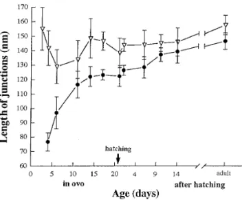
a gradual process, as indicated by the following data. Figure Peripheral couplings also vary in the shape and luminal 5 shows the size of peripheral couplings measured from the content of the SR vesicles forming them. Where feet are
profiles seen in thin sections. Two sets of data are plotted: not visible, the SR vesicles have a variable shape and little triangles represent the total length of the junction, regard-content (Figs. 3A, 3D, 3G, 3E, and 3H, left side of the junc- less of the presence or absence of feet; e.g., the length of tion). On the other hand, where the junctional gap is
zip-membrane between arrows in Fig. 3A. Circles represent the pered by arrays of feet, the SR has a characteristic flat shape length of the junction zippered by feet, i.e., either the zip-and a dense content aligned to the junctional face of the
pered portions of partially complete junctions (left side of vesicles (Figs. 3C, 3E, 3F, 3H, and 3I). This content, termed junction in Fig. 3B and right side of junctions in Figs. 3E ‘‘junctional granules’’ (Sommer and Johnson, 1979;
Som-and 3H) or the whole width of complete junctions (Figs. 3C, mer, 1995), presumably denotes an association of calseques- 3F, and 3I).
trin with the RyR-containing membrane. Formation of the
From these data, we obtain the following information. (i) junctional granules follows aggregation of feet: at E2.5 and The total length of the junctions remains approximately E4, junctions have no dense content even when aggregates
constant during development. The difference between aver-of 2–4 feet are seen (Fig. 3B), while at E6, most junctions aged total length at E4–E14 (four hearts) versus that at D10 with arrays of feet have a quite visible content, even when to adult (three hearts) is not significant (P Å 0.27). (ii) The few feet are present (Fig. 3C and see also below).
length of zippered junctions, on the other hand, increases with age. The difference between the averaged zippered
Time Course of Peripheral Coupling Development length at E4–E14 (four hearts) versus that at D10 to adult
(three hearts) is extremely significant (P õ 0.0001). (iii) The Changes with age in the relative frequencies of the
vari-ous types of junctions are indicative of a gradual evolution. zippered length is shorter than the total length of junctions
FIG. 1. Confocal images of cryosections from the left ventricle of chick myocardium, at ages E7 (A and B), E10 (C and D), D1 (E and F),
and adult (G and H) immunolabelled for DHPRs (left) and RyRs (right). Discrete foci of the two proteins mark the profiles of single myocytes. Note the increase in size of myocytes between E7 and adult and the presence of RyR foci in the interior of the adult fiber. Bars, 10mm.
270 Protasi, Sun, and Franzini-Armstrong
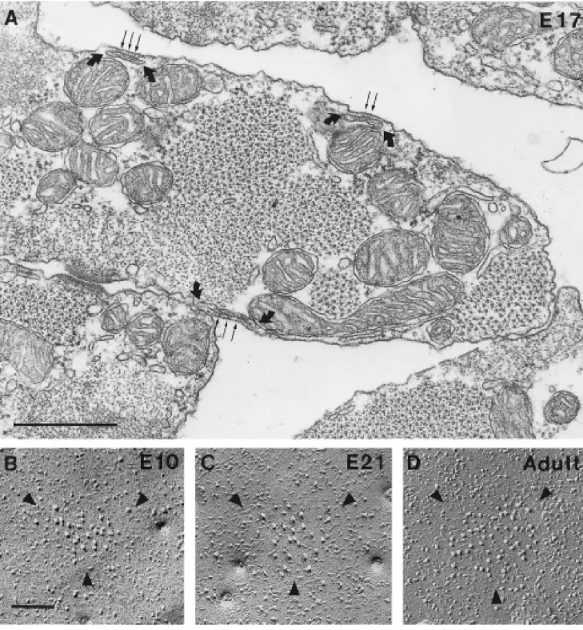
FIG. 2. (A) Cross section of cardiac myocytes at E17, showing peripheral couplings (between curved arrows) at the periphery of the
muscle fibers. Feet (small arrows) occupy the junctional gap between sarcoplasmic reticulum cisternae and the plasma membrane. (B–D) Aggregates of large, tall intramembrane particles in junctional domains of the plasma membrane are outlined by arrowheads in these freeze-fracture images. The size of the domains increases with age (E10, E21, and adult are shown). Bars, (A) 0.5mm; (B–D) 0.1mm.
at early time points, but becomes similar at late time points. dent’s t test of the difference between total and zippered lengths at D10 to adult (three hearts) indicates a marginally The difference between total and zippered lengths at E4–
E17 (five hearts) is extremely significant (P õ 0.0001). Stu- significant difference (P Å 0.1), with the caution that this
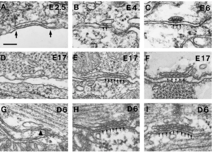
FIG. 3. Junctions between peripherally located cisternae of sarcoplasmic reticulum and the plasmalemma show various appearances. (A,
D, and G) Incomplete junctions have either no feet (A and D) or, perhaps, a single foot (arrowhead in G) in the junctional gap. The size of the gap in these junctions is on the average smaller than the height of a foot. The content of the vesicles is ill defined. (B, E, and H) In partially complete junctions, evenly spaced feet (small arrows) are present in a portion of the junctional gap, but are missing elsewhere. The content of the SR vesicle (probably representing calsequestrin) forms a density associated with the feet-bearing portion of the junctional SR in E and H, but not in B. (C, F, and I) Complete junctions are totally zippered by arrays of evenly spaced feet and have a periodic dense content throughout. Bar, 0.1mm.
is not precise, because the standard deviation is higher for and infrequent. Groups of two feet are detected at E2.5, but rarely. They were measured starting at E4 and their length one set of data (the triangles). Note that very few incomplete
also increases with age. The images in Fig. 6 give a visual junctions (õ10%) are present at the late stages.
impression of this development, showing increasing sizes of junctional domains (left) and arrays of feet (right) at E6, E17, and D6.
Parallel Development of Zippers and Particle
Figure 7A compares the measured surface areas of
junc-Groups
tional domains (open circles) and the surface areas of feet The sizes of zippered junctions and plasma membrane arrays (solid circles), calculated from the data in Fig. 5 (see junctional domains increase in parallel. Junctional domains Materials and Methods). The two surface areas remain ap-of the plasma membrane containing large, tall particles are proximately equal to each other while more than doubling detected starting at E4 and become larger and more frequent in size between E4 and adult. The difference in areas of with age (outlined by arrowheads in Figs. 2B–2D, 6A, 6D, junctional domains between E4 and E14 and D10 and adult and 6F). Groups of large particles are not detected at E2.5, (220 junctions, four hearts versus 193 junctions, three possibly because the cells are very small, the membrane hearts) is extremely significant (P õ 0.0001). The statistics for the diameters of feet arrays were done for the data in outlines very convoluted, and the junctional domains small
272 Protasi, Sun, and Franzini-Armstrong
FIG. 4. (A) Changes in the relative frequency of incomplete (solid circles), partially complete (open circles), and complete (triangles)
junctions with age. The frequency of incomplete junctions is very high at E2.5, and it declines to insignificant levels at E11. Partially complete junctions are more abundant in early than in later stages. Complete junctions are not present at E2.5 and E4, they are a minor component at E6, and they become predominant at E11 or later. The numbers are given as the percentage of total. Each time point on the graph is derived from a total of 38 to 184 junctions from one myocardium. (B) Comparison of the relative frequency of junctions with two or more closely spaced feet in the junctional gap (solid circles, partially complete and complete junctions) and of junctions that have a visible association of the SR vesicle content (presumably calsequestrin) with the feet-bearing SR membrane (open circles; see Figs. 3E, 3F, 3H, and 3I). At early time points, some vesicles have feet but lack a dense content. Sample size is the same as for A.
Fig. 5, and this also shows a significant difference between While the size of peripheral couplings increases with age, the spacing between feet and the density of particles in E4 and E14 (four heart) and D10 and adult (P õ 0.0001). The
two sets of areas cannot be compared statistically with each the junctional domains remain approximately constant (Fig. 7B). The particle density remains between 1086 and 1452 other, because the areas of feet arrays are extrapolated from
their diameters (see legend to Fig. 7). particles/mm2
, and the feet spacings are between 30.7 and 34.6 nm in myocardium from E4 to adult. Feet spacings and particle densities at E6–E14 are not significantly different from the spacings at D10 to adult. For feet spacings, P Å 0.14, 69 junctions, five hearts versus 74 junctions, three hearts. For particle densities, P Å 0.2, 220 junctions, four hearts versus 193 junctions, three hearts. Since the densities of the two proteins and the areas they occupy remain con-stant, their ratio also remains constant.
Maturation of the e – c Coupling Apparatus
As the heart develops, its fibers become more filled with contractile material, and the calcium handling capacity creases. One expression of this maturation process is the in-crease in frequency of peripheral couplings at the fiber periph-ery (Fig. 8): the number of junctions per unit length of plasma membrane increases by a factor of about 2.5 between E4 and E19 and then remains approximately constant. The difference in the frequency of peripheral couplings between E2.5 and E6
FIG. 5. Comparison between the total length of peripheral cou- (300 counts and three hearts) and D19 and D3 (three hearts
pling profiles (triangles) and the length of profiles occupied by
and 300 counts) is extremely significant (P õ 0.0001), showing arrays of feet (circles). The ‘‘overall’’ length of junction remains
a large increase in the number of junctions. approximately constant with age, but the length of junction
‘‘zip-A second maturation event is the development of internal pered’’ by feet increases with age. Triangles, 38–184 junctions from
feet-bearing SR, not associated with the plasma membrane one myocardium at each time point (27 junctions at E2.5). Solid
(extended junctional SR, Jewett et al., 1971). Extended junc-circles, 32–179 junctions from one myocardium at each time point
(22 and 18 junctions for E4 and E6). Error bars, 1 SEM. tional SR in chicken muscle first appears after hatching and
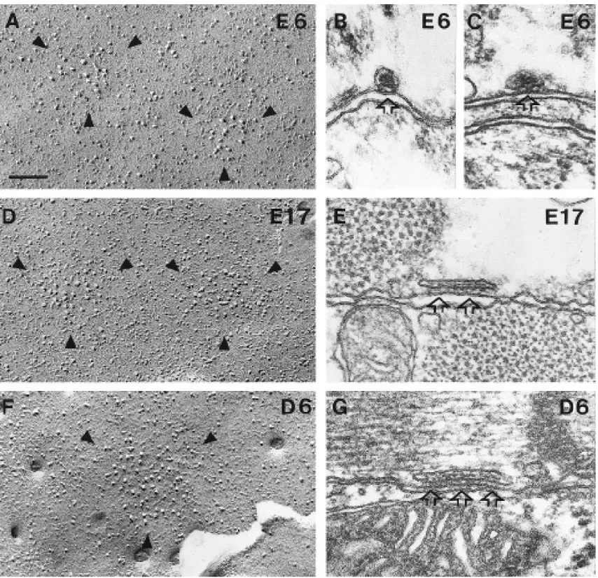
FIG. 6. Development of plasma membrane domains and peripheral couplings. (A, D, and F) The overall size of junctional domains
increases with age, but the density of particles in them does not (E6, E17, and adult). (B, C, E and G) Arrays of feet within peripheral couplings also increase in size (B and C, E6; E, E17; G, D6). See also Figs. 7A and 7B. Bar, 0.1mm.
increases in time (J. Sommer, personal communication; see surround a plasma membrane domain of the type outlined by arrowheads in the enlargements (Figs. 9A and 9C). Most of also Fig. 1).
Finally, as in skeletal muscle, junctional SR is initially ran- these domains are located near the Z lines, although there are some clusters in different positions. Peripheral couplings (seen domly disposed relative to the cross striation, but it gradually
acquires a specific location, starting around hatching time. in thin sections) and clusters of DHPRs and RYRs (detected by immunohistochemistry) also show an increased tendency Figure 9B shows the surface of a cell at D14: the sarcomere
spacing is clearly visible in the replica and the position of the for a location in the proximity of the Z line during develop-ment (see Sun et al., 1995, for more details).
274 Protasi, Sun, and Franzini-Armstrong
FIG. 7. (A) Comparison between areas of plasma membrane domains, measured from freeze-fracture replicas (open circles) and areas of
arrays of feet (solid circles), calculated from data from Fig. 5. Averages for the two areas increase in size during development, but remain approximately equal to each other. The data for the feet arrays do not show a value for SEM, because sample variability includes the position of the section relative to the area, as well as the variability in the population of junctions. Sample size: open circles, 32–90 domains from one myocardium for each point; solid circles, 32–179 junctions from one myocardium (but 22 and 18 junctions at E4 and E6). Error bars, 1 SEM. (B) Spacing between feet (open circles, in nm) and density of junctional particles in plasma membrane domains (no symbol, number/mm2) remain approximately constant throughout development. Thus, the densities of the two components of the
junctions have a constant ratio. Sample size: 32–90 membrane domains and 14–63 feet arrays from one myocardium for each point. Error bars, 1 SEM.
chinery that is used in the adult myocardium. RyRs have
DISCUSSION
been detected in their ryanodine-binding tetrameric form as early as E4 (Dutro et al., 1993). We find clusters of RyRs The heart is a developmentally precocious organ: in the
and DHPRs at E7 by using immunohistochemistry. Using chick embryo, it starts to contract feebly and locally at
electron microscopy, we also detect closely spaced feet (or about 30–33 hr of incubation, and it beats shortly after that
RyRs) beginning at E2.5 and more clearly apparent at E4 and (Romanoff, 1960). One important question is whether early
clusters of large plasma membrane particles (presumably myocardial activity relies on the same e–c coupling
ma-DHPRs) at E4. We conclude that essential components of the e–c coupling machinery are not only present, but are also arranged within peripheral couplings (Carl et al., 1995; Sun et al., 1995) at a very early age, shortly after beating starts. However, we cannot be sure whether smaller, less easy to detect e–c coupling assemblies are present when local contractions are first seen.
The time course of formation of peripheral couplings gives clues to the development and function of the e–c coupling apparatus in cardiac muscle. Peripheral couplings form by a gradual accrual of feet within a narrow space (the junctional gap), which is formed by the docking of SR vesicles to the plasma membrane. This conclusion is based on three obser-vations. (a) Entire junctions at early ages and portions of junctions at later ages have no visible feet and a gap which is too narrow to accommodate feet (see Radermacher et al., 1994 for the foot height). (b) SR/surface junctions with few or no feet gradually decline in frequency, while junctions zippered by increasingly larger groups of feet become more numerous during development. The similarity in the average
FIG. 8. Frequency of peripheral coupling profiles along the
periph-size of partially zippered and fully zippered junctions during ery of myocardial cells. The number of couplings per unit length
early and late stages of development supports the gradual (100mm) of plasmalemma profile increases by a factor of about 2.5
filling hypothesis. (c) The disposition of feet in partially com-between E4 and adult. Sample size: 100 counts (0.8-mm segments)
and one heart for each time point. Error bars, 1 SEM. plete junctions is uneven: feet are grouped on one side of the
FIG. 9. (B) Relationship between cross striation and the position of surface domains in a myocardial cell at D14. Each semicircle surrounds
a group of particles of the type shown, enlarged and outlined by arrowheads in A and C. Note a preferential location of these domains along and in proximity to the Z lines, whose position is indicated by arrows. Bars, (A and C) 0.1mm; (B) 1mm.
276 Protasi, Sun, and Franzini-Armstrong
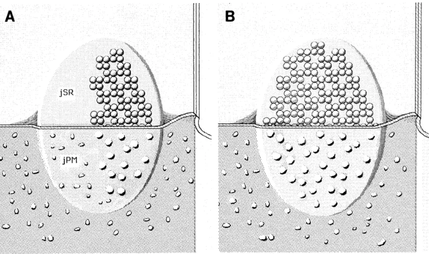
FIG. 10. (A) Reconstruction of a partially complete junction, extrapolated from our findings. The upper half of the image shows the
junctional surface of an SR vesicle (jSR), which is partially covered by an array of feet. In the lower half, the image shows the split protoplasmic (inner) leaflet of the plasma membrane, facing the SR vesicle (jPM). Large membrane particles are restricted to the portion of jPM facing the array of feet. (B) Reconstruction of a complete junction. jSR and jPM are completely occupied by an array of feet and a cluster of large particles, respectively.
junction, leaving the other side empty. This is probably due cance of the RyR–DHPR proximity has been discussed in terms of current hypothesis of cardiac e–c coupling (Sun et to high affinity of the feet for each other, since feet form
arrays in the absence of DHPRs and other muscle-specific al.,1995; Cannell et al., 1995). We notice, however, that the large particles of junctional domains in cardiac muscle (pre-junctional proteins, when RyRs are expressed in CHO cells
(Takekura et al., 1995b). Incomplete and partially complete sumably representing DHPRs) do not have a disposition indi-cating a link to the feet. This is in contrast to skeletal muscle junctions have also been observed in skeletal muscle and in
a cell line of skeletal muscle origin (Edge, 1970; Marks et al., (compare Sun et al., 1995 with Block et al., 1988; Franzini-Armstrong and Kisch, 1995). In view of the possible lack of 1991; Takekura et al., 1995a).
Regarding the association of DHPRs with the junction, direct interactions between particles and feet, the mechanism by which large particles are retained in the junctional domains we note that jSR (containing feet) and jPM (containing large
particles) grow in synchrony and that the ratio of feet to of cardiac muscle remains a mystery.
We suggest that the formation of arrays of RyRs (feet) is particles remains constant through development. Thus,
ac-crual of the two major components of peripheral couplings the leading event in junction formation and that trapping of DHPRs (large particles) rapidly follows. This is supported is strictly coordinated. Figures 10A and 10B summarize our
findings. In a developing junction (Fig. 10A), the array of by the following observations in the literature: feet form arrays independently of the presence of DHPRs in dysgenic feet and the group of large particles are limited to the same
side of the junction. In the fully formed junction (Fig. 10B), skeletal muscle and in CHO cells (Franzini-Armstrong et
al.,1991; Takekura et al., 1995b); grouping of DHPR fails feet and large particles completely occupy the facing
junc-tional areas of SR and plasma membrane. in the absence of feet in dyspedic muscle (Takekura et al., 1995a); and clusters of RyRs and DHPRs are colocalized The close proximity of RyRs and DHPRs and the controlled
ratio of the two proteins are early events in differentiation of through development and in adult skeletal and cardiac mus-cle (Jorgensen et al., 1989; Flucher et al., 1990; Yuan et al., cardiac muscle. We deduce that these two factors are crucial
to the contractile function of early myocardium. The signifi- 1991; Carl et al., 1995; Sun et al., 1995).
calization of sarcolemmal dihydropyridine receptor and sarco-The electron-dense content of the SR, due to the presence
plasmic reticular triadin and ryanodine receptor in rabbit ventri-of calsequestrin (Jorgensen et al., 1985), is associated with
cle and atrium. J. Cell Biol. 129, 673–682. the feet-bearing areas of SR, indicating some interaction
Catterall, W. A. (1991). Excitation–contraction coupling in verte-between feet and the protein responsible for linking
brate skeletal muscle: A tale of two calcium channels. Cell 64, calsequestrin to the junctional membrane. However, this 871–874.
association is detectable with a slight delay relative to the Dolber, P. C., and Sommer, J. R. (1984). Corbular sarcoplasmic feet. reticulum of rabbit cardiac muscle. J. Ultrastruct. Res. 87, 190–
The relative frequencies of various types of junctions are 196.
indicative of the relative length of their lifespan. Our data Dutro, S. M., Airey, J. A., Beck, C. F., Sutko, J. L., and Trumble, W. R. (1993). Ryanodine receptor expression in embryonic avian suggest that during very early development (up to E6), filling
cardiac muscle. Dev. Biol. 155, 431–441. of the gap by feet is slow relative to the rate of junction
Edge, M. B. (1970). Development of apposed sarcoplasmic reticulum formation, perhaps due to low levels of RyR expression.
at the T system and sarcolemma and the change in orientation After E6, filling of the gap with feet is rapid.
of triads in rat skeletal muscle. Dev. Biol. 23, 634–659. The maturation events in cardiac muscle membranes are
Flucher, B. E., Morton, M. E., Froehner, S. C., and Daniels, M. P. an increase in the frequency of peripheral couplings, the
(1990). Localization of thea1anda2subunits of the
dihydropyri-after hatching appearance of the subsidiary internal calcium dine receptor and ankyrin in skeletal muscle triads. Neuron 5, release system (extended junctional SR; Bossen et al., 1978; 339–351.
Jorgensen et al., 1993; Junker et al., 1994; Sommer, 1995), Franzini-Armstrong, C., and Jorgensen, A. O. (1994). Structure and and the redeployment of peripheral couplings from a ran- development of e–c coupling units in skeletal muscle. Annu.
Rev. Physiol.56, 509–534.
dom to a striation-related position. This latter event is quite
Franzini-Armstrong, C., and Kisch, J. W. (1995). Alternate disposi-similar to the striking rearrangement of transverse tubules
tion of tetrads in peripheral couplings of skeletal muscle. J. Mus-and triads from a longitudinal to a transverse disposition in
cle Res. Cell Motil.16, 319–324.
skeletal muscle (Franzini-Armstrong and Jorgensen, 1994;
Franzini-Armstrong, C., Pincon-Raymond, M., and Rieger, F. Takekura et al., 1994b).
(1991). Muscle fibers from dysgenic mouse in vivo lack a surface component of peripheral couplings. Dev. Biol. 146, 364–376. Giannini, G., Conti, A., Mammarella, S., Scrobogna, M., and
Sor-ACKNOWLEDGMENTS
rentino, V. (1995). The ryanodine receptor/calcium channel genes are widely and differentially expressed in murine brain and pe-ripheral tissue. J. Cell Biol. 128, 893–904.We thank Drs. J. A. Airey, J. L. Sutko, M. Takahashi, and H.
Inui, M., Saito, A., and Fleischer, S. (1987a). Purification of the Takeshima for antibodies and Mrs. Nosta Glaser for excellent
tech-ryanodine receptor and identity with feet structures of junctional nical help. Supported by National Institute of Health Grant R01
terminal cisternae of sarcoplasmic reticulum from fast skeletal HL48093.
muscle. J. Biol. Chem. 262, 1740–1747.
Inui, M., Saito, A., and Fleischer, S. (1987b). Isolation of the ryanod-ine receptor from cardiac sarcoplasmic reticulum and identity
REFERENCES
with the feet structure. J. Biol. Chem. 262, 15637–15642.Jewett, P. H., Sommer, J. R., and Johnson, E. A. (1971). Cardiac Airey, J. A., Beck, C. F., Murakami, K., Tanksley, S. J., Deerinck, muscle: Its ultrastructure in the finch and hummingbird with T. J., Ellisman, and Sutko, J. L. (1990). Identification and localiza- special reference to the sarcoplasmic reticulum. J. Cell Biol. 49, tion of two triad junction foot protein isoforms in mature avian 50–65.
fast twitch skeletal muscle. J. Biol. Chem. 265, 14187–14194. Jewett, P. H., Leonard, S. D., and Sommer, J. R. (1973). Chicken Anderson, K., Lai, F. A., Liu, Q-Y., Rousseau, E., Erickson, H. P., and cardiac muscle. J. Cell Biol. 56, 595–600.
Meissner, G. (1989). Structural and functional characterization Jorgensen, A. O., Shen, A. C-Y., and Campbell, K. P. (1985). Ultra-of the purified cardiac ryanodine receptor-Ca2/ release channel
structural localization of calsequestrin in adult rat atrial and ven-complex. J. Biol. Chem. 264, 1329–1335. tricular cells. J. Cell Biol. 101, 257–268.
Ashley, C. C., Mulligan, I. P., and Lea, T. J. (1991). Ca2/and
activa-Jorgensen, A. O., Shen, A. C-Y., Arnold, W., Leung, A. T., and tion mechanisms in skeletal muscle. Quart. Rev. Biophys. 24, Campbell, K. P. (1989). Subcellular distribution of the 1,4-Dihy-1–73. dropyridine receptor in rabbit skeletal muscle in situ: An immu-Berridge, M. J., and Irvine, R. F. (1989). Inositol phosphates and cell nofluorescence and immunocolloidal gold-labeling study. J. Cell
signaling. Nature (London) 341, 197–205. Biol.109, 135–147.
Block, B. A., Imagawa, T., Campbell, K. P., and Franzini-Armstrong, Jorgensen, A. O., Shen, A. C-Y., Arnold, W., McPherson, P. S., and C. (1988). Structural evidence for direct interaction between the Campbell, K. P. (1993). The Ca2/release channel/ryanodine
re-molecular components of the transverse tubule/sarcoplasmic re- ceptor is localized in junctional and corbular sarcoplasmic reticu-ticulum junction in skeletal muscle. J. Cell Biol. 107, 2587–2600. lum in cardiac muscle. J. Cell Biol. 120, 969–980.
Bossen, E. H., Sommer, J. R., and Waugh, R. A. (1978). Comparative Junker, J., Sommer, J. R., Sar, M., and Meissner, G. (1994). Extended stereology of the mouse and finch left ventricle. Tissue Cell 10, junctional sarcoplasmic reticulum of avian cardiac muscle con-773–779. tains functional ryanodine receptors. J. Biol. Chem. 269, 1627– Cannell, M. B., Cheng, H., and Lederer, W. J. (1995). The control 1634.
of calcium release in heart muscle. Science 268, 1045–1049. Lai, F. A., Erickson, H. P., Rosseau, E., Liu, Q-Y., and Meissner, Carl, S. L., Felix, K., Caswell, A. H., Brandt, N. R., Ball, W. J., G. (1988). Purification and reconstitution of the calcium release channel from skeletal muscle. Nature (London) 331, 315–319. Vaghy, P. L., Meissner, G., and Ferguson, D. G. (1995).
Immunolo-278 Protasi, Sun, and Franzini-Armstrong
MacLennan, D. H., and Toyofuku, T. (1992). Structure–function of membranes involved in excitation-contraction coupling of car-diac muscle. J. Cell Biol. 129, 659–671.
relationships in sarcoplasmic or endoplasmic reticulum type
Ca//pumps. Ann. N.Y. Acad. Sci. 671, 1–10. Takei, K., Mignery, G. A., Mugnaini, E., Sudhof, T. C., and De
Camilli, P. (1994). Inositol 1,4,5-triphosphate receptor causes for-Marks, A. R., Taubman, M. B., Saito, A., Dai, Y., and Fleisher, S.
mation of ER cisternal stacks in transfected fibroblasts and in (1991). The ryanodine receptor/junctional channel complex is
cerebellar Purkinkje cells. Neuron 12, 327–342. regulated by growth factors in a myogenic cell line. J. Cell Biol.
Takekura, H., Bennet, L., Tanabe, T., Beam, K. G., and
Franzini-114, 303–312.
Armstrong, C. (1994a). Restoration of junctional tetrads in dys-Meissner, G. (1994). Ryanodine receptor/Ca2/release channels and
genic myotubes by dihydropyridine receptor cDNA. Biophys. J. their regulation by endogenous effectors. Annu. Rev. Physiol. 56,
67, 793–804.
485–508.
Takekura, H., Sun, X-Y., and Franzini-Armstrong, C. (1994b). De-Pozzan, T., Rizzuto, R., Volpe, P., and Meldolesi, J. (1994).
Molecu-velopment of the excitation–contraction coupling apparatus in lar and cellular physiology of intracellular calcium stores.
Phys-skeletal muscle. Peripheral and internal calcium release units
iol. Rev.74, 595–636.
are formed sequentially. J. Muscle Res. Cell Motil. 15, 102–118. Radermacher, M., Rao, V., Grassucci, R., Frank, J., Timerman,
Takekura, H., Nishi, M., Noda, T., Takeshima, H., and Franzini-A. P., Fleisher, S., and Wagenknecht, T. (1994). Cryo-electron
Armstrong, C. (1995a). Abnormal junctions between plasma microscopy and three-dimensional reconstruction of the calcium
membrane and sarcoplasmic reticulum in skeletal muscle with release channel/ryanodine receptor from skeletal muscle. J. Cell
a mutation targeted to the ryanodine receptor. Proc. Natl. Acad.
Biol.127, 411–423.
Sci. USA92, 3381–3385.
Romanoff, A. L. (1960). ‘‘The Avian Embryo. Structural and
Func-Takekura, H., Takeshima, H., Nishimura, S., Takahashi, M., Ta-tional Development.’’ Macmillan, New York.
nabe, T., Flockerzi, V., Hoffman, F., and Franzini-Armstrong, C. Sandow, A. (1965). Excitation-contraction coupling in skeletal
mus-(1995b). Co-expression in CHO cells of two muscle proteins in-cle. Pharmacol. Rev. 17, 265–320.
volved in e–c coupling. J. Muscle Res. Cell Motil., in press. Sommer, J. R. (1995). Comparative Anatomy: In praise of a powerful
Yoshida, A., Takahashi, M., Nishimura, S., Takeshima, H., and approach to elucidate mechanisms translating cardiac excitation
Kokubun, S. (1992). Cyclic phosphorylation and regulation of the into purposeful contraction. J. Mol. Cell Cardiol. 27, 19–35.
cardiac dihydropyridine-sensitive Ca2/channel. FEBS Lett. 309,
Sommer, J. R., and Johnson, E. A. (1979). Ultrastructure of cardiac
343–349. muscle. In ‘‘Handbook of Physiology’’. (R. M. Berne, N.
Spere-Yuan, S., Arnold, W., and Jorgensen, A. O. (1991). Biogenesis of lakis, and S. R. Geiger, Eds.), pp. 113–186. American
Physiologi-transverse tubules and triads: Immunolocalization of the 1,4-cal Society, Bethesda, MD.
dihydropyridine receptor, TS28, and the ryanodine receptor in Stern, M. D., and Lakatta, E. G. (1992). Excitation–contraction
cou-rabbit skeletal muscle developing in situ. J. Cell Biol. 112, 289– pling in the heart: The state of the question. FASEB J. 6, 3092–
301. 3100.
Sun, X-H., Protasi, F., Takahashi, M., Takeshima, H., Ferguson, Received for publication July 17, 1995 Accepted October 10, 1995 D. G., and Franzini-Armstrong, C. (1995). Molecular architecture