Journal of Craniovertebral
Junction and Spine
J J C V S Editor-in-Chief : Atul Goel (INDIA) Open Access HTML Format
For entire Editorial Board visit : http://www.jcvjs.com/editorialboard.asp
97
Letter to Editor
Intervertebral disc
calcification in childhood:
Case report and review
of relevant literature
Sir,
Idiopathic IDC of childhood is a rare benign self-limited condition. It is generally asymptomatic and incidentally diagnosed during evaluations of nonspecific clinical
complaints.[1] Clinical relevance of such finding is limited to
rare symptomatic cases.
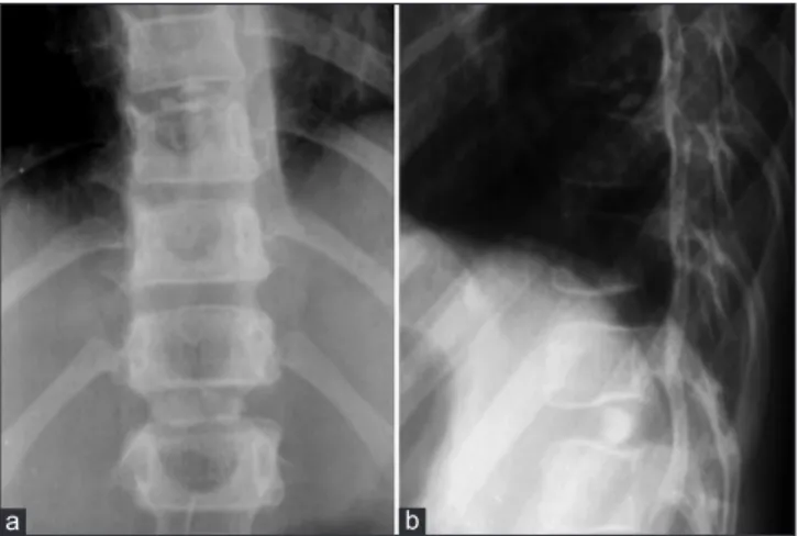
We briefly mention the case of a 7-year-old child who reported a recent episode of short-term mild back pain not preceded by trauma and not associated with local changes in temperature, dysesthesia, and/or blood tests alteration. Spinal X-ray performed on that occasion showed calcifications in two dorsal intersomatic spaces [Figure 1]. Magnetic resonance imaging (MRI) confirmed the X-ray findings and ruled out any alteration of vertebral canal, paravertebral soft tissues, or spinal cord [Figure 2].
Idiopathic IDC of childhood are probably related to developmental changes of the disc or flogosis triggered by
moderate traumas.[2] Such findings show average presenting
age of about 7 years,[3] slightly higher frequency among
males. They typically involve the nucleus pulposus and cervical spine is their most common location, although every tract of the vertebral column can be affected and multiple
localization is not rare.[4] In adulthood, IDC involve the
annulus fibrosus and mostly occur at several levels of the
thoracolumbar spine.[5]
Symptomatic cases are usually characterized by abrupt onset with pain, low-grade fever, and slight increase in flogosis indexes. Mostly, no trauma is reported because children find it difficult to describe the pain onset. A fast spontaneous symptom resolution usually coincides with resorption of the calcific foci. Long-lasting persistent calcifications may be associated with chronic back pain, scoliosis, loss of vertebral height and osteophytes. Surgical therapy is required in rare cases associated with neurological symptoms due to posterior herniations causing medullary/nervous compression, while conservative therapy is usually satisfactory.[1]
Plain X-ray, often the first diagnostic step, allows detection of IDC and provides evidences for differential diagnosis with inflammatory/post-inflammatory calcifications. IDC of children appear as intensely radiopaque
oval/round-shaped centrally-located spots affecting one or more intervertebral spaces [Figure 1]. Light changes in the adjacent vertebral bodies (e. g., endplate impressions) are
frequently reported.[4] IDC take several months to become
radiologically evident, while their resorption is earlier and usually preceded by fragmentation.
Computed tomography (CT) allows an overall evaluation of the involved spinal tract and a better definition of the disc. Mild deformities of adjacent vertebral bodies, disc herniations and involvement of spinal cord, nerve roots or
adjacent ligament can also be demonstrated.[1] Although
CT is better tolerated because of shorter duration, MRI is better as second-level-method because it doesn’t imply children exposition to ionizing radiations and allows better evaluation of discs, adjacent soft structures, spongy bone
edema, and spinal cord/nerve roots compression.[4] On
Figure 1: Spine X-ray showed disc calcifications in D8-D9 and D11-D12 as oval radiopaque spots in the rear part of intervertebral spaces (a) Adjacent vertebral body plates showed mild impressions coinciding with calcifications (b)
a b
Figure 2: Sagittal T1-weighted (a) T2-weighted (b) and fat-suppression (c) MR images showed hypointense spots in the rear portion of the discs in D8-D9 and D11-D12 (a and b). Paravertebral soft tissues, spinal cord, and neighboring vertebral bodies showed normal signal (c) MR = Magnetic resonance
98
Journal of Craniovertebral Junction and Spine 2015, 6: 25 Letter to Editor MRI, idiopathic IDC of children apper as hypointense disc
spots on T1- and T2-weighted images. Hyperintensity due to edema can be observed in neighboring vertebral bodies on fat-suppression sequences. No enhancement is observed after intravenous injection of gadolinium. In our case report, IDC showed typical MRI appearance [Figure 2]. Unlike other methods, MRI allows accurate detection and assessment of inflammatory involvement of adjacent structures, such as vertebral spongy bone edema and active enthesopathy of the posterior longitudinal ligament.
Gianrocco Favia, Andrea Salvati
1,
Luigi Chiumarulo
1, Franca Dicuonzo
1Departments of Imaging, and 1Neuroimaging, Hospital Policlinico, Bari, Italy
Corresponding author: Dr. Gianrocco Favia, Corso Alcide De Gasperi - 380 70125, Bari, Italy. E-mail: [email protected]
Journal of Craniovertebral Junction and Spine 2015, 6:25
Access this article online
Quick Response Code:
Website:
www.jcvjs.com
DOI:
10.4103/0974-8237.156074
REFERENCES
1. Girodias JB, Azouz EM, Marton D. Intervertebral disk space calcification. A report of 51 children with review of the literature. Pediatr Radiol 1991;21:541-6.
2. Swick HM. Calcification of intervertebral discs in childhood. J Pediatr 1975;86:364-9.
3. Dai LY, Ye H, Qian QR. The natural history of cervical disc calcification in children. J Bone Joint Surg Am 2004;86-A:1467-72.
4. Beluffi G, Fiori P, Sileo C. Intervertebral disc calcifications in children. Radiol Med 2009;114:331-41.
5. Chanchairujira K, Chung CB, Kim JY, Papakonstantinou O, Lee MH, Clopton P,
et al. Intervertebral disk calcification of the spine in an elderly population:
Radiographic prevalence, location, and distribution and correlation with spinal degeneration. Radiology 2004;230:499-503.