Università degli Studi del Piemonte Orientale “Amedeo Avogadro”
Dipartimento di Scienze del FarmacoPhD program in Chemistry & Biology Curriculum: Drug discovery and development
XXIX ciclo a.a. 2013-2016 SSD MED/04
Inflammation and cancer:
relevance of myeloid cells recruitment and plasticity
in tumor biology
Francesca Maria Consonni
Supervised by Prof. Antonio Sica
Contents
Chapter 1
Introduction
1.1 Inflammation and cancer 1.2 Myeloid cells in cancer 1.3 Myeloid-derived suppressor cells
1.3.1 MDSCs phenotype
1.3.2 Mechanisms of MDSCs suppressive activity 1.3.3 Expansion and activation of MDSCs in cancer 1.3.4 Therapeutic strategies targeting MDSCs
1.4 Tumor-associated macrophages
1.4.1 Macrophage polarization
1.4.2 Origin, recruitment and activation of TAMs 1.4.3 Pro-tumoral mechanism of TAMs
1.4.4 TAMs as therapeutic targets
1.5 The transcriptional factor NF-κB
1.5.1 NF-κB family and activation 1.5.2 NF-κB and cancer
1.6 Retinoid-related orphan receptors (RORs)
1.6.1 RORs structure and activity 1.6.2 RORγ and cancer
Bibliography
Chapter 2
Outline of the thesisChapter 3
"Homing regulation of distinct macrophage subsetsin infection and cancer"
1 3 6 9 9 10 13 15 17 18 21 26 30 33 33 36 38 40 42 43 61 67
Chapter 4
"RORC1 regulates tumor-promoting "emergerncy"granulo-monocytopoiesis"
Chapter 5
DiscussionChapter 6
List of pubblications 109 167 1771.1 Inflammation and cancer
The relationship between inflammation and cancer was described for the first time in 1863, when Rudolf Virchow observed leukocyte infiltration in neoplastic tissues (“lymphoreticular infiltration”) and hypothesized that the origin of cancer was at site of chronic inflammation. In the last decade several lines of evidences – based on a range of findings from epidemiological studies of patients to molecular studies of genetically modified mice – demonstrated the critical role of infection and chronic inflammatory diseases in tumorigenesis, and some of the underlying molecular mechanisms have been elucidated [1-3]. Inflammation is a well-coordinated process fundamental either in physiological conditions or in protecting body against different exogenous and/or endogenous treats. Any disorder in tissue homeostasis activates innate immune cells that are a first line of defense designed to “heal” the afflicted tissue. In response to chemotactic cytokines, the innate immune cells (macrophages, mast cells, dendritic cells and natural killer cells) migrate from the venous system to sites of damage, where initiate the inflammatory response by releasing cytokines, chemokines, matrix-remodeling proteases, and reactive oxygen and nitrogen species, leading to the elimination of pathogens and repair of tissue damage [4]. The key concept is that physiological inflammation, for example inflammation associated with wound healing, is a strictly controlled and self-limiting process; however, any deregulation in the specific control of immune components can lead to chronic inflammation that may favor the initiation and progression of cancer. In this scenario, a sustained inflammatory microenvironment provides a constant supply of a variety of factors (i.e. cytokines, chemokines and growth factors) which can counteract cell death and/or repair programs, resulting in genomic instability and predisposes tissues for cancer development [5, 6].
Hence, the mediators and cellular effectors of inflammation are important components of the local environment of tumors. It is estimated that 20% of all
cancers is associated with chronic infection and inflammation [7], indicating inflammation as the “Seventh hallmark of cancer” (Fig. 1) [8, 9].
Figure 1: Inflammation as the seventh hallmark of cancer.
In 2000, Hanahan and Weinberg proposed a model to define the six hallmarks that a tumor acquires: unlimited replicative potential, ability to develop blood vessels, evasion of programmed cell death, self-sufficiency in growth signals, insensitivity to growth inhibitors and tissue remodeling and metastasis. Next studies suggest that this model should be revised to include cancer-related inflammation as an additional hallmark [10].
The connection between inflammation and cancer can be viewed as consisting of two pathways; in some types of cancer, inflammatory conditions are present before a malignant change occurs (extrinsic pathway) [7, 11]. Conversely, in other types of cancer, an oncogenic change induces an inflammatory microenvironment that promotes the development of tumors (intrinsic pathway) [12, 13]. These two pathways converge, resulting in the activation of transcription factors, among which nuclear factor-κB (NF-κB), signal transducer and activator of transcription 3 (STAT3) and hypoxia-inducible factor 1α
activate various leukocytes, most notably cells of the myelo-monocytic lineage, and activate the same key transcription factors in inflammatory cells, stromal cells and tumor cells, resulting in more inflammatory mediators being produced and a cancer-related inflammatory microenvironment being generated. This uncontrolled and non self-limiting cancer-related inflammation has many tumor-promoting effects; in fact, it aids in the proliferation and survival of malignant cells, promotes angiogenesis and metastasis, subverts adaptive immune responses and alters responses to chemotherapeutic agents [3, 7].
However, despite these evidences, genetic studies of mouse models have demonstrated that innate immune cells activate an adaptive immune response capable of eliminating rising tumors [14]. It is generally accepted that immune cells continuously recognize and destroy nascent tumor cells but, due to the genetic, epigenetic and metabolic instability that characterizes neoplastic cells, the arising of new variants able to evade the immune surveillance results in tumor establishment and progression (immunoediting process) [15]. Several studies have emphasized that the “smouldering” inflammation associated with tumors is mainly oriented to tune the adaptive immune response. Thus, tumor-infiltrating lymphocytes have functional roles in promoting tumor immune escape by producing immunosuppressive cytokines and generating immunosuppressive networks. Indeed, myelo-monocytic cells recruited in tumors express an alternative M2 functional phenotype mainly oriented towards the suppression of the adaptive immune response [16, 17]. In agreement, clinical studies suggest that an established type-2 “suppressive” immunological profile correlates with poor prognosis, as shown in colorectal, hepatocellular and pancreatic carcinomas and in Hodgkin’s lymphoma [18].
Hence, the immune system plays a dual role in cancer: not only it can suppress tumor progression by destroying cancer cells or inhibiting their outgrowth, but it can also promote tumor development by establishing favorable conditions within the tumor microenvironment that facilitate tumor growth and metastasis [19].
1.2 Myeloid cells in cancer
Myeloid cells are the most abundant hematopoietic cells in the human body and are a collection of distinct cell populations with different function. All myeloid cells arise from multipotent hematopoietic stem cells (HSCs) that develop into mature myeloid cells through sequential steps of differentiation. The three groups of terminally differentiated myeloid cells (macrophages, dendritic cells (DCs) and granulocytes) are essential for the normal functions of the innate and adaptive immune systems [20].
Steady-state myelopoiesis is a continuous homeostatic process, occurring in the bone marrow, for replacing hematopoietic cells that are lost to normal programmed cell death. In contrast, emergency (or stress) hematopoiesis is an episodic modification of the magnitude and composition of the hematopoietic output that occurs during immunological stress, including tumor progression, to ensure proper supply of immune cells to increased demand [21].
In this scenario, compelling evidences in literature indicate that dynamic changes in myeloid cell functions occur in parallel to tumor development and that cancer fuels myeloid cells heterogeneity by promoting sustained myelopoiesis. It is well recognized that tumor-derived factors (TDFs), such as cytokines, chemokines, metabolites and inflammatory messengers like prostaglandins, act in paracrine or systemic manner to induce tumor-reprogrammed myeloid cells not only to create a widespread tolerogenic environment by blocking T cell functions and proliferation, but also to drive tumor growth by promoting cancer stemness, angiogenesis, stroma deposition and metastasis formation. Consequently, chronic exposure of the bone marrow microenvironment to non-physiologic levels of ordinarily tightly regulated myelopoietic-like growth factors corrupts the normal process of myeloid cell development and differentiation [8, 20]. This phenomenon drives the increase of circulating myeloid cells in tumour-bearing hosts and it is associated with a
of highly immunosuppressive, immature myeloid cells (iMCs). Indeed, many TDFs are myelopoietic factors making the myeloid compartment a major target of this ‘tumor reconditioning’ [22]. For example, granulocyte colony-stimulating factor (G-CSF) and granulocyte/macrophage colony-colony-stimulating factor (GM-CSF) were reported to drive “emergency” myelopoiesis by securing supply of neutrophils and macrophages from bone marrow and extra-medullary hematopoietic stem cell niches (HSCs) [23, 24].
In addition, the macrophage colony-stimulating factor (M-CSF) promotes macrophage differentiation from marrow precursors cells and is required for differentiation and expansion of tissue macrophages involved in tissue homeostasis and tumor progression [25]. Investigation of signaling pathways controlling hematopoiesis revealed that G-CSF-induced granulopoiesis is mediated through the transcription factors c-EBPβ [26, 27], SOCS3 [28], Bcl3 [29] and STAT3 [30], whereas M-CSF supports monocyte differentiation through activation of the transcription factors PU.1 and IRF8 [31]. Of relevance, interleukin-17A (IL-17A) promotes G-CSF- and stem-cell-factor-mediated neutrophilia [32] and supports G-CSF-driven ‘‘emergency’’ myelopoiesis [33]. Importantly, reciprocal regulation of macrophage versus neutrophil/granulocyte differentiation might control tissue homeostasis; indeed, depletion of tissue macro- phages leads to exacerbated G-CSF-mediated granulopoiesis [34]. Thus, investigation of the molecular networks that dictate this reciprocal regulation appears to be crucial, as it may affect tissue homeostasis during cancerogenesis. Myeloid deficiencies can occur at developmental and/or functional levels in essentially all myeloid lineages. To distinguish “normal” myeloid cells from their dysfunctional counterparts, the latter populations have been variously renamed myeloid-derived suppressor cells (MDSCs), tumor-associated macrophages (TAMs), tumor-associated neutrophils (TANs), immature DCs or tolerogenic DCs [35]. Among these, TAMs and MDSCs represent the major orchestrators of the immunosuppressive circuits arising in tumor bearers (Fig. 2) [36, 37].
Figure 2: Myeloid cells in cancer.
Factors produced in the tumor microenvironment by tumor cells and stromal cells promote the aberrant differentiation of myeloid lineage cells. The dotted lines show the normal pathways of myeloid cell differentiation from immature myeloid precursor cells to dendritic cells, macrophages and granulocytes. The solid bold lines indicate the aberrant pathways of myeloid cell differentiation that occur in cancer, in which the tumor environment can promote the development of various immunosuppressive populations, including monocytic MDSCs, polymorphonuclear MDSCs, suppressive DCs and TAMs and TANs [20].
1.3 Myeloid-derived suppressor cells
MDSCs are a heterogeneous population of cells of myeloid origin that comprises myeloid progenitor cells and immature macrophages, immature granulocytes and immature dendritic cells [38]. They expand during cancer, inflammation and infection and have a remarkable ability to suppress T-cell responses. In particular, MDSCs are generated in the bone marrow and, in tumor-bearing hosts, migrate to peripheral lymphoid organs and tumor tissues contributing to the formation of the pro-tumor microenvironment [39, 40]. Although initial observations and most of the current information on the role of MDSCs in immune responses has come from studies in the field of cancer research, accumulating evidence has shown that MDSCs also regulate immune responses during bacterial and parasitic infections, acute and chronic inflammation and sepsis [38].
1.3.1 MDSCs phenotype
MDSCs are characterized by a morphological mixture of granulocytic and monocytic cells lacking the expression of cell-surface markers associated with fully differentiated monocytes, macrophages or dendritic cells [41]. In particular, MDSCs include a group of immature mononuclear cells, which are morphologically and phenotypically similar to monocytes (M-MDSCs), and immature polymorphonuclear (PMN) cells, which are morphologically and phenotypically similar to neutrophils (PMN-MDSCs). In mice, MDSCs are identified by co-expression of myeloid lineage differentiation antigens Gr-1 and CD11b [42]. The two major subsets of MDSCs can be distinguished by different expression of the Gr-1 marker (Gr1high cells are mostly PMN-MDSCs and Gr1low cells are mostly M-MDSC). However, the Gr-1 marker is a combination of the Ly6C and Ly6G markers, so these subsets can be more accurately identified based on Ly6C and Ly6G expression (M-MDSCs as
CD11b+Ly6ChighLy6Glow and PMN-MDSCs as CD11b+Ly6G+Ly6Clow). In humans, MDSCs are identified in the mononuclear fraction as cells that express the CD33 marker but lack the expression of markers of mature myeloid and lymphoid cells and the HLA-DR. The identification of human MDSCs subsets is, actually, really difficult because of their heterogeneity. However, human PMN-MDSCs are defined as CD14–CD33+CD15+ HLA-DR–/low cellsand M-MDSCs as CD14+HLA-DR–/lowCD33+CD15- cells [43-46]. These cells represent ≈0.5% of peripheral blood mononuclear cells in healthy individuals with a 10-fold increase in cancer patients, such as those with renal cell carcinoma and colorectal carcinoma [47].
Several biochemical and genomic features distinguish MDSCs from neutrophils and monocytes. In particular, MDSCs are characterized by increased expression of of NADPH oxidase (Nox2), resulting in increased production of ROS [48]; increased expression of arginase 1 (arg1) and nitric oxide synthase 2 (nos2) genes, resulting in increased production of Arg1 and NO [49]; increased expression of the transcriptional factors c/EBP β [27] and STAT3 [50] and decreased expression of IRF8 [51]. As a result, MDSCs display decreased ability to differentiate into mature myeloid cells and their immune suppressive effects are due to the hyper production of ROS, NO and Arg1 [20].
1.3.2 Mechanisms of MDSCs suppressive activity
MDSCs exert their immunosuppression through several mechanisms. In particular, they can mediate suppression of T-lymphocytes in an antigen-specific and -nonantigen-specific manner, deploying strategies that can be either direct or indirect, leading to the generation or expansion of regulatory cell populations, such as CD4+
CD25+
Tregs. Main mechanisms of action include:
- ARG1, iNOS and peroxynitrite: the suppressive activity of MDSCs has been associated with the metabolism of L-arginine, which serves as a substrate for
arginine into urea and L-ornithine. MDSCs express high levels of both arginase and iNOS, and the direct role for both of these enzymes in the inhibition of T-cell function is now well established [52]. In fact, high levels of Arg1 expression by MDSCs can accelerate the depletion of L-arginine in the tumor microenvironment, which subsequently promotes T cell proliferation arrest and functional inhibition by down regulation of the CD3ζ chain in the T cell receptor (TCR) complex. [53]. NO suppresses T-cell function through a variety of different mechanisms that involve the inhibition of JAK3 and STAT5 in T cells [54], the inhibition of MHC class II expression [55] and the induction of T-cell apoptosis [56]. NO is mainly produced by M-MDSCs while both subsets express Arg1. More recently, it has emerged that reactive nitrogen species, such as peroxynitrite (ONOO-), are byproducts of the combined activity of iNOS, ARG1 and NOX2 that can alter the formation of a correct peptide-MHC complex in MHCI molecules and interfere with T cell migration and viability. In fact, they induce the nitration and nitrosylation of the amino acids cystine, methionine, tryptophan and tyrosine in many types of T cells, driving their un-responsiveness to tumor antigens [57], and modify trafficking of leukocytes through aromatic amino acid nitration and nitrosylation of chemokines (CCL2, CCL5, CCL21, CXCL12) or chemokine receptors (CXCR4) [58].
- Reactive oxygen species (ROS): ROS production is the major regulator of the suppressive activity of the PMN-MDSCs in both murine models and human cancers; it affect T-cell fitness by down regulating CD3ζ chain expression and reducing cytokine secretion [59].
- TGFβ: TGFβ is an immunosuppressive cytokine that has been associated with MDSCs functions and with the regulation of tumor induction and expansion [60].
- Induction of regulatory T cells (Tregs): emerging evidence indicates that MDSCs are involved in Tregs recruitment and differentiation through the production of several chemokines (acting on CCR5 and CCR6) and cytokines (i.e. IFN-γ, IL-10) and/or through direct cell-cell contacts (including
CD40-CD40L interactions) [61]. Moreover, to sustain the immune-suppressive environment, MDSCs also skew TAMs activation towards an M2 polarized phenotype, characterized by impaired production of functional IL-12 supporting immune escape and tumor growth, through a cell contact–dependent mechanism [40]. MDSCs also promote immune dysfunction by expressing membrane surface ligands of T cell–inhibitory receptors, such as programmed cell death ligand 1/2 (PD-L1/2), which bind programmed death 1 (PD-1), and B7-1/2, which bind to cytotoxic T lymphocyte antigen 4 (CTLA4) and CD28 as well as FASL [62, 63].
Hence, in tumor-bearing hosts MDSCs inhibit the immune response through four processes: 1) driving the differentiation of immune cells toward regulatory cells; 2) interfering with T cell migration; 3) altering T cell functionality by production of NO, ROS and RNS; and 4) depleting essential metabolites for T lymphocyte fitness. Several studies indicate that M-MDSCs are highly immunosuppressive and exert their effects largely in an antigen-nonspecific manner, whereas PMN-MDSCs are moderately immunosuppressive and promote T cell tolerance via antigen-specific mechanisms (Fig. 3) [41].
MDSCs suppress the immune system by distinct mechanisms including increased Tregs proliferation, direct actions of MDSC on T cells by increased NO, nitrotyrosine and ROS secretion, and decreased l-arginine production [64].
1.3.3 Expansion and activation of MDSCs in cancer
Accumulating evidence from tumor-bearing mice and human cancers indicates that the expansion and activation of MDSCs in the tumor microenvironment requires the integration of at least two types of signals: signals promoting accumulation of immature myeloid cells, followed by signals providing for the pathological activation of these cells [43].
MDSCs are generated in the bone marrow from common myeloid progenitor cells (CMP). The expansion of immature myeloid cells is mediated largely by granulocyte-macrophage CSF (GM- CSF), macrophage CSF (M-CSF), granulocyte CSF (G-CSF) and other factors, such as pro-inflammatory cytokines and damage-associated molecular pattern molecules (DAMPs), produced by tumor cells and stroma (e.g. cyclooxygenase 2, prostaglandins, IL-6, IL-17, C5a, VEGF) [65-70]. MDSCs expansion is facilitated by triggering a cascade through the signaling molecules that regulate cell survival, proliferation, differentiation and apoptosis, which are known as family members of Janus tyrosine kinase and STAT3 [71]. In recent years, more evidence has emerged regarding the mechanism of MDSCs regulation by STAT3. Indeed, it was reported that MDSCs from tumor-bearing mice have markedly increased levels of phosphorylated STAT3 compared with Immature Myeloid Cells (IMCs) from naive mice. Several evidence in vitro indicate also that the exposure of hematopoietic progenitor cells to the supernatant from tumor-cell cultures activate JAK2 and STAT3 in parallel with an expansion of MDSCs, and that this expansion is abrogated when STAT3 expression in hematopoietic progenitor cells is inhibited [72]. Moreover, ablation of STAT3 expression through the use of conditional knockout mice or selective STAT3 inhibitors
markedly reduced the expansion of MDSCs and increased T-cell responses in tumor-bearing mice [73].
The effect of STAT3 on MDSCs accumulation is mediated by C/EBPβ and IRF8 and is associated with increased survival and proliferation of myeloid progenitor cells, probably through the up-regulation of the expression of B-cell lymphoma XL, cyclin D1, MYC and survivin. So, abnormal and persistent activation of STAT3 in myeloid progenitor cells prevents their differentiation into mature myeloid cells and thereby promotes MDSCs expansion [27, 51]. However, the activity of MDSCs does not only require factors that promote their expansion but also activating factors that exert their effect through multiple signaling pathways including STAT6, STAT1 and NF-κB. These factors are mainly produced by activated T cells and tumor stromal cells and include IFN-γ, ligands for TLRs, IL-13, IL-4 and TGF-β [20]. STAT1 is the major transcription factor that is activated by IFN-γ and drives the up-regulation of Arg1 and iNOS expression in MDSCs within the tumor microenvironment. MDSCs from STAT1-knockout mice could not up-regulate Arg1 and iNOS expression and subsequently had no inhibitory effect on T cells [74, 75]. Moreover, also the signaling pathway that involves IL-4 receptor α-chain (IL-4Rα) and STAT6 (which is activated by the binding of either IL-4 or IL-13 to IL-4Rα) is important for MDSCs activation, but probably only in some tumor models [76]. Hence, the impact of MDSCs in cancer is characterized both by an abnormal myelopoiesis and recruitment of MDSCs into the tumor site, resulting from the persistent stimulation of the myeloid compartment with signals coming from tumors, and by an active MDSCs cytokine production and cell–cell interactions within the environment that promote tumor immune evasion by limiting T-cell responses and infiltration into the tumor microenvironment [39, 77].
MDSCs are also capable of supporting tumor growth through nonimmune-related mechanisms designed to remodeling tumor microenvironment. They have been shown to produce several mediators implicated in the
fact, MDSCs promote the angiogenic switch, by producing proteases (cathepsin and matrix metallo-proteinases 9 (MMP9)) and enhancing VEGF bioavailability [79]. In addition, MDSCs also play an active role in promoting the spread of distal tumor cells. In a murine model of melanoma, it was reported that MDSCs promote cancer cell dissemination by inducing epithelial–to-mesenchymal transition (EMT) through the production of TGF-β in primary tumor [80]. Moreover, mouse models have shown that MDSCs appear in the lungs as early as two weeks prior to the appearance of metastases in parallel with decreased immune function in the lungs. Importantly, myeloid-specific deletion of MMP9 essentially eliminated metastasis, suggesting indispensable role of MDSCs in the establishment of the premetastatic niche [81-83].
1.3.4 Therapeutic strategies targeting MDSCs
The fact that MDSCs strongly contribute to the establishment of an immunosuppressive tumor-microenvironment has stimulated the search for a way to therapeutically target these cells in order to allow for increased antitumor immunity. Several therapeutic strategies are currently being tested in clinic with the aim to elimination, deactivation, or skewing of myelopoiesis away from the accumulation of MDSCs [43, 84].
MDSCs can be eliminated with some first-generation chemotherapeutic agents, probably because these cells are more sensitive than tumor cells to low-dose chemotherapy [85]. In this scenario, gemcitabine has been shown to specifically deplete splenic MDSCs in tumor-bearing mice, resulting in enhanced antitumor response and prolonged survival [86, 87].
Similar success in selectively MDSCs targeting has been achieved with a single administration of 5-fluorouracil, which resulted in increased CD8+ T-cell responses [88]. Moreover, it was shown that selective up regulation of TRAIL receptor DR5 on mouse MDSCs induce their depletion selectively, resulting in tumor growth inhibition [89].
MDSCs can be functionally inactivated by targeting their suppressive machinery. In particular, the up regulation by a synthetic triterpenoid of Nf-E2– related factor 2 (NRF2), that plays an important role in protecting cells against free radical damage, has been shown to reduce the production of ROS by MDSCs ex vivo [90]. Moreover, phosphodiesterase-5 (PDE-5) inhibitor is able to inhibit MDSCs functions by decreasing their production of both Arg1 and iNOS restoring the in vitro T-cell proliferation in multiple myeloma and head and neck cancer patients [91, 92].
Finally, also nitroaspirin has been shown to down regulate NO and ROS production and thus eliminates the suppressive functions of MDSCs [93]. Other compounds have been reported to affect MDSCs accumulation in cancer; for example all-trans-retinoic acid (ATRA) at therapeutic levels has been shown to induce MDSCs differentiation into DCs and macrophages, leading to a subsequent reduction in the number of MDSCs in renal and lung carcinoma patients and mice [94, 95]. Additionally, sunitinib, which inhibits STAT3, VEGF, c-kit, and M-CSF signaling, has been shown to reduce MDSCs accumulation in renal cell carcinoma patients and may provide a strategy for improving antitumor immunity in these patients [96].
Therefore, different approaches have been explored to harness the potency of the immune system to target cancer. However, till now, efforts to actively stimulate the immune system against tumors in patients have been largely disappointing despite substantial evidence that peripheral immune responses against tumor antigens can be generated. Moreover, immune-modulating activities of chemotherapeutic agents are often very complex to understand, in fact, the same molecules may play opposite roles depending on tumor type, immune contexture, and/or precise therapeutic strategy. For example, gemcitabine and 5-fluorouracil, have been reported to deplete immunosuppressive MDSCs but also to induce the release of cathepsin B from lysosomes and the activation of the NLRP3 inflammasome and caspase-1,
which causes IL-1β secretion from MDSCs, resulting in IL-17 production by T-cells and promotion of tumor growth [97].
These complexities underscore the need for an ever more profound comprehension of the dynamic changes in the tumor microenvironment and in systemic immune responses as tumors evolve, progress, and respond to therapy, in order to facilitate the rational design of highly efficient, synergistic regimens that combine anticancer agents and immunotherapies.
1.4 Tumor-associated macrophages
Macrophages are a group of terminally differentiated tissue-resident myeloid cells found in all tissues, derived from monocytes circulating in peripheral blood [98]. They are key players in tissue homeostasis maintenance, tissue repair and immune surveillance, with important functions in both innate and acquired immunity. Resident macrophages provide immediate defense against foreign pathogens contributing to the balance between antigen availability and clearance through phagocytosis and subsequent degradation of senescent or apoptotic cells, microbes and possibly neoplastic cells. Macrophages also coordinate leukocyte infiltration and their role is essential for triggering, instructing and terminating the adaptive immune response [99].
Macrophages are the most plastic cells of the hematopoietic system able to finely modulate their programs in response to different microenvironmental conditions [100]. In the last decade, there have been numerous reports about the relationship between macrophages and tumors. In fact, tumor-associated macrophages (TAMs) represent the major population of leucocytes infiltrating tumors and, despite their potential anti-tumor activities, there is an extensive literature demonstrating that in both mouse and man, TAMs are co-opted during malignancy to facilitate tumor development and invasion [101].
1.4.1 Macrophage polarization
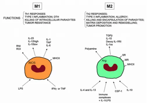
Macrophages are highly plastic cells that can adopt different phenotypes or activation states in response to different microenvironmental signals (e.g. cytokines, pathogen-associated molecular patterns, danger stimuli) [102, 103]. This plasticity increases the heterogeneity of macrophage populations in a given tissue and in particular, the M1/M2 dichotomy is the widely used model for describing their different functional states (Fig. 4) [104, 105].
Figure 4: M1 and M2 macrophages.
In the presence of IFN–γ, LPS and other microbial products, monocytes differentiate into M1 macrophages. In the presence of macrophage colony-stimulating factor (CSF-1), IL-4, IL-13, IL-10 and immunocomplexes in association with either IL-1R or TLR-ligands, monocytes differentiate into M2 macrophages. M1 and M2 subsets differ in term of phenotype and functions. M1 cells have microbial activity, immuno-stimulatory functions and tumor cytotoxicity. M2 cells have high scavenging ability, promote tissue repair and angiogenesis and favour tumor progression [106].
“Classical activated” M1 macrophages are potent antigen presenter cells involved in T helper 1 (Th1)-cell-mediated immune resolution of infection and exert their cytotoxic activities by secreting nitric oxide and ROS. Typical
products (LPS). These signals trigger the activation of NF-κB- and STAT1- pathways with subsequent transcription of NF-κB- and STAT1- dependent proinflammatory cytokines (e.g. IL-12, IL- 23 and TNFα) [107, 108].
On the contrary the “alternative activated” M2 macrophages secrete anti-inflammatory cytokines and are involved in scavenge debris, tissue remodelling and repair, angiogenesis and humoral immunity and are able to tune inflammatory response [109]. They show strong activation of arginase pathway with generation of ornithine and polyamines. Important drivers of M2 activation are the Th2 cytokines, such as IL-4 and IL-13, the anti-inflammatory cytokines IL-10 and TGF-β, hormones (e.g. glucocorticoids), and immune complexes. These M2-polarizing signals generally inhibit the expression of M1 cytokines and chemokines. These inhibitory effects principally relay on STAT-3 dependent mechanisms and the direct inhibition of NF-κB [110].
In vivo, macrophages can be exposed to a large number of stimuli that may induce opposing signaling pathways and thus result in mixed functional states. Nevertheless, there is considerable plasticity between distinct types. For this reason macrophage activation should not be seen as existing of discrete states M1 and M2, but rather as a continuum that emphasize the extremes of macrophage plasticity (Fig. 5) [109]. M1 and M2 extremes exhibit specific characteristic expression of metabolic enzymes (iNOS vs. ARG1), cytokines (IL-12highIL-10low vs. IL-12lowIL-10high), chemokines (CXCL9 and CXCL10 vs. CCL17 and CCL22), as well as transcription factors (NF-κB, STAT1 and IRF5 vs. STAT6, MYC, IRF4 and PPARγ) [104].
A large body of literature indicates that monocytes and macrophages associated with established tumors show an immunosuppressive, M2 phenotype which supports immune escape, tumor growth and malignancy exerting crucial tumor-promoting functions [109, 111]. However, when applied to TAMs the M1/M2 dichotomy is too simplistic since their phenotype varies significantly between tumors or even among different areas of the same tumor [112, 113]. Monocytes
from patients with renal cell carcinoma indeed coexpress proinflammatory genes, such as TNFα, together with tumor-promoting genes VEGFA, MMP9 and CXCR4 [44]. Moreover, similar mixed phenotypes have been observed in various other tumor models [114]. Thus, defining TAMs polarization status require integration of a multiparameter analysis of cell surface markers, and comparison of the TAMs transcriptome with the gene profile of resident macrophages isolated from the same tissues [17, 104].
1.4.2 Origin, recruitment and activation of TAMs
As mentioned above, TAMs are the main orchestrators of inflammation associated with cancer and constitute the dominant myeloid cell population, both in terms of number and functions, in most solid tumors. However, the details regarding TAMs origins and the stimuli that drive their differentiation and functional states are still being defined [25, 105, 115].
It has long been held that macrophages originate from the blood compartment and that chemotactic signals originating from tumor cells or from normal cells present in the cancer microenvironment recruit monocytic precursors at the primary and metastatic tumor sites [116]. Recent studies, however, have shown that most tissue macrophages, although with some exceptions such as intestine, arise during early embryonic development from yolk sac and do not require input from the bone marrow (BM) but rather maintain their populations via local proliferation. In contrast, macrophages involved in pathogen responses appear to come from circulating BM monocytes (Fig. 6) [98, 117-120].
Figure 6: Macrophage ontogeny in mice.
The mononuclear phagocytic system in adults derives from at least three sources. The first is the yolk sac (YS) that results in progenitors that populate all tissues and their progeny persist throughout life as F4/80 bright resident macrophages. These lineages are largely regulated by CSF1R. The second from the fetal liver is less well defined but seems to contribute to adult LCs
perhaps through a progenitor derived from the YS. The third lineage derives from the bone marrow (BM) to give circulating monocytes and their progeny F4/80 low macrophages and DCs. In this case the Ly6C + monocytes give rise to classical DCs under the regulation of FLT3 and these are continuously replenished. Other macrophages that are F4/80low also emanate from Ly6C+ monocytes and in some cases such as kidney and lung, co-exist with those derived from YS to give chimeric organs. The exact role of the patrolling Ly6C + macrophages remains unclear, as is the contribution of fetal liver to adult tissue macrophages [98].
Hence, in light of these recent findings, macrophage ontogeny in carcinogenesis has been revisited. A relevant question is whether TAMs are derived from the local tissue macrophage pool or whether they are newly recruited BM-derived cells. To date, developmental origins of TAMs have been best studied in mouse tumor models of the breast and lung and experimental indications suggest a monocyte origin for TAMs [112, 121-124]. In this regard, a recent study by Franklin et al., conducted in the polyoma middle T (PyMT) oncogene-driven mouse model of breast cancer (MMTV-PyMT), showed that these monocyte-derived TAMs are continuously replaced via peripheral recruitment. In fact, they pointed out that the major TAMs population (MHCII highCD11blow), as well as the mammary tissue-resident macrophage population ((MTMs) MHCIIhighCD11bhigh) originates from Ly6ChighCCR2+ monocytes. However, the role of local renewal cannot be excluded, since they observed that TAMs also expand their population through in situ proliferation [122]. In line with these results, in another spontaneous breast cancer model MMTV-Neu, two TAMs populations were identified (CD11bhighF4/80lowMHCIIhigh and CD11blowF4/80highMHCIIint) and both were also found to be derived from monocytes, with the CD11blow population heavily dependent on in situ proliferation [123].
Recent experimental evidences conducted in mouse models of lung carcinoma, suggest that TAMs also in the lung tumor are monocyte-derived, similar to their counterparts in breast cancers [121, 124]. A study by Cortez-Retamozo et al. showed that in the genetic KrasLSL–G12D/+p53fl/fl lung carcinoma model, fluorescently tagged monocyte precursors were found to differentiate into
instead of coming directly from the BM, a substantial fraction of TAMs arise from extra-medullary hematopoiesis within the spleen, which may function as a reservoir that continuously supplies the tumor with fresh progenitors [121], although the relative contribution of BM and spleen to the monocyte reservoire and tumor trafficking is not clear and might be tumor dependent [125].
Together, these findings suggest that TAMs can arise from tissue-resident macrophages, originating either from embryonic precursors which seed peripheral locations and self-sustain or from circulating monocytes, that may undergo a change in phenotype and/or activation state during carcinogenesis, or from inflammatory Ly6C+ circulating monocytes that undergo a distinct differentiation step to become macrophages in response to tumor microenvironment, and that these two populations may both be present simultaneously in the tumor microenvironment potentially having differential roles.
TAMs differentiation and localization is not a defined and preserved track but depends on both anatomical location and the tumor stage: cancers with different histology are infiltrated by TAMs with phenotypic and functionally distinct properties [112]. Interestingly, TAMs heterogeneity may be due to the nature of the monocytic precursor that is recruited to the tumor. Clear indications emerging from several tumor models suggest that Ly6Chigh monocytes, which rely on the CCL2-CCR2 axis, are the major precursor of TAMs [112, 121, 122, 126].
In addition, a smaller subset of TAMs, encompasses monocytes that express the angiopoietin-2 receptor TIE2, may arise from Ly6C lowCCR2- monocytes. These pro-angiogenic TAMs, known as Tie2-expressing monocytes (TEMs), are recruited to the tumor by angiopoietin-2, localize preferentially in areas of angiogenesis, aligned along the abluminal surface of blood vessels, and play important non-redundant roles in tumor neovascularization [114, 127].
The development of macrophages from monocytes is regulated by several cytokines, such as IL-6, and myelopoietic growth factors, such as M-CSF and
GM-CSF [128]. Among these, M-CSF, also known as colony stimulating factor 1 (CSF1), is the major lineage regulator of macrophages regardless their arising from the yolk sac or BM, and in addition it is a chemotactic factor for macrophages [129]. In this regard, in several models of cancer, genetic deletion of CSF1 or CSF-1R signaling inhibition with anti-CSFR1 antibodies, results in reduced number of TAMs recruited in the tumor microenvironment associated with slowed tumor initiation or decreased disease progression and distal metastatic spread [130-132]. Indeed, elevated CSF1 levels correlated with marked macrophage infiltration in human metastatic breast cancer [25]. In addition, the transcription factor PU.1, which among other functions controls the expression of CSF1R, regulates differentiation of progenitors to the macrophage lineage [133].
Moreover, in a xenograft model of skin cancer, it was reported that also VEGFA recruits macrophage progenitors that then differentiate to TAMs under IL-4 influence and that loss of these VEGF-recruited TAMs inhibited tumor growth, angiogenesis and invasion [134]. These observations indicate that CSF1 and VEGFA can be independent recruiters of macrophages to tumors in mouse models and that this effect could be explicated via recruitment of monocytes and/or through proliferation of recruited or resident cells.
Compelling evidence indicate that these growth factors act collaboratively in the tumor microenvironment with locally synthesized TDFs, such as cytokines (e.g. IL-10, IL-4, IL-13, IFN-γ, IL-1β), chemokines (e.g. CCL2, CXCL12, CCL5) as well as growth factors and noncanonical chemotactic peptides (e.g. TGF-β, PGE2), to drive the monocyte-to-macrophage differentiation and the macrophage activation state inside tumors [122, 126, 135-137].
Several factors controlling TAMs phenotype coincide with signals driving "alternative" macrophage activation such as IL-10, IL-4 and IL-13. These latter, bind different receptors sharing the IL-4Rα chain that is responsible for
involved in the immune-suppressive program [138]. Recently, genetic evidence in the mouse suggested that complement components, C5a in particular, play an important role in recruitment and functional polarization of TAMs [139].
Interestingly, macrophages may also be affected by the metabolic environmental signals that are associated with malignant neoplasms. The tumor microenvironment is typically hypoxic and characterized by a high concentration of lactate due to the ‘Warburg effect’ namely the metabolic shift occurring in highly proliferating cells which predominantly convert glucose into lactate even in the presence of oxygen (aerobic glycolysis) [140]. This results in substantial production of lactic acid, which was recently shown to polarize TAMs activation toward an M2 state and to influence their spatial dissemination within specific areas of tumors [141]. In particolar, TAMs accumulate preferentially in poorly vascularized regions of tumors, suggesting that oxygen availability has a role in guiding their localization and function. Hypoxia promotes the metabolic adaptation of TAMs through the activation of hypoxia-inducible factors HIF-1α and HIF-2α. HIF-1α influences the positioning and function of tumor cells, stromal cells and TAMs by up-regulating their expression of CXCR4. Moreover, HIF-1α activation can have a role in the induction of the CXCR4 ligand CXCL12, a chemokine involved in cancer metastasis [142-144].
Despite these several indications, the detailed mechanisms for the recruitment and tumor-promoting function of TAMs are not fully understood; thus, further investigation is required to identify new targets for cancer immunotherapy.
1.4.3 Pro-tumoral mechanism of TAMs
For decades, solid tumors have been known to be strongly infiltrated by inflammatory leukocytes and accumulating evidences have clearly demonstrated in various mouse and human malignancies, that despite high levels of infiltration, macrophages are unable to stimulate an effective antitumor response and are instead generally associated with poor patient prognosis [4, 145, 146]. One of the most important characteristic of TAMs include their ability to directly affect tumor growth through promotion of tumor angiogenesis as well as the survival and metastasis of tumor cells [147, 148].
As already discussed, in terms of cytotoxicity and expression of inflammatory cytokines, we can say that TAMs resemble the M2 macrophages: both are poor producers of NO and of ROIs [149]; both are poor antigen presenting cells and not only they are unable to trigger Th1 polarized immune responses, but also they induce Treg cells and suppress T cell activation and proliferation [111]. Moreover, TAMs express high levels of both scavenger receptor-A (SR-A) and the mannose receptor (MR) together with other M2 markers like Arginase I, YM1, FIZZ1 [150].
In agreement with the M2 signature, TAMs were reported to express low levels of inflammatory cytokines (e.g. IL-12, IL-1β, TNFα, IL-6) [109].
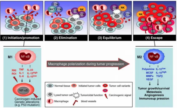
Activation of the transcriptional factor NF- κB is a necessary event promoting transcription of several proinflammatory genes. TAMs display a defective NF-κB activation in response to M1 polarizing signals LPS and TNFα [151]. The defect in κB was shown to be due to the over expression of nuclear p50 NF-κB homodimers which inhibit the transcription of proinflammatory genes [152]. The defective NF-κB activity was seen in TAMs isolated from tumors characterized by advanced stages and is in apparent contrast with TAMs NF- κB dependent pro-tumorigenic functions observed in murine models of inflammation-associated liver and colorectal cancer [153, 154]. This
during the transition from early-neoplastic events to advanced tumor stages, which would result in progressive modulation of the NF- κB activity expressed by infiltrating inflammatory cells and progressive conversion of the TAMs from an M1 to an M2 macrophage phenotype (Fig. 7) [15].
Figure 7: Macrophage polarization in tumor immunoediting and progression.
During tumor progression a gradual switching of macrophage polarization, M1 versus M2, is paralleled by the gradual inhibition of NF-κB activity. These events concur to establish permissive conditions for tumor growth and spread [100].
Interestingly, over-expression of p50 NF-κB has also been reported in endotoxin tolerant human monocytes which show defective TNFα production but overexpress IL-10, similar to the TAMs [155]. However, while in sepsis the over-expression of the p50 NF- κB represents a protective response against deregulation of the inflammatory process, in cancer it may be part of the immunosuppressive mechanisms associated with tumor growth [37].
TAMs favor tumor growth through non-immune and immune processes. Angiogenesis is an M2-associated function, which represents a key event in
tumor progression. Indeed, in several human cancers, TAMs accumulation has been associated with angiogenesis and with the production of angiogenic factors such as VEGF and platelet-derived endothelial cell growth factor [1, 156]. Additionally, TAMs participate to the proangiogenic process by producing the angiogenic factor thymidine phosphorylase, which promotes endothelial cell migration in vitro and whose level of expression is associated with tumor neovascularization [157].
TAMs also play an active role in promoting the spread of distal tumor cells. They express enzymes that regulate the digestion of the extracellular matrix, such as MMPs, plasmin, urokinase-type plasminogen activator [158] and the uPA receptor. In mammary tumors, TAMs promote metastatic diffusion via a paracrine loop involving CSF1 and EGF, which induces macrophages and tumor cells to cluster around blood vessels, where macrophages create a gate for tumor cell intravasation into the circulation, thus producing a tumor microenvironment for metastasis [35, 148, 159, 160].
As mentioned above, the most important pathogenic activity of TAMs is the suppression of anticancer immune responses. This ability is due at least partially on their reduced immunostimulatory properties. For instance, TAMs produce low levels of IL-12, which triggers tumoricidal actions of natural killer cells and the generation of cytotoxic CD4+ T-cells, and high levels of immunosuppressive factors such as IL-10, TGFβ and PGE2, which recruit Treg-cells [150]. In particular, TAMs-derived IL-10 negatively regulates the production of IL-12 by tumor-associated myeloid cells and thus indirectly stimulates the differentiation of Th2 cells that release high levels of IL-4 and IL-13 reinforcing TAMs protumor phenotype [161].
TAMs can also direct suppress the cytotoxic functions of T-cells and this immunosuppression is mediated, at least in part, by nitrosylation of T-cell receptors via ARG1, iNOS and peroxynitrite, inducing T cell apoptosis [20, 162, 163]. Moreover, TAMs may also promote apoptosis of T-cells by
expressing on their cell surface the inhibitory B7 family molecules PD-L1 and B7-H1, which trigger checkpoint blockade in T cells [164].
Finally, TAMs maintain the cancer cell reservoir by providing a niche for cancer stem cells (CSC). Indeed, TAMs can sustain CSC proliferation by releasing proinflammatory cytokines such as TNF-α and IL-6, which reinforce tumor cell proliferation through NF-κB and STAT3 signaling pathways, and by producing milk fat globule EGF factor 8 (MFGE8), which favored CSC reservoir survival during chemotherapeutic treatment [165-167].
1.4.4 TAMs as therapeutic targets
It is now clear that cells of the monocyte-macrophage lineage are an essential element of the inflammatory component in the tumor contest that play a key role in supporting cancer development [168, 169]. In addition to these pro-tumoral activities, TAMs can also modulate the efficacy of various form of anticancer therapy [162, 170]. Based on this, both the recruitment and activation of TAMs are considered putative targets for therapeutic intervention. In particular, current therapeutic approaches affecting TAMs compartment are aimed either at reeducating their functional activation to an antitumor, M1-like phenotype or inhibiting their recruitment and/or survival in tumors [171].
As mentioned above, mediators involved in macrophage recruitment in tumors include for instance CCL2, CCL5, complement components, CSF-1 and VEGF and the inhibition of these molecules with specific monoclonal antibodies or antagonist prevent TAMs recruitment, reducing tumor growth and dissemination [122, 126]. Indeed, compelling evidences indicate that inhibition of CCL2 with specific antibodies reduced tumor growth in several experimental models such as prostate, melanoma, breast and lung cancer; moreover, when administered in combination with chemotherapy, anti-CCL2 antibodies improved the therapeutic efficacy [172-174]. However, it has been also shown in a breast cancer mouse model, that recession of anti-CCL2 treatment increased the mobilization and recruitment of circulating monocyte, thus accelerating lung metastasis [175].
Analysis of leukocyte migration in colon cancer (CRC) metastasis revealed overproduction of CCL5, a TAMs attractant also responsible for their functional skewing. Recent clinical studies reveal that treatment with a CCR5 antagonist result in biological and clinical responses in a small cohort of advanced CRC patients, indicating the CCL5-CCR5 axis another possible therapeutic target [176].
The CSF-1 receptor (CSF-1R) is exclusively expressed by the monocytic lineage, thus representing an obvious target to hit TAMs; accordingly, anti-CSF-1R neutralizing antibodies or small molecule inhibitors interfering with this pathway have been developed and tested in preclinical models [177, 178]. Ries et al. reported that treatment with anti-CSF-1R antibody (RG7155) strongly reduced TAMs infiltration in tumors accompanied by an increase of the CD8+/CD4+ T cell ratio, in animal models; in addition, administration of RG7155 to diffuse-type giant cell tumor (Dt-GCT) patients led to reductions of macrophages in tumor biopsies [131].
Moreover, TAMs depletion by anti-CSF1 antibodies enhanced the efficacy of combination chemotherapy (cyclophosphamide, methotrexate, and 5-fluoro-uracil) in chemoresistant, human breast cancer xenografts grown in immunodeficient mice [162]. Similarly, TAMs depletion improved the efficacy of paclitaxel in a transgenic mouse model of mammary adenocarcinoma [179]. Small molecule inhibitors to CSF1R have also been shown to deplete some populations of TAMs and to dramatically enhance responses to chemotherapy. This effect is at least in part consequent to the removal of macrophage-mediated immunosuppression leading to the increased of CD8 + T cell infiltration during the tumor recovery period [162, 170].
Trabectedin is an anticancer drug licensed in Europe and several other countries for the treatment of soft tissue sarcoma patients and ovarian carcinoma patients; it was initially identified for its potent cytotoxic and antiproliferative activity against malignant cells [180]. Further clinical and experimental evidences indicate that trabectedin not only hits neoplastic cells but also importantly modulate tumor microenvironment; in particular, it activates a caspase-dependent pathway of apoptosis selectively in cells of the monocyte lineage, causing a partial depletion of circulating monocytes and TAMs. In murine tumors and in human sarcomas Trabectedin-induced TAMs reduction was associated with decreased angiogenesis and increased T-cell infiltration [62, 181, 182].
The contribution of TAMs to the modulation of tumor responses to chemotherapy can vary markedly among different cytotoxic agents and tumor models and intriguingly, increasing data suggest that the efficacy of some forms of immunotherapy may also depend on effective reprogramming of TAMs toward an M1-like phenotype. For example, the antitumor activity of the taxane docetaxel involves the depletion of immunosuppressive (M2-like) TAMs and the concomitant activation or expansion of antitumoral (M1-like) monocytes in 4T1-Neu mammary tumor implants. Indeed, in vivo T cell assays showed that docetaxel-treated monocytes/MDSCs are able to enhance tumor-specific cytotoxic T cell responses [183].
More specific macrophage targeting came from the administration of an anti-CD40 antibody in a preclinical model of pancreatic cancer, where alternatively activated, M2-like macrophages were re-educated in the tumor microenvironment to acquire antigen-presenting capabilities, leading to re-establishment of tumor immune surveillance and reduction of tumor progression [184].
Finally, recent clinical studies suggest that usage of nonsteroidal anti-inflammatory drugs, aspirin in particular, is associated with protection against occurrence of many tumors and metastasis and that this protective function relies on the inhibition of prostaglandin production [185, 186]. Ineed, PGE2 is well-known to have immune-suppressive effects and to favour M2-like polarization of TAMs [187, 188].
Overall these reports strongly indicate that macrophage-targeting strategies have the potential to complement and synergize with cytoreductive therapies, anti-angiogenic agents and immunotherapy [171].
1.5 The transcription factor NF-
κ
B
NF-κB family has been considered the central mediator of the inflammatory process and a key participant in innate and adaptive immune responses; moreover during the last years it has been demonstrated that NF-κB could play a crucial role in cancer development [189].
NF-κB is evolutionarily conserved and plays a critical role in many biological systems, above all the immune system, where it acts as the major orchestrator of the transcriptional responses to many different stimuli. The engagement of several immune receptors such as B and T cell receptor (BCR, TCR), TLRs, Tumor Necrosis Factor Receptor (TNFR) or CD40 [190] triggers NF-κB activation which in turn results in the expression of cytokines, growth factors and effector enzymes. At present, more than 150 genes under control of NF-κB have been identified, as a demonstration of its vast spectrum of biological functions [191, 192].
Because NF-κB activation drives expression of key genes in inflammation, immunity, cell survival and proliferation its mis-regulation, such as constitutive activation, could be associated with pathological conditions such as rheumatoid arthritis, asthma, intestinal bowel diseases (IBDs), multiple sclerosis and cancer [193-196]. Given this great variety of biological roles, a better understanding of NF-κB pathways could provide the basis for the development of therapeutic strategies with a relevant impact on human diseases.
1.5.1 NF-
κ
B family and activation
NF-κB family includes five members: RELA (p65), RELB, cREL, NF-κB1 (p105-p50) and NF-κB2 (p100-p52). All these proteins possess a conserved 300-amino acid REL homology domain (RHD) that is located toward the N-terminus of the proteins and is responsible for dimerization, binding to inhibitors of nuclear factor κB (IκBs) and binding to DNA. Instead, the
carboxy-terminal non-homologous transactivation domain (TAD), which strongly activates the transcription of targeted genes, is present only in cREL, RELB and RELA [197, 198]. p50 and p52 are generated by proteolytic degradation of p105 and p100 precursors and they lack the transactivation domain, therefore if they form homodimers they still bind the DNA consensus sites, but they do not activate transcription [199, 200]. Each member of NF-κB family, except for RELB, can form homodimers as well as heterodimers with one another and the main activated form of NF-κB is the heterodimer containing p65 together with p50 or p52 (Fig.9) [110].
Figure 9: Mammalian NF-κB-family members.
NF-kB family comprises five members: RELA (p65), cREL, RELB, p105/p50 and p100/p52. Proteolytic processing of p105 and p100 at residues 435 and 405 (as indicated by arrows), respectively, generates the p50 and p52 NF-κB proteins. The glycine-rich region (GRR) and the carboxy-terminal sites of inducible phosphorylation (in the DSVCDS and EVKEDSAYGS sequences for p105 and p100, respectively) are required for processing. Phosphorylation of RELA at Ser276, Ser529 and Ser536 is important for its transactivation activity. The size of each human protein is shown on the right (number of amino acids) [110].
In resting conditions, IκBs exhert their regulatory function binding NF-κB proteins masking their Nuclear Localization Sequence (NLS). So, the
the cytoplasm in inactive forms. Triggering of many different receptors can induce NF-κB activation that is initiated upon phosphorylation of IκBs by IκB Kinases (IKK). IKK is a complex made by kinase subunits IKKα and IKKβ and the regulatory subunit IKKγ or NEMO (NF-κB Essencial Modifier). Hence, upon activation of IKKβ, IκB is phosphorylated and degraded by the proteasome, so the released NF-κB dimers can go to the nucleus and activate gene transcription [201-204].
NF-κB could be activated through two different pathways: classical and alternative[190]. The classical pathway is particularly involved in innate immunity, is activated predominantly by the subunit IKKβ in a NEMO dependent manner and is mainly mediated by Toll like receptors (TLRs), scavenger receptors and complement system. Signalling through TLRs leads to activation of canonical IKKs complexes, degradation of IκBs and activation of RELA and cREL containing NF-κB dimers. The released NF-κB dimers, that in this pathway are predominantly p65-p50 heterodimers, go to the nucleus and activate gene transcription. The beginning of an inflammatory response is strictly dependent on NF-κB classical pathway. Signals coming from the environment lead to the recruitment and activation of effector cells, initially neutrophils and later macrophages and other leukocytes, resulting in the tissue changes characteristic of inflammation – rubor, calor, dolor and tumor (redness, heat, pain and swelling, respectively) [199, 203, 205, 206].
The alternative pathway is particularly active in cells of the adaptive immunity, such as B and T lymphocytes, is independent of IKKβ and NEMO but it is dependent of IKKα homodimers, which selectively phosphorylate p100 associated with RELB. Therefore, the consequence is the release of active RELB-p52 heterodimers [207, 208]. Activation of NF-κB downstream B cell receptor (BCR) and T cell receptor (TCR) is a critical step for mounting adaptive immune responses allowing antigen specific maturation and proliferation of lymphocytes into effector cells [209].
1.5.2 NF-
κ
B and cancer
Several evidence suggest that NF-κB is a molecular bridge between inflammation and cancer. Indeed, among all the different signaling pathways activated by inflammation and infection, NF-κB is the major activator of genes encoding for proteins important for cell proliferation (e.g. cyclin D1, c-Myc) survival (BCL-2, c-FLIP) adhesion, and angiogenesis (e.g. CXCL8, VEGF) [210].
In fact, as a master regulator of inflammation, NF-κB triggers the transcription of several proinflammatory mediators such as IL-1β, TNFα, IL-6 and IL-8. These factors are themself able to induce higher NF-κB activation, thus providing a positive feedback loop at the site of inflammation. This inflammatory environment favours DNA damage, cell proliferation, transformation and survival and consequently cancer initiation, growth and progression [154, 192].
The pro-inflammatory stimuli, such as bacterial products via Toll-like receptors or inflammatory cytokines (e.g. TNFα and IL1β), activate NF-κB that translocates into the nucleus inducing the expression of cytokines (such as TNFα and IL6) and chemokines, which contribute to the inflammation-related tissue damage. This elevated IKK/ NF-κB activity may also lead to aberrant up-regulation of certain tumorigenic, adhesion proteins, chemokines, and inhibitors of apoptosis that promote cell survival [211].
Hence, NF-κB is involved not only in tumor development at early stages, but also in the migration, invasion and metastasis of malignant cells [154]. For instance, the invasive capacity of cancer cells can increase in the presence of inflammatory cytokines such as TNFα, IL-1β and IL6 [3]. In particular, TNFα is a potent stimulator of epithelial-mesenchimal transition by breast cancer cells for its ability on activate NF-κB activation [212]. NF-κB was also found to promote metastatization in a genetic mouse model of prostate cancer, in which
Moreover, compelling indications demonstrate that in many cancers, NF-κB is constitutively active, even if the exact mechanism that sustain this activation is not fully understood and several mechanisms have been proposed, such as IL-1β and TNFα production, shorter IκBα half life or IκBα mutations [214, 215]. For these reasons, NF-κB represents an ideal therapeutic target for the development of new anti-tumor strategies.
p50/NF-
κ
B1
The NF-κB1 gene encodes two functional proteins: p50 and p105. In particular, p105 is the precursor of p50, which is the active form of the protein and could form dimers with itself or with other NF-κB subunits. The role of p50 and its precursor in cell physiology and function is very complex. Indeed, although originally considered a repressor of transcription, p50 could also be a transcriptional activator and the balance between pro- and anti- inflammatory activity of p50 depends on cell type and environmental conditions [216, 217]. The nuclear translocation of p50 homodimers deeply controls functions of myeloid cells in cancer. In fact, it has been demonstrated that in LPS-tolerant macrophages increased expression of the p50 subunit of NF-κB directly results in the downregulation of LPS-induced TNFα production, whereas in p50-/- macrophages long-term pre-treatment with LPS was unable to induce tolerance [100, 218]. Accordingly, as mentioned above, our group has demonstrated that TAMs display a defective NF-κB activation in response to the M1 polarizing signals (LPS and TNFα) and that this phenotype is due to the over expression of nuclear p50 NF-κB homodimers which inhibits the transcription of pro-inflammatory genes. On the contrary, we have shown that LPS stimulated p50 -/-TAMs recover an M1 (IL-12 highTNFαhighIL-10low) phenotype that correlates, in vivo, with tumor growth inhibition [219]. Further, a detailed analysis of the role of p50 NF-κB homodimer in macrophage functions revealed that its nuclear accumulation, both in TAMs and LPS-tolerant macrophages, not only mediates
a status of unresponsiveness (tolerance) toward pro-inflammatory signals, but actually plays a role as key regulator of M2-driven inflammatory responses [152]. Hence p50 NF-κB regulates the orientation of macrophage polarization, playing a crucial role in the control of M1- vs. M2-driven inflammation [100]. Moreover, recently our group demonstrated that nuclear accumulation of p50 NF-κB promotes a tolerogenic phenotype in DCs, affecting both their survival and capacity to drive effective activation of effector T cells. In fact, lack of p50 in murine DCs promoted increased lifespan, enhanced level of maturation associated with increased expression of the pro-inflammatory cytokines IL-1, IL-18 and IFN-β, enhanced capacity of activating and expanding CD4+ and CD8+ T cells in vivo and decreased ability to induce differentiation of FoxP3 + regulatory T cells [220].
So targeting p50 could represent a novel fascinating strategy to revert the immunosuppressive phenotype of tumor infiltrating myeloid cells and boost anti-tumor immunity.
1.6 Retinoid-related orphan receptors RORs
As mentioned above, it is well known that cancer is associated with a profound perturbation in myelopoiesis and that circulating hematopoietic stem and progenitor cells from patients with solid tumors have “myeloid-biased differentiation” [23]. Moreover, while “emergency” myelopoiesis to infection or trauma increases rapidly the inflammatory neutrophil and monocyte/macrophage pools, chronic cancer inflammation-driven myelopoiesis converges in splenic accumulation of immature MDSCs and TAMs recruitment at the tumor site [22, 37, 221]. While the pro-tumor functions of MDSCs and TAMs are well characterized [20], a large gap remains in our understanding of the mechanisms that translate persistent inflammation into reactive “emergency”


![Figure 8: Pro-tumoral mechanisms of TAMs [101].](https://thumb-eu.123doks.com/thumbv2/123dokorg/4807460.49667/35.774.110.671.384.776/figure-pro-tumoral-mechanisms-of-tams.webp)


