Pisa
Structure Function Studies of
Rotavirus NSP5
Thesis submitted for the Degree of Doctor Philosophiae
(Perfezionamento in Genetica Molecolare e Biotecnologie)
Academic Year 2006-2007
Candidate
Supervisor
Mojej Córce Martynie,
Pamięci Mego Ojca,
CONTENTS………...3 ABSTRACT………...6 LIST OF ABBREVIATIONS....………..…....7 INTRODUCTION……….…………..……….8 1.1. Rotavirus classification.………...9 1.2. Structure of rotavirus………..………..………….11 1.2.1. Rotavirus architecture………...……..……....11
1.2.2. The outer capsid layer………...…………..11
1.2.3. The intermediate layer………14
1.2.4. The inner layer, subcore and genome structure………..14
1.3. Organization of rotavirus genome segments….……….…………16
1.4. Structural proteins………..………18 1.4.1. VP1………..18 1.4.2. VP2………..19 1.4.3. VP3………..20 1.4.4. VP4………..20 1.4.5. VP6………..24 1.4.6. VP7………..25 1.5. Nonstructural proteins………27 1.5.1. NSP1………...27 1.5.2. NSP2………...28 1.5.3. NSP3………...32 1.5.4. NSP4……….…..34 1.5.5. NSP5……….…..35 1.5.6. NSP6………...41
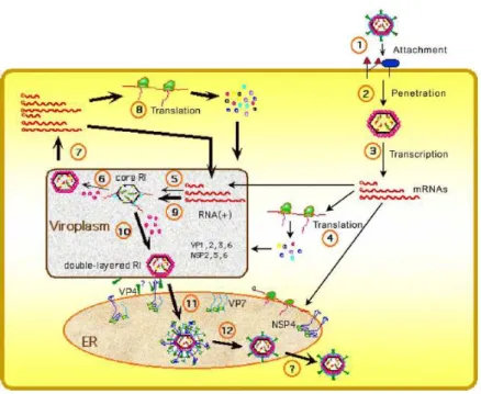
1.6. The replication cycle of the virus………...42
1.6.1. Cell binding and entry……….42
1.6.2. Virus uncoating, transcription and translation of proteins………..43
1.6.3. Replication and RNA packaging……….44
1.6.4. Virus release………47
1.8.2. Mass spectrometry………..54
Importance of mass spectrometry in biological research………..54
Main applications of MS in biological sciences………...…..……..55
Principles of the method and instrumentation……….………..56
2. MATHERIALS AND METHODS………61
2.1. Nsp5 wt and mutants constructs preparation and production of anti NSP5 serum………61
2.2. Tissue culture……….62
2.3. Transient transfection of MA104 cells………...62
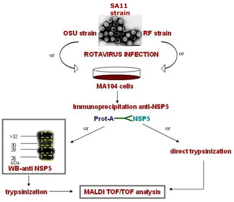
2.4. Virus infection and propagation………...62
2.5.Western blotting………..63
2.6. Sample preparation for MALDI TOF/TOF analysis………...64
2.6.1. Protein extraction from SDS-PAGE gel……….65
2.6.2. Direct trypsinization………....66
2.7. MALDI TOF/TOF analyses………...67
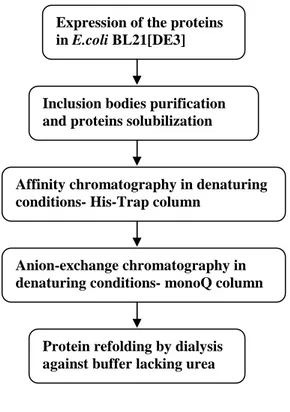
2.8. Expression of NSP5 and its mutants………..67
2.9. Proteins purification and refolding………...….68
2.9.1. Inclusion bodies preparation………...68
2.9.2. FPLC purification using AKTÄ system………..69
2.9.3. Proteins refolding………..…..70
2.10.Size exclusion chromatography………...………...70
2.11. Trypsin limited proteolysis……….………….70
2.12. Circular dichroism measurements………71
2.13. Binding assay based on ELISA………...71
2.14. Peptides synthesis for the NSP5 phosphorylation by CK1α………72
2.15. Expression and purification of His-6-tagged proteins for the in vitro kinase assay with recombinant CK1α………...……….………73
2.16. In vitro CK1α phosphorylation assay……….……….74
3. RESULTS………75
Part 1: Studies on hyperphosphorylation mechanism of NSP5…………..………....76
Ser-67 and the Activation Function………..78
Ser-67 Phosphorylation and CK1……….80
Part 2: Post-translational modification studies: mapping phosphorylation and glycosylation regions in NSP5………83
Part 3: Structural studies on NSP5………...…………..102
Expression of NSP5wt in E. coli……….102
NSP5wt purification and refolding……….106
ELISA-based binding assay………109
Trypsin limited proteolysis assay………110
Expression, purification and refolding of NSP5 mutants………...111
Gel filtration of purified NSP5 wt and mutants………..115
Circular dichroism far-UV experiments……….117
DISCUSSION………123
Rotaviruses, causative agents of gastroenteritis in young animals and humans, are large icosahedral viruses with a complex architecture. The double-stranded RNA (dsRNA) genome composed of 11 segments, that codes for 6 structural and 6 non-structural proteins, is enclosed within three concentric capsid layers.
NSP5, a non structural protein, is encoded by segment 11. It is produced early in infection and localizes in ‘viroplasms’, cytoplasmic inclusion bodies in which viral RNA replication and packaging take place. NSP5 is essential for the replicative cycle of the virus since, in its absence, viroplasms are not formed and viral RNA replication and transcription do not occur.
NSP5 is known to undergo two different types of posttranslational modifications, a cytoplasmic O-glycosylation and phosphorylation, which lead to the formation of proteins differing in electrophoretic mobility. Although the hyperphosphorylation process of NSP5 seems to be very complex, its role in the replicative cycle of rotavirus is unknown.
We demonstrated that NSP5 operates as an auto-regulator of its own phosphorylation as a consequence of two distinct activities of the protein: substrate and activator. In the first part of the thesis we have shown, that phosphorylation of Ser-67 within the SDSAS motif (amino acids 63-67) was required to trigger hyperphosphorylation by promoting the activation function. The evidence coming from iv vitro experiments, including kinase assay with recombinant casein kinase 1α from zebrafish, proved that this enzyme is responsible for a key phosphorylation step that initiates the whole hyperphosphorylation cascade of NSP5.
In the second part of the dissertation, using MALDI TOF/TOF spectroscopy, we added new data to the information about theposttranslational modifications of NSP5. We confirmed that the region of the protein encompassing Ser-67 is phosphorylated in vivo. Additionally we managed to map which parts of NSP5 sequence carries N-acetyloglucosamine and which regions bear phosphorylated serines or threonines.
There is no evidence about structure of NSP5 so far. In the last chapter we focused on investigating the structural organization of this crucial viral protein. To achieve this, in addition to the full length protein, one point mutation and two different truncation mutants were constructed, expressed, purified and refolded. The secondary structure of the different proteins was analyzed by circular dichroism spectroscopy and general information about protein conformation was provided. Our findings, together with an analysis of NSP5 sequence indicate that NSP5 can be an intrinsically unfolded/disordered protein.
3’CS 3’consensus sequence
5’CS 5’consensus sequence
aa amino acids
Ala alanine
Asp aspartic acid
ATP adenosine triphosphate
bp base pair
BSA bovine serum albumin
CD circular dichroism
CID collision-induced dissociation
CK1 casein kinase 1
CK2 casein kinase 2
C-terminal carboxy-terminal
DLP double layered particles
DMEM Dulbecco’s modified Eagle’s medium
dsRNA double stranded RNA
DTT dithiothreitol
EDTA diaminoethanetetraacetic acid
EGFP eukaryotic green fluorescent protein
eIF4GI eukaryotic initiation factor 4GI
ER endoplasmic reticulum
ESI electrospray ionization
FPLC fast protein liquid chromatography
GST glutathione-S-transferase
GTP guanosine triphosphate
HIT histidine triad
HPLC high performance liquid chromotography
HRP horse radish peroxidase
IEC ion exchange chromatography
IMAC immobilized metal affinity chromatography
IPTG Isopropyl β-D-1-thiogalactopyranoside
kDa kilo Dalton
Km Michaelis constant
MALDI matrix-assisted laser desorption/ionization
min minute
m.o.i. multiplicity of infection
MS mass spectrometry
N-terminal amino-terminal
NSP viral nonstructural protein
ORF open reading frame
PABP polyA binding protein
PAGE polyacrylamide gel electrophoresis
PBS phosphate buffer saline
PFU plaque forming units
p.i. post infection
PKC protein kinase C
RPHPLC reverse phase HPLC
SDS sodium dodecyl sulfate
Ser serine
SLP single layered particle
ssRNA single stranded RNA
TLP three layered particle
TOF time-of-flight
UTR untranslated region
VLP virus like particle
VLS viroplasm like structure
VP viral structural protein
1. INTRODUCTION
Until the 1970s, the etiological agents of diarrhea were not specified. Bacterial, viral, or parasitic agents could be detected in only 10% to 30% of children with diarrhea. In 1973, Bishop et al. noticed a 70 nm virus particle while using electron microscopy to detect infection in the duodenal epithelium of children hospitalized for treatment of acute diarrhea [11]. It soon became clear that the 70 nm particle, subsequently called rotavirus for its wheel-like appearance (Fig.1) (Latin, rota=wheel), was a causing agent of acute infantile gastroenteritis. Within 5 years after that discovery, rotavirus was recognized as the most common cause of diarrhea in infants and young children worldwide, responsible for approximately 600 000 infant deaths annually [12].
Fig.1. Rotavirus particles visualized by immune electron microscopy in stool filtrate from child with acute gastroenteritis. 70-nm particles possess distinctive double-shelled outer capsid. Bar = 100 nm [13].
1.1. Rotavirus classification
The dsRNA viruses are classified into six major families that are distinguished by they genome organization, strategies for protein coding, virion structures, host range and other differences. While the simplest members of the Totiviridae family posses single dsRNA segment, more complex viruses like members of the Reoviridae family contain 10-12 segments of dsRNA. Rotaviruses (RV) are classified as a genus within this family. Common morphological, and biochemical features of Rotaviruses are listed in table 1.
Tab.1. General characteristics of Rotaviruses[1].
Rotavirus strains are classified by three antigenic specificities- group, subgroup, and serotype/genotype. There are seven RV groups (A-G), determined by serological specificity to the structural protein of the inner capsid, VP6, predominant group antigen. The human pathogens belong to groups A, B, and C, the most important of which are those in group A.
Structure
65- to 75-nm icosahedral particles Triple-layered protein capsid
Nonenveloped (resistant to lipid solvents)
Capsid contain all enzymes for mRNA production
Genome
11 segments of dsRNA
Purified RNA segments are not infectious Each RNA segment codes for at least one protein
RNA segments from different viruses reassort at high frequency during dual infections of cells
Replication
Cultivation facilitated by proteases Cytoplasmic replication
Inclusion body formation
Unique morphogenesis involves transient enveloped particles
Group B viruses have been associated with outbreaks of severe adult diarrhea in China, those in group C cause rare and sporadic outbreaks of disease in children in many countries [14] [14]. RVs in groups D-G infect only animals.
Subgroup is also determined by epitopes of VP6, and is divided into two categories, marked as I and II. Further classification of RVs involves a binary system based on two proteins, VP7 and VP4. The outermost capsid glycoprotein, VP7, determines the G (G=glycoprotein) type of a RV strain. G type refers to either VP7 serotype or genotype as determined by antibody neutralization or nucleic acid sequence, respectively. To date, all identified G serotypes and genotypes are concordant. Thus, the G type of RV is expressed as a single digit, for example, G2. There are 15 G types found in humans but most abundant are those from G1 to G4.
The RV spike protein, VP4, determines the P type (protease-sensitive) of a strain. P types also consist of serotype and genotype, though each is denoted separately in current nomenclature. P serotype is listed first followed by P genotype in brackets, e.g. P1[8]. Eleven P serotypes have been identified while twenty one P genotypes are known. Among them seven are found in humans [15].
RV field isolates are monitored worldwide and have provided useful information about RV genetic variation. Prevalent strains can differ between regions in the same country, as well as from year to year in the same region. Additionally, no correlation exists between disease severity and serotype [15]. Over the past 30 years, the majority of RV disease in North America, Europe, and Australia has been caused by P[8]G1 strains [14].
Summarizing, classification of rotaviruses is based on scheme: group, designed by roman capital letters (A-G), subgroup, represented by Roman numerals and serotype (G or P), designed by Arabic number. However emergence of an increasing number of unusual strains that cannot be classified in either subgroup or are classified to both subgroups [16] have been described. This highlights the importance of RV strain surveillance in vaccine development.
1.2. Structure of rotavirus
1.2.1. Rotavirus architecture
Tree- dimensional structures of different members of Reoviridae family, as bluetongue virus (BTV) of Orbivirus genus and several strains of Reovirus genus were obtained using electron cryomicroscopy (Cryo-EM) and computer image reconstruction techniques [17]. The overall organization is similar among these viruses.
Rotavirus is the best characterized virus of Reoviridae. The first Cryo-EM reconstruction (made up to a resolution of 40Å) of rotavirus was demonstrated by Prasad et al. in 1988. These studies were performed on the simian strain SA11. Two years later reconstruction (at higher resolution) of another rotavirus strain, rhesus RVV, was showed [4]. The analysis of these two rotavirus strains revealed very similar structural features [1].
The virion of rotavirus is, as all virions of dsRNA viruses except bacteriophages from Cystoviridae family [18], non-enveloped. It is relatively large, has a diameter of around 1000Å and a left handed T=13 icosahedral symmetry, characterized by 132 aqueous channels and 60 surface spikes [19]. Capsid is composed of three concentric protein layers. The complete rotavirus virions are called TLPs (triple-layered particles), particles that lacks the outer layer are named DLPs (double-layered particles) and, in contrast to TLPs, are non-infectious. Particles that lack two the outer shells are called SLPs (single-layered particles) or cores. The outer layer is made of two proteins- spike-protein VP4 and VP7. The intermediate one is built of VP6 trimers and surrounds the inner shell, core, composed of the structural protein VP2. The layer of VP2 protects two structural viral proteins involved in virus replication and transcription (VP1 and VP3) and eleven dsRNA segments, the rotavirus genome, that encode six structural and six nonstructural proteins (Fig. 2).
1.2.2. The outer capsid layer
The outer capsid layer plays the crucial role in first phase of virus infection as is implicated in host cell attachment, membrane penetration and cell entry [20].In rotavirus, the outermost shell is made up of VP4 and VP7 proteins. Cryo-EM studies indicate that the 780
molecules of VP7, arranged as 260 trimers and located at the local and strict three-fold axes of a T = 13 (left-handed) icosahedral lattice, are uniformly distributed and form a smooth surface. From the surface protrude 60 spikes, bilobed at the distal end and 100- 120Å long. The spikes are composed of dimeric VP4 protein, so in the whole virion there are 120 copies of VP4 [19] [21]. Subsequent cryo-EM studies discovered that VP4 has a large globular domain that is buried inside the inner layer, making the total length of the spike about 200Å .The buried part interacts with the intermediate layer protein VP6 [22] [23].
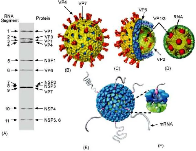
Fig 2. Architectural features of rotavirus. (A) PAGE gel showing 11 dsRNA segments comprising the rotavirus genome. The gene segments are numbered on the left and the proteins they encode are indicated on the right. (B) Cryo-EM reconstruction of the rotavirus triple-layered particle. The spike proteins VP4 is colored in orange and the outermost VP7 layer in yellow. (C) A cutaway view of the rotavirus TLP showing the inner VP6 (blue) and VP2 (green) layers and the transcriptional enzymes (shown in red) anchored to the VP2 layer at the five-fold axes. (D) Schematic depiction of genome organization in rotavirus. The genome segments are represented as inverted conical spirals surrounding the transcription enzymes (shown as red balls) inside the VP2 layer in green.(E and F) Model from Cryo-EM reconstruction of transcribing DLPs. The endogenous transcription results in the simultaneous release of the transcribed mRNA from channels located at the five-fold vertex of the icosahedral DLP [20].
The characteristic feature of rotavirus is the presence on the surface of not only VP4 spikes but also 132 aqueous channels (Fig.2F, 3 and 4) that link the outer surface with the inner core [17]. The channels, about 140 Å deep, are localized at all the five- and six-coordinated positions of the T = 13 lattice. There are three types of channels that can be distinguished according to their position and size (12 type I, 60 type II and 60 type III) [1]. Type II and III are about 55Å wide while type I have a narrower and more circular opening around 40Å in diameter and they are defined only by VP6.
The base of the type I channels is closed in the center by the VP2 pavement [24] [25]. Type I channels are involved in importing metabolites required for RNA transcription and exporting the nascent mRNA (Fig 2E, F) [24].
Fig. 3. Surface view of the 37Å rhesus rotavirus reconstruction viewed along two fold symmetry axes display T = 13 icosahedral lattice symmetry. The characteristic features of the outer surface are the 60 prominent spikes extending over 100 Å from the virion surface and a relatively smooth, spherical outer capsid perforated by 132 holes of three types: 12 type I holes at the icosahedral vertices; 60 type II holes at the peripentonal positions; and 60 type III holes encircling the icosahedral threefold axes of symmetry [4].
Fig. 4. Aqueous channels.
Central (equatorial) sections of the 37Å,
reconstructions viewed along a twofold direction are displayed with reverse contrast (bright regions correspond to high mass density). The three types of holes identified in Fig.4 are labeled. Icosahedral symmetry axes that lie within the equatorial plane are also shown [4].
1.2.3. The intermediate layer
Intermediate layer of rotavirus virion is formed by 260 trimers of structural protein VP6, the most abundant protein of the virion. The distribution of the protein mass in the second layer of the virion is not uniform and has a bristly appearance in contrast to the smooth VP7-VP4 surface. The trimers are assembled, like the outer shell, on T=13 lattice, and interact with outer shell proteins in a way that the channels in these shells are in register [19] [4]. All the surfaces of VP6 that are implicated in interactions with VP4, VP7 and VP2 contain the most conserved residues. Extensive lateral interactions between trimers and outer shell involve charged residues, whereas contacts with VP2 are mainly hydrophobic [26].
1.2.4. The inner layer, subcore and genome structure
The most inner shell of rotavirus is composed of 120 molecules of VP2 protein (60 dimers), possess T=1 symmetry and is quite smooth on the exterior [27] [1].The layer is interrupted by small pores that connect the core interior with environment. Whereas type I and type II channels are terminated at this layer, the type III channels continue beyond this shell [1]. This surface provides a structural platform for assembly of VP6 trimers, preventing aggregation of the core (VP1 and VP3 are highly hydrophobic proteins). This is the only rotavirus structural protein that has ability to self-assemble into a native-like icosahedral structure on the physiological conditions when expressed in insect cells. This suggests, that VP2 can be the scaffold for three layered particle formation [28] [29]. The two other proteins that are part of the core, VP1, the RNA-dependent RNA polymerase [30], and VP3, the guanylyl and methyl transferase (mRNA capping) [31] [32], are ten times less abundant than VP2 [33], and provide enzymatic functions required for producing the capped mRNA transcripts and genome replication. Biochemical and structural studies have shown that VP1 and VP3 are located in close proximity to each other and that form a heterodimer [34] [25]. VP2 is known to bind RNA through its N-terminal residues, what was observed in cryo-EM structure of DLS. The N-terminal region of VP2 protrudes inward at the fivefold axis to form a pentagonal shape and is also implicated in anchoring VP1/VP3 complex. This position is
consistent with releasing of nascent mRNA though the type I channel [35]. It is still not completely clear, but the N-terminus residues of one VP2 subunit appears to be occupied in transcription enzyme holding, while the N-terminal residues of the other subunit may be involved in interaction with the underlying genomic RNA [20].
The precise structural organization of rotavirus genome is not completely clear. It was however demonstrated that the genome forms concentric layers, separated by a distance of 28-30Å [36], where each segment of RNA is spooled around transcription enzymes complex, located at the icosahedral vertices. Possible model suggested by Goeyt et. al. for the bluetongue virus [37] seems to be very realistic also for the rotavirus [5] ( Fig. 2D, 5).
This model allows 12 independent transcription complexes, each attached to an individual dsRNA segment. It is concordant with the fact that till now no dsRNA virus with more than 12 segments is known, however most of the members of this family have not 12 but 10 or 11 segments [20].
Fig. 5. Model of structural organization of rotavirus genome. Each dsRNA segment is coiled in a cone shape at the fivefold vertex [5].
1.3. Organization of rotavirus genome segments
The total length of rotavirus genome is about 18 550 kbp. It consists 11 segment of dsRNA with a size range of 0.6 to 3.3 kbp [38]. Deproteinated rotavirus dsRNA segments are not infectious, confirming the need for presence of RNA-dependent RNA polymerase to transcribe the individual RNA segments into active mRNA. Purified dsRNA segments can be resolved by polyacrylamide gel electrophoresis and, according to the order of migration (length) of segments, are numbered from 1(the slowest) to eleven (the fastest) (Fig. 2A). The sequence of different genome segments, also derived from distinct rotavirus strains, show common characteristics (Fig. 6.) [1]:
- sequences are A+U rich (58 to 67%) - segments are base-paired end to end
- the positive RNA strands contain cap structures at the 5’ end, while lacking 3’ terminal poly (A ) tails (distinctly to the most of cellular mRNAs) [39]
- each (+) RNA segment starts at 5’-prime end with guanidine, followed by a set of conserved nucleotides that are part of 5’-noncoding sequences (called untranslated 5’ region, 5’UTR), then followed by usually unique open reading frame (ORF) for the protein product, that ends with the stop codon, and then finished by set of noncoding conserved 3’ sequences (3’UTR) with two terminal cytosines at the very end
- all the sequenced genes possess at least one long ORF initiated by a strong initiation codon based on Kozak’s rules, although some of the genes contain additional in-phase ORF (segments 7, 9 and 10) or out of phase alternative ORF (segment 11)
- all genes are monocistronic, except gene 11, that codes for two different proteins. The roles of the UTRs are not completely elucidated but they are probably targets for rotavirus RNA-binding proteins that participate in RNA synthesis, regulation of gene expression and packaging. In addition to primary sequences, secondary structures present in the UTRs may serve as component to the recognition signals for the RNA-binding proteins [40] [41].
As all the 11 mRNAs are replicated by the same VP1 polymerase, they must share common cis-acting signals and these signals are likely to be formed by secondary structures rather than by primary sequences. Some of the cis-acting signals for rotavirus RNA replication and translation have been identified but packaging rules still have to be clarified. Each mRNA has to contain a unique signal because 11 mRNAs must be distinguished from one another. Possible explanation can depend on secondary structure of positive RNA strands because secondary panhandle-like, and especially, different in each (+)RNA, stem-loop structures, have already been predicted [42].
Fig. 6. Major features of rotavirus gene structure. Schematic shows the overall structure of rotavirus genes from the published sequences of genes 1 to 11. All eleven rotavirus genes lack a polyadenylation signal, they are A+U rich, and they contain conserved consensus sequences (UTRs) at their 5’ and 3’ ends [1].
1.4. Structural proteins
1.4.1. VP1
The structural protein VP1 is codified by the gene 1 of rotavirus. It is a basic protein and for the bovine rotavirus (RF strain) has an apparent molecular mass of 125 kDa [43]. It is present in the cores only in a few copies and together with VP3 forms a heterodimer. VP1 has two recognized functions:
- acts as a transcriptase - using as a template minus strand of dsRNA synthesizes primary viral transcripts which are extruded into the cell’s cytoplasm through the I- class channels
- functions as a replicase synthesizing minus strand on the template of (+)RNA strand. These functions were found on the basis of several observations. It reveals non specific affinity to RNA [44] and it is the only protein of rotavirus that shows sequence-specific recognition of viral RNA (binds to 3’end of gene 8 mRNA) [45]. Moreover, it shares the four common motifs conserved among sequences of all RNA- dependent RNA polymerases of other RNA viruses [43]. VP1 binds nucleotides and was demonstrated that crosslinking of photoreactable nucleotide azido-adenosine triphosphate (azido-ATP) to VP1 inhibits transcription [30].
Additionally its temperature sensitive mutant of do not synthesize ssRNA at the non permissive temperature [46]. Reconstitution experiments with baculovirus-expressed protein have shown that VP1 requires VP2 for replicase activity and presence of VP2 stimulates VP1 replicase activity several fold. VP1 is able to bind viral mRNA in the absence of any other viral proteins but its replicase activity requires previous VP2 interaction with RNA [45] [47].
Moreover it was demonstrated several times that VP1 is able to interact with NSP2 [48, 49] and can be chemically crosslinked in living cells with NSP5 [49]. However, the experiments performed recently in our laboratory showed, that the interaction of NSP5 with VP1 was found to be stronger than the interaction of NSP5 with NSP2[50].
1.4.2. VP2
VP2 is the most abundant protein of the rotavirus core (around 90%). It is encoded by gene 2 and yields the 882aa protein with a molecular mass of 102.5 kDa. Between amino acid 536 and 686 are localized two leucine zippers that may play a role in VP2 oligomeryzation into the core where it is present in the form of 60 dimers [1] .VP2 interacts with VP6, genomic dsRNA and with VP1/VP3 replication-transcription complex. Contact with the VP6 layer is created on the base of hydrophobic interactions [26]. VP2 is the only structural protein that has the ability to self-assembly into a native like icosahedral structure, when expressed in insect cells. This observation strongly indicates that it may play as structural scaffold for the proper assembly of other viral proteins. Co-expression in baculovirus of VP2 together with VP6 leads to formation of VP2/6 double layered complex called Virus Like Particles (VLPs) [29].Same results were obtained expressing these two proteins in vaccinia system [51] [52].
It is known that VP2 interact with VP1 and VP3 by its N-terminal region. That was deduced from biochemical studies on recombinant VLPs, containing VP2 with amino-terminal deletions, co-expressed with VP6, VP1 and VP3. Lack of 25 residues on the N-terminus completely prevents incorporating of VP1/VP3 replication/transcription complex [53] [20].
Further evidence suggests that the amino terminus of VP2 plays a major role in organizing the major components of the endogenous transcription apparatus, the genomic dsRNA and the VP1-VP3 enzyme complexes, within the core of the virion. It was found that the N-terminal 132aa domain of VP2 is implicated in RNA binding. Through that region VP2 is able to bind single and double stranded RNA [54]. Subsequent experiments have shown that removal of as few as the first 25 amino acids from the amino terminus of VP2 completely abolishes RNA binding activity, suggesting that the integrity of the complete amino-terminal domain is critical for the conformation of the RNA binding site. The predicted amino acid sequence of this domain contains several motifs which could potentially form the site of interaction between VP2 and RNA [55] [56]. In support of these observations, the structure of the native double-layered particle (DLP) determined by electron cryomicroscopy shows significant interactions between the inner surface of VP2 and the genomic dsRNA near the icosahedral fivefold axes and minor interactions along the icosahedral twofold axes [25]. These interactions result in nearly 25% of the genomic dsRNA adopting a highly ordered
conformation. Clearly the VP2 capsid layer is highly instrumental in organizing the genomic dsRNA within the core.
1.4.3. VP3
VP3 is a 98kDa (835aa) basic protein coded by genome segment 3. It is the minor component of the core that together with VP1 forms a homodimer- transcription/replication complex. Encapsidation of VP1 and VP3 in the inner core depends on interaction with the N-terminal of VP2 [53]. VP3 expressed in rotavirus covalently bound GTP what suggest that VP3 alone is a guanyltransferase [32]. Subsequent studies showed that VP3 interacts with GTP [57] and forms covalent VP3-GMP adducts [58] that confirmed the previous finding. It was also noticed that VP3 has ability to bind ssRNA but does not bind dsRNA. The ssRNA-binding is sequence independent, but higher interaction affinity was found for uncapped than for capped RNA [47] [44]. The guanylyltransferase activity of VP3 is nonspecific as it can cap plus- and minus-strand viral RNAs, non viral RNAs, and RNAs initiating with G and A residues, but not dsRNA [32]. It was also demonstrated by VP3 binding to S-adenosyl-l-methionine (SAM), a substrate necessary for cap methylation of RNA, that open rotavirus cores possess active methylotransferase activity.
All the data indicate that VP3 is a multifunctional capping enzyme as it shares characteristics with capping enzymes of other members of the Reoviridae, including the VP4 protein of bluetongue virus (BTV) and the λ2 protein of reovirus [44].
1.4.4. VP4
VP4 is a structural non glycosilated protein, encoded by genome segment 4. It is known to be the main component of rotavirus cell entry apparatus therefore plays essential role in the virus life cycle. Indeed antibodies against VP4 neutralize the virus and block cell entry [59].
It is a very distinctive element of rotavirus architecture as it forms characteristic 60 spikes on the surface of the virion. Recent studies discovered that VP4 is a very flexible
protein and during virus cell entry process performs series of molecular rearrangements that change its conformation. Initially, prior to trypsin cleavage, VP4 has a flexible conformation. Trypsin digestion rigidifies VP4 molecules and prime rotavirus to infect the cell. In this state VP4 is organized in a dimer that forms a rigid spike, third VP4 molecule remain flexible (Fig. 9A). This is a crucial moment in rotavirus infection as without proteolytic activation virus can not enter the cell. During digestion, VP4 (88 kDa) is cleaved into VP8* (28 kDa, aa 1–247) and VP5* (60 kDa, aa 248–776), the cleavage products remain associated in the virion [60]. The VP8* fragment contains a globular domain, the VP8* core, which forms the ‘head’ of the spike. Portions of both VP8* and VP5* make up the ‘body’ that is linked by an asymmetric ‘stalk’ to a ‘foot,’ which is buried beneath the VP7 shell (Fig.7.) [23] [22].
VP8* masks the hydrophobic apex of VP5* on primed spikes [9]. It contains a hemagglutination motif and sialic acid (SA) binding site in the strains that use this receptor to enter the cell. These strains are able to agglutinate red blood cells by binding SA on the surface of erythrocytes [60]. Involvement of SA during rotavirus infections is not an essential step for all rotavirus strains. In many rotavirus strains, including human rotaviruses, cell entry is SA independent [61]. In these viruses, the majority of neutralizing monoclonal antibodies that recognize VP4 select mutations in VP5* [62], suggesting that cell entry is mediated mainly by the VP5*. The VP8* core is an important target of neutralizing antibodies against rotavirus and major determinant of P serotype for both SA-dependent and SA-independent strains [63]. VP8* can also play intracellular role in virus replication as it has been shown to activate cell signaling pathway upon binding the tumor necrosis factor receptor-associated factors (TRAFs) [64].
Fig. 7. VP4 domains scheme .Head (H), body (B), stalk (S) and foot (F) regions of the VP4 spikes are indicated. The drawing is based on an electron cryomicroscopy image reconstruction from [4]. Copied from [9].
VP8* has a β-sandwich fold of the galactines- a family of sugar binging proteins. The crystal structure reveals that the core is organized in a single domain composed from two β-sheets, formed by five-stranded and six-stranded β-strands. β-strands are flanked by two α-helixes, a shorter one, laying in the inter-sheet loop and C-terminal, bridging parallel β-structure and extended β-ribbon. The central part of a β-structure is composed by the cleft situated between two β- sheets. The tight fold of β-sandwich with short loops between strands and dense hydrophobic cores between the major structural elements suggest a rigid structure that is unable to undergo major rearrangements during cell entry. The compactness favors protease resistance [65]. Although crystal structure of the equivalent VP8* domains from the sialic acid-independent rotavirus strains differ functionally, share the same galectin-like fold. Differences in the groove region that corresponds to the SA binding site make it unlikely that SA-independent rotavirus binds an alternative carbohydrate ligand in this location [66].
Another, not yet elucidated event triggers VP4 to change the conformation for a second time (Fig. 8 B.). Two VP5* subunits fold-back and join a third subunit to form a tightly associated trimer. VP8* domain dissociates (interactions with VP5* are due to hydrophobic interactions) and expose a hydrophobic apex of VP5* that is a potential membrane interaction region [7].
Fig. 8. Models of two VP4 conformations. (A) The trypsin primed state. Two rigid subunits form the spike visible in electron cryomicroscopy, a third subunit is flexible. VP8* is gray, with an N-terminal tether and a globular head creased by the sialoside-binding site. The VP5* antigen domain is green bean-shape, with a red membrane interaction region and a yellow GH loop. An additional b-strand C-terminal to the antigen domain is also yellow. The spike body includes the VP5* antigen domain, part of the VP8* tether, and the GH loop. The foot is blue, as is a protruding region that rearranges into the coiled-coil. (B) The putative post-membrane penetration state. VP8* has dissociated; the yellow parts of each subunit have joined in a b-annulus; the a-helical triple coiled-coil has zipped up; and the VP5* antigen domain has folded back [7].
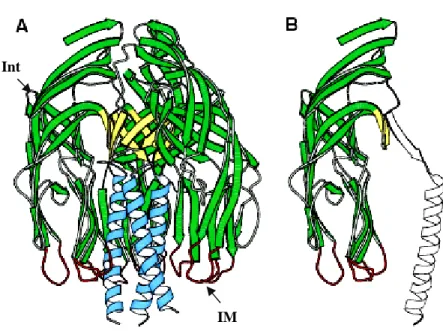
The structure of VP5* fragment, VP5CT, was solved by crystallization. It is the strongest evidence confirming trimeric, folded-back state of VP5*. VP5CT is a protease cleaved fragment of rhesus rotavirus (RVV) that consists of residues A248 to L525 (or 528); the missing C-terminal is a part of the ‘foot’ that is buried in the VP7 layer and interacts with VP6. VP5CT is a well-ordered homotrimer that resembles a folded umbrella (Fig. 9A). The ‘post’ of the umbrella is a C-terminal, α-helical, triple coiled-coil. Each of the three panels comprising the ‘shade’ of the umbrella represents an N-terminal globular domain. The core of each globular domain is an eight-stranded anti-parallel β-sandwich. The flexible tip of one of the β-hairpins is exposed to the solvent and contain a sequence motif (DGE), probably implicated in rotavirus binding to α2α1 integrin (Fig.9A). Another interesting aspect of this structure is that the tips of loops projecting from the bottom edge of globular domains possess a hydrophobic region that may function in membrane penetration. The sequence of these loops shares a sequence similarity with alphavirus loops implicated in membrane disruption but the loop and domain structures are different. Thus, the sequence similarity probably reflects selection for hydrophobicity and flexibility in both loops, but not common ancestry [9].
Fig. 9. VP5CT and the VP5* antigen domain. (A) Ribbon diagram of the VP5CT trimer, colored to match Figure 1. VP5CT does not include the foot region. Int- integrin binding motif; IM- potential membrane interaction loop (B) Ribbon diagram of a single VP5CT subunit. The part that forms the VP5* antigen domain is green, yellow, and red. Modified from [9] [7].
Int
Apart from cell entry VP4 may be implicated in other interactions inside of infected cell. It was demonstrated that VP4 interacts with VP7 and NSP4 [67]. Newly synthesized VP4 particles are present in the space between periphery viroplasms and outside of ER [68]. It was found at the plasma membrane and it colocalizes with the cytoskeleton of infected cells. Recently it was shown that it is strongly associated with lipid raft microdomains and binds to two cellular proteins, GTPase Rab5, and to prenylated Rab acceptor (PRA1) that regulate the vesicular traffic and the motility of early endosomes along microtubules. These results suggest that Rab5 and PRA1 may be involved in the localization and trafficking of VP4 in infected cells [69]. The precise goal and mechanism of all these interactions has to be already entirely clarified.
1.4.5. VP6
VP6 is encoded by segment 6 of the genome. It is a highly immunogenic, very hydrophobic 45 kDa protein that is conserved among most of rotavirus strains. It is the most abundant structural component of the virus. It forms, organized in 260 very stable trimers, the intermediate layer of virion structure and the most outer shell in DLP in the way so that the aqueous channels in the two T = 13 layers are in register. In the overall organization of the rotavirus, VP6 appears to integrate the two principal functions of the virus, cell entry and endogenous transcription, through its interactions with the outer layer proteins VP7 and VP4, and the inner layer protein VP2. Biochemical studies indicated that none of the component of DLP alone is able to transcribe the dsRNA but VP6, despite of lack of enzymatic function, is essential for endogenous transcription of the genome [70] [71].
The structure of VP6 was revealed by crystallography [26]. Each VP6 monomer has two distinct domains, defined as B and H. Domain B is present at the base of the molecule and is composed of a bundle of eight α-helices that derive from two different segments of polypeptide chain. The first segment comes from N-terminal part of VP6 while remaining three helices are formed from C-terminus end of the sequence. In domain B the only β-sheet element consists of β-hairpin that connects two α-helices. That β-hairpin motif of contains a highly conserved sequence that protrudes into the channel I and plausibly may play a functional role in the translocation of the nascent mRNA transcripts during endogenous transcription. The electrostatic repulsion between the negatively charged channel surface and the nucleic acid is likely to facilitate the extrusion of the transcript by increasing its fluidity.
Domain H making up the top of the molecule, folds into β-sandwich with the jelly-roll topology. Hydrophobic interactions in this domain are responsible for trimerization of VP6, as mutant lacking that domain can not form the trimer while deletion mutants that lack the domain B are capable to trimerize [72]. The characteristic feature of the VP6 trimer structure is a Zn2+ ion that essentially contributes to the trimer stabilization.
VP6 trimers interact specifically with itself and with all three structural proteins in the virion. Interactions with VP2 are mainly hydrophobic whereas contacts between VP4 at the sides of domain H and VP7 at the top of domain B utilize charged residues. The lateral interactions between the trimers that involve charged residues are not sufficient to form the closed shell. Therefore it is likely that a proper scaffold for the assembly of VP6 into T=13 icosahedral organization is provided by VP2 layer. All the interaction surfaces relay on the most conserved amino acids of polypeptide chain. The experiments like detailed mutational analysis based on pseudo-atomic model of VP6 [71] and cryo-EM structural studies on DLP-anti (VP6) MAb complexes [34] [73] clearly indicate that not only the observed proper assembly of VP6 trimers on VP2 but also intact dynamics of interaction between these two proteins are absolutely required for correct endogenous transcription.
1.4.6. VP7
VP7 (37 kDa) is encoded by genome segment 9 in SA11 strain. It is the second, after VP6, most abundant structural protein of rotavirus. Together with VP4 it is responsible for forming the most outer layer of the virion implicated in cell attachment and entry.
VP7 is a glycoprotein that contains three N-glycosylation sites, from which only two are apparently used. Biochemical studies indicate that modifications contain N-linked adding of high mannose oligosaccharides, which are processed by trimming [74] [75] [76]. Man8GlcNAc2 and Man6GlcNAc2 oligosaccharide residues are found on intracellular VP7
while Man6GlcNAc2 is found on mature virus particles [77] [74]. VP7 is cotranslationally
glycosylated as it is inserted into the membrane of the ER, and insertion is directed by a cleavable signal sequence localized at the amino-terminus of the protein [78] [79] [74]. The ORF of 326 amino acids begins with an initiation codon with a weak consensus sequence. A second, in-frame initiation codon precedes two regions of hydrophobic amino acids (H1 and H2), which can act as the signal sequence to direct VP7 to the ER, although the second is thought to be the major species used in cells. The site of cleavage of signal peptide is
glutamine 51 [80]. VP7 lacks the characteristic KDEL sequence important in conferring ER [81] retention but contains a conserved region with hydrophobic amino acids (consensus peptide LPXTG) which acts as the signal sequence in directing VP7 to ER [82].
Although these residues are critical for retention, the method by which VP7 remains in the ER is unresolved. After its insertion into membranes, VP7 is resistant to digestion with proteolytic enzymes, suggesting it is not a membrane-spanning protein [74] [76]. It remains membrane associated after high salt treatment and release of microsomal contents at alkaline pH what suggest that it is an integral membrane protein with a luminal orientation 120 [74].
VP7 in infected cells forms oligomers with VP4 and NSP4 [67].These interactions appear to be important for the assembly of VP7 into outer capsid. Mature VP7 lacks all amino acids proximal to Gln 51 but many conserved amino acids are present before the second hydrophobic domain. Proper folding of VP7, in which are involved highly conserved cysteins, requires ATP [83].It was found that this process requires also cellular factors like ER-associated chaperone calnexin, interacting also with NSP4 [84].
The precise role of VP7 during early interactions of the virus with the cell is not clear, but it has been postulated that VP7 may modulate the function of VP4 during the attachment and entry process [85]. It may interact with cell surface molecules after the interaction is initiated by VP4 [86]. It was found that VP4 contain sequences that bind integrins. It has two sequences motifs that may be important in this process, CNP that interact with integrin αvβ3 [87] and GPRP that is bound by integrin αxβ2 [88]. VP7 in its sequence containes pralines that binds calcium and the sensitivity of virions to low calcium concentrations is strain-dependent [89] [90]. Several studies also have suggested calcium-driven conformational changes in VP7 [91]. Studies on baculovirus-expressed recombinant VP7 have shown a requirement for calcium in the formation of VP7 trimers, which crystallize into hexagonal plates mimicking the arrangement of VP7 on the capsid [92]. Thus, while appropriate levels of calcium help maintain the structural integrity of the VP7 layer, low calcium concentrations, similar to those in the cytoplasm, trigger the disassociation of VP7 trimers leading to uncoating of the VP7 layer and releasing transcriptionally competent form of rotavirus -DLP.
1.5. Nonstructural proteins
1.5.1. NSP1
Rotavirus NSP1 (55kDa) is the product of the genome segment 5. It is known to be the least abundant protein of rotavirus. Although in infected cells it localizes throughout the cytoplasm [93] in contrast to most other rotavirus proteins that concentrate in viroplasms, it is able to interact with NSP3, NSP5 and NSP6 [94]. Moreover NSP1 was found associated with the cytoskeleton when analyzed by subcellular fractionation [93].
NSP1 is also the least conserved protein among all rotavirus nonstructural proteins. The only conserved part is the N-terminus. In its sequence there are present eight cysteins and two histidines that form the part of zinc finger domain. That domain has an affinity to 5’ region of all viral mRNAs [93] [95].
NSP1 apparently is not required for rotavirus replication. Strains isolated from animals and from both immune-deficient and immune-competent children containing rearrangements in gene 5 that result in the synthesis of truncated NSP1 have showed to replicate in cell culture to titers close to those of their wild-type counterparts [96]. Also knockdown experiments using RNA interference have confirmed that the protein is not needed for virus replication [97]. Although NSP1 is not crucial for replication, production of other viral proteins and formation of viroplasms, it seems to be essential for proper rotavirus cell-to-cell spread [98] [99].
Analysis of interaction with cellular proteins showed that NSP1 interact with interferon regulatory factor 3 (IRF-3) [96] [100]. This protein during normal cell state accumulates as an inactive monomer in the cytoplasm. Events associated with virus infection like production of double-stranded RNA and expression of viral proteins trigger the innate immune response mechanisms that include the phosphorylation of IRF3 by cellular kinases. That modification initiates structural changes in IRF3 that results in its dimerization [101]. The dimer, an active form of IRF3, is translocated to the nucleus, where it interacts with specific promoter elements, stimulating expression of IFN-α and IFN-β. The secreted IFNs promote the production and activation of antiviral proteins in neighboring cells blocking the virus cell-to-cell spread [102]. It was demonstrated that NSP1 mediates IRF3 degradation through a
proteasome-dependent pathway. The role of this interaction in rotavirus infection could be diminishing the cellular interferon response [96].
The fact that NSP1 protein is much more conserved among rotaviruses infecting the same host may explain in part why rotavirus strains causing severe gastroenteritis in a homologous animal model are usually less infectious and asymptomatic in a heterologous animal model [103].
1.5.2. NSP2
NSP2, encoded by segment 8, is a highly basic protein of relative molecular mass 35kDa. In vivo studies have shown that NSP2 and NSP5 together with VP1, the RNA polymerase, are co-localized in the viroplasms of infected cells and are the main constituents of the replication intermediates [104] [105]. Indeed several facts demonstrate that NSP2 is essential for viral replication. Temperature-sensitive mutants of NSP2 fail to replicate the genome and produce mostly empty particles, which implicates NSP2 in genome replication and packaging [106] [107]. This experiment is concordant with the observation that silencing NSP2 synthesis by RNA interference causes complete blocking of viroplasms formation, production of viral proteins and rotavirus replication [97]. Additionally functional and structural similarities were described among NSP2, bluetongue protein NS2 and reovirus
σNS, suggests that they are functional homologs [108].
NSP2 selfassembles into stable doughnut-shaped octamers, formed by the tail-to-tail interaction of two tetramers [10]. The overall architecture of such octamers is highly conserved even among distantly related groups of rotaviruses [109].
Many biochemical studies on recombinant NSP2 have specified its roles in genome replication and packaging. The octamers have single-stranded sequence-independent ssRNA-binding activity thanks to which are capable of destabilizing RNA–RNA duplexes by an ATP and Mg2+ independent mechanism [110] [111]. In addition, the octamers have a Mg2+-dependent nucleosidetriphosphate phosphohydrolase (NTPase) activity that cleaves the γ-β phosphoanhydride bond of any nucleoside triphosphate (NTP), yielding the products NDP and Pi. Following cleavage, the γP is transferred to NSP2, generating a short-lived phosphorylated form of the protein [110] [112]. It was demonstrated that hydrolytic activity
of NSP2 is essential for genome replication [109]. Moreover NSP2 octamers in the presence of NTPs undergo a conformational transition, shifting from a relaxed to a more condensed state as is typical of molecular motors [113].
All together, these properties have led to the suggestion that the NSP2 octamer may facilitate genome packaging and replication by relaxing secondary structures in viral template RNAs that impede polymerase function and by assisting in the translocation of viral RNAs into pre-virion cores in genome replication and packaging [114].
The understanding of these mechanisms has been much facilitated by solving the structure of NSP2 by X-ray crystallography to a resolution of 2.6A° [10]. NSP2 crystallizes as an octamer using crystallographic 4-2-2 symmetry, resulting in one monomer per asymmetric unit. The NSP2 monomer has two distinct domains (Fig. 10), an N-terminal domain from residue 1 to 141 and a C-terminal domain from 151 to 313. A striking feature of the monomer is a presence of electropositive 25 A° deep cleft that lies between the two domains.
The N-terminal domain has a novel fold. Although predominantly α -helical, has two pairs of anti-parallel β -strands towards the N terminus. In this domain can be noticed two sub-domains that are connected by a 24-residue-long predominantly basic loop (Fig. 10, arrow). There are three highly conserved prolines (53–55) at the start of this loop, which may be essential for its conformation. This loop lines the prominent grooves that run diagonally across the 2-fold axes of the doughnut-shaped octamer (Fig 11), and contributes to their basic character.
The C-terminal domain has an α /β fold. The important feature of this domain is the twisted anti-parallel β-sheet flanked by helices, formed by residues from 150 to 245, in a
β3αβ2α configuration. The anti-parallel β -strands, between residues 186 and 191 (β7), and
between 226 and 230 (β9), along with the loop, between residues 221 and 226, constitute the base of the cleft. The interdomain loop and the helix α5 from the N-terminal domain form a major part of one side of the cleft, whereas the other side of the cleft is made of helix α 11 and a loop between residues 245 and 260. The remainder of the C-terminal region, from residues 260 and 311, consists predominantly of α -helices (α 12– α16). That domain has a significant, limited only to the tertiary structure level, homology with the protein kinase C interacting protein (PKCI), a member of the Histidine Triad (HIT) familyof nucleotide-binding proteins [115] [10].
On that base the location of the NTP-binding site was initially proposed. Now it is known that it is localized in 25 Å cleft, between the two domains of the monomer. Although lacking a precise signature of HIT motif, mutagenesis studies have indicated that conserved
basic residues in the cleft form a HIT-like motif, responsible for the binding and hydrolysis of NTPs [112]. More recently, data obtained by co-crystallization of NSP2 with nucleotide analogs has shown that His225 is the catalytic residue of the motif [116].
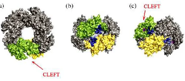
Extending diagonally across the NSP2 octamer surface (Fig. 11.) are four highly basic grooves, 30 Å wide and 25 Å deep, which function as ssRNA-binding sites. [10] [117]. Each groove is lined by two 24-residue electropositive loops, originating between the two subdomains of the N terminus of each monomer (Fig. 10, arrow). The location of these loops is such that they position the electropositive residues at the entrance of the clefts containing the HIT-like motif.
Fig. 10. The ribbon representation the monomeric subunit of NSP2. The secondary structural elements in the monomer are coloured: α-helices in red, β-strands in green and loops in blue. The basic loop in the groove between the subdomains in the N-terminal domain is shown by an arrow [10].
Fig.11. Structural proximity of the NTP-binding cleft and the RNA-binding grooves. Surface representation of the NSP2 octamer viewed along the 4-fold (a) and 2-fold axes ((b) and (c)). Two monomers have been identified in the octamer (green and yellow). The image in (b) represents a 90° rotation along the x-axis with respect to the image in (a). Residues 53 to 76 that form part of the electropositive loops, which line the RNA-binding grooves, are denoted in blue. (c) The image in (b) was rotated 45° clockwise along the y-axis to allow visualization into the cleft. Residues within the cleft that make up the active site for NTP hydrolysis are shown in purple [114].
Recently it was demonstrated that that the NSP2 octamer has also RTPase activity and that the RTPase and NTPase activities of the octamer utilize the same HIT-like motif and generate indistinguishable phosphorylated intermediates. It was also shown that ssRNA is preferred as a substrate over NTPs in hydrolysis reactions, likely due to the higher affinity of the octamer for ssRNA. It was discovered that NSP2 uses a HIT-like motif to direct both RTPase activity and NTPase activity[114].
The donut-shaped NSP2 octamer also displays a central hole with a diameter of 35 Å.
One interesting possibility is that the central hole of the NSP2 octamer could be used as a protective environment for newly synthesized dsRNA emerging from VP1 or to serve as a passive conduit for its packaging during the assembly of VP2 capsid layer [117]. It looks as the octameric structure of NSP2 forms a tempting platform or a scaffold around which the replication complex is organized. It is possible that the hydrophobic side of the octamer, around the four-fold axis, may bind to the VP1. Furthermore the molecular partner of NSP2, NSP5, is an acidic protein and the basic grooves of the NSP2 octamer may be the sites for its binding. In fact recent observation utilizing cryo-electron microscopy has indicated that a truncated species of NSP5 binds along the same electropositive grooves involved in RNA
binding, showing that both NSP5 and RNA share the same binding site on the NSP2 octamer [117].
1.5.3. NSP3
NSP3, encoded by gene seven is a basic nonstructural 36 kDa protein that localizes in cytoplasm of infected cells [118]. The experimental evidence shows that NSP3 is responsible for shutting off cellular protein synthesis, while ensuring at the same time the translation of viral mRNAs. Rotaviruses rely on the host translation machinery to produce the proteins encoded by their genome. Rotaviral transcripts are capped at their 5’ end by the action of the structural protein VP3 during transcription but their 3’ ends are not poly-adenylated like most of cellular mRNAs. In the host cells only poly-adenylated and capped messages are known to be efficiently translated. This is brought about by recognition of the 5’ cap by eIF4E (cap binding protein) and the poly-A tail by PABP (poly-A binding protein) which then interacts with a cellular factor eIF4GI. This factor is a multipurpose adaptor protein that is responsible for assembling the cellular translational complex composed by the proteins eIE4E, eIF4A (helicase), PABP, eIF3 as well as bound and circularized by the complex mRNA. The complex then is delivered to the ribosome where the translation begins. Rotaviruses overcome the lack of a poly-A tail by possessing on 3’ ends of theirs mRNAs a consensus sequence (UGACC) which is specifically bound by NSP3 [119] [120] [121] [122]. While the N-terminal domain of NSP3 binds this consensus sequence, the C-N-terminal half interacts with eIF4GI with an affinity greater than cellular PABP to give translation of the rotavirus messages a selective boost following infection [120].
Resolving the structure of NSP3 by X-ray crystallography has helped to understand better mechanisms of these actions. NSP3 consists of two readily separable domains divided by a dimerization domain [94] [121]. N-terminal domain forms a heart-shaped, asymmetric homodimer with a medial basic deep cleft on the surface that creates a basic dead end tunnel that binds the 3’ terminal consensus nucleotides of rotavirus mRNA. This tight interaction not only promotes translation of the rotavirus mRNA but also prevents degradation of the rotavirus message by cellular nucleases and excludes the possibility of cellular sequences recognition. Biophysical studies demonstrate a high affinity binding leading to increased thermal stability and slow dissociation kinetics, consistent with the NSP3 functions [123, 124]. On the other hand C-terminal domain is an α-helical symmetric homodimer. It
recognizes a fragment of eIF4GI and binds it with an affinity greater than PABP [125] in pockets at the dimmer interface. Site-directed mutagenesis and isothermal titration calorimetry documented that NSP3 and PABP use analogous eIF4GI recognition strategies, despite marked differences in tertiary structure[126, 127].
Recent studies [128] seem to revise mentioned model questioning how NSP3 blocks cellular protein synthesis and ensures at the same time the translation of viral mRNAs. In this work, the expression of NSP3 in infected cells was knocked down using RNA interference. Unexpectedly, under these conditions the synthesis of viral proteins was not decreased, while the cellular protein synthesis was restored. Also, the yield of viral progeny increased, which correlated with an increased synthesis of viral RNA. Silencing the expression of eIF4GI further confirmed that the interaction between eIF4GI and NSP3 is not required for viral protein synthesis. These results indicate that NSP3 is neither required for the translation of viral mRNAs nor essential for virus replication in cell culture.
These newly found discoveries can be elucidated in different ways. Even though the interactions of NSP3 with eIF4GI and the 3’ end of viral mRNAs have been clearly established, there is no direct evidence that NSP3 engages simultaneously in these two interactions to promote the circularization of viral mRNAs, and although it is generally accepted, there is also no evidence that these interactions favor the translation of viral mRNAs.
Moreover interaction of NSP3 with eIF4GI is not necessary for viral translation. Furthermore, an increased level of viral RNA synthesis (both single stranded and double stranded) was detected in cells where NSP3 was silenced, suggesting that rather than promoting the translation of viral mRNAs, the interaction of NSP3 with the 3’ end of viral mRNAs might prevent them from being selected for replication.
The explanation why eIFG4I silencing decrease total cellular protein production only slightly can be explained by the fact that eIF4GII can functionally complement eIF4GI [129]. Thus, the small reduction of total protein synthesis when the expression of eIF4GI was silenced could result from complementation of eIF4GII under these conditions. The fact that in standard rotavirus-infected cells (where NSP3 is expressed at normal levels) a more severe shutdown of cellular protein synthesis is observed suggests that NSP3 binds to both eIF4GI and eIF4GII. Indeed, the region of eIF4GI that interacts with NSP3 is very similar, if not identical, in eIF4GII [127]. Thus, although not formally proven, it might be expected that NSP3 could bind both factors, displacing PABP from both eIF4GI and eIF4GII, resulting in the severe shutoff of cell protein synthesis. It has not been already ruled out, but rotaviruses
might use more than one mechanism to control the translation machinery of the cell. It is possible that virus needs such a sophisticated mechanism to shut off the synthesis of a particular set of cellular proteins that could interfere with the replication cycle and/or propagation of the virus in vivo. The inhibition of protein synthesis could also be required to impair the structural integrity of the cell, facilitating cell lysis and the release of progeny viruses [128].
1.5.4. NSP4
NSP4 encoded by gene segment 10 is a multifunctional protein with roles in viral assembly and pathogenesis of infection. The 28 kDa glycoprotein has three hydrophobic domains on N-terminus and a C-terminal coiled-coil domain. NSP4 is inserted into the ER bilayer by its N-terminus. Its firs hydrophobic domain that is exposed to the ER lumen contains two N-linked high-mannose oligosaccharide residues. The transmembrane domain anchor the protein in ER bilayer (aa 22-44) [130]. The basis of ER retention of the protein is unknown, as NSP4 contains no characterized retention signals [131]. The coiled-coil domain is responsible for oligomerization of the protein that form dimers and tetramers stabilized by Ca2+ [132] what is interesting because of the fact that calcium mobilization by NSP4 is considered to be one of the mechanisms by which that protein fulfils its enterotoxic function. The cytoplasmic C-terminus (aa 45-175) interacts also with viral and cellular proteins. Residues 161-175 bind VP6 on the surface of DLPs [133]. During rotavirus maturation, NSP4 acts as a receptor for these subviral particles budding into the ER lumen, where the outer coat and spike protein assemble [134]. Furthermore residues 112-148 of the C terminus bind VP4 and VP7 during outer layer assembly [135]. Another studies have shown that last 54 residues of NSP4 act as a microtubule binding domain that despite of lack of homology with other microtubule associated proteins, seems to play a role in microtubules binding and preventing the traffic between ER and Golgi [136]. Recent studies that used RNA interference indicated that silencing of NSP4 in rotavirus-infected cells disrupts assembly, evidenced by a 75% reduction in progeny and the alteration in synthesis and redistribution of other rotaviral proteins. While there was a strong production decrease of VP2, VP4, VP7, NSP2 and NSP5
the synthesis level of NSP3 was twofold increased. Also the redistribution of some proteins in the cell was changed [137].
NSP4 displays enterotoxigenic activities pertinent to rotavirus pathogenesis. Purified NSP4 or even peptide 114-135 is sufficient to induce Cl- secretion and diarrhea in neonatal mice by a Ca2+ mediated mechanism [138] [139].
NSP4 induces intracellular Ca2+ mobilization by at least two mechanisms. When added exogenously to uninfected Sf9 and HT-29 cells, as well as to isolated mouse intestinal villi and crypt epithelia, the enterotoxin induces Ca2+ release via a receptor-mediated phospholipase C cascade with phosphatidylinositol 4,5- bisphosphate cleavage to release inositol trisphosphate [140]. Endogenously expressed NSP4 induces a PLC-independent mechanism for mobilizing Ca2+ [141]. NSP4 also promotes plasma membrane Ca2+ permeability, resulting in influx of Ca2+ from extracellular sources [140]. Thus, NSP4 stimulates increased intracellular Ca2+ by multiple mechanisms and is postulated to do so for varying purposes. Proposed model [142] in which intracellular NSP4 induces Ca2+ permeability of the plasma membrane and Ca2+ ions mobilization early in rotavirus infection to promote an ionic environment desirable for virus maturation [143]. It is well documented that high calcium concentration is required for structural stability of the outer capsid [142] [144]. During subsequent cycles of replication, NSP4 is proposed to be secreted from infected cells to exert enterotoxic effects on neighboring uninfected cells via PLC-dependent Ca2+ ions mobilization with accompanying Cl- secretion. Other findings suggest NSP4 forms a pore in the ER membrane to elicit Ca2+ release. This model arose from crystallographic analysis of the NSP4 oligomerization domain (aa 95- 137) which reveled a core metal Ca2+-binding site in homotetramers [132]. Clearly, NSP4 contributes to rotavirus pathogenesis through a complex interplay with Ca2+ that may initiate secretory diarrhea.
1.5.5. NSP5
NSP5, formerly called NS26, is a nonstructural acidic protein encoded by genome segment 11. Its sequence has 196-198aa, depending on a strain, and is characterized by high serine (24%) and threonine content (4.5%) [145, 146]. NSP5 is produced early in infection and was originally characterized to have the mass of 26kDa on SDS-PAGE butfurther studies demonstrated that this form of the protein is a precursor for subsequent modifications.

![Fig. 16. Schematic representation of the NSP5 mutants constructed. The ability of mutant to produce mobility shift, to be phosphorylated in vivo and to form VLS is indicated [Fabbretti, 1999 #102]](https://thumb-eu.123doks.com/thumbv2/123dokorg/4928583.51607/39.892.196.689.734.1016/schematic-representation-mutants-constructed-mobility-phosphorylated-indicated-fabbretti.webp)