1
UNIVERSITY OF SIENA
Department of Biotechnology, Chemistry, and Pharmacy Doctorate in Chemical and Pharmaceutical Sciences
Coordinator: Prof. Stefano Mangani XXXII Cycle
MARIE SKŁODOWSKA-CURIE ACTIONS
TRAining in Cancer mechanisms & Therapeutics (TRACT) Innovative Training Networks (ITN): H2020-MSCA-ITN-2016 Project Coordinator: Prof. Daniela Zisterer, Trinity College Dublin, Ireland
Early Stage researcher (ESR) 6
Development of New Drugs for the Treatment of Oral Cancers
Doctorate Supervisors: Doctorate Fellow:
Prof. Giuseppe Campiani Akella Prasanth Saraswati Prof. Stefania Butini
2 1.1.2. Histone modifications
1.1.3. Non coding RNA
1.2. Histone deacetylases (HDACs) 1.2.1. HDAC family
1.2.2. Mechanistic aspects: mechanism of lysine deacetylation 1.2.3. HDAC6, a unique deacetylase
1.2.4. Functions of HDAC6
1.2.4.1. Deacetylase-dependent functions of HDAC6 1.2.4.2. Ubiquitin-dependent functions of HDAC6 1.3. HDAC6 and cancer
1.3.1. HDAC6 is required for oncogenic cell transformation
1.3.2. HDAC6 modulates tumour development through non-histone substrates 1.3.3. HDAC6 and apoptosis
1.3.4. The essential role of HDAC6 in the regulation of immunity in cancer 1.4. HDAC inhibitors
1.5. Selective inhibitors of HDAC6 1.5.1. Tubacin
1.5.2. Mercaptoamides 1.5.3. Thiolates
Chapter 2
Rational design of novel HDAC6 inhibitors
2.1. Background 2.1.1. Tubastatin
2.1.2. The hydroxamate group 2.2. Aim of the thesis
2.2.1. Spiroindoline based HDAC6 inhibitors 2.2.2. Quinolone based HDAC6 inhibitors
3
Chapter 3 Chemistry
3.1. Spiroindoline based HDAC6 inhibitors 3.1.1. Synthesis of compounds 14-19 3.1.2. Synthesis of compound 20 3.1.3. Synthesis of compounds 21-23 3.2. Quinolone based HDAC6 inhibitors 3.2.1. Synthesis of compounds 24-27
3.2.2. Synthesis of compounds 28-30, 35 and 39 3.2.3. Synthesis of bromo-derivatives 56 and 59 3.2.4. Synthesis of compounds 31, 32, 36 and 37 3.2.5. Synthesis of compound 33
3.2.6. Synthesis of compounds 34 and 38
Chapter 4
Results and discussion
4.1. Spiroindoline series 4.2. Quinolone series Chapter 5 Cellular assays 5.1. Spiroindoline series 5.2. Quinolone series
5.2.1. Mutagenicity profile evaluation of 25, 36 and 38 5.2.2. Lossen Rearrangement study on compound 38 5.3. Conclusions Chapter 6 Experimental Section Abbreviations list Bibliography Publications
4 successo dell'approvazione della FDA di quattro farmaci. Tuttavia, a causa della natura pan-inibitoria di questi composti e dei loro effetti collaterali, vi è un urgente bisogno di identificare inibitori selettivi delle differenti isoforme. A questo proposito, l'unicità dell'isoforma HDAC6 ha attirato l'attenzione diffusa su vari programmi di scoperta di farmaci. A differenza di altre isoforme HDAC, HDAC6 contiene due domini catalitici e quindi agisce sia su substrati istonici che non-istonici. La sovraespressione di HDAC6 è stata collegata a molte condizioni patologiche come cancro, neurodegenerazione, autoimmunità, malattie infettive e disturbi rari. Inoltre, recenti studi che evidenziano la bassa incidenza di letalità nei modelli di topi knockout per HDAC6 sottolinea i benefici dell'uso di inibitori selettivi di HDAC6 rispetto agli inibitori pan o parzialmente selettivi. Sulla base di questi fatti, molti gruppi di ricerca hanno sviluppato attivamente numerosi inibitori selettivi dell'HDAC6 per varie terapie.
Questo lavoro di tesi si concentra sulla porgettazione razionale, fatta presso il notro laboratirrio di chimica computazionale, sullo studio delle ralazioni struttura-affinità (SAR) sulla sintesi e lo svuluppo di nuovi inibitori potenti e selettivi per HDAC6 come agenti antitumorali. Saranno anche discussi i risultatii ottenuti relativamente agli studi a raggi X e alla valutazione biologica dei composti sviluppati e oggetto del presente lavoro di tesi. In particolare, sulla base della struttura modulare deio composti sviluppati, sono stati sintetizzati due nuovi “cap-groups” che hanno consentito l’inserimento di diverse decorazioni per ottenere elevata potenza e selettività nei confronti di HDAC6. I composti con le migliori prestazioni sono stati sottoposti a screening in varie linee cellulari tumorali per valutarne la loro citotossicità, i loro effetti sul ciclo cellulare e il potenziale pro-apoptotico. Inoltre, sulla base dei rapporti che indicano l'uso di inibitori di
5 HDAC6 su STAT3, alcuni dei composti sono stati valutati per la loro attività inibitoria su STAT3. Inoltre, è stato valuatato il potenziale di mutagenicità e citotossicità per i composti per determinare il loro profilo di sicurezza e tossicità. In conclusione, questo lavoro ha consentito lo sviluppo di una nuova serie di inibitori potenti e selettivi per HDAC6 come agenti antitumorali dotati di promettenti profili terapeutici.
6 The groundbreaking discovery of the double-stranded structure of the DNA by James Watson and Francis Crick represents one of the most pivotal milestones in modern science.1 Following this discovery, several remarkable developments have been made to elucidate the “central dogma” involving the transcription of genetic information contained in DNA into RNA, and its subsequent translation into proteins.2 Half a century later, this central dogma remains a guiding principle to study environment-genome interaction that is necessary to understand the effect of stimuli on cellular functions and gene expression. These events paved the way for the development of “epigenetics” (the term coined by Waddington is 1942), which represents one of the most innovative areas of research in modern biology and medicine. Epigenetics involves elucidating the modifications to DNA and/or its associated proteins in response to external stimuli. Such modifications often lead to inheritable or non-inheritable phenotypic changes and can occur anywhere in the human genome. Since these are phenotypical changes, they do not alter the underlying sequence of the DNA. However, they can have profound effects on gene transcription, especially on cell lineage, fate and function. In most cases “epigenetic memory” is conferred in response to external stimuli that allow the genetic information and the associated phenotype to pass through the cells. In addition to the DNA template, epigenetic mechanisms work by stabilizing gene expression programs thereby canalizing cell-type identities. The widespread success of the Human Genome Project coupled with various technological advancements such as chromatin immunoprecipitation and next-generation sequencing (ChIP-seq) have facilitated scientists to perform “epigenomic profiling” of both normal and abnormal cells. Epigenomic profiling has also allowed defining the critical DNA control elements
7 namely, gene enhancers or promoters. Furthermore, in combination with DNA sequence analyses, deeper insights into the disease processes have been gained. Considering the reversibility of the chromatin epigenetic modifications, several promising therapies have evolved based on the adaptive nature of epigenetic control.
Epigenetics comprises of a series of mechanisms that can alter the DNA and its associated, or other crucial protein substrates chemically or structurally. Chemical alterations involve modification to histones and DNA, whereas structural alterations happen via chromatin remodelling and inter/intra-chromosomal DNA interactions. In general, three main epigenetic modifications include DNA methylation, histone modifications and gene silencing associated with non-coding RNA (ncRNA).
1.1.1. DNA methylation: DNA methylation involves the enzymatic addition of a methyl (CH3)
group onto the cytosine rings of DNA with both spatial and temporal precision. In the human genome, 60-80% DNA methylation occurs in the 28 million CpG dinucleotides, to form 5-methylcytosine (5-mC).3 In contrast to the previously established concept of DNA methylation being a stable heritable genetic trait, recent reports indicate that methyl groups can be erased or added dynamically.4 DNA methylation in the promoter genes, particularly in the
transcription suppression sites (TSSs), suppresses the downstream genes expression via the recruitment of DNA binding proteins and histone modifiers that ultimately suppress transcription.5-8 Interestingly, the DNA methylation process can also take place in gene bodies, however, unlike methylation of promoters, it is involved in active transcription.9 Imprinting of the insulin-growth factor 2 gene (IgF2) in humans is an excellent example of methylation influencing gene expression. The dynamic DNA methylation process is characterized by the generation of methylation marks which can be synthesized de novo, maintained, or removed. An intricate balance between DNA methyltransferases (DNMTs) and DNA methylases is crucial for the mediation of such processes.
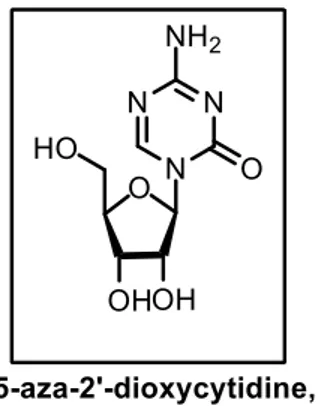
8 2’-deoxycytidine (5-Aza, 1, Figure 1), a cytosine nucleoside analogue that inhibits DNMTs, has been extensively used for the investigation of DNA methylation in many cellular functions necessary for mechanistic and translational studies.13 Specifically, due to its ability to activate
tumor suppressor genes in leukemic cells, 5-Aza (1) has been employed clinically as a first-line antileukemic agent and is considered as the first “epigenetic drug”.14, 15
Figure 1. 5Aza, a DNMT inhibitor used as a first-line antileukemic agent.
Ten-eleven translocation methylcytosine dioxygenase (TET) family proteins, catalyze the major pathway mediating DNA demethylation, wherein the methyl group of the 5-methyl cytosine (5-mC) gets oxidized to yield 5-hydroxymethyl cytosine (5-hmC).16 This on further oxidation affords 5-formylcytosine and 5-carboxylcytosine. These derivatives of 5-mC represent novel epigenetic markers with potentially new biological roles.
1.1.2. Histone modifications: histones, the core components of the nucleosome, undergo
more than 130 post-translational modifications (PTM), including methylation, acetylation, phosphorylation, sumoylation, and ubiquitination.17 Histone PTMs are extensively distributed
9 all over the human genome forming the histone code, that can control the accessibility of DNA.18 Also, it recruits transcription factors (TFs) and coactivators/suppressors to result in active, poised or silenced transcriptional states. Distinct histone modifications can result in activation or inactivation of the adjacent genes by recruitment of regulatory mechanisms such as chromatin remodeling complexes, TFs, and transcriptional coactivators/suppressors.19 Transcriptional activities are affected by histone modifications by two main mechanisms. Firstly, histone PTMs can modify the structure and conformation of chromatin. For example, H3K27 acetylation can decrease the positive charge on the histones, thereby reducing DNA binding and in turn increasing their accessibility. Secondly, histone PTMs can deliver signals to the ‘reader’ enzymes to recruit transcriptional activators/repressors. For instance, H3K27 trimethylation gets recognized by the polycomb repressive complex 1 (PRC1), that mediates ubiquitination of histone H2A, a key process required for transcriptional repression.20
For each histone PTM, three specific enzymes catalyze the “writing”, “reading” and “erasing” of the modifications. “Writers” include histone acetyltransferases (HATs), histone methyltransferases (HMTs), and protein arginine methyltransferases (PRMTs). Whereas “erasers” are histone deacetylases (HDAC) and lysine demethylase (KDM). The “reader” proteins that contain distinct domains (bromodomains, chromodomains) recognize the differentially modified histones.
1.1.3. Non-coding RNA (ncRNA): even though three-quarters of the human genome can be
transcribed, only a limited portion of the genes can be translated into proteins.21 Non-coding RNAs (ncRNAs) are the RNAs that cannot be translated into proteins. ncRNAs can be clustered based on their size, namely small ncRNA (that includes small interfering RNA (siRNA), microRNA (mRNA), piwi-interacting RNA (piRNA), transfer RNA (tRNA), and small nucleolar RNA (snRNA)), and long ncRNA (lncRNA). lncRNAs are the most crucial in the epigenetic regulations and are the least understood. They are more than 200 bases in length and
10 recruitment of proteins involved in the assembly of ribonucleoprotein complexes, that modulate histone markers by acting on chromatin.
1.2. Histone deacetylases (HDACs)
1.2.1. HDAC family
Protein acetylation balance maintained by histone acetyltransferases (HAT) and deacetylases (HDACs) play a crucial role in post-translational modifications.24 HDACs are enzymes
responsible for deacetylating lysine residues from histones and other large sets of proteins involved in various functions, from gene expression to protein activity. Some of these proteins include transcription factors, HATs, metabolic enzymes and proteins involved in cell signaling, apoptosis, DNA recombination, repair, and replication.25 HDACs perform their functions by forming large multiprotein complexes and act as a scaffold for different temporal and spatially regulated interactions.26 Deacetylation by HDACs confers a tag for epigenetic repression and plays a crucial role in cell cycle progression, transcriptional regulation and developmental events. Their involvement has also been implicated in infection and inflammation. Moreover, deregulation of HDAC has been observed in disease conditions such as cancer, autoimmunity, and neurodegeneration.27-29
In mammals, a total of 18 HDACs have been identified to date. Basing on their homology to yeast HDACs, cellular localization and enzymatic actions, HDACs are clustered into four classes. While classes I, II and IV are Zn2+ dependent, class III (also called sirtuins, SIRT1-7)
11 are NAD+ dependent. Class I contains nuclear enzymes HDAC1, 2, 3 and 8, whereas class II
is further subdivided into two classes of enzymes with nucleocytoplasmic shuttling capability, namely class IIa (HDAC4, 5, 7 and 9) and class IIb (HDAC6, 10). class IV includes only one member, HDAC11.30 (Figure 2)
Figure 2. Phylogenic tree of human HDACs established with tools available at
http://www.phylogeny.fr/. Wherein, the branch length is proportional to the number of substitutions per site, that is the number of changes or 'substitutions' divided by the length of the sequence.
Structurally, HDACs are heterogeneous in length ranging from 347 amino acid residues (shortest, HDAC11) to 1215 residues (longest, HDAC6) and contain a conserved deacetylase domain.31, 32 Their deacetylase activity on the lysine residues on histones and other proteins is due to the binding of the Zn2+ present in the active site with the N-ℇ acetylated lysine. On
activation, the N-acetyl group gets attacked by a water molecule, resulting in an N-ℇ free lysine and acetic acid. HDACs differ based on the different residues present at the entry of the active site. Specifically, class I HDACs have an additional internal cavity for water entry and acetic acid removal. However, class II HDACs are devoid of this secondary pocket, wherein the water
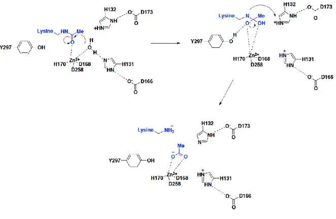
12 studies on coordination in the catalytic site and the mechanism of zinc protease chelation.33-35 The most intriguing feature in the structure of HDAC8 is the encircling of the Zn2+ by two
dipeptides (His131-Asp166 and His132-Asp173). It is linked to a tyrosine (Tyr297) and coordinated with Asp258, Asp168, and His170 for proton assistance.
In Class I enzymes, a Tyr-His pair is accessible for proton assistance, whereas in Class II enzymes histidine is present in place of tyrosine for proton assistance.36 Although the bacterial HDAC (HDAH) is classified as Class II HDACs, they share structural similarity with Class I HDACs with two potassium ions bound as in the case of HDAC8. However, HDAH has more similarities with HDAC6 and therefore thought to be a tubulin deacetylase.37 The first step of
the proposed mechanism is the intermediate chelation of the assisted water molecule and the carbonyl group of the acetyl moiety to the zinc atom. Once the Zn2+ is chelated, the protonation of His 132 occurs. His 132 deprotonates water to produce the oxide ion that attacks the carbonyl of the acetyl group. The anion formed leads to the formation of an acetate ion and a terminal ammonium ion on the side chain of lysine. At the final stage of protection and deprotection, His131 gets protonated while His132 does not.
There exists a coplanar arrangement between the His131-Asp166 pair (HDLP) with the Asp carboxylate group at 2.5 Å from the basic imidazole ring.34 Inversely, the His132-Asp173 pair
(HDLP) does not have a similar coplanar arrangement with the Asp carboxylate group at 2.8 Å from the basic imidazole ring, with a deviation of 90º being acidic. The binding of SAHA (2) to HDLP, HDAH, and HDAC8 generates the same geometry. His843 is 10 Å far from the
13 metal ion and in case of Class II HDACs the tyrosine residue for proton transfer is replaced.36, 38 The second dyad is formed by Asn712 similar to HADH (Asn185). In the case of TSA
(Trichostatin A, 3) bound to HDAC7, the two dyads display a planar relation. HDAC4 contains an Asn845 in one of the dyads.39 In the case of HDAC6 modeled with tubacin, the catalytic site represents the dyad pair (His112-Asp149 and His113-Asn156), both being distorted. All the histidine residues surrounding the central metal ion are not found, one being pushed away.
Figure 3. Mechanism of lysine deacetylation proposed by Finnin.
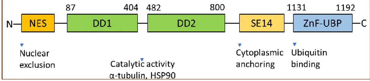
14 at the N-terminal tail (Figure 4).41, 42 These features allow clear identification of its orthologs
in other species namely, Caenorhabditis elegans, Drosophila melanogaster, and others.43 HDAC6 in class IIb distinguishes itself from the class IIa HDACs owing to the presence of the two domains DD1 and DD2. However, the exact role of the dual catalytic domain organization concerning the deacetylase activity of the enzyme is not yet established. Some studies indicate the indispensability of both the domains for tubulin deacetylase activity, whereas other investigations have ascribed all the activity to DD2 alone, which is inhibited by tubacin, a selective HDAC6 inhibitor.44, 45 To support this, experiments conducted with purified HDAC6 suggested that DD2 was solely essential for its catalytic activity for histone and α-tubulin substrates.46 Further studies led to the conclusion that the spacer region linking both domains of the protein is crucial for the complete activity of HDAC6. Also, amino acid insertion or deletion in the spacer region significantly affected tubulin deacetylase activity.41
HDAC6, in addition to the two catalytic domains, also contains peptide regions necessary for its intracellular localization. Human HDAC6 is characterized by the unique SE14-repeat domain-containing eight consecutive tetradecapeptide repeats contributing to its stable cytoplasmic retention.47, 48 Also, the accumulation of HDAC6 in the nucleus is prevented by an NES located on the N-terminus side of DD1.42 HDAC6 undergoes nucleocytoplasmic shuttling in response to certain cellular signals, and NES and SE14 are key factors in its active and stable maintenance in the cytoplasm.
15 Another unique feature of HDAC6 is the presence of a zinc finger motif, with conserved regions rich in cysteine- and histidine at the C-terminus end.40 The central part of this motif resembles the regions found in several ubiquitin-specific proteases (UBPs), therefore referred as ZnF-UBP.49 ZnF-UBP can bind specifically to mono- and poly-ubiquitin chains.49-51 This ubiquitin-binding activity of HDAC6 is critical for the transport of ubiquitinated proteins along the microtubule tracks to pericentriolar structures, known as aggresomes, that enables cells to deal with aberrant accumulations of misfolded proteins.52
Figure 4. HDAC6 structure: domain organization and functions.
1.2.4. Functions of HDAC6
1.2.4.1. Deacetylase-dependent functions of HDAC6
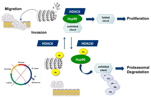
HDAC6 regulates diverse functions by deacetylating multiple targets, such as histone, tubulin, cortactin, and HSP90 (Figure 5).53, 54 By far its most studied interaction with a substrate is with α-tubulin subunit of microtubules.44, 55 α-Tubulin acetylation at lysine 40 is common in
microtubules, but it is uncertain how the acetylating enzyme gains access to this site.56, 57
Recent reports have indicated that acetylated α-tubulin plays a crucial role in motor-based trafficking in mammalian cells.58, 59 Reed et al. demonstrated that the tubulin-binding and motility of motor protein, kinesin-1, are controlled by acetylation of α-tubulin and HDAC6 inhibition induces the kinesin-1 cargo protein transport, JNK-interacting protein 1 (JIP1) transport to neurites and the aggregation of JIP1 in neuritis.59 This process highlights the fact
16 studies shed light on the tubulin deacetylase activity of HDAC6 as an important factor of microtubule-dependent intracellular trafficking.
Recently, a cytoplasmic protein involved in the structural dynamics of the actin cytoskeleton, referred to as cortactin has been identified as another non-histone HDAC6 substrate regulating the control of intracellular trafficking.61 Cortactin acts on the filamentous F-actin to increase its polymerization and branching, however, acetylation of cortactin precludes its attachment. This activity indicates the role of cortactin as a modulator of filament-based trafficking of actin, which consequently may affect autophagy.62 Intriguingly, a novel study showed that HDAC6 can also link actin filaments with microtubule dynamics by interacting with formin homology protein mDia2, thereby controlling actin polymerization.63
HSP90 was recognized as the second substrate of HDAC6 which suggested that HDAC6 could also act on cellular circuits. HDAC6-dependent HSP90 acetylation influences the binding of an essential HSP90 cochaperone, p23 and subsequently the maturation of the glucocorticoid receptor.64 HSP90 consists of multiple acetylation sites, and its acetylation activity is a key factor in the binding of cochaperones and client proteins. HDAC6 is sensitive to ubiquitinated cellular aggregates and thereby induces the expression of major cellular chaperones. This is achieved by promoting the dissociation of a repressive HDAC6-heat shock transcription factor 1 (HSF1)–HSP90 complex and consequently inducing HSF1 activation.65
17
Figure 5. HDAC6 and its role in the acetylation status of HSP90 and α-tubulin
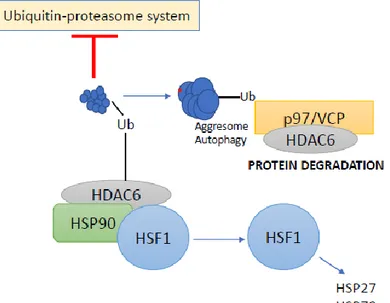
1.2.4.2. Ubiquitin-dependent functions of HDAC6
The first indications of the ubiquitin-dependent functions of HDAC6 were observed after the study of its ubiquitin-binding activity and its localization in ubiquitinated aggresomes.52 Aggresomes are inclusion bodies juxtaposed to the nucleus in the vicinity of the microtubule-organizing center and represent the termination point of microtubule-dependent transport of misfolded polyubiquitinated protein aggregates.66 Aggresomes also employ motor proteins that transport misfolded or aggregated proteins to chaperones and proteasomes for consequent destruction.67 HDAC6 plays a crucial role in aggresomes degradation. The ubiquitin-binding activity of HDAC6 mediates the transport of ubiquitinated proteins along microtubule tracks to aggresomes. HDAC6 binds to polyubiquitinated proteins and dynein proteins, thereby recruiting protein cargo to dynein motors that transport misfolded proteins to aggresomes. This microtubule-dependent intracellular trafficking potential of HDAC6 is associated with its tubulin deacetylase activity. HDAC6 binds ubiquitin with an equilibrium constant (Kd) of 60 nM, which is much higher in comparison to other ubiquitin-binding proteins (range: 5 to 500 μM).49-51, 68 Such high binding affinity enhances polyubiquitin chain stability and the inhibition
18 accumulation of polyubiquitinated proteins. Therefore, a precise equilibrium of cellular concentrations of HDAC6 and p97/VCP seems to be a critical factor that determines whether polyubiquitinated proteins are subjected to proteasomal degradation by p97/VCP or for sequestration into the aggresome by HDAC6 (Figure 6).
Figure 6. HDAC6-Ubiquitin interplay.
19
1.3.1. HDAC6 is required for oncogenic cell transformation
Anchorage-independent proliferation enables cells to survive by escaping anoikis, a special kind of programmed cell death, resulting from disengaging of the cell from the extracellular matrix and the surrounding basement membrane.69 Lee et al. in their studies suggested that HDAC6 promotes tumorigenesis and oncogenic transformation by expediting anchorage-independent proliferation in transduced cells. Furthermore, HDAC6 knockdown in MCF7 breast cancer, SKOV3 ovarian cancer, and SKBR3 breast carcinoma cell lines decreased anchorage-independent growth to 3–20%.69 Further in vivo studies were performed using
steadily expressed HDAC6-scrambled control and specific shRNA cells that were injected separately into immunocompromised and severe combined immunodeficient-Beige mice, respectively. After two weeks, the mice injected with HDAC6-shRNA showed fewer tumors than control mice.
Another interesting aspect regarding HDAC6 was observed in the inflammatory breast cancer (IBC) cells. Even though HDAC6 is not overexpressed in the IBC cells, its activity is considerably higher in IBC cells in comparison to the non-IBC cells.70 The HDAC6 inhibitor ACY1215 (ricolinostat, 4) was observed to significantly inhibit IBC cell proliferation, both in
vitro and in vivo, but was less sensitive in the case of non-IBC cells.70 This shows that in cancer cells, HDAC6 functions involve alterations in its expression and activities by controlling its cellular deacetylation.
1.3.2. HDAC6 modulates tumor development through non-histone substrates
HDAC6 participates in cell movement by acting on the non-histone substrates. Increased cell mobility results in a) microtubule depolymerization during cellular movement and b) remodeling of new adhesions at the constantly forming front of the spreading cells. This leads to an enhancement in tumor cell movement, metastasis, and invasion.45, 71 Being an
estrogen-20 through α-tubulin deacetylation and promoting CYLD and BCL3 interaction.75, 76
HDAC6 is highly expressed in malignant melanoma. In cases where HDAC6 is silenced or knocked down, acetylated α-tubulin increases, accumulation of acetylated MTs take place, and CYLD is translocated to the perinuclear region, resulting in a reduced interaction between CYLD and BCL3.77, 78 BCL3 is therefore increased in the cytoplasm, and its transfer into the nucleus is reduced. Low levels of BCL3 in the nucleus prevents the transcriptional activity of NF-κB, resulting in decreased expression of cyclin D1 and a significant interruption of the cell cycle in the G1/S phase.75 Thus, the HDAC6 mediated regulation of α-tubulin leads to enhanced cell motility and mitosis, which consequently affects proliferation, metastasis, and invasion.79, 80
In lung cancers, epidermal growth factor receptor (EGFR) and activation of its downstream pathways lead to cell proliferation.81, 82 Therefore, altering the synthesis and degradation of EGFR may affect the role of EGFR in tumors. Gao et al. demonstrated that HDAC6 expression is tightly involved in cell endocytosis and regulates EGFR trafficking and degradation via α-tubulin deacetylation.83 Loss of HDAC6 results in accumulation of acetylated α-tubulin, resulting in the dysregulation of microtubule-dependent endocytic vesicle trafficking, and hastening EGFR degradation.84-86
Mounting evidence suggests that HSP90 is essential for the stability and function of proteins involved in tumor metastasis, and HSP90 affects the growth of tumor cells by stabilization of
21 key chaperone protein levels, in particular, AKT.87 HSP90, when bound to AKT, shields AKT
from phosphates thereby maintaining AKT phosphorylation and activity. Accordingly, AKT binding of HSP90 safeguards HSP90 from proteasomal degradation.88 Also, as HSP90 influences the functional stability of AKT, it affects the PI3K/AKT signaling pathway, thereby playing a role in cell survival, migration, differentiation, and angiogenesis.88, 89 Selective inhibition of HDAC6 has been linked to elevated HSP90 acetylation, leading to a decrease HSP90 and ATP binding, thereby decreasing the combination of chaperone and oncogene, which could be beneficial for cancer treatment.90
1.3.3 HDAC6 and apoptosis
The role of HDAC6 in the regulation of apoptosis has been widely reported. The acetylation status of cytoplasmic Ku70 and its interaction with the proapoptotic Bax (Bcl-2-associated X protein) protein is regulated by HDAC6. In the acetylated state, Ku70 no longer interacts with BAX, which leads to the activation of apoptosis. In contrast, HDAC6 mediated deacetylation of the Ku70 results in BAX sequestration and consequently the inhibition of apoptosis.91 Moreover, dissociation of acetylated Ku70 from the antiapoptotic protein FLIP (FLICE [FADD-like IL-1 β-converting enzyme] inhibitory protein) results in proteasomal degradation and induction of apoptosis.92 HDAC6 involvement with PI3K (phosphoinositide 3-kinase)/AKT and mitogen-activated protein kinase (MAPK)/ERK signaling pathways have also been reported.69 HDAC6 knockdown or its catalytic inhibition promotes dephosphorylation of AKT and ERK with reduced cell proliferation and induction of cancer cell death. HDAC6 inhibition also disrupts its interaction with protein phosphatase 1 that may lead to dephosphorylation of its targets, phospho-AKT and phospho-ERK. Also, HDAC6 inhibition promotes HSP90 hyperacetylation resulting in decreased levels of phosphorylated AKT and ERK.93-95
22 been reported to regulate the expression of specific tumor-associated antigens, MHC class I proteins, co-stimulatory molecules, and cytokine production.98, 99 In the case of human melanoma cell lines, melanoma antigens TYRP1, TYRP2, gp100, and MART1 are elevated at the mRNA level on treatment with HDAC6 inhibitors. Genetic disruption of HDAC6 also leads to an increase in protein expression of gp100 and MART1.99
Interestingly, HDAC6 has been reported as an important regulator of the STAT3 pathway.100 Being a key transcriptional promoter, STAT3 is involved in various processes such as pathogenesis and sustainable development of many malignancies, induction and maintenance of tumor immune tolerance.101 Recent reports have elucidated that STAT3 modulates PD-L1 expression in antigen-presenting cells (APCs) and also in several tumor cells, including melanoma and lung cancer, to inhibit the tumor immune response.102 Woan et al. have reported that HDAC6 participates in antitumor immunity through STAT3-PD-L1 pathway.103 Increased
HDAC6 expression results in STAT3 phosphorylation and nuclear ectopia at an invariant acetylation level, without an acetylation change in PP2A, Shp-2, and JAK2 proteins, which are key factors in phospho-STAT3 homeostasis.104 On entering the nucleus, pSTAT3 and HDAC6 are recruited to PD-L1 promoter leading to the activation of PD-L1 gene transcription. However, in the case of HDAC6 knockdown, STAT3 remains undetected at the PD-L1 promoter, implying HDAC6’s requirement for STAT3-mediated PD-L1 expression.103 In the case of antigen-presenting cells (APCs), HDAC6 binds to STAT3 via the 503–840 amino acid region in HDAC6, and this complex binds to a specific sequence in the promoter region
23 of the immunosuppressive and anti-inflammatory cytokine IL-10, to enhance its gene expression.105 Reduced HDAC6 expression leads to decreased IL-10 by lowering STAT3 phosphorylation and by inducing inflammatory APCs, that efficiently activating antigen-specific naive T cells and improve the reactive ability of CD4+ T cells.100, 105
Recent preclinical trials have also shown that HDAC6 inhibitor ACY-241 (citarinostat, 5) in combination with PD-L1 antibody augments pDC-induced T- and NK cell-mediated cytolytic activities in the cells of multiple myeloma patient.106
1.4. HDAC inhibitors
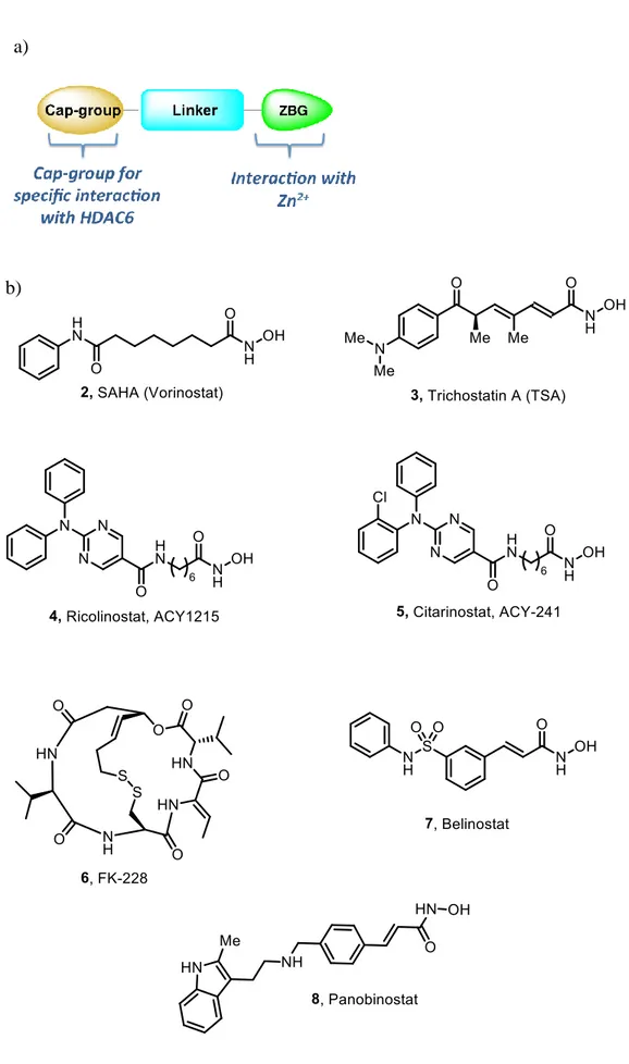
The active site of zinc-dependent HDACs consists of four distinct binding domains: a) surface binding domain, b) hydrophobic channel, c) the catalytic zinc-binding domain, and d) the adjacent internal cavity. The general pharmacophoric model of HDAC inhibitors (HDACi) (Figure 7a) encompasses three structural domains: a zinc-binding group (ZBG) interacting with the zinc ion at the catalytic pocket, a linker occupying the hydrophobic channel and a cap group. In some HDACi, an internal cavity motif is also present.107, 108 The cap group generally consists of an aromatic or heteroaromatic hydrophobic moiety which mediates the interaction with the amino acids at the surface of the enzyme, responsible for the HDAC isoforms selectivity.109, 110 On the other hand, modification of the ZBG can lead to a change in the potency of the inhibitors.111
Based on the chemical structures of the ZBGs, HDACi can be clustered into five main classes: hydroxamates, cyclic peptides, benzamides, short-chain fatty acids, ketones, and others.112, 113
24
Figure 7. a) General structure of HDACi b) Representative structures of HDACi.
25 Among these, hydroxamate containing HDACi are the most potent and are extensively investigated.114 However, most hydroxamates possess pan-inhibitory activities, whereas the
benzamides class of inhibitors are more selective towards class I enzymes. To date, four HDACi, vorinostat (SAHA, 2), romidepsin (FK-228, 6), belinostat (PXD-101, 7) and panobinostat (LBH-589, 8), (Figure 7b) have received FDA approval for the treatment of cutaneous T-cell lymphoma, T-cell lymphoma, and multiple myeloma.115 However, the use of such nonselective or partially selective HDACi result in undesirable side effects such as fatigue, nausea/vomiting, and cardiotoxicity.116, 117 Therefore, an increasing number of research efforts are being focused on the development of isotype-selective HDAC inhibitors to study the complex interactions of these proteins involved in the transcriptional regulation.25, 118, 119
1.5. Selective inhibitors of HDAC6
Recent reports have shown that when compared to the lethal effect of HDAC1-3 genetic ablation, HDAC6-knocked out mice are not affected by severe symptoms, suggesting that HDAC6 selective inhibitors may have lesser side effects than HDAC1-3 isoform-selective inhibitors or pan-HDACi.120 Selective HDAC6i have been widely investigated for a range of therapeutic purposes, such as cancer, autoimmunity, neurodegenerative diseases, infectious diseases, and rare diseases.90, 121-123 Design of selective HDAC6i has been primarily dependent
on the homology model of HDAC6, until the report of the crystal structures of HDAC6 in the previous years.124, 125 Zinc-dependent HDACi have been discovered from many different structural classes, such as hydroxamic acids, electrophilic ketones, cyclic peptides, short-chain fatty acids, and benzamides. However, they inhibit all or many subtypes of classes I, II or IV. In particular, hydroxamic acid derivatives, such as TSA and suberoylanilide hydroxamic acid (SAHA, 2) inhibit HDAC isoforms. Only a limited number of HDAC6 selective inhibitors have
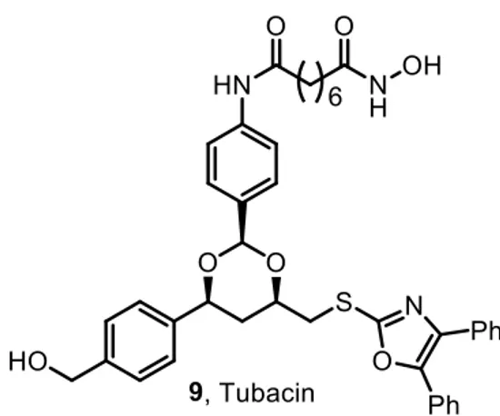
26 chemical genetic screen of 7392 small molecules.45, 126 Tubacin induces α-tubulin acetylation (EC50 = 2.9 μM) without producing a significant increase in acetylation of histones (EC50=217
μM).126 Moreover, tubacin does not cause HSP90 acetylation.127 Besides this, it does not affect
gene expression profiling, global histone deacetylation, or cell cycle progression. In terms of selectivity, tubacin exhibits 4-fold selectivity for α-tubulin over HDAC1 and HDAC4.128 Tubacin has been employed in many experiments to elucidate several functions of HDAC6, that have shed light on the many effects of tubacin on apoptosis, cancer-cell motility, and axonal vesicular transport.60, 72, 129 These findings indicate that HDAC6i could be effective as novel antitumor and neuroprotective agents. Also, synergistic cytotoxicity of the proteasome inhibitor bortezomib with tubacin was demonstrated in multiple myeloma cell lines.129
Figure 8. Chemical structure of tubacin.
1.5.2. Mercaptoacetamides
Itoh et al. and other groups have demonstrated that potent HDAC inhibition can be achieved with compounds bearing a mercaptoacetamide moiety that chelates the zinc ion in the active
27 site of HDACs.130-133 Recent functional analytical studies have confirmed that
mercaptoacetamide compounds show potent HDAC6 selectivity (EC50 = 0.2–2 μM).
Intriguingly, mercaptoacetamides also protected the cortical neurons from oxidative stress-triggered death in culture.134 These results imply that HDAC6-selective inhibition by mercaptoacetamides is neuroprotective in nature.
1.5.3. Thiolates
Itoh, Suzuki, and their co-workers generated a set of different thiol-containing derivatives based on small-molecule HDAC6-selective substrates: NCT-10a, NCT-14a, and S-isobutyryl prodrugs NCT-10b and NCT-14b.133, 135-137 NCT-10a (EC50 = 29 nM) and NCT-14a (EC50 =
82 μM) show high selectivity for HDAC6 over HDAC1 and HDAC4 in enzymatic assays.133, 135 NCT-10b and NCT-14b induced a dose-dependent increase in acetylation of α-tubulin
without causing a major increase in histone H4 acetylation, indicating that these compounds selectively inhibit HDAC6. Also, biological experiments indicate that thiolate-analogue based HDAC6-selective inhibitors are potential anticancer agents.133, 135
28
2.1.1. Tubastatin A
The potential toxicity associated with the inhibition of some HDAC isoforms discourages the use of pan-HDAC inhibitors. With regard to HDAC6 enzyme, its specific involvement in many diseases (in the areas of both oncology and neurological disorders) underscores the importance of selectively targeting this isoform with ad-hoc developed inhibitors (Figure 9).
In this scenario, tubastatin A (TubA, 10) represents an ideal reference molecule owing to its potency and selectivity (HDAC6 IC50 =15 nM, HDAC1 IC50 = 16 μM), and to its chemical
tractability and derivatization potential.
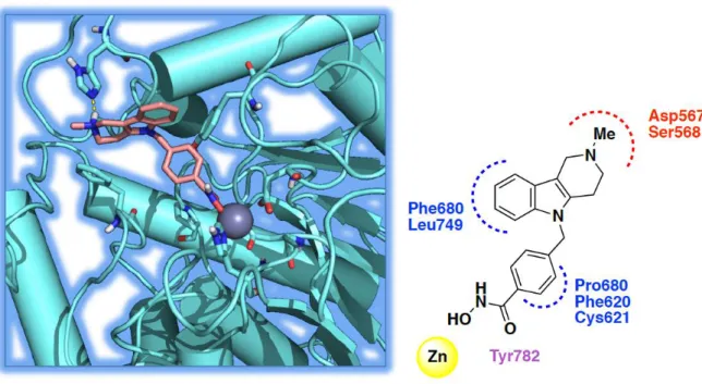
The selectivity of 10 was studied by comparison of α-tubulin acetylation (cytosolic localization) and histone acetylation (nuclear localization). At 2.5 μM, 10 preferentially induced tubulin acetylation with a strong in vitro preference for HDAC6 rather than HDAC1. Moreover, 10 was examined on an oxidative stress model induced by homocysteic acid (HCA) demonstrating dose-dependent protection against HCA-induced stress. Kozikowski et al. commenced their rational design of selective HDAC6 inhibitors by comparing the structures of HDAC6 and HDAC1 since these two enzymes display diverse phylogeny and belong to two distinct classes (I for HDAC1, IIb for HDAC6). The bioinformatic analysis of the two catalytic pockets showed that the catalytic channel rim dimension is different in the two enzymes. In the case of HDAC6, the channel appears wider and shallower, therefore a larger cap-group can fit better into the rim region.
29 Structurally, 10 contains a) tricyclic ring system bearing a tertiary amine, that further enlarges the dimensions of the cap group favoring the interactions with HDAC6, b) tolyl linker and hydroxamate as ZBG. Notably, the replacement of the traditional alkyl chain in the linker (as in tubacin and SAHA) with bulkier and shorter aromatic moieties led to a clear improvement of selectivity.
HDAC6 channel is mainly lined by apolar residues that explain its ability to accommodate the tolyl linker. The carbazole moiety establishes a π-π stacking with the aromatic residues surrounding the rim cavity (His500, His611, Phe620, and Phe680). Similarly, the indole moiety retains the aromatic contacts with His611 and Phe680 and elicits additional polar interactions between the ammonium head, and Asp567 and Ser568.125
Figure 9. The docked pose of tubastatin A, 10 into HDAC6 and its interactions with the
residues in the active site (polar interaction are represented in red, aromatic interaction in blue and hydrogen-bond in violet).
30 hydroxamate is the possibility of indiscrimination among the different classes of metalloenzymes and vulnerability to cross-reactions. However, a recent study conducted by Day and Cohen demonstrated that metal-binding groups for Zn2+ ions such as hydroxamic acids and thiols will not indiscriminately inhibit Zn2+ metalloenzymes in the absence of favorable
inhibitor backbone-protein interactions.138
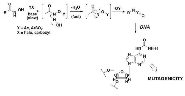
Other issues often related to the use of hydroxamates are toxicity and pharmacokinetic liabilities. In this regard, a recent perspective by Shen and Kozikowski has shed light on the potential mutagenicity of the hydroxamate group. The mutagenic properties of hydroxamates are associated with their ability to undergo the Lossen rearrangement, a reaction which converts an activated hydroxamate into the corresponding isocyanate (Figure 10). Isocyanates can readily react with the nucleophilic groups of the DNA and cause subsequent DNA damage.139 The rate-limiting step of the Lossen rearrangement is the formation of an activated hydroxamate since the rate of reaction is directly proportional to the acidity of the conjugate acid of the leaving group.140 Under physiological conditions, the activation of the hydroxamic
group is a key step for the rearrangement that takes place by O-acetylation due to the presence of acetyl-CoA present in bacteria. Most rearrangements take place in a basic medium; however, the low acidity of hydroxamates (pKa ≈ 8.5) does not allow deprotonation at the physiological pH.141
31
Figure 10. Mechanism of mutagenicity by HDACi
Soon, a metal-assisted mechanism was then proposed, described in Figure 11 wherein a zinc-triggered rearrangement takes place involving two hydroxamic groups that bind zinc as neutral or anionic ligands. Thereafter, hydrogen migration from nitrogen to oxygen leads to the formation of an unsaturated complex followed by its rearrangement to form the more stable isocyanate.142
Figure 11. Lossen rearrangement: metal-assisted mechanism.
However, driven by this evidence and the importance of hydroxamate for its ability to chelate zinc as well as its isoform selectivity, the rational design of less activated hydroxamates can be beneficial to synthesize compounds that do not display mutagenicity potential. This is supported by many reported HDAC inhibitors bearing hydroxamate that do not exert mutagenic
32
Figure 12. Chemical structures of Givinostat and Abexinostat.
2.2. Aim of the thesis
Aim of the thesis can be divided into four main objectives:
a) Rational design, synthesis, structure-activity relationships (SAR) of novel HDAC6 inhibitors as anticancer agents by introducing new cap groups with structural modifications aimed at increasing the potency and selectivity towards HDAC6 isoform. This would be achieved by linking the cap group with suitable aromatic linkers followed by the attachment of hydroxamate ZBG.
b) Characterization of the newly synthesized compounds utilizing 1H and 13C NMR spectroscopy, LC-MS, and also X-ray crystallography (performed by Prof. David Christianson at the University of Pennsylvania).
c) Biological evaluation of the newly developed HDAC6 inhibitors using binding affinity studies on HDAC1, HDAC6, HDAC8 enzymes and cell-based assays on different cancer cell lines (performed in collaboration with Prof. Vincent Kelly, Dr. Jeff O’sullivan and Prof. Daniela Zisterer at Trinity College Dublin, Dr. Richard Turkington
33 at Queens University Belfast, Prof. Manfred Jung at University of Freiburg and Dr. Giovina Ruberti at the Institute of Biochemistry and Cell Biology, IBBC).
d) Assessment of toxicity by determining cytotoxicity and mutagenicity (performed by Dr. Stefania Lamponi at the University of Siena).
2.2.1. Spiroindoline-based HDAC6 inhibitors
To develop novel HDAC6i, our group recently identified a highly potent and selective spiroindoline-capped HDACi (13, Table 1) with promising anticancer activity against several cancer cell lines.145 This previous work permitted us to narrow down the spiroindoline scaffold as an optimal cap group for developing selective HDAC6 inhibitors. In this work, we examined the effect of a strategical overturning of the linker and the ZBG moieties from the indoline nitrogen (as in compound 13) to the piperidine nitrogen (compounds 14-23). The prototype of this novel series of compounds (14, Table 1) exhibited a promising HDAC6 inhibition potency with an IC50 value of 264.4 nM and a selectivity index of 85 over HDAC1 and 7 over hHDAC8.
To get a deeper insight into the binding mode of this compound, a 2.09 Å-resolution X-ray crystal structure was determined of the complex between 14 and the catalytic domain of HDAC6 from Danio rerio (zebrafish). The active site features of zebrafish HDAC6 is identical to that of the human HDAC6 isoform, and zebrafish HDAC6 yields crystals of higher quality compared to the human HDAC6 crystals.146 Subsequently, molecular modeling approaches
were utilized to analyze the binding mode and the structural requirements to design novel “reversed” spiroindolines with an increased HDAC6 inhibitory profile and greater selectivity index. This was realized following two key approaches: i) synthesizing derivatives with bulkier cap-groups, and ii) controlling the outdistancing between the cap-group and the ZBG by inserting amide, urea and carbamate functionalities to the linker portion. The resulting compounds (15-23) were then tested for their ability to inhibit the HDAC1, 6 and 8 isoforms.
34 Cmpd R1 R2 X Ar 14 H -CH3 -CH2- 15 H -CH2C6H5 -CH2- 16 H -COC6H5 -CH2- 17 C6H5 -CH3 -CH2- 18 C6H5 -CH2C6H5 -CH2- 19 C6H5 -COC6H5 -CH2- 20 H -CH3 -CH2- 21 H -CH2C6H5 -COCH2- 22 H -CH2C6H5 -CONHCH2- 23 H -CH2C6H5 -COOCH2-
2.2.2. Quinolone based HDAC6 inhibitors
Numerous natural products have been identified as potent anticancer agents, that have inspired chemists to utilize them as building blocks in the synthesis of novel chemotherapeutics.147 In this context, functionalized 4-arylquinolin-2(1H)-ones represent a versatile scaffold for developing biologically active molecules, that includes an orally active anticancer agent
35 currently under clinical investigation.148 Based on a comprehensive literature search, we
identified the quinolone moiety as a potential cap group due to lipophilic nature in vitro. This system fits perfectly in the cap region of the HDACi pharmacophore.149
Several quinolone containing compounds are well known as antibiotics (e.g. ciprofloxacin, norfloxacin) and anticancer agents (voreloxin).150 Quinolone-based anticancer agents exhibit their activity influencing the DNA intercalation process, inhibiting topoisomerase II activity and tubulin polymerization.151 In our quest to identify novel cap groups to design new potent and selective HDAC6i, we exploited the potential of a naturally occurring compound namely, viridicatin (3-hydroxy-4-phenylquinolin-2(1H)-one), containing the quinolone skeleton as an effective and versatile cap-group suitable for a wide variety of scaffold decorations.152, 153 Inspired from a multi-component protocol to construct the 3-hydroxy-4-arylquinolin-2(1H)-ones, we exploited this method to synthesize a varied set of cap groups.154 With this methodology, we were able to develop novel HDAC6i bearing N- or O-appended linker moieties (Table 2) and an array of focused modifications at both their heterocyclic core and the aromatic portions. Consequently, we aimed to obtain compounds with high selectivity index over HDAC6 and effectively screen them over different cancer cell lines.
36 Cmpd R X 24 H CH 25 Me CH 26 CH 27 H N 28 H CH 29 Me CH 30 CH 31 CH 32 CH 33 CH 34 CH 35 Me N 36 N 37 N 38 N 39 - -
37
Chapter 3
Chemistry
3.1. Spiroindoline based HDAC6 inhibitors
For the synthesis of compounds 14-23, four key-steps were employed to obtain the desired products which include: i) a Fischer indole synthesis, starting from suitable arylhydrazines and
N-Boc-piperidine-4-carboxaldehyde providing 3,3-disubstituted indolenines, ii) reduction of
the imine bond of the indolenines to get the respective indolines, iii) appropriate substitution at the N-1 position of the indoline, and, iv) insertion at the piperidine nitrogen with suitable linkers.
3.1.1. Synthesis of compounds 14-19
Synthesis of compounds 14-19 is described in Scheme 1. Commercially available 1-Boc-4-piperidone (40) was converted to its homologated enol ether derivative by Wittig reaction using (methoxymethyl)triphenylphosphonium chloride in the presence of NaHMDS as the base.21 The corresponding enol ether was hydrolyzed with cerium(III)chloride in MeCN to obtain 41 in high yield. Compound 41 and the suitable phenyl hydrazine were heated in AcOH at 80 ºC to obtain the corresponding spiroindolenines, by applying a Fischer indole synthetic protocol.155, 156 These intermediates were then reduced by catalytic hydrogenation to provide thet spiroindolines 42a and 42b. A reductive amination of these latter with the suitable aldehydes in the presence of NaBH3CN or treatment with benzoyl chloride in the presence of
TEA afforded the N-substituted derivatives, which, upon Boc-deprotection, provided amines
43a-f. A second reductive amination protocol, performed on the piperidine using methyl
4-formyl benzoate followed by treatment of the resulting ester derivatives with NH2OH and
38
Reagents and conditions: a) Ph3P(Cl)CH2OMe, NaHMDS, THF, 0 to 25 ºC, 30 min; b)
CeCl3·7H2O, NaI, 40 ºC, MeCN, 16 h; c) phenylhydrazine or
(1,1-biphenyl)-2-ylhydrazine·HCl , AcOH, 80 ºC, 2 h; d) H2, Pd/C, MeOH, 25 ºC, 3 h; e) paraformaldehyde or
benzaldehyde, NaBH3CN, MeOH, 25 ºC, 8 h; f) benzoyl chloride, TEA, DCM, 25 ºC, 1 h; g)
1 N HCl in MeOH, 25 ºC, 15 min; h) methyl 4-formyl benzoate, NaBH3CN, MeOH, 25 ºC, 8
h; i) NH2OH (50% wt in H2O), 4 M KOH in MeOH, DCM/MeOH, 25 ºC, 2 h
3.1.2. Synthesis of compound 20
Synthesis of compound 20 is described in Scheme 2. Aldehyde 45 was obtained from the bromo derivative 44 upon reaction with 4-methylmorpholine N-oxide (NMO) in MeCN. The successive reductive amination reaction involving 43a and 45 afforded the methyl ester intermediate, which was converted to the hydroxamate 20.
39
Scheme 2
Reagents and conditions: a) NMO, MeCN, 25 ºC, 12 h; b) 43a, NaBH3CN, MeOH, 25 ºC, 8
h; c) NH2OH (50% in H2O), KOH, DCM, MeOH, 25 ºC, 2 h.
3.1.3. Synthesis of the compounds 21-23
Synthesis of the compounds 21-23 is described in Scheme 3. 2-(4-(methoxycarbonyl)phenyl)acetic acid was coupled with amine 43b and the resulting methyl ester intermediate was converted to the corresponding hydroxamic acid 21. To synthesize the urea 22, the key intermediate was methyl 4-(isocyanatomethyl)benzoate which was reacted with 43b to obtain the methyl ester intermediate subsequently converted to the hydroxamic acid 22. Finally, the carbamate 23 was prepared by reacting amine 43b with the commercially available methyl 4-(hydroxymethyl)benzoate in the presence of CDI in DCM to afford the methyl ester intermediate needed for subsequent conversion into the corresponding hydroxamic acid 23.
40
Reagents and conditions: (a) 2-(4-(methoxycarbonyl)phenyl)acetic acid, EDCI, HOBt,
DIPEA, DCM, 0º to 25 ºC, 24 h; b) NH2OH (50% in H2O), KOH, DCM, MeOH, 25 ºC, 2 h; c)
methyl (isocyanatomethyl)benzoate, TEA, dry THF, 45 ºC, 2 h; d) methyl 4-(hydroxymethyl)benzoate, CDI, DCM, 0º to 25 ºC, 6 h.
3.2. Quinolone based HDAC6 inhibitors
In order to achieve the synthesis of the quinolone core of the compounds 24-39, the ring expansion procedure described by Tangella et al was applied.154 It is a multicomponent reaction involving: an aldehyde, activated with p-toluenesulfonyl hydrazide (PTSH), and a N-substituted isatin in the presence of K2CO3 as base.
3.2.1. Synthesis of compounds 24-27
In Scheme 4 the synthesis of the compounds 24-27 is described. Isatin (46) was alkylated with methyl 4-(bromomethyl)benzoate and the resulting product 47 was subjected to the ring expansion procedure with benzaldehyde or 3-pyridinecarboxaldehyde obtaining compounds
48a,b, respectively. As expected, a transesterification with EtOH occurred in this step.
Intermediates 48a and 48b were converted to their corresponding hydroxamic acid derivatives
24, 27 after treatment with a strong excess of NH2OH in the presence of KOH. Intermediate
48a was also submitted to O-alkylation with MeI or benzyl bromide providing derivatives 49a,b, respectively. These latter were converted into final compounds 25, 26 upon reaction
41
Scheme 4
Reagents and conditions: (a) methyl 4-(bromomethyl)benzoate, NaH, DMF, 0 to 25 °C, 12
h; (b) PTSH, benzaldehyde or 3-pyridinecarboxaldehyde, K2CO3, EtOH, 80 °C, 12 h; (c)
NH2OH, KOH, DCM, MeOH, H2O, 25 °C, 3 h; (d) MeI, NaH, THF, 0 to 25 °C, 12 h; or BnBr,
KI, K2CO3, DMF, 80 °C, 12 h.
3.2.2. Synthesis of compounds 28-30, 35 and 39
In Scheme 5 is reported the synthesis of compounds 28-30, 35 and 39. The ring expansion performed on isatin (46) in the presence of benzaldehyde or 3-pyridinecarboxaldehyde provided quinolone derivatives 50a,b, respectively. These latter were subjected to alkylation reaction with methyl 4-(bromomethyl)benzoate in the presence of K2CO3 as the base to furnish
the O-alkylated intermediates 51a,b. Treatment of 51a with NH2OH in the presence of KOH
led to compound 28. Alternatively, alkylation of the lactam nitrogen of 51a,b with MeI or propargyl bromide followed by reaction with NH2OH, led to the N-alkyl derivatives 29, 30,
and 35. For the synthesis of compound 39, isatin was alkylated with MeI and successively subjected to the ring expansion reaction with cyclohexanecarboxaldehyde obtaining compound
42
Reagents and conditions: (a) PTSH, benzaldehyde or 3-pyridinecarboxaldehyde, K2CO3,
EtOH, 80 °C, 12 h; (b) Methyl 4-(bromomethyl)benzoate, K2CO3, KI, DMF, 80 °C, 12 h; (c)
NH2OH, KOH, DCM, MeOH, H2O, 25 °C, 3 h; (d) MeI/ propargyl bromide, NaH, THF, 0 to
25 °C, 12 h.
3.2.3. Synthesis of bromo-derivatives 56 and 59.
In Scheme 6 is reported the synthesis of bromides 56 and 59 used as alkylating agents for the synthesis of final compounds 32, 34 and 38. 4-Pyridinecarboxaldehyde (54) was reduced to alcohol 55 with NaBH4 and then converted to the corresponding bromide 56 after treatment
with PBr3. Instead, for the synthesis of the intermediate 59, 4-(bromomethyl)benzonitrile (57)
was reduced with DIBAL to the corresponding benzaldehyde 58, that was successively protected to its dioxolane derivative 59 with ethylene glycol in the presence of PTSA.
43
Scheme 6
Reagents and conditions: (a) NaBH4, MeOH, 0 to 25 to 25 °C, 2 h; (b) 48% HBr, 100 °C, 4
h then PBr3, DCM, 45 °C, 4 h; (c) DIBAL, DCM, -78 to 0 °C, 1 h; (d) ethylene glycol, PTSA,
toluene, 110 °C, 12 h.
3.2.4. Synthesis of compounds 31, 32, 36, and 37
In Scheme 7 the synthesis of final compounds 31, 32, 36, and 37 is described. Isatin (46) was alkylated with benzyl bromide or bromide 56 using NaH as the base to generate intermediates
60a,b. These compounds were subjected to the previously described ring expansion procedure
with benzaldehyde or 3-pyridinecarboxaldehyde to provide the desired quinolones 61a-d that were converted to the final compounds 31, 32, 36, and 37 in the presence of NH2OH as
44
Reagents and conditions: (a) BnBr or 56, NaH, DMF, 0 to 25 °C, 12 h; (b) PTSH,
benzaldehyde or 3-pyridinecarboxaldehyde, K2CO3, EtOH, 80 °C, 12 h; (c) methyl
4-(bromomethyl)benzoate, K2CO3, KI, DMF, 80 °C, 12 h; (d) NH2OH, KOH, DCM, MeOH,
H2O, 25 °C, 3 h.
3.2.5. Synthesis of compound 33
The synthesis of compound 33 is described in Scheme 8. Since alkylation of the isatin with 1-benzyl-4-bromo or iodopiperidine failed in all the attempted conditions, N-alkylated isatin 65 was prepared starting from piperidone (62) which was first N-benzylated and then subjected to a reductive amination protocol with aniline in presence of NaBH(OAc)3 providing intermediate
64. This compound was treated with oxalyl chloride, obtaining an unstable intermediate that
was immediately treated with AlCl3 in DCM to afford isatin derivative 65 under Friedel-Craft
conditions. Ring expansion, alkylation and final reaction with NH2OH were then performed as
45
Scheme 8
Reagents and conditions: (a) benzyl bromide, K2CO3, DMF, 80 °C, 12 h; (b) aniline,
NaBH(OAc)3, DCM, AcOH, 0 to 25 °C, 12 h; (c) oxalylchloride, DCM, 25 °C, 2 h; then AlCl3,
DCM, 40 °C, 2 h; (d) PTSH, benzaldehyde, K2CO3, EtOH, 80 °C, 12 h; (e) methyl
4-(bromomethyl)benzoate, K2CO3, KI, DMF, 80 °C, 12 h; (f) NH2OH, KOH, DCM, MeOH,
H2O, 25 °C, 3 h.
3.2.6. Synthesis of compounds 34 and 38
In Scheme 9 the synthesis of the compounds 34, 38 is described. Isatin was alkylated with bromide 59 affording intermediate 66. This compound was subjected to the previously described ring expansion procedure to get the desired quinolones 67a,b. These intermediates were alkylated with methyl 4-(bromomethyl)benzoate obtaining 68a,b which were subsequently deprotected in acidic medium, and the corresponding free aldehydes were subjected to a reductive amination protocol with diethylamine in the presence NaBH(OAc)3,
affording the corresponding diethylamino derivatives, that were converted to the final compounds 34, 38 as previously described.
46
Reagents and conditions: (a) 59, NaH, DMF, 0 to 25 °C, 12 h; (b) PTSH, benzaldehyde or
3-pyridinecarboxaldehyde, K2CO3, EtOH, 80 °C, 12 h; (c) methyl 4-(bromomethyl)benzoate,
K2CO3, KI, DMF, 80 °C, 12 h; (d) 6 N HCl, THF, 25 °C, 1 h; (e) diethylamine, NaBH(OAC)3,
47
Chapter 4
Results and discussion
4.1. Spiroindoline series (compounds 14-23)
Table 3. Inhibitory activity of compounds 14-23 and reference compounds (tubastatin A)
against hHDAC1, as IC50 (µM), and hHDAC6, as IC50 (nM).
Cpd R1 R2 X Ar HDAC1 IC50 (µM)a or % of inhibition at 1 µM HDAC6 IC50 (nM)a or % of inhibition at 1 µM HDAC1/HDAC6 14 H -CH3 -CH2- 22.4 ± 6 264.4 ± 45 85 15 H -CH2C6H5 -CH2- 6.5 ± 0.8 561.0 ± 203 12 16 H -COC6H5 -CH2- 10.1 ± 2.1 155.0 ± 26 65 17 C6H5 -CH3 -CH2- 8.5% 50.0% n.d. 18 C6H5 -CH2C6H5 -CH2- 2.9% 29.7% n.d. 19 C6H5 -COC6H5 -CH2- 4.7 ± 0.5 465.0 ± 122 10 20 H -CH3 -CH2- 6.6 % 51.3% n.d. 21 H -CH2C6H5 -COCH2- 10.2 ± 1 227.0 ± 97 45 22 H -CH2C6H5 -CONHCH2- 3.6 ± 0.3 110.0 ± 19 33 23 H -CH2C6H5 -COOCH2- 6.8 ± 0.3 48.5 ± 23 140 Tubastatin A - - - - 1.91 ± 0.42 30.4 ± 2.1 63
aAll compounds were assayed at least two times, and the results are expressed with standard
48 interactions with catalytically important residues accounts for the generally high affinity of hydroxamate-based inhibitors in the HDAC6 active site.
The para-substituted phenyl linker makes favorable offset - interactions in the aromatic crevice defined by F583 and F643. The piperidine ring adopts a chair conformation and the piperidine nitrogen forms a hydrogen bond with a water molecule that in turn hydrogen bonds with the backbone carbonyl of R798. The spiroindoline capping group is oriented toward the L2 pocket at the mouth of the active site. There, the indoline nitrogen hydrogen bonds with a water molecule that in turn hydrogen bonds with N645 and a second water molecule; a third water molecule completes a hydrogen bond network between the indoline nitrogen and Zn2+
ligand H614. Although the inhibitor makes no direct enzyme-inhibitor hydrogen bonds apart from those made with the hydroxamate moiety, it is interesting that three water molecules comprise a “wet” hydrogen bonded interface in such a high-affinity enzyme-inhibitor pair. It is relatively rare to see inhibitor capping groups bind in the L2 pocket, since most tend to bind in the L1 pocket on the opposite side of the active site. 124, 157-160 It appears that the chair
conformation of the piperidine ring combined with the molecular structure of the novel spiro-fused indoline moiety yields a structure and a conformation that is ideal for binding within the L2 pocket.
49
Figure 13. Stereoview of a Polder omit map of the HDAC6-14 complex for which the atomic
coordinates of 14 were omitted from the structure factor calculation (PDB 6V7A; contoured at 5.0 σ). Atoms are color-coded as follows: C = light blue (HDAC6 catalytic domain 2), light gray (symmetry mate), or wheat (inhibitor), N = blue, O = red, Zn2+ = gray sphere, and solvent
= small red spheres. Metal coordination and hydrogen bond interactions are indicated by solid and dashed black lines, respectively.
The in vitro inhibitory profile of the newly developed compounds 14-23 (Table 4) was evaluated against both hHDAC1 and 6 isoforms. SAR studies were performed by taking into consideration the data obtained from in vitro, X-ray and computational studies. These calculations outlined the preferential binding of the “reversed” spiroindoline compounds towards HDAC6. To get a better understanding of the behavior of the compounds in the binding sites of hHDAC1 and 6 we performed docking studies based on a previously reported protocol.152, 153 It was observed that the hindrance imposed by a bulkier cap group allowed the
compound to be better accommodated into the HDAC6 enzyme with respect to the HDAC1 isoform. Accordingly, we herein report the docking outputs of 14 (Figure 14) and 23 (Figure
15), two of the most selective derivatives of the series with respective selectivities for HDAC
50
Figure 14. Docked poses of 14 into HDAC1 (panel A) and HDAC6 (panel B). Compound 14
is represented by purple sticks while the residues in the active sites are represented by lines and the protein is represented in cartoon form. Zn2+ is represented by a gray sphere. H-bonds are shown as by black dotted lines, while the red solid lines represent the metal coordination bonds.
Based on these studies, limited contacts were established by 14 within the HDAC1 binding site (Figure 14A) compared to the contacts established within the HDAC6 binding site (Figure
14B). In fact, 14 was able to coordinate with the metal ion in HDAC1 by its hydroxamic moiety
through polar contacts with the backbone of G149 and the sidechain of Y303. In addition, we observed only a π-π stacking with H141 and some hydrophobic interactions with Y204, F205 and L271. On the contrary, the docking output of 14 into HDAC6 showed an improvement of the number of the contacts. In this accommodation, the hydroxamic acid moiety in addition to the metal coordination bond with the Zn2+ ion, established a series of supplementary H-bonds
51 with the sidechain of Y782 and H610 and with the backbone of G619. Moreover, the benzyl linker was able to establish a double π-π stacking with F620 and H651. We also noted relevant hydrophobic interactions with F679, F680, M682 and L749. This pattern of interaction perfectly supported the selectivity of 14 towards HDAC6 over HDAC1 (IC50 HDAC1 = 22.4
µM; IC50 HDAC6 = 264.4 nM).
Compounds 15-20 were mainly synthesized in order to investigate the role of the cap-group and its bulkiness on HDAC potency and selectivity, while maintaining unvaried the benzyl moiety of the linker.
Compound 15, bearing a pendant benzyl group on the indoline nitrogen of the cap portion, was found to be largely solvent exposed owing to its bulkiness thereby reducing the ligand efficiency. In particular, it showed a π-π stacking with F150 through its benzyl moiety. 15 showed a decreased number of the contacts with HDAC6 with the loss of the key π-π stacking with F680 (IC50 HDAC1 = 6.5 µM; IC50 HDAC6 = 561 nM). The N-benzoyl functionality led
to a similar pattern of interaction as for 15 with the exclusion of a π-π stacking with F150. As regards HDAC6, 16 was able to restore the π-π stacking with F680. Moreover, the carbonyl group belonging to the benzoyl functionality established an H-bond with the sidechain of F680 (IC50 HDAC1 = 10.1 µM; IC50 HDAC6 = 155 nM). In compound 17, the bulk was increased
by introducing a phenyl ring on the indoline aromatic moiety, while inserting a methyl group on the indoline nitrogen. In this case, we observed a reduction in hydrophobic contacts as well as a loss of key H-bond interaction, namely with the sidechain of Y303 in HDAC1 and the sidechain of H610 in HDAC6. This pattern accounted for a weak inhibition towards both enzymes with respect to compound 14 with a percentage of inhibition of 8.5% for HDAC1 and of 50% for HDAC6 when tested at 1 µM. The introduction of a N-benzyl moiety as in compound 18 resulted in the least potent compound of the series. In full agreement with in vitro data, the cap group was found completely solvent exposed for both enzymes. Notably, 19,