Synthesis and biological evaluation
Roberto Romeo*1,§, Caterina Carnovale1, Salvatore V. Giofrè*1,¶, Maria A. Chiacchio2,
Adriana Garozzo3, Emanuele Amata2, Giovanni Romeo1 and Ugo Chiacchio2
Full Research Paper
Open AccessAddress:
1Dipartimento Scienze del Farmaco e dei Prodotti per la Salute, University of Messina, Via S.S. Annunziata, 98168 Messina, Italy, 2Dipartimento di Scienze del Farmaco, University of Catania, Via A. Doria 6, 95125-Catania, Italy and 3Dipartimento di Scienze Bio-Mediche, University of Catania,Via Androne 81, 95124 Catania, Italy
Email:
Roberto Romeo* [email protected]; Salvatore V. Giofrè* [email protected]
* Corresponding author
§ Tel.: +39-090-356230; fax: +39-090-6766474 ¶ Tel.: +39-090-6766566; fax: +39-090-6766474
Keywords:
antitumor activity; click chemistry; 1,3-dipolar cycloaddition; nucleic acids; 2’-oxa-3’-aza-4’a-carbanucleoside analogs
Beilstein J. Org. Chem. 2015, 11, 328–334.
doi:10.3762/bjoc.11.38
Received: 04 December 2014 Accepted: 20 February 2015 Published: 09 March 2015
This article is part of the Thematic Series "Nucleic acid chemistry".
Guest Editor: H.-A. Wagenknecht
© 2015 Romeo et al; licensee Beilstein-Institut. License and terms: see end of document.
Abstract
A novel series of 2’-oxa-3’-aza-4’a-carbanucleosides, featured with a triazole linker at the 5’-position, has been developed by exploiting a click chemistry reaction of 5’-azido-2’-oxa-3’-aza-4’a-carbanucleosides with substituted alkynes. Biological tests indi-cate an antitumor activity for the synthesized compounds: most of them inhibit cell proliferation of Vero, BS-C-1, HEp-2, MDCK, and HFF cells with a CC50 in the range of 5.0–40 μM. The synthesized compounds do not show any antiviral activity.
Introduction
Synthetic modified nucleosides are of great interest as potential new lead structures in particular as antiviral or anticancer agents [1-8]. As analogues these compounds can interfere in nucleic acid synthesis or block nucleosides- and/or nucleotide-depen-dent biological processes by mimicking natural nucleosides and serving as inhibitors or building units [9-12]. Many structural variations of the natural nucleosides have been exploited. In general, the performed modifications included the replacement of the furanose moiety by other carbon or heterocyclic systems
[13,14] or even acyclic fragments [15,16], the substitution of pyrimidine or purine natural nucleobases with unnaturally-substituted heteroaromatics or homoaromatic systems, or the modification of the phosphate P(O)–O–C bond with the non–hydrolyzable phosphonate P(O)–C linkage [17,18]. In this context, nucleoside analogues, where different carbon or heterocyclic systems replace the furanose ring, have been reported as anticancer or antiviral agents [19,20]. In particular,
2’-oxa-3’-aza-4’a-carbanucleosides 1–4, characterized by the presence of an isoxazolidine ring, represent a scaffold of modi-fied dideoxynucleosides endowed with interesting physiolog-ical features (Figure 1) [21-27].
Figure 1: 2’-Oxa-3’-aza-modified nucleosides and
2’-oxa-3’-aza-modi-fied nucleotides.
2’-Oxa-3’-aza-4’a-carbanucleosides 1–4 can be considered as mimics of natural nucleosides and act as terminators of the viral DNA chain. Their antiviral activity is linked to the competitive reversible inhibition of the reverse transcriptase. Furthermore, as antimetabolites, they can interact with intracellular targets to induce cytotoxicity [28-32].
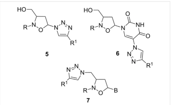
Several functionalities have been inserted as linkers on the 2’-oxa-3’-aza-4’a-carbanucleoside skeleton in order to confer novel mechanisms of action for nucleoside mimics: in this context, the 1,2,3-triazole unit assumes particular interest according to its easily access and the well-known biological activity of many derivatives. In these last years, in fact, tria-zoles have gained considerable attention in medicinal chem-istry, bioconjugation, drug-delivery, and materials science [33-38]. Moreover, the 1,2,3-triazole motif is exceedingly stable to basic or acidic hydrolysis and interacts strongly with biological targets through hydrogen bonding to nitrogen atoms as well as through dipole–dipole and π-stacking interactions [39]. Recently, a synthetic approach towards 3-hydroxymethyl-5-(1H-1,2,3-triazol)-isoxazolidines 5 has been described [40]: the obtained compounds inhibit the growth of anaplastic and follic-ular human thyroid cancer cell lines, with IC50 values in the range of 3.87–8.76 μM. In the same context, novel 1,2,3-tria-zole-appended 2’-oxa-3’-azanucleoside analogs 6 were devel-oped [41]: Some of these compounds show a good anticancer activity against the anaplastic (8305C) and the follicular (FTC-133) human thyroid cancer cell lines, and especially on the U87MG human primary glioblastoma cell line (Figure 2).
Figure 2: Triazolyl-2’-oxa-3’-aza-4’a-carbanucleosides.
Accordingly, considering that the incorporation of the triazole moiety can lead to interesting biological properties, we report in this paper the preparation of a small library of nucleoside analogues 7 (Figure 2), where the furanose ring is substituted by an isoxazolidine system and a triazole unit replaces the phos-phodiester linker at 5’ position of the 2’-oxa-3’-aza-4’a-carbanucleoside. However, in order to maintain the six-bond periodicity of the oligonucleotides and thus the flexibility of the oligonucleotide chain the methylene bridge at the pseudo-5’-position was retained. The obtained compounds have shown to be endowed with an interesting antitumor activity: most of them inhibit cell proliferation of Vero, BS-C-1, HEp-2, MDCK, and HFF cells by 50% (CC50) at concentrations in the range of 5.0–40.0 μM. No antiviral activity against both RNA and DNA viruses was observed.
Results and Discussion
Chemistry
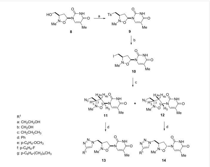
The synthetic route to 5’-triazolyl-2’-oxa-3’-aza-4’a-carbanu-cleosides 13 and 14 is described in Scheme 1 (and Table 1). (3′RS,5′SR)-2′-N-methyl-3′-hydroxymethyl-1′,2′-isoxazolidin-5′-ylthymine 8, obtained as the main compound, in a two-step process, by 1,3-dipolar cycloaddition of vinyl acetate to
C-[(tert-butyldiphenylsilyl)oxy]-N-methylnitrone, followed by
Hilbert–Jones nucleosidation using silylated thymine and TBAF [42-44], was converted into the corresponding iodo-derivative
10 by sequential tosylation and iodination.
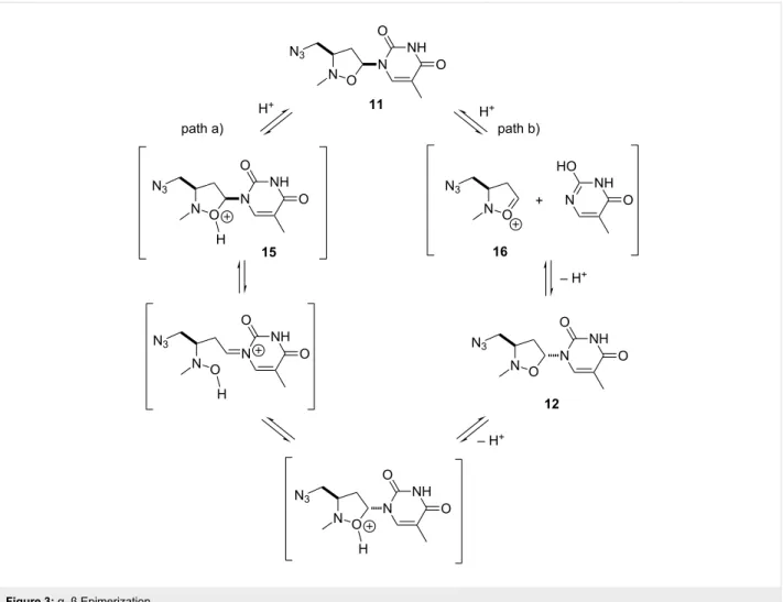
The subsequent reaction of 10 with sodium azide, performed at 50 °C in CH3CN/H2O (1:10) in the presence of NH4Cl for 48 h afforded two azides, 11 and 12, epimeric at C-5, in a relative ratio 2:1 with a global yield of 85%. Two azides were sep-arated by flash chromatography (CH2Cl2/MeOH 98:2 as eluent). Compound 12 originates from 11: its formation can be rationalized by considering that the acidic medium of the reac-tion, linked to the presence of NH4Cl, promotes an equilibrium process which starts from 11 and leads to a mixture of α- and
Scheme 1: Synthesis of triazolyl isoxazolidinyl-nucleosides 13 and 14. Reagents and conditions: a) Tosyl chloride, TEA, CH2Cl2, rt, 24 h; b) NaI, acetone, reflux, 72 h; c) NaN3, CH3CN/H2O (1:10) in the presence of NH4Cl, 50 °C for 48 h; d) substituted alkynes, 17a–g, CuSO4·5H2O, sodium ascorbate, TEA, rt, 5 h.
Table 1: C-5’-Triazolyl-2’-oxo-3’-aza-4’a-carbanucleosides 13a–g and 14a–g produced via click chemistry.
Alkyne R1 Product Yielda Product Yielda
17a –CH2CH2OH 13a 88 14a 79
17b –CH2OH 13b 84 14b 81
17c –CH2CH2CH3 13c 80 14c 83
17d 13d 78 14d 82
17e 13e 78 14e 82
17f 13f 85 14f 84
17g 13g 89 14g 85
Figure 3: α–β Epimerization.
β-anomers, via the intermediate oxonium ion 15 (path a) or 16 (path b) (Figure 3). As reported in similar systems [45], in the equilibrium mixture the β-anomer 11, thermodynamically more stable, predominates.
The structure of the obtained compounds was determined by spectroscopic data and MS analysis: the main product of the reaction was the cis derivative. NOE measurements confirm the assigned stereochemistry. For compound 11, the cis isomer, ir-radiation of the H-5 resonance at 5.99 ppm (as doublet of doublets) induced a positive NOE effect on H-3 resonance at 3.85–4.00 ppm (as a multiplet) and on H-4b proton (2.34–2.42 ppm, multiplet) (Scheme 1). Accordingly, in the
trans derivative 12, on irradiating H-5 resonance (6.14 ppm;
doublet of doublets), a positive NOE effect was detected only for the H-4a proton that resonates at 2.18 ppm as a doublet of doublet of doublets.
5’-Azido-2’-oxa-3’-aza-4’a-carbanucleosides 11 and 12 were independently engaged in a CuI-catalyzed Huisgen [3 + 2] cycloaddition reaction with a series of substituted alkynes 17,
according to the procedure described by Sharpless [46] (Scheme 1 and Table 1). The click chemistry process, carried out with equimolar amounts of the respective dipolarophiles, afforded in all the cases the corresponding C-5’-triazolyl-2’-oxa-3’-aza-4’a-carbanucleosides 13 and 14 in good yields (79–89%). According to other copper-catalyzed azide–alkyne cycloadditions, no traces of 1,5-regioisomers were observed [47,48].
The structure of the obtained compounds was assessed according to 1H NMR, 13C NMR and MS data. In particular, the 1H NMR spectra of 5-methyl-1-[(3RS,5SR)-2-methyl-3-(1H- 1,2,3-triazol-1-ylmethyl)isoxazolidin-5-yl]pyrimidine-2,4(1H,3H)diones 13 and 5-methyl-1-[(3RS,5RS)-2-methyl-3- (1H-1,2,3-triazol-1-ylmethyl)isoxazolidin-5-yl]pyrimidine-2,4(1H,3H)diones 14 show, besides the resonances of the protons of the isoxazolidine unit, diagnostic resonances at 7.25–7.75 ppm, as a singlet, for the proton of the triazole system, and at 4.50–5.10 and 4.25–4.75 ppm, respectively in 13 and 14, as a doublet of doublets, for the methylene group at C-4’ position.
Biological tests
The antiproliferative effect of the obtained derivatives was tested on a panel of cell lines: african green monkey kidney cells (Vero and BS-C-1), human epidermoid carcinoma larynx cells (HEp-2), Madin–Darby canine kidney (MDCK), and human foreskin fibroblast cells (HFF). In these assays the cells were in the logarithmic phase of growth.
Inhibition of cell proliferation, with a CC50 ranging from 5 to 40 µM (Table 2), has been observed for all the new synthesized compounds. In particular, compound 14d showed a high level of inhibitory activity with CC50 values of 5 μM for all the utilized cell lines, while compounds 13c, 13e, 13d, 14c, 14e,
14f and 14g show the same CC50 values only for HFF cells.
Table 2: Biological activity of
C-5’-triazolyl-2’-oxa-3’-aza-4’a-carbanu-cleosides 13a–g and 14a–g.
CC50 μMa
Compound VERO HEp2 MDCK HFF BS-C-1
13a 10 40 10 10 10 14a 20 40 20 20 20 13b 40 40 30 30 30 14b 20 40 20 20 10 13c 20 40 20 5 20 14c 20 40 20 5 20 13d 20 20 20 5 20 14d 5 5 5 5 5 13e 20 40 20 5 20 14e 20 40 20 5 20 13f 10 40 10 40 10 14f 20 40 20 5 20 13g 10 20 10 5 10 14g 10 20 10 5 10
aCC50: Concentration which inhibited cell growth by 50% as compared with control cultures. Values are mean ± 0.5 S.D. (estimated maximal standard deviation) of three separate assays.
Noteworthy, the relative cis, trans configuration of 13 and 14 does not seem to affect the biological effect. The cytostatic activity of the compounds was particularly exploited against HFF cell proliferation.
According to our initial hypothesis, the presence of the triazole linker at C-5’ position in the 2’-oxa-3’-aza-4’a-carbanucleoside skeleton induces a different biological effect with respect to 2’-oxa-3’-aza-4’a-carbanucleosides devoid of the triazole unit, such as compounds 2 and 8, which are endowed with antiviral activity, but do not show any cytotoxicity
The ability of compounds 13a–g and 14a–g to interfere with the replication of different DNA and RNA viruses was also
evalu-ated, by using the subsequent cell-virus tests: (a) Vero cell for poliovirus 1, human echovirus 9, herpes simplex type 1 (HSV-1); (b) HEp-2 cell for Coxsackievirus B1, adenovirus type 2; (c) human foreskin fibroblast cells (HFF) for cytomegalovirus (CMV); (d) BS-C-1 cell (African green monkey kidney) for varicella-zoster virus (VZV); (e) Madin–Darby canine kidney (MDCK) for influenza virus A/Puerto Rico/8/34 H1N1 (PR8). Acyclovir was used as the reference compound. For the synthe-sized compounds, no inhibitory activity against any virus was detected until 250 μM.
Biological assays
Cells. Biological assays have been performed on African green
monkey kidney cells (Vero and BS-C-1), human epithelial type 2 cells (HEp-2), human foreskin fibroblast cells (HFF), Madin-Darby canine kidney (MDCK). All cell lines were obtained from the American Type Culture Collection. The cell cultures were maintained at 37 °C in a humidified atmosphere with 5% CO2 and grown in D-MEM (Dulbecco's modified Eagle’s Minimum Essential medium) supplemented with 10% FCS (fetal calf serum, 2 mM/L glutamine, 0.1% sodium bicarbonate, 200 μg/mL of streptomycin and 200 units/mL of penicillin G. The maintenance medium (DMEM with 2% heat inactivated FCS) was used to culture the viruses.
Cell viability. The cytotoxicity of the tested compounds was
evaluated by measuring the effect created on cell morphology and/or cell growth (cytostatic activity). Cell monolayers were prepared in 24-well tissue culture plates and exposed to various concentrations of the compounds. Cytotoxicity was recorded as morphological variations (such as rounding up, shrinking and detachment) at 24, 48, 72 and 96 h, using light microscopy. Cytotoxicity was expressed as the minimum cytotoxic concen-tration (MCC) that caused a microscopically detectable varia-tion of cell morphology. The extent of cytostatic activity was measured as inhibition of cell growth using the MTT method, as previously described [49,50]. The 50% cytotoxic dose (CC50) is the compound concentration required to reduce cell prolifera-tion by 50% relative to the absorbance of the untreated control. CC50 values were estimated from graphic plots of the percentage of control as a function of the concentration of the test compounds.
Test compounds. Compounds 13 and 14 were dissolved in
DMSO and diluted in maintenance medium to achieve the final required concentration. The final dilution of test com-pounds contained a maximum concentration of 0.01% DMSO, which had no effect on the viability of the cell lines. Stock solu-tions of acycloguanosine (Sigma, USA) were prepared in distilled water, filtered through 0. 2 μm filter and stored at 4 °C until use.
Viruses. In the antiviral assays the following viruses were used:
Poliovirus 1 (Sabin strain: VR-1562), Human echovirus 9 (VR-1050), Herpes simplex type 1 (HSV-1: VR-260), Coxsack-ievirus B1 (VR-28), adenovirus type 2 (VR-1080), Cytomegalovirus (CMV: VR-538), varicella-zoster virus (VZV: VR-1367), influenza virus A/Puerto Rico/8/34 H1N1 (PR8). Viruses were obtained from the American Type Culture Collec-tion. The tests on the antiviral activity were carried out by the 50% plaque reduction assay or by 50% virus-induced cytopath-ogenicity, as previously described [51]. The concentration of the compound that inhibit the formation of viral plaques or virus-induced cytopathogenicity by 50% is expressed as EC50.
Conclusion
In summary, starting from 5’-azido-2’-oxa-3’-azanucleosides, a new series of C-5’-triazolyl-2’-oxo-3’-aza-4’a-carbanucleo-sides has been synthesized by using a CuI-catalyzed Huisgen [3 + 2] cycloaddition with substituted alkynes. The biological assays indicate that these compounds inhibit the cell prolifera-tion of Vero, BS-C-1, HEp-2, MDCK, and HFF cells by 50% (CC50) at concentrations in the range of 5.0–40.0 μM. No antiviral activity at subtoxic concentrations was observed.
Supporting Information
Supporting Information File 1
Preparation and analytical data of compounds 9–14. Copies of 1H and 13C NMR spectra of all new compounds. [http://www.beilstein-journals.org/bjoc/content/ supplementary/1860-5397-11-38-S1.pdf]
Acknowledgements
We gratefully acknowledge the Italian Ministry of Education, Universities, and Research (MIUR), the Universities of Messina and Catania, and the Interuniversity Consortium for Innovative Methodologies and Processes for Synthesis (CINMPIS) for partial financial support.
References
1. Mehellou, Y.; De Clercq, E. J. Med. Chem. 2010, 53, 521–538. doi:10.1021/jm900492g
2. Štambaský, J.; Hocek, M.; Kočovský, P. Chem. Rev. 2009, 109, 6729–6764. doi:10.1021/cr9002165
3. Galmarini, C. M.; Popowycz, F.; Joseph, B. Curr. Med. Chem. 2008,
15, 1072–1082. doi:10.2174/092986708784221449
4. Balestrieri, E.; Matteucci, C.; Ascolani, A.; Piperno, A.; Romeo, R.; Romeo, G.; Chiacchio, U.; Mastino, A.; Macchi, B.
Antimicrob. Agents Chemother. 2008, 52, 54–64.
doi:10.1128/AAC.00470-07
5. De Clercq, E. Nat. Rev. Microbiol. 2004, 2, 704–720. doi:10.1038/nrmicro975
6. Galmarini, C. M.; Mackey, J. R.; Dumontet, C. Lancet Oncol. 2002, 3, 415–424. doi:10.1016/S1470-2045(02)00788-X
7. Pathak, T. Chem. Rev. 2002, 102, 1623–1668. doi:10.1021/cr0104532 8. Ferrero, M.; Gotor, V. Chem. Rev. 2000, 100, 4319–4348.
doi:10.1021/cr000446y
9. Saag, M. S. Top. Antivir. Med. 2012, 20, 162–167. 10. Bonate, P. L.; Arthaud, L.; Cantrell, W. R.; Stephenson, K.;
Secrist, J. A.; Weitman, S. Nat. Rev. Drug Discovery 2006, 5, 855–863. doi:10.1038/nrd2055
11. Hatse, S.; De Clercq, E.; Balzarini, J. Biochem. Pharmacol. 1999, 58, 539–555. doi:10.1016/S0006-2952(99)00035-0
12. Lauria, F.; Benfenati, D.; Raspadori, D.; Rondelli, D.; Zinzani, P. L.; Tura, S. Leuk. Lymphoma 1993, 11, 399–404.
doi:10.3109/10428199309067932
13. Romeo, G.; Chiacchio, U.; Corsaro, A.; Merino, P. Chem. Rev. 2010,
110, 3337–3370. doi:10.1021/cr800464r
14. Merino, P. Curr. Med. Chem. 2006, 13, 539–545. doi:10.2174/092986706776055779
15. Hirota, K.; Monguchi, Y.; Sajiki, H. Synthesis of Purine Acyclonucleosides via Ribofuranose-Ring Cleavage of Purine Nucleosides by Diisobutylaluminum Hydride. In Recent Advances in
Nucleosides: Chemistry and Chemotherapy; Chu, C. K., Ed.; Elsevier:
Amsterdam, 2002; pp 57–70. doi:10.1016/B978-044450951-2/50003-5 16. Littler, E.; Zhou, E. E. In Comprehensive Medicinal Chemistry II;
Taylor, J. B.; Triggle, D. J., Eds.; Elsevier, 2006; Vol. 7, pp 295–327. 17. Sharma, P. L.; Nurpeisov, V.; Hernandez-Santiago, B.; Beltran, T.;
Schinazi, R. F. Curr. Top. Med. Chem. 2004, 4, 895–919. doi:10.2174/1568026043388484
18. Bortolini, O.; Mulani, I.; De Nino, A.; Maiuolo, L.; Nardi, M.; Russo, B.; Avnet, S. Tetrahedron 2011, 67, 5635–5641.
doi:10.1016/j.tet.2011.05.098
19. Piperno, A.; Chiacchio, M. A.; Iannazzo, D.; Romeo, R.
Curr. Med. Chem. 2006, 13, 3675–3695.
doi:10.2174/092986706779026110
20. Maiuolo, L.; Bortolini, O.; De Nino, A.; Russo, B.; Gavioli, R.; Sforza, F.
Aust. J. Chem. 2014, 67, 670–674. doi:10.1071/CH13511
21. Merino, P.; Tejero, T.; Unzurrunzaga, F. J.; Franco, S.; Chiacchio, U.; Saita, M. G.; Iannazzo, D.; Piperno, A.; Romeo, G.
Tetrahedron: Asymmetry 2005, 16, 3865–3876.
doi:10.1016/j.tetasy.2005.11.004
22. Chiacchio, U.; Genovese, F.; Iannazzo, D.; Librando, V.; Merino, P.; Rescifina, A.; Romeo, R.; Procopio, A.; Romeo, G. Tetrahedron 2004,
60, 441–448. doi:10.1016/j.tet.2003.11.007
23. Chiacchio, U.; Corsaro, A.; Pistarà, V.; Rescifina, A.; Iannazzo, D.; Piperno, A.; Romeo, G.; Romeo, R.; Grassi, G. Eur. J. Org. Chem.
2002, 1206–1212.
doi:10.1002/1099-0690(200204)2002:7<1206::AID-EJOC1206>3.0.CO ;2-0
24. Chiacchio, U.; Corsaro, A.; Iannazzo, D.; Piperno, A.; Procopio, A.; Rescifina, A.; Romeo, G.; Romeo, R. Eur. J. Org. Chem. 2001, 1893–1898.
doi:10.1002/1099-0690(200105)2001:10<1893::AID-EJOC1893>3.0.C O;2-K
25. Romeo, R.; Carnovale, C.; Giofrè, S. V.; Monciino, G.; Chiacchio, M. A.; Sanfilippo, C.; Macchi, B. Molecules 2014, 19, 14406–14416. doi:10.3390/molecules190914406
26. Romeo, R.; Navarra, M.; Giofrè, S. V.; Carnovale, C.; Cirmi, S.; Lanza, G.; Chiacchio, M. A. Bioorg. Med. Chem. 2014, 22, 3379–3385. doi:10.1016/j.bmc.2014.04.047
27. Romeo, R.; Giofrè, S. V.; Garozzo, A.; Bisignano, B.; Corsaro, A.; Chiacchio, M. A. Bioorg. Med. Chem. 2013, 21, 5688–5693. doi:10.1016/j.bmc.2013.07.031
28. Romeo, R.; Carnovale, C.; Giofrè, S. V.; Romeo, G.; Macchi, B.; Frezza, C.; Marino-Merlo, F.; Pistarà, V.; Chiacchio, U.
Bioorg. Med. Chem. 2012, 20, 3652–3657.
doi:10.1016/j.bmc.2012.03.047
29. Piperno, A.; Giofrè, S. V.; Iannazzo, D.; Romeo, R.; Romeo, G.; Chiacchio, U.; Rescifina, A.; Piotrowska, D. G. J. Org. Chem. 2010, 75, 2798–2805. doi:10.1021/jo902485m
30. Chiacchio, U.; Borrello, L.; Iannazzo, D.; Merino, P.; Piperno, A.; Rescifina, A.; Richichi, B.; Romeo, G. Tetrahedron: Asymmetry 2003,
14, 2419–2425. doi:10.1016/S0957-4166(03)00525-1
31. Chiacchio, U.; Corsaro, A.; Iannazzo, D.; Piperno, A.; Rescifina, A.; Romeo, R.; Romeo, G. Tetrahedron Lett. 2001, 42, 1777–1780. doi:10.1016/S0040-4039(00)02325-X
32. Romeo, R.; Giofrè, S. V.; Iaria, D.; Sciortino, M. T.; Ronsisvalle, S.; Chiacchio, M. A.; Scala, A. Eur. J. Org. Chem. 2011, 5690–5695. doi:10.1002/ejoc.201100767
33. Singhal, N.; Sharma, P. K.; Kumar, N.; Duhe, R. Chem. Biol. Interface
2011, 1, 338–348.
34. Singh, R. J.; Singh, D. K. E-J. Chem. 2009, 6, 796–800. doi:10.1155/2009/419214
35. Moorhouse, A. D.; Moses, J. E. ChemMedChem 2008, 3, 715–723. doi:10.1002/cmdc.200700334
36. Lutz, J.-F. Angew. Chem., Int. Ed. 2007, 46, 1018–1025. doi:10.1002/anie.200604050
37. Angell, Y. L.; Burgess, K. Chem. Soc. Rev. 2007, 36, 1674–1689. doi:10.1039/b701444a
38. Tome, A. C. In Product class 13: 1,2,3-triazoles; Stor, R.; Gilchrist, T., Eds.; Science of Synthesis, Vol. 13; Thieme: New York, 2004; pp 415–601.
39. Rowan, A. S.; Nicely, N. I.; Cochrane, N.; Wlassoff, W. A.;
Claiborne, A.; Hamilton, C. J. Org. Biomol. Chem. 2009, 7, 4029–4036. doi:10.1039/b913066g
40. Romeo, R.; Giofrè, S. V.; Carnovale, C.; Campisi, A.; Parenti, R.; Bandini, L.; Chiacchio, M. A. Bioorg. Med. Chem. 2013, 21, 7929–7937. doi:10.1016/j.bmc.2013.10.001
41. Romeo, R.; Giofrè, S. V.; Carnovale, C.; Chiacchio, M. A.; Campisi, A.; Mancuso, R.; Cirmi, S.; Navarra, A. Eur. J. Org. Chem. 2014, 5442–5447. doi:10.1002/ejoc.201402106
42. Carnovale, C.; Iannazzo, D.; Nicolosi, G.; Piperno, A.; Sanfilippo, C.
Tetrahedron: Asymmetry 2009, 20, 425–429.
doi:10.1016/j.tetasy.2009.02.026
43. Chiacchio, U.; Rescifina, A.; Iannazzo, D.; Piperno, A.; Romeo, R.; Borrello, L.; Sciortino, M. T.; Balestrieri, E.; Macchi, B.; Mastino, A.; Romeo, G. J. Med. Chem. 2007, 50, 3747–3750.
doi:10.1021/jm070285r
44. Iannazzo, D.; Piperno, A.; Pistarà, V.; Rescifina, A.; Romeo, R.
Tetrahedron: Asymmetry 2002, 58, 581–587.
doi:10.1016/S0040-4020(01)01161-9
45. Ward, D. I.; Jeffs, S. M.; Coe, P. L.; Walker, R. T. Tetrahedron Lett.
1993, 34, 6779–6782. doi:10.1016/S0040-4039(00)61700-8
46. Kolb, H. C.; Finn, M. C.; Sharpless, K. B. Angew. Chem., Int. Ed. 2001,
40, 2004–2021.
doi:10.1002/1521-3773(20010601)40:11<2004::AID-ANIE2004>3.0.CO ;2-5
47. Spiteri, C.; Moses, J. E. Angew. Chem., Int. Ed. 2010, 49, 31–33. doi:10.1002/anie.200905322
48. Tornøe, C. W.; Christensen, C.; Meldal, M. J. Org. Chem. 2002, 67, 3057–3064. doi:10.1021/jo011148j
49. Denizot, F.; Lang, R. J. Immunol. Methods 1986, 89, 271–277. 50. Cutrì, C. C. C.; Garozzo, A.; Siracusa, M. A.; Sarvà, M. C.;
Tempera, G.; Geremia, E.; Pinizzotto, M. R.; Guerrera, F.
Bioorg. Med. Chem. 1998, 6, 2271–2280.
doi:10.1016/S0968-0896(98)80007-2
51. Garozzo, A.; Cutrì, C. C. C.; Castro, A.; Tempera, G.; Guerrera, F.; Sarvà, M. C.; Geremia, E. Antiviral Res. 2000, 45, 199–210. doi:10.1016/S0166-3542(00)00072-3
License and Terms
This is an Open Access article under the terms of the Creative Commons Attribution License
(http://creativecommons.org/licenses/by/2.0), which permits unrestricted use, distribution, and reproduction in any medium, provided the original work is properly cited. The license is subject to the Beilstein Journal of Organic
Chemistry terms and conditions:
(http://www.beilstein-journals.org/bjoc)
The definitive version of this article is the electronic one which can be found at: