University of Siena – Department of Medical Biotechnologies
Doctorate in Genetics, Oncology and Clinical Medicine (GenOMeC)
XXXIII cycle (2017-2020)
Coordinator: Prof. Francesca Ariani
Sequencing-based approaches for the
study of Lung-related diseases
Scientific disciplinary sector: MED/06 – Medical Genetics
Tutor
PhD Candidate
Dr. Silvestro Conticello
Dr. Filippo Martignano
Academic Year 2019/2020
FILIPPO MARTIGNANO 04.05.2021 17:17:50 UTC
Preface
My thesis is focused on sequencing-based methods for lung diseases monitoring. Modern sequencing techniques allow us to comprehensively characterize nucleic acids obtained from patient-derived biological material, with potential applications in both basic research and clinical practice.
The thesis is divided in two sections:
In Section 1: “Evidence for host-dependent RNA editing in the transcriptome of SARS-CoV-2” I describe the presence of RNA editing events in SARS-CoV-2, by analysing publicly available second generation RNA sequencing data from infected patients. In Section 2: “Analysis of copy number variations from cell-free DNA of lung cancer patients via Nanopore sequencing” I have developed a customized workflow to exploit Nanopore sequence for the analysis of plasmatic cell-free DNA. The technique has been tested on plasma samples obtained from lung cancer patients, with the aim of detecting tumor-specific copy number variations. The approach has been subsequently validated by comparing it with the current standard technique (second generation sequencing: Illumina).
LIST of abbreviations
ACE-2: angiotensin-converting enzyme 2. ADAR: Adenosine Deaminases acting on RNA. APC: Antigen-presenting cell.
ApoB: Apolipoprotein B.
APOBEC1: Apolipoprotein B messenger RNA Editing Enzyme Catalytic Subunit 1. ARDS: Acute Respiratory Distress Syndrome.
BALF: bronchoalveolar lavage fluid. cfDNA: cell-free DNA
CGH: Comparative Genomic Hybridization. CNV: copy number variation.
COVID-19: novel coronavirus disease 2019 ctDNA: circulating tumor DNA.
CTs: number of amplification cycles ddPCR: Digital droplet PCR.
dNTP: deoxynucleoside triphosphate. dsRNA: double stranded RNA. E: envelope protein.
ERGIC: endoplasmic-reticulum-Golgi intermediate compartment. HE: hemagglutinin esterase protein.
HIV: human immunodeficiency virus. HLA: Human leukocyte antigen. HMW: high molecular weight LMW: low molecular weight. M: membrane protein.
MERS-CoV: middle east respiratory syndrome coronavirus. MHC: Major Histocompatibility Complex.
miRNA: micro-RNA.
MLPA: Multi Ligation-dependent Probe Amplification. mRNA: messenger RNA.
N: nucleocapsid phosphoprotein. NSP: nonstructural protein.
ONT: Oxford Nanopore Technologies. ORF: open reading frame.
qPCR: Quantitative real-time PCR. RBD: receptor binding domain. RC: read count.
RdRp: RNA-dependent RNA polymerase RTC: replicase-transcriptase complex. S: spike protein.
SARS-CoV-2: severe acute respiratory syndrome coronavirus 2. SARS-CoV: severe acute respiratory syndrome coronavirus. SBS: sequencing by synthesis.
SGS: Second Generation Sequencing. SNP: Single-Nucleotide Polymorphism. SNV: Single nucleotide variation.
ssRNA: single-stranded RNA. SV: structural variation.
SWGS: Shallow Whole Genome Sequencing. WGS: Whole genome sequencing
Summary
Section 1: Evidence for host-dependent RNA editing in the transcriptome of
SARS-CoV-2 ... 6
Introduction ... 6
Origin and characteristics of SARS-CoV-2 ... 6
Life-cycle and infection mechanisms of SARS-CoV-2 ... 10
Evolution of SARS-CoV-2 ... 11
Pathogenesis and host response ... 13
RNA editing... 15
Sequencing-based detection of RNA editing ... 18
Rationale ... 20 Methods ... 21 Sequencing data ... 21 Data preprocessing... 22 SNV calling ... 22 Data manipulation ... 26
Sequence context analysis ... 26
SNV calling in genomic data from SARS-CoV-2, SARS, and MERS ... 27
SNV annotation ... 27
Statistical analysis ... 27
Results ... 28
Discussion... 37
Section 2: Analysis of copy number variations from cell-free DNA of lung cancer patients via Nanopore sequencing ... 45
Introduction ... 45
Copy number variations and their impact on human diseases ... 45
Importance of cancer monitoring ... 47
Liquid biopsy ... 49
Molecular-based methods for the study of CNVs ... 53
Sequencing-based CNV analysis from cfDNA ... 57
Third generation sequencing ... 58
Rationale ... 62
Methods ... 62
Sample collection and cfDNA isolation ... 64
Nanopore library preparation and analysis ... 64
Illumina library preparation and analysis ... 66
Segmentation comparison ... 68
Results ... 68
Sequencing yield and quality control... 68
CNV profiling and artefact removal ... 70
Illumina and Nanopore result comparison ... 76
Detection of lung cancer-related CNVs ... 77
Discussion... 81
Section 1: Evidence for host-dependent
RNA editing in the transcriptome of
SARS-CoV-2
Introduction
Origin and characteristics of SARS-CoV-2
Emerging viral infections represent a threat to global health, and the recent outbreak of novel coronavirus disease 2019 (COVID-19) caused by severe acute respiratory syndrome coronavirus 2 (SARS-CoV-2) exemplifies the risks (1, 2).
Coronaviruses are enveloped viruses, members of the subfamily Coronavirinae in the family Coronaviridae and the order Nidovirales; they have a positive nonsegmented single-stranded RNA (ssRNA) genome with a length ranging from 26 to 32 kb (3), which is structurally similar to eukaryotic messenger RNAs (mRNAs) in having 5'caps and 3′ poly-adenine tails (4). The coronavirus genome codes for membrane (M), spike (S), envelope (E) and hemagglutinin esterase (HE, not always present) structural proteins. These are responsible for cell infection/entry mechanisms and virion assembly, and are exposed on the surface of the virion giving it its distinctive “spiked” shape (Figure 1) (4). The structural nucleocapsid phosphoprotein (N) is on the other hand located inside the inner membrane of the virion, and it is associated with viral genomic RNA forming a
ribonucleoprotein with a helical structure (5, 6).
Figure 1: Stylized representation of coronavirus virion. Viral envelope is constituted by structural proteins M, S, H, E and HE, inserted in a lipid bilayer. Viral RNA genome is protected inside the envelope and associated with N structural proteins forming the helical ribonucleoprotein. Figure taken from (4) .
The 5’-most end of the genome is occupied by open reading frame (ORF) 1a and ORF1b, which constitute almost two-thirds of the entire region and code for 16 nonstructural proteins (NSPs) responsible of viral genes transcription and genome replication. Finally, coronaviruses possess a variety of accessory proteins whose number depends on the strain (Figure 2). Despite being dispensable, accessory proteins may confer biological advantages for the coronaviruses in the infected host cells (4).
Figure 2: Schematic representation of human-infecting coronavirus genomes. Numbers and letters indicate structural, nonstructural and accessory proteins, Figure taken from (4) .
Despite being enveloped viruses, coronaviruses are far from being fragile or quickly inactivated; they are more robust than, for example, Human Immunodeficiency Virus (HIV)-1 and their infectivity can persist after 1-4 days on the relatively harsh environment of hard surfaces (4).
The genera Alphacoronavirus and Betacoronavirus are of particular interest due to their ability to infect mammals, usually causing respiratory illness in humans and gastroenteritis in animals (3). In particular, Severe Acute Respiratory Syndrome Coronavirus (SARS-CoV) and Middle East Respiratory Syndrome Coronavirus (MERS-CoV) are responsible respectively of the china outbreak in 2002–2003 and the Middle East outbreak in 2012 (7). The emergence and re-emergence of coronaviruses is thought to be facilitated by increased contact of humans with wildlife (in particular in developing regions), accompanied often by lack of strict regulamentations, or local uses and costumes which encourage close contacts with natural reservoirs of novel viruses (4).
During the 2002 outbreak, the majority of early SARS-CoV cases were people attending chinese wildlife markets, in close contact with wild animals such as palm civets.
Subsequently, many coronaviruses phylogenetically related to SARS-CoV were discovered in bats from different chinese provinces; hence bats have been identified as the natural reservoir for SARS-CoV, with palm civet as the intermediate host. Spillover to humans was likely caused by multiple mutations acquired by the virus during infection in palm civets. Similarly, for MERS-CoV, bats are considered to be the natural reservoir and dromedary camels are the intermediate host (3, 8). Interestingly, most of the SARS-related viruses able to infect humans are found in China; it is therefore generally believed that bat-derived coronaviruses could re-emerge in future, causing outbreaks, with China being a likely hotspot (9).
SARS-CoV-2 is the last discovered member of the genus Betacoronavirus known to infect humans. Many theories have been proposed but, to date, the exact origin of SARS-CoV-2 is still a matter of debate (3, 7). Phylogenetic comparison of coronavirus sequences from the patients of different geographical regions, and climatic conditions supports the natural origin of SARS-CoV-2 (10-14).
Life-cycle and infection mechanisms of SARS-CoV-2
The first step of SARS-CoV-2 infection is the binding of viral extracellular S protein to Angiotensin-converting enzyme 2 (ACE-2) receptors on host cellular membrane, that provokes the fusion of viral and host cells’ membranes (4, 15). Human coronaviruses often differ for Receptor Binding Domains (RBDs) of S protein, which binds to different human receptors; notably both SARS-CoV and SARS-CoV-2 bind to ACE-2, and their RBDs are nearly identical, furtherly supporting a close evolutionary relationship between the two viruses (16).
Another pivotal step for viral entrance, is the cleavage of S protein by Transmembrane Serine Protease 2: after the binding of ACE-2 with S protein, the latter exposes the fusion peptide that is close to the cleavage site, to finally achieve the fusion of the viral membrane with the cellular membrane (17, 18). Protein fragments resulting from the cleavage of S protein, are released in the extracellular space and serve as decoys for the inhibition of antibody-mediated neutralization, enhancing the chance of a successful infection (19).
After entry, the viral genome is released into the cellular cytoplasm and becomes available for transcription/translation, in a process termed “uncoating” (4, 15).
Subsequently, coronavirus takes advantage of host translational machinery to translate the polycistronic gene ORF1, directly from the positive sense viral genomic RNA.
The translation of ORF1, which is composed by ORF1a and ORF1b can generate two polyproteins: pp1a resulting from the canonical translation of ORF1a, and pp1ab resulting from a minus 1 (-1) ribosomal frameshift which bypasses ORF1a stop codon, leading to a fusion protein including both ORF1a and ORF1b (20).
The cleavage of pp1a/pp1ab generates 16 NSPs; among them, the proteases NSP3 and NSP5 are responsible for the autoprocessing of pp1a/pp1ab itself (21, 22).
Most of the NSPs assemble into the replicase-transcriptase complex (RTC) which creates a favorable environment for viral RNA synthesis. For this purpose, NSP3, NSP4 and NSP6 exploit endoplasmic reticulum (ER) membranes to produce vesicles, to which RTC is bound via their transmembrane domains (23). Viral genome and RNA synthesis factors (including RTC and hundreds of hijacked host proteins) are concentrated in these organelle-like vesicles, which protect the virus from host defence mechanisms and exonucleases (24, 25). NSP1 is not included in such vesicles due to its ability to hamper translation by interacting with 40S ribosomal subunit and causing premature mRNA degradation. Indeed, coronavirus exploit NSP1 to hamper the translation of host mRNAs while redirecting the translation machinery towards the production of viral proteins (26).
NSP12 contains the RdRp domain, responsible for viral RNA transcription producing both genomic and (smaller) subgenomic RNAs. The transcription involves the production negative strand intermediates which are used as a template for the transcription of positive strand RNAs, and represent only the 1% of the total viral RNA. Genomic RNAs are exact copies of the viral genome which will constitute the viral progeny; while subgenomic RNAs are portions of the viral genome, sharing the 3’ end, used as mRNA for the translation of structural and accessory proteins. After the translation, structural proteins S, E and M are inserted into RE membrane and, subsequently, reach the endoplasmic-reticulum-Golgi intermediate compartment (ERGIC). Finally, viral genome is encapsidated by N protein and included into ERGIC membranes, forming mature virions which are transported to the cell surface and released by exocytosis (4, 27).
Evolution of SARS-CoV-2
The accumulation of mutations and homologous/nonhomologous recombination events (occurring in intermediate hosts and natural resevoirs) are decisive factors linked to the ability
of viruses to cross the species barrier and, in this context, to affect humans (27-30). The nature of viral genetic material is an important factor with regard to propensity for emergence. Roughly 85% of emerging viruses posses ssRNA genomes, and this may be related to their mechanism of replication which is highly error-prone. Indeed, the error rate of RNA genome replication is generally about 10-4, and order of magnitude higher than DNA viruses (~10-5). Such high error rate is due to RNA polymerase, responsible of viral replication, which lacks the proofreading and post-replication mismatch repair features, fuelling ssRNA viruses’ predisposition to mutate and evolve (4, 31, 32). Due to its strand switching ability, viral RNA polymerase is responsible also of homologous and nonhomologous recombination events (27). Notably, in Coronaviruses, Nsp14 mediates a form of error correction which helps reducing the overall mutational rate (33).
SARS-CoV-2 shares ~75–80% of its viral genome with SARS-CoV; and its genome 96% identical to the bat SARS-like coronavirus strain BatCov RaTG13 genome. This suggests that, once again bats, are likely to be reservoir hosts for this strain (8). Currently, the most likely intermediate host is the Malytan pangolin: an illegally trafficked species which is very popular in China for traditional medicine. Pangolin-derived coronavirus samples show a 85.5-92,4% homology with SARS-CoV-2 (34-36).
With regards to single viral proteins: SARS-CoV-2 and SARS-CoV S proteins show ~77% identity in the aminoacidic sequence (37-39).
Furthermore, the S protein RBD of SARS-CoV-2 and pangolin coronavirus are extremely close in terms of sequence similarity (99%) (36, 40). Such evidences suggest that SARS-CoV-2 may be the result of the recombination of two viruses, without any trace of human-mediated genetic manipulation.
SARS-CoV-2 genomes isolated from different patients show more than 99.9% sequence identity, suggesting a very recent host shift of this virus to humans (12, 14, 41).
According to a phylogenetic network analysis of 160 complete human SARS-CoV-2 genomes, it is already possible to define three main variants (A, B and C, with A being the ancestral type according to the bat outgroup coronavirus. Such variants have been defined ) on the basis of aminoacidic changes. The A and C types belonged to the Europeans and Americans while the B type is the most common type in East Asia (42).
Pathogenesis and host response
The pathogenesis of SARS-CoV-2 is currently under the spotlight of a large section of the scientific community. However, SARS-CoV-2 specific studies are needed since most of our knowledge still derives from previous studies regarding similar viruese such as SARS-CoV and MERS-CoV. SARS-CoV-2 typically infects epithelial cells of the upper respiratory tract (i.e. oral an nasal cavities) which represent the first site of viral replication; during the disease progression it eventually reaches the conducting airways, where it infects primary ciliated cells. Most of the patients (~80%) have a mild course limited to upper and conducting airways. Alternatively, similarly to SARS-CoV, the virus can proceed infecting alveolar type II pneumocyte cells which comprise 10-15% of total lung cells and are responsible for the maintenance of surface tension in alveolar walls by producing surfactant.
Those cells are also important players in the maintenance of the lung epithelium after injury through epithelial regeneration. SARS-CoV-2 infection cause apoptosis of alveolar type II pneumocyte leading to serious injury of the lungs, impairing gas exchange which is hypothesized to lead to Acute Respiratory Distress Syndrome (ARDS) (7, 43, 44).
Intestinal enterocytes are another possible target of SARS-CoV-2 infection, which, in a subset of patients, can cause gastrointestinal symptoms (45). Notably, ACE-2 receptor is highly expressed in both enterocytes and pneumocytes, making these cellular types the preferred targets of infection (45). ACE-2 is a strong discriminant to determine the infectability of
human cells; indeed, Jia et al. reported the ability of SARS-CoV-2 to infect also adipose cells (which express ACE-2) (46). Also, ACE-2 expression is often reduced infected lung cells; its downregulation is associated with acute lung injury probably contributing to the development of ARDS (45, 47, 48).
During the infection, the activation of the body's humoral and cellular immunities is mediated by virus-specific B and T cells. In particular, studies on SARS-CoV show that cytotoxic T lymphocytes recognize viral antigens presented mainly via class I Major Histocompatibility Complexes (MHC) on Antigen-Presenting Cells (APCs). Different Human Leukocyte Antigen (HLA) genotypes are possibly linked to differences in susceptibility to the virus. HLA-B*46:01 allele has been associated with more severe manifestations of SARS-CoV infection; however, this relationship it has not been assessed yet with regard to SARS-CoV-2 (7, 49, 50).
On the other hand, innate immune system against coronaviruses is activated thanks to the recognition of viral genome fragments by toll like receptors 3 and 7, cytosolic RNA sensor, and RIG1/MDA5. Dendritic cells are widespread in the respiratory mucosa and are among the main contributors to innate response by producing type I IFNs and IL-6; also, they can serve as APCs to trigger adaptive immunity (45).
Immune system activation is characterized by a massive production of pro-inflammatory cytokines such as TGFβ, TNF-α, IFN-γ, IFN-α, IL-1β, IL-6, IL-8, IL-12, IL-18, and IL-33. The aim of cytokine production is the restrain of viral infection; however, the excessive and uncontrolled production of cytokines, termed “cytokine storm”, has deleterious effect on the patient (45). Indeed, the high levels of type I IFN, IL-2, IL-6, IL-7, IL-8, IL-10, M1A, IP-10, G-CSF, MCP-1, and TNF-α has been associated to the to the progression of mild inflammation to severe inflammation in critical patients (51-53). The cytokine storm causes lung-tissue damage by activating the immune inflammatory cells to attack the alveoli and
produce fibrotic tissue in the lung. Also, it can lead to multiple-organ failure, which aggravates the health status of the patients, involving dysfunction of the kidneys, liver, heart, and other end organs (54).
RNA editing
RNA editing is a cellular mechanism involving post-transcriptional RNA modifications that cause single nucleotide variations (SNVs) in the mature transcript.
With regards to mRNAs, RNA editing can have a recoding function creating novel start/stop codons (55-57) or open reading frames (58); while editing of transfer RNAs can affect their function and structure (59-61).
RNA editing typically involves endogenous RNAs but, if targeting viral RNA, it is potentially deleterious for virus’ viability itself, by generating premature stop codons and missense mutations in the viral genome. On the other hand, RNA editing on positive strand genomic RNAs, could fuel virus evolution by increasing the basal mutational rate. With regards to negative strand intermediate RNA: it is possible that the presence of edited bases leads to base mis-incorporations by the RdRp, which result in mutations in the progenie. However, to my knowledge, there are still no evidences that edited bases affect base incorporation specificity of RdRps (as they do with canonical DNA polymerases).
Two deaminase enzymes are responsible of RNA editing in higher eukaryotes:
- Apolipoprotein B messenger RNA Editing Enzyme Catalytic Subunit 1 (APOBEC1) (62): APOBEC1 catalyses the deamination of Cytidine to Uridine (C-to-U) on single stranded RNA (ssRNA), with cytosine 6666 of Apolipoprotein B (ApoB) mRNA as the main canonical target. Editing on ApoB happens only in the small intestine, causing the formation of a premature stop codon and leading to the correct maturation
of ApoB’s mRNA (55, 56). For years, ApoB has been considered the only target of APOBEC1; more recently, additional targets have been identified: Neurofibromatosis type 1 in human peripheral nerve-sheath tumors (63), N-Acetyl-Transferase 1 in mouse and rabbit livers (64), and hundreds of transcripts in murine immune cells where APOBEC1 is strongly expressed (65-68).
Besides recoding functions, the effect of RNA editing on transcripts is not completely understood; it has been reported that it can affect mRNA fate by modifying micro-RNAs (mimicro-RNAs) binding sites (68).
APOBEC1 belongs to a larger family of deaminases (comprising AID, APOBEC1, APOBEC2, and APOBEC3 subgroups) (69).
APOBEC3A and APOBEC3G are the only other members of the APOBECs family able to edit RNA; most of their targets have been identified in white blood cells, and are involved in viral restriction pathways (70-72). Notably, the APOBEC3 sub-family is closely related to viral restriction; they have been proved effective against many viral species in experimental conditions, yet, until now, their mutational activity in clinical settings has been shown only in a handful of viral infections (73-80) through DNA editing. To date, the only evidence of RNA editing in viruses regards rubella virus (81).
- Adenosine Deaminases acting on RNA (ADAR) (82): The ADAR proteins catalyse the deamination of Adenine to Inosine (A-to-I) in double stranded RNAs (dsRNAs) via a hydrolytic mechanism (82-85). The catalytically active proteins ADAR1 and ADAR2 are ubiquitously expressed in all vertebrate tissues (86), with higher levels in the brain (87), where ADAR-mediated editing regulates neural signaling via recoding
of neurotransmitter receptors’ and ion channels’ mRNA (88). Only the 1% of human A-to-I editing sites affect coding regions (89); indeed, most of the targets include:
A) Non coding repetitive elements (90-92), with a possible role in transposable elements restriction.
B) miRNAs: affecting their specificity and the efficiency of their processing (93-96).
C) Introns and untranslated regions: affecting transcript stability (97-101).
ADARs’ relationship with viral infections is quite contradictory: on one hand ADAR-related editing has been found in RNA viruses such HIV, Epstein-Barr and herpes virus (102, 103), potentially hampering viral integrity; on the other hand, they seem to have an inhibitory effect on immune system activation against exogenous dsRNA (104-106).
Considering the relationship between deaminases and immunity, and their ability to target viral genomes, it would not be surprising if they also play a role in coronavirus restriction via RNA editing. Indeed, expression of APOBEC3s is induced by mediators of
inflammation, possibly reflecting their role as a first line of defense against invading viruses. In particular type I IFNs have been reported to enhance the expression of APOBEC3A, and APOBEC3G in monocyte, macrophages and plasmacytoid dendritic cells. Such IFN-mediated induction is mainly related to TLR activation. The massive production of cytokines, including type I IFNs, during coronaviruses infection may lead to APOBECs activation, along with their RNA editing activity. Also ADAR1 expression can be triggered by type I; in
particular possesses an IFN-inducible variant (p150), which is induced following detection of viral infection (107). It has been reported that A-to-I RNA editing can facilitate TLR7/8
sensing of phagocytosed viral RNA (108). On the other hand, ADAR1 plays a role in
avoiding overproduction of IFN by competing with RIG1 for the binding of exogenous RNA, and consequently preventing its activation (109, 110). These evidences suggest an active role of host deaminases during coronavirus infection, but it still has to be demonstrated if, in this context, their activation is associated with RNA editing activity on SARS-CoV-2
Sequencing-based detection of RNA editing
Second Generation Sequencing (SGS) is the technology of choice for the study of RNA editing, with Illumina being the leading company in the field.
Thanks to its high throughput and low error rate (~0.24%) (111), SGS allows de-novo detection of rare mutational events without prior knowledge of their genomic position, which make it particularly indicated for the study of noncanonical editing sites and off targets. Illumina sequencers are based on the sequencing by synthesis (SBS) technology: a library of DNA fragments (genomic DNA or cDNA) is bound to a physical support (flow-cell); a sequencing cycle is composed by 4 steps, in each step a different fluorescently labeled deoxynucleoside triphosphate (dNTP) is added to the flow cell and incorporated in the growing filament. These dNTP are reversible terminators hence, for each template, only a single dNTP is added at the end of a sequencing cycle, and the base-specific fluorescence is detected photographically.
Before the start of a new sequencing cycle, terminator dNTPs are cleaved to allow incorporation of the next base (111).
It is possible to sequence one or both ends of a single DNA fragment (termed, respectively, single-end and paired-end sequencing) and a typical Illumina run is composed of 150 cycles for each fragment end (resulting in 150 bp long reads).
As a result, each read (or each couple of paired-end reads) represents part of the sequence of an input DNA fragment.
In a process called alignment (or mapping), the reads are compared with a reference sequence to identify the reference position they belong to, based on the degree of similarity between a candidate portion and the read itself: the higher the similarity (the lower the mismatches) the higher the probability the read is assigned to the correct position (112).
Once determined the most likely reference position, any eventual mismatch between the reference and the read represents a SNV (a mutation or, in this particular context, a RNA editing event). This concept is at the base of the so called “callers”: tools for the detection of SNVs from Illumina reads, which mostly differ in filtering approaches for the removal of false positive calls (typically sequencing errors). The filtering strategy is usually based on the application they have been designed for: germline single nucleotide polymorphisms (SNPs), somatic mutations, editing events etc… (113-117)
The reliability of SNV calling strictly depends on sequencing coverage: coverage is the count of reads that include a specific reference position; in other words, it’s the number of times a position has been sequenced and, hence, observed. It is then intuitive that, the higher the coverage the higher the accuracy.
The concept of coverage is closely related to the concept of “allelic fraction” (AF) which is calculated as following:
Where ALT is the number of reads carrying the mismatch (i.e. the alternative allele) and COV is the coverage in that specific position.
As sequenced reads are a proxy of input DNA fragments, the percentage of reads carrying the alternative allele represents the abundance of DNA carrying the SNV, allowing a quantitative
analysis (115, 118). With regards to RNA editing, AF calculation is of great importance as it can be interpreted as the frequency of an editing event and, as a rule of thumb, allows to discriminate an acquired SNV (typically low AF) from a germline SNV (typically AF ~ 50-100%).
Rationale
The aim of the study is to investigate ADAR and APOBEC-induced RNA editing on the coronavirus genome during infection in humans. From public repositories, we downloaded transcriptomic Illumina data obtained via RNA-sequencing of bronchoalveolar lavage fluid (BALF) samples from coronavirus infected patients. BALF is a diagnostic method of the lower respiratory system in which a bronchoscope is inserted in the lungs, with a measured amount of fluid introduced and then collected for examination (119). The fluid recovered is used to perform transcriptome analysis and has higher sensitivity compared to oropharyngeal and nasopharyngeal swabs (usually used for diagnostic purposes), for the detection of SARS-CoV-2 RNA (7, 120). It is hence the ideal technique to investigate viral genome sequences. We indeed detected putative RNA editing events from Illumina reads using two different softwares. Subsequently, we employed public genomic sequences of SARS, MERS and SARS-CoV-2 to assess the frequency of RNA editing events in the coronaviruses populations and the effect of both transcriptomic and genomic RNA editing events on protein traduction has been investigated.
A paper, including the following content, entitled “Evidence for host-dependent RNA editing in the transcriptome of SARS-CoV-2”, DOI: 10.1126/sciadv.abb5813, is available at https://advances.sciencemag.org/.
Methods
Sequencing data
RNA sequencing data available from projects PRJNA601736, PRJNA603194, and PRJNA605907 were downloaded from the National Center for Biotechnology Information (NCBI; https://www.ncbi.nlm.nih.gov/sra/) using the FASTQ-dump utilities from the SRA-toolkit with the following command line:
prefetch -v SRR* && fastq-dump --outdir /path_dir/ | --split-files /path_dir/SRR*.sra
Table 1: Samples characteristics. * These samples were not considered because either the sequening depth was too low or the error rate was too high.
All the data has been produced by RNA-sequencing of BALF samples from SARS-CoV-2 infected patients. More details about the case series are available through the NCBI repository. Because most of the reads of samples from PRJNA605907 were missing their
Run BioProject Library
Selection Instrument Total reads Mapped reads (%) Mean coverage Median coverage Error rate SNVs count
SRR10903401 PRJNA601736 RANDOM Illumina
MiSeq 953264 3.09 136.65 120 0.17% 25
SRR10903402 PRJNA601736 RANDOM Illumina
MiSeq 1353388 8.32 535.28 455 0.16% 163
*SRR10971381 PRJNA603194 RANDOM Illumina
MiniSeq 56565928 0.36 602.64 412 0.78% NA SRR11059940 PRJNA605907 RT-PCR Illumina HiSeq 2500 79687 99.42 245.75 177 0.22% 24 *SRR11059941 PRJNA605907 RT-PCR Illumina HiSeq 2500 13710 92.85 22.53 15 0.31% NA SRR11059942 PRJNA605907 RT-PCR Illumina HiSeq 2500 2043855 99.85 6,991.56 2384 0.34% 208 *SRR11059943 PRJNA605907 RT-PCR Illumina HiSeq 2500 190094 98.60 1,114.19 192 0.49% NA SRR11059944 PRJNA605907 RT-PCR Illumina HiSeq 2500 1462225 99.11 4,345.56 2642 0.32% 111 SRR11059945 PRJNA605907 RT-PCR Illumina HiSeq 2500 262312 98.21 578.24 53 0.41% 82 SRR11059946 PRJNA605907 RT-PCR Illumina HiSeq 2500 7829225 99.59 22,582.02 12935 0.35% 238 SRR11059947 PRJNA605907 RT-PCR Illumina HiSeq 2500 95405300 99.94 287,341.54 178543 0.29% 59
mate, forward-reads and reverse-reads from these samples have been merged in a single FASTQ, which is treated as a single-end experiment. Details of the sequencing runs are summarized in Table 1.
Data preprocessing
SRR11059940, SRR11059941, SRR11059942, and SRR11059945 showed a reduced quality of the sequencing in the terminal part of the reads. We used TRIMMOMATIC (121) to trim the reads of those samples to 100 base bp, with the following command line:
rimmomatic SE SRR*.fastq SRR*.trimmed.fastq CROP:100
We aligned the FASTQ files using Burrows-Wheeler Aligner (112) using the official sequence of SARS-CoV-2 (NC_045512. 2) as reference genome. After the alignments, BAM files were sorted using SAMtools (113).
The command line used for paired-end samples is as follows:
bwa mem NC_045512.2.fa SRR*_1.fastq SRR*_2.fastq | samtools sort –O BAM -o SRR*_.bam
The command line used for single-end samples is as follows:
bwa mem NC_045512.2.fa SRR*.fastq | samtools sort –O BAM -o SRR*_.bam
The aligned bams have been analyzed with QUALIMAP (122). Because of a high error rate reported by QUALIMAP, samples SRR11059943 and SRR10971381 have been removed from the analysis.
SNV calling
A diagram of the entire pipeline is shown in Figure 3. We used REDItools 2 (116, 123) and JACUSA (117) to call the SNVs using the following command line:
python2.7 reditools.py -f SRR*.bam -o SRR10903401_stat_table_allPos.txt -S -s 0 -os 4 -m /homol_site/SRR*_homopol.txt -c SRR*_homopol.txt -r
/Reference/NC_045512.2.fa -a SRR*_stat_table_allPos.txt -q 25 -bq 35 -mbp 15 -Mbp 15
Figure 3: Schematic representation of the worflow for the detection of RNA editing events. (A) Mutation calling and filtering approaches via REDItools 2 and JACUSA. (B) Venn diagram of the SNVs identified by REDItools 2 and JACUSA.
With regard to REDItools 2, we removed all SNVs within 15 nucleotides from the beginning or the end of the reads to avoid artifacts due to misalignments.
To avoid potential artifacts due to strand bias, we used the AS_StrandOddsRatio parameter, calculated following GATK guidelines (https://gatk.broadinstitute.org/hc/en-us/articles/360040507111-AS-StrandOddsRatio), and any mutation with an AS_StrandOddsRatio > 4 has been removed from the dataset.
Bcftools (113) has been used to calculate total allelic depths on the forward and reverse strand (ADF and ADR) for AS_StrandOddsRatio calculation, with the following command line:
mpileup a FORMAT/AD,FORMAT/ADF,FORMAT/ADR,FORMAT/DP,FORMAT/SP O v A C -I -d 1000000 -q 25 -Q 35 -f NC_045512.2.fa -o SRR*.vcf SRR*.srt.bam
Mutations common to the datasets generated by REDItools 2 and JACUSA were considered (n = 910; Figure 3). The percentage of concordant mutations doesn’t depend on samples’
Figure 4: Relationship between samples’ coverage and calling software concordance. Each sample is a dot, the percentage of concordance is calculated as follows: common SNVs/(Total Reditools2 SNVs + Total Jacusa SNVs).
coverage (Figure 4). The threshold we used to filter the SNVs is based on minimum coverage (20 reads), number of supporting reads (at least four mutated reads), allelic fraction (0.5%), quality of the mapped reads (>25), and base quality (>35). In the dataset, there were only six SNVs with allelic fractions in the range of 30 to 85% (C>T, 1; T>C, 3; G>T, 2). Because there were no SNVs with higher allelic fractions, we presume that all samples originated from the same viral strain. Recurring SNVs have been defined as the SNVs present in at least two samples. To overcome the problem of samples with lower sequencing depth, we used the positions of the SNVs common to both REDItools 2 and JACUSA to call again the SNVs irrespectively of the number of supporting reads.
Data manipulation
R packages (Biostrings, rsamtools, ggseqlogo ggplot2, and splitstackshape) and custom Perl scripts were used to handle the data.
Sequence context analysis
Logo alignments were calculated using ggseqlogo, using either the pooled dataset or the dataset of recurring SNVs. Logo alignments of the human edited sites were performed using ADAR sites from REDIportal (98) that were shared by at least four samples. SARS-CoV-2, SARS, and MERS genomic data were prepared for the Logi alignment using the GenomicRanges R package (124).
Normalized logo enrichment plots were generated with “two sample logos” (http://www.twosamplelogo.org/). Sequence contexts around Cs and As from reference genome were used as a control set when analysing respectively C-to-U and A-to-I editing sites.
SNV calling in genomic data from SARS-CoV-2, SARS, and MERS
The viral genomic sequences of MERS (taxid:1335626) and SARS (taxid:694009) were selected on NCBI Virus (https://www.ncbi.nlm.nih.gov/labs/virus/vssi/#/) using the following query: Host : Homo Sapiens (human), taxid:9606; -Nucleotide Sequence Type: Complete. They were aligned using the “Align” utility. Consensus sequences of SARS and MERS genomes were built using the “cons” tool from the EMBOSS suite (http://bioinfo.nhri.org.tw/gui/) with default settings. SARS-CoV-2 genomic sequences were downloaded from GISAID (https://www.gisaid.org/) and aligned with MUSCLE (125). SNVs have been called with a custom R script, by comparing viral genome sequences to the respective consensus sequence or, for SARS-CoV-2, to the NC_045512.2 reference sequence.
SNV annotation
SNVs (from both genomic and somatic SNV sets) occurring on coding sequences have been annotated with custom R scripts to determine the outcome of the nucleotide change
(nonsense/missense/synonymous mutation). A summary is reported in Table 2.
Statistical analysis
fisher.test() function from the R base package has been used for all the statistical tests. To test the significance of C-to-U bias on the positive strand, we compared C>T/G>A SNV counts to the count of C/G bases on the reference genome. For P values of “RNA vs Reference,” “DNA vs Reference,” and “genome vs RNA,” 2 × 2 contingency tables have been generated as shown in Table 2.
Results
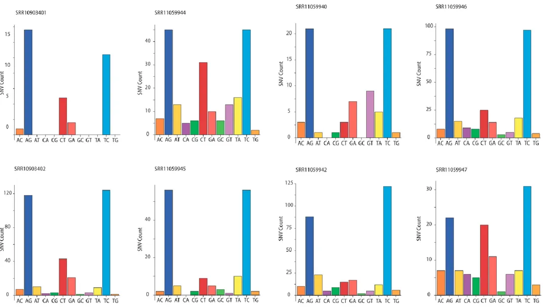
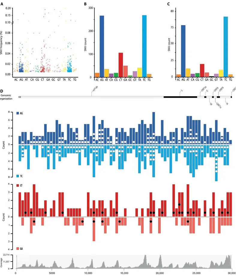
To assess whether RNA editing could be involved in human host responses to SARS-CoV-2 infections, we started from publicly available RNA sequencing datasets from BALF obtained from patients diagnosed with COVID-19. While transcriptomic data for all samples could be aligned to the SARS-CoV-2 reference genome, the quality of the sequencing varied and only eight samples had coverage and error rates suitable for the identification of potentially edited sites (Table 1). We called SNVs on these eight samples (126, 127) using REDItools 2 (116, 123, 128) and JACUSA (117) using the following thresholds: reads supporting the SNV ≥4, allelic fraction ≥0.5%, coverage ≥20, quality of the reads >25, base quality >35 (Figure 3 A). The two pipelines gave comparable results with ~50% of the SNV positions called by both (Figure 3 B, Figure 5 and Figure 6). We identified 910 SNVs common to REDItools 2 and JACUSA, ranging from 24 to 238 SNVs per sample (Figure 7). Given the thresholds used to call the SNV, samples with lower sequencing depths displayed lower numbers of SNVs. While the weight of each SNV type varies across samples (Figure 7), a bias toward transitions is always present, which is even more evident when all mutational data are pooled (Figure 8 A and B). This pattern holds true even when only SNVs recurring in more samples are considered (Figure 8 C).
The SNV allelic fraction (also referred to as frequency) and number of transversions are compatible with the mutation rates observed in coronaviruses [10–6/−7; (129)] and commonly associated to the RdRp. RdRps are error prone and are considered the main source of mutations in RNA viruses. However, the coronavirus NSP14-ExoN gene provides a form of error correction (33), which is probably the reason mutation rates in coronaviruses are lower than those observed in RNA viruses with smaller genomes. The mutational spectrum in SARS quasispecies presents a very weak bias toward U-to-G.
Figure 7: SNVs identified in SARS-CoV-2 transcriptomes after the intersection of Reditools 2 and JACUSA results. The bar charts show the number of SNVs identified in each SARS-CoV-2 transcriptome for each SNV type (e.g., A>C, AC). The sequencing depth for each sample is indicated.
Inactivation of NSP14-ExoN error correction reveals the mutational spectrum of the RdRp, which is quite different from the pattern we observe (i.e., main changes are C-to-A, followed by U-to-C, G-to-U, A-to-C, and U-to-G) (130). Hence, we would consider that SNVs deriving from RdRp errors represent a marginal fraction of the SNVs in the SARS-CoV-2 samples. The bias toward transitions—mainly A>G/T>C changes—resembles the pattern of SNVs observed in human transcriptomes (97) or in viruses (131-133), where A>G changes derive from deamination of A-to-I mediated by the ADARs. It is thus likely that the A>G/T>C changes seen in SARS-CoV-2 are also due to the action of ADARs.
C>T and G>A SNVs are the second main group of changes and could derive from APOBEC-mediated C-to-U deamination. Unlike A-to-I editing, C-to-U editing is a relatively rare phenomenon in the human transcriptome (97), and with regard to viruses, it has been associated only with positive-sense ssRNA rubella virus (81), where C>T changes represent the predominant SNV type. The observation that only A-to-I editing is present in RNA viruses that infect nonvertebrate animals, where RNA-targeting APOBECs are not present (131, 132), supports the hypothesis that APOBECs are involved in the RNA editing of this human-targeting virus.
A third group of SNVs, A>T/T>A transversions, is also present in these samples. While this type of SNV has been reported in other genomic studies (134), its origin is still unknown. A>G and T>C changes are evenly represented with respect to SNV frequency (Figure 8 A), the number of unique SNVs (Figure 8 B and C), and their distribution across the viral genome (Figure 8 D). As ADARs target dsRNA, this suggests that dsRNA encompasses the entire genome. While dsRNA in human transcripts is often driven by inverted repeats, the most likely source of dsRNA in the viral transcripts is replication, where both positive and negative strands are present and can result in wide regions of dsRNA.
Figure 8: Total SNV identified in SARS-CoV-2 transcriptomes. (A) Allelic fraction and (B) number of SNVs for each nucleotide change in the entire dataset and (C) for SNVs recurring in at least two samples. (D) Distribution of SNVs across the SARS-CoV-2 genome. A-to-G (blue) and C-to-U (red) SNVs are grouped in 400-nucleotide (nt) bins and plotted above (AG and CT) or below the line (TC and GA) based on the edited strand. Dots (white/black) indicate recurring SNVs. Genetic organization of SARS-CoV-2 (top). The dark/white shading indicates the viral coding sequences; coverage distribution of all analyzed samples (bottom).
Unlike A-to-I changes, C-to-U changes are biased toward the positive-sense strand (Figure 8 B to D; P < 0.0001). Because ADARs and APOBECs selectively target dsRNA and ssRNA, this distribution could arise from the presence at all times of RNA in a dynamic equilibrium between double-strandedness—when negative-sense RNA is being transcribed—and single-strandedness—when nascent RNA is released. Although some areas seem to bear fewer SNVs, these reduced SNV frequencies might be related to lower sequencing depth in those regions.
Figure 9: Logi alignment for SARS-CoV-2 RNA edited sites. (A) Local sequence context for A-to-I and C-to-U edited sites in the viral transcriptome and (B) for recurring sites.
As APOBEC deaminases preferentially target cytosines within specific sequence contexts, we analyzed the nucleotide context of A-to-I and C-to-U SNVs in the viral genome (Figure 9 A and B, Figure 10 A, B, C, D). A slight depletion of G bases in position −1 is present at A-to-I edited positions. This depletion is not as strong as the signal previously reported in human transcripts (91, 135-137). The low editing frequencies we observe resembles the editing present on human transcripts containing Alu sequences, which were found in a limited number in those early datasets. After the logi alignment, there is no evidence of a sequence context preference if we use a larger dataset such as REDIportal (98), which includes >1.5 M sites in Alu repeats (Figure 9). When normalising sequence contexts around A-to-I editing sites on sequence contexts around As in reference genome, a GC[A]S motif has been identified; however such motif is different than the ones reported in literature (91, 135, 137, 138).
With regards to the APOBECs, C-to-U changes preferentially occur downstream from uridines and adenosines, within a sequence context that resembles the one observed for APOBEC1-mediated deamination ([AU]C[AU]) (66, 139). However, no nucleotide enrichment was detected after sequence context normalization (Figure 10 F); raising the question of wether such sites could derive from random events rather than motif-specific mutations.
Figure 10. Additional logi alignments: logi aligments showing the information content of the position (bits) for (A) A-to-I edited sites, (B) reference genome sequence context around As, (C) C-to-U edited sites, (B) reference genome sequence context around Cs. Enrichment and depletion plot of sequence context of edited sites normalized against reference genome sequence contexts around candidate base for (E) A-to-I editing and (F) C-to-U editing.
A
B
D
C
E
F
-2 -1 +1 +2 -2 -1 +1 +2 -2 -1 +1 +2 -2 -1 +1 +2 -2 -1 +1 +2 -2 -1 +1 +2We then aligned available genomes from SARS-CoV-2, Middle-East respiratory syndrome– related coronavirus (MERS-CoV), and SARS-CoV to test whether RNA editing could be responsible for some of the mutations acquired through evolution. The genomic alignments reveal that a substantial fraction of the mutations in all strains could derive from enzymatic deaminations (Figure 11 A to C), with a prevalence of C-to-U mutations, and a sequence context compatible with APOBEC-mediated editing also exists in the genomic C-to-U SNVs (Figure 11 D to F).
Discussion
Our data source—metagenomic sequencing—raises the question whether the low-level editing we observe (~1%) reflects the actual levels of editing of viral transcripts within human cells. Aside from a small fraction of cellular transcripts edited at high frequency, most ADAR-edited sites in the human transcriptome (typically inside Alu sequences) present editing levels of ~1% (97, 140, 141). It has been shown that a fraction of the cellular
Figure 11: Nucleotide changes across Coronaviridae strains. (A to C) Number of SNVs for each nucleotide change and (D to F) local sequence context for C-to-U edited sites in genome alignments from SARS-CoV-2 (A and D), human-hosted MERS-CoV (B and E), and human-hosted SARS-CoV (C and F).
While we were unable to observe hyperedited reads in the metagenomic samples, it is possible that hyperedited transcripts fail to be packaged into the virus.
With regard to APOBEC-mediated RNA editing, its detection in the viral transcriptomes is already indicative, as this type of editing is almost undetectable in human tissues (97). This enrichment points either toward an induction of APOBECs triggered by coronavirus infection or to specific targeting of the APOBECs to the viral transcripts. APOBECs have been proved effective against many viral species in experimental conditions, yet, until now, their mutational activity in clinical settings has been shown only in a handful of viral infections (73-80) through DNA editing and, in rubella virus, on RNA (81).
Kim et al. (145) observed 41 recurrent base-modification sites in SARS-CoV-2 RNAs; it is possible that base modifications may lead to nucleotide misincorporation during PCR amplification, acting as a confounding factor and introducing biases in our analysis.
However, most of the base-modification sites fall approximately at 29000kb of viral genomic RNA (within N protein coding region), and we don’t observe an enrichment of APOBEC and ADAR-related mutations in that region (Figure 8 D).
As in rubella virus, we observe a bias in APOBEC editing toward the positive-sense strand. This bias and the low editing frequencies might be indicative of the dynamics of the virus, from transcription to selection of viable genomes. It is reasonable to assume that sites edited on the sense strand will result in a mid-level editing frequency, as not all negative-sense transcripts will be edited (Figure 12 A). On the other hand, editing of the positive-negative-sense strand can occur upon entry of the viral genome, thus yielding high-frequency editing (Figure 12 B), or after viral genome replication, resulting in low-frequency editing (Figure 12 C). The lack of a sizable fraction of highly edited C>T SNVs suggests that APOBEC editing occurs late in the viral life cycle (Figure 12 C). Yet, because they occur earlier, G>A SNVs should be closer in number to C>T SNVs and with higher levels of editing, which is not what
we observe (Figure 8 A to C). The overrepresentation of C>T SNVs could be due to an imbalance toward positive-sense transcripts, as these are continuously generated from the negative-sense ones (and double-stranded hybrid RNAs are lost). However, the editing frequencies of G>A SNVs should be much higher, as G>A SNVs are generated upstream to the C>T ones. A more fitting explanation is that editing of the negative-sense transcripts results somehow in a loss of the edited transcript (Figure 12 D), lowering the chances of the edited site to be transmitted. Despite the fact, according to our data, that APOBEC-related mutations seem to be more deleterious than ADAR-related ones, it is possible that ADAR mediates hyper-editing (144), introducing a large amount of mutations on the same transcript. Such a load of mutations it is more likely to affect gene functionality rather than a single mutation, and it might be undetectable because of the loss of the edited (and not functional) transcript.
Taking in account our observation, with low editing frequency of both C>T and G>A mutations and with a higher number of unique C>T SNVs, the most likely compatible model
Figure 12: Model of APOBEC RNA editing on SARS-CoV-2 transcriptome. The four panels model the editing frequencies and the C>U/G/A ratios expected from four different scenarios: (A) C-to-U editing on the negative-sense transcripts, (B) “early” editing on the viral genomes before viral replication, (C) “late” editing after viral replication, and (D) “late” editing after viral replication with loss of negative-sense transcripts. Red dots indicate editing on the positive-sense transcript; orange dots indicate editing on the positive-sense transcript. Green and blue segments indicate positive- and negative-sense viral transcripts, respectively.
Because most of the APOBECs are unable to target RNA, the only well-characterized cytidine-targeting deaminases are APOBEC1, mainly expressed in the gastrointestinal tract, and APOBEC3A (70), whose physiological role is not clear. As with A-to-I editing, it will be important to assess the true extent of APOBEC RNA editing in infected cells.
The functional meaning of RNA editing in SARS-CoV-2 is yet to be understood: In other contexts, editing of the viral genome determines its demise or fuels its evolution. For DNA viruses, the selection is indirect, as genomes evolve to reduce potentially harmful editable sites [e.g., (131)], but for RNA viruses, this pressure is even stronger, as RNA editing directly affects the genetic information and efficiently edited sites disappear.
A comparison of the SNV datasets from the transcriptomic and genomic analyses reveals a different weight of A-to-I and C-to-U changes (Figure 8 B and Figure 11 A), with an underrepresentation of A-to-I in the viral genomes. As our analysis underestimates the amount of editing due to the strict parameters used, the underrepresentation of A-to-I changes could be explained by the possibility that A-to-I editing is more effective in restricting viral propagation, thus reducing the number of viral progeny showing evidence of these changes. In contrast, the remnants of less effective C-to-U editing are retained in viral progeny and get fixed during viral adaptation.
An analysis of mutation outcomes is difficult due to the low numbers of events collected so far, but there are some possibly suggestive trends (Table 2). C-to-U changes leading to stop codons are overrepresented in the transcriptomic data but—as expected—disappear in the genomic dataset. This might point—again—to an antiviral role for these editing enzymes. There is also an underrepresentation of C>T missense mutations, but its meaning is difficult to interpret.
Last, this analysis is a first step in understanding the involvement of RNA editing in viral replication, and it could lead to clinically relevant outcomes: (i) If these enzymes are relevant in the host response to coronavirus infection, a deletion polymorphism quite common in the Chinese population, encompassing the end of APOBEC3A and most of APOBEC3B (146, 147), could play a role in the spread of the infection. (ii) Because RNA editing and selection act orthogonally in the evolution of the viruses, comparing genomic sites that are edited with those that are mutated could lead to the selection of viral regions potentially exploitable for therapeutic uses.
Section 2: Analysis of copy number
variations from cell-free DNA of lung
cancer patients via Nanopore sequencing
Introduction
Copy number variations and their impact on human diseases
Copy number variations (CNVs) are structural variants involving genomic segments of more than 1kb in length, which are represented in a variable number of copies compared to the normal ploidy of the organism (148).
For years, the weight of CNVs on human genome has been underestimated, also due to a lack of an adequate technology for their study. With the advent of large population studies, supported by more accessible and refined experimental approaches, CNVs have been recognised as a big contributor to inter-individual variation in the genomes of healthy individuals, along with single nucleotide polymorphisms (148, 149).
However, the presence of CNVs can influence the phenotype of the cells by altering the expression of the genes affected by the CNV or located nearby its boundaries (probably via alteration of adjacent regulatory sequences (148, 150, 151), and by generating to fusion-genes and, consequently, production of aberrant proteins (152).
It is hence not surprising that CNVs have been associated with a variety of diseases classified as 'genomic disorders'. Unlike mutation-driven pathologies which usually depend on variations on single genes, CNVs often involve large genomic regions affecting a set of genes as, for example, in Prader-Willi syndrome (15q11-q13 deletion) and Williams-Beuren syndrome (7q11.23 deletion) (153, 154).
However, this is not always the case: Smith-Magenis syndrome is caused by a deletion in chromosome 17p11.2; despite its size (on average 3.7 Mb) is variable among patients, a common 1.5 Mb portion has been identified. This “critical” portion includes the retinoic acid induced 1 gene, which is considered the main responsible of the pathologic phenotype (149, 155).
Germline inherited and de-novo CNVs have been associated with a wide spectrum of human diseases including:
- Infectious and autoimmune diseases: asthma, Chron’s disease, HIV infections, systemic lupus erythematosus and anti-neutrophil cytoplasmic antibody-associated vasculitis (148, 156-168).
- Nervous system diseases: autism, schizophrenia, epilepsy, Parkinson’s disease, amyotrophic lateral sclerosis and autosomal dominant Alzheimer’s disease (169-177).
- Metabolic and cardiovascular diseases: familial hypercholesterolemia, atherosclerosis and coronary artery disease (178-180).
- Cancer (181-189).
This project is focused in particular in the detection of cancer related CNVs.
Cancer development is a multistep process characterized by the accumulation of genetic alterations eventually leading to the acquirement of the malignant phenotype (190). In contrast to most of the aforementioned pathologies, such alterations (including CNVs) can be both germline, representing a predisposing factor for the development of cancer, and somatic, contributing to the load of alterations necessary for the transformation (148, 191).
The relationship between CNVs and cancer development can be explained in part by the Kudson’s “two hit” hypothesis (192): a homozygous deletion can lead to the loss of a tumor-suppressor gene, while a heterozygous one can be deleterious when the other allele is altered by an inactivating mutation or an additional deletion. On the other hand, amplifications can lead to overexpression of oncogenes. Notably, germline CNVs are typically more abundant in individuals from cancer-prone families, in particular among TP53 mutant carriers, suggesting that CNVs are not always a contributing cause to cancer, but rather a consequence of genomic instability (149, 193, 194).
Specific CNVs have been associated with cancer types and outcome: for example, in prostate cancer, loss of 8p23.2 is associated advanced stage disease, and gain at 11q13.1 is predictive predictive of post-operative recurrence; while heritable CNV at chromosome 1q21.1 is associated with neuroblastoma (189).
The recurrence of CNVs is not limited to single genes, but also to entire pathways such as the ERBB2, EGFR and PI3K pathways, which have been reported to be enriched in CNVs and SNVs in both breast and colorectal cancer (188).
Importance of cancer monitoring
During cancer development, malignant cells gain specific genotypic, phenotypic and epigenetic features, making cancer one the most heterogeneous human diseases.
Even within the same cancer type, it is possible to identify an large number of molecular subtypes defined by gene/protein expression patterns and alterations of the genome. Such molecular heterogeneity often results in different aggressivity, invasivity, response to treatment and, consequently, overall outcome (195-197).
For example, triple negative breast cancer patients have typically worse outcomes compared to HER2, progesterone and estrogen receptor expressing ones (198); EGFR mutations confer resistance to Tyrosine Kinase Inhibitor therapies in lung cancer patients (199); AR amplifications are linked to the development of castration resistance prostate cancers (200); and MGMT promoter methylation is an important prognostic biomarker, influencing the response to radio therapy in glioblastoma patients (201). The goal of the so-called “precision oncology” is to define personalized treatment strategies based on cancer molecular features, aiming at maximizing the efficacy against a specific subtype. In this context, it is pivotal to accurately detect biomarkers for a proper (correct) subtype identification and patient stratification (195, 196).
Typically, bioptic samples or surgical resections are necessary for biomarker investigation. Tissue sections are typically used for techniques which are based on eye inspection such as: immunohistochemistry for protein expression, in-situ hybridization or RNA-scope for gene expression and fluorescent in-situ hybridization for structural variations (SV) detection. In addition, DNA and RNA can be extracted from tissue samples for gene expression, DNA methylation, mutation and copy number variation analyses (199, 202-210).
However, a significant limitation of tissue sampling is that it fails to comprehensively capture intra-tumoral heterogeneity. Indeed, cancer heterogeneity is not limited to inter-patient molecular diversity and a tumoral mass is often composed by subclones carrying different features. Hence, the portion of mass which is sampled may not fully represent the entire tumoral bulk (sometimes not even the major clone), with an high risk of missing clinically relevant alterations. Moreover, collection of tissue samples is usually invasive, requires trained medical staff and can be harmful for the patient. (210, 211).
Notably, cancer is an extremely dynamic disease: malignant cells are constantly under selective pressure, competing for nutrients against other cells or escaping human defense mechanisms, be them physiological (immune system) or artificial (drugs and treatments) (212). Consequently, the evolutionary path of each tumor can take different directions due to such pressure. It is hence important to monitor cancer development at multiple timepoints to closely follow its evolution, aiming at driving clinical decisions during the entire patient’s history (195, 196). Unfortunately, the risks and invasiveness of conventional biopsy make it unsuitable for repeated sampling (213).
Liquid biopsy
A valid and non-invasive alternative to tissue sampling is represented by liquid biopsy. The principle behind liquid biopsy is that tumour masses shed cellular material into the bloodstream, urine or stool. It is therefore possible to analyse the blood to investigate tumor-related analytes to obtain information about the characteristics of the tumor (214). This concept is definitely not new as many protein-based serum biomarkers have proven useful for cancer diagnosis (215, 216). The most emblematic example is prostate specific antigen which is currently the first-line screening biomarker for prostate cancer early detection (216).
The recent emergence of cutting edge techniques with increased sensitivity and reliability allowed to extend blood-based analyses beyond proteic biomarkers.
It is currently possible to analyze tumor-derived nucleic acids (e.g. non coding RNAs, DNA) (213), vesicles (e.g. exosomes) (217) and circulating tumor cells (218) from blood samples.
In particular, I’m focused on the study of circulating cell-free DNA (cfDNA) which is extracellular DNA released into the bloodstream during cell death. CfDNA is extracted from plasma (less frequently from serum) obtained via blood centrifugation. Since we are interested in extracellular DNA, the goal of centrifugation is to remove intact blood cells whose DNA is non-informative and would reduce the sensitivity of the approach (219).
In healthy individuals, cfDNA belongs mainly from myeloid and lymphoid apoptotic cells due to the physiological turnover of hematopoietic cells (with minimal contributions from other tissues) (220, 221) while, in cancer patients, a fraction of the total cfDNA, termed circulating tumor DNA (ctDNA), comes from neoplastic lesions (222). CtDNA is very informative for the study of oncological pathologies as it harbours tumor-specific genetic alteration that reflects the genomic status of the malignant cell of origin (223-227).
However, several technical aspects make the study of cfDNA extremely challenging: CfDNA concentration is very low and typically higher in advanced cancer patients rather than healthy subjects and low grade patients; this is one of the aspects that complicate cfDNA analysis, in particular for early-stage applications (214).
Moreover, the percentage of ctDNA among the totality of cfDNA can be very low (0.01-60%) [15–18] and depends on different tumor features such as tumor volume, stage, vascularization, proliferation rate, and cell death rate (223, 228-231).
For these reasons, cfDNA study requires highly sensitive techniques compatible with very low input DNA.
Figure 13: Typical fragmentation patterns in cfDNA. (A) Monophasic pattern showing only the ~160-167 bp peak. (B) Biphasic pattern showing ~160-167, ~320 and ~480 bp peaks. (C) Example of contamination by HMW DNA, likely due to blood cells lysis. Figure taken from (232).
CfDNA is also highly degraded with an enrichment in low molecular weight (LMW) DNA fragments: the typical cfDNA fragmentation profile is composed by a major peak at ~160-167 bp and two smaller peaks at ~320 and ~480 bp (not always detectable) (Figure 13) (221, 232). This particular pattern is due to nucleosome protection from degradation: due to cell death, DNA is degraded by DNAses, which cleave the filament mostly in linker regions between nucleosomes, generating mainly ~167 bp fragments corresponding to chromatosomes length (nucleosome + linker histone). Less frequent
scenarios, in which 2-3 chromatosomes are still intact, result in the weak peaks of longer DNA fragments (e.g. 320/480 bp) mentioned previously (221, 232).
The presence of high molecular weight (HMW) DNA is indicative of blood cell lysis, which can happen during sample transportation, blood withdrawal and centrifugation, or of an incorrect plasma collection, in which blood cells are accidentally resuspended by pipetting. HMW DNA is a dangerous contaminant for cfDNA, as it belongs to healthy blood cells and is hence uninformative (219).
Despite these technical challenges, liquid biopsy has some important advantages over conventional biopsy (211, 233), it is:
- Noninvasive: The entire procedure is completely unharmful and painless, without any complication that may arise with conventional biopsies.
- Simple: It is based on a simple blood withdrawal and it can be performed without particular training or instrumentation. This has also a positive impact on per-sample costs.
- Repeatable: This aspect is closely related to the aforementioned simplicity and non-invasivity that make liquid biopsy highly repeatable (even on a month-basis), just like any other routinary blood-based test.
- Versatile: The feasibility of tissue sampling depends on tumor characteristics - Versatile and comprehensive: The feasibility of tissue sampling depends on
tumor characteristics and location, meaning that not all tumors may be succesfully sampled. Virtually any cancer cell releases DNA into the circulation (as soon as it is vascularized), minimizing the risk of sampling biases. In addition, liquid biopsy provides a comprehensive profile of tumor, which include alterations from all the subclones and even from different tumor/metastasis sites.