Scuola Dottorale di Biologia
Sezione di Scienze Biolmolecolari e Cellulari
XXV Ciclo“The role of the ospB-phoN2 operon in the mechanism of pathogenicity of S. flexneri”
“Il ruolo dell’operone ospB-phoN2 nel meccanismo di patogenicità di S. flexneri” Supervisor I Prof. M.Casalino PhD Student Daniela Scribano Supervisor II Prof. M.Nicoletti PhD School Coordinator Prof. P.Mariottini
3
Index
Abstract 5 Riassunto 7 Introduction 9 Results 15OspB is involved in the modulation
of host inflammatory response 15
Expression of S. flexneri ospB gene in infected
HeLa cell monolayers and OspB localization in the host cell 18 OspB activates the MAPK signalling pathways
in Caco-2 cultured intestinal epithelial cells 20 Periplasmic PhoN2 (apyrase) polarly localizes
in S. flexneri and E. coli cells 23
PhoN2 localizes beneath the IcsA cap 27
Characterization of PhoN2 domains
involved in its polar localization 28
The PPPP motif controls PhoN2 stability 33
The polyproline PPPP motif indirectly influences
S. flexneri virulence 36
The poly-proline region is necessary for PhoN2 3D structure 38
PhoN2 binds to the OM protein A (OmpA) 39
In vivo cross-linking experiments 41
4 Interaction between PhoN2 and OmpA:
a computational model 44
Role of OmpA in the mechanism
of pathogenicity of S. flexneri. 47
Construction of the ompA mutant strain HND92 47
The ompA mutation does not affect IpaABCD secretion 48 The ompA mutant presents an altered invasive phenotype 49
Membrane permeability of the ompA mutant 57
OmpA is not required for host cytosolic
phospholipase A2 activation 58
Discussion 60
Conclusion 70
5
Abstract
In this thesis we have studied the role of the ospB-phoN2 bicistronic operon in the virulence of Shigella flexneri. OspB is, a not fully characterized yet, type III secretion system (T3SS) effector secreted into host cells to subvert cellular and immune functions and promote infection. Here we report evidences that OspB participates at the induction of the severe inflammatory reaction, the hall-mark of bacillary dysentery, at early stages of infection. We found that once secreted into the cytoplasm of host cells, OspB rapidly induces phosphorylation of mitogen-activated protein kinases (MAPKs) Erk1/2 and p38. Early activation of Erk1/2 and p38 leads to the induction of the inflammatory response culminating in massive polymorphonuclear leukocytes (PMNs) infiltration within the colonic mucosa. PMN infiltration, by breaching the epithelial barrier, initially promotes bacterial colonization. On the other hand, 30 min post-infection OspB accumulates within the host cell nucleus where it exerts its functions by modulating expression of pro-inflammatory genes. We propose a working model to try to explain the role of OspB in PMNs transmigration. Proper protein localization is critical for bacterial virulence. The virulence-associated PhoN2 protein is a periplasmic ATP-diphosphohydrolase (apyrase), involved in S. flexneri IcsA-mediated actin-based motility (ABM). IcsA is an autotrasporter protein that is inserted and exposed on the outer membrane (OM) at the old bacterial pole where promote ABM. Here we show that PhoN2 strictly localizes at bacterial poles, where the great majority of bacteria (more that 90%) presenting PhoN2 localized just beneath IcsA. PhoN2 was found to be polarly localized also in a phoN2-complemented Escherichia coli K-12 strain, indicating a conserved mechanism of PhoN2 delivery across species. Analysis of deletion and point mutations encompassing the N-terminal polyproline (41ILPPPPAE48) and C-terminal regions of PhoN2 indicated that the PPPP motif and the Y155 residue play a pivotal role in the polar localization of PhoN2, in its proper folding and stability, in the correct exposition of IcsA and in apyrase activity. Two-hybrid and cross-linking experiments proved OmpA as a strong interactor of PhoN2. Interestingly, neither IcsA nor OmpA are required for PhoN2 polar localization, while the lack of OmpA induces IcsA exposition over the entire bacterial length. A model is presented to explain how the PhoN2-OmpA interaction allows proper IcsA exposition and ABM.
Next, the finding of a PhoN2-OmpA interaction prompted us to investigate the role of OmpA on the virulence of S. flexneri. An ompA mutant of
wild-6 type S. flexneri 5a strain M90T (strain HND92) was shown to be severely impaired in cell-to-cell spread since it failed to plaque on HeLa cell monolayers. Nevertheless, the ompA mutant displayed IcsA exposed across the entire bacterial surface although it was able to produce proper F-actin comet tails, indicating that the aberrant IcsA exposition at bacterial lateral surface did not affect proper activation of actin-nucleating proteins, and suggesting that the absence of OmpA likely unmasks IcsA at bacterial lateral surface. Moreover, the ompA mutant was able to invade and to multiply within HeLa cell monolayers, although internalized bacteria were found to be entrapped within the host cell cytoplasm. We found that the
ompA mutant produced significantly less protrusions than the wild-type
strain, indicating that this defect could be responsible of its inability to plaque. Complementation of the ompA mutation clearly indicated that a functional OmpA protein is required and sufficient for proper IcsA exposition, plaque and protrusion formation.
7
Riassunto
In questa tesi abbiamo studiato il ruolo dell’operone bicistronico
ospB-phoN2 nel meccanismo di virulenza di Shigella flexneri. OspB è un
effettore secreto dal sistema di secrezione di tipo terzo (T3SS) il cui ruolo non è stato ancora del tutto caratterizzato. OspB, quando viene rilasciato all’interno della cellula ospite, è in grado di modulare la fisiologia cellulare e la risposta immunitaria promuovendo l’instaurarsi dell’infezione. Il nostro studio evidenza che OspB ha un ruolo nell’induzione della imponente reazione infiammatoria, caratteristica saliente della dissenteria bacillare, agendo negli stadi precoci dell’infezione. Quando viene secreto nel citoplasma della cellula ospite OspB induce rapidamente la fosforilazione e quindi l’attivazione di Erk1/2 (Extracellular response kinases) e p38 che rappresentano due principali MAP (mitogen-activated protein) chinasi coinvolte nella attivazione della risposta cellulare ad uno stimolo infiammatorio. Infatti, quando Erk1/2 e p38 sono attivate, attraverso una cascata di trasduzione del segnale, inducono l’attivazione della risposta infiammatoria che culmina con il potente richiamo di leucociti polimorfonucleati (PMN) nel sito d’infezione. L’infiltrazione dei PMN nel tessuto causa la distruzione dell’integrità dell’epitelio intestinale, aprendo vie d’accesso alla sottomucosa, sfruttate dai batteri per la colonizzazione del tessuto, nelle fasi precoci dell’infezione. Successivamente OspB si localizza nel nucleo della cellula ospite e in questo sito esplica la sua funzione modulando l’espressione dei geni pro-infiammatori. I nostri risultati ci hanno permesso di proporre un modello che spiega il meccanismo attraverso il quale OspB attiva la risposta infiammatoria dell’ospite.
L’azione di alcuni fattori di virulenza dipende dalla loro specifica localizzazione all’interno della cellula batterica. PhoN2 è una proteina periplasmica associata alla virulenza di S. flexneri, è una ATP difosfoidrolasi che è coinvolta nel meccanismo di movimento di S. flexneri. Tale meccanismo si basa sulla polimerizzazione d’actina della cellula ospite (ABM), e richiede l’espressione del fattore di virulenza IcsA che si inserisce e si espone sulla membrana esterna, al polo vecchio del batterio. Il nostro studio dimostra che PhoN2 si localizza, nel periplasma, ai poli batterici, inoltre la maggior parte dei batteri presenta PhoN2 localizzata sotto la proteina IcsA. La localizzazione polare di PhoN2 è mantenuta anche in un ceppo di Escherichia coli, a cui è stato inserito in trans, il gene
phoN2, indicando che esiste un meccanismo di esporto al polo batterico
8 proline nella regione N-terminale (41ILPPPPAE48) e i domini del sito catalitico nella regione C-terminale. Attraverso delezioni e sostituzioni amminoacidiche sul motivo PPPP e nel sito catalitico abbiamo dimostrato che il motivo PPPP e la tirosina 155 (Y155) sono residui necessari alla struttura tridimensionale di PhoN2. Di conseguenza tali residui sono necessari nella stabilità della proteina, nell’attività enzimatica e indirettamente nel processo di localizzazione di IcsA al polo batterico. Tramite il saggio del doppio ibrido in lievito abbiamo dimostrato che PhoN2 interagisce con la proteina della membrana esterna OmpA. Sia OmpA che IcsA non sono coinvolte nel meccanismo di localizzazione polare di PhoN2, viceversa la mancanza della proteina OmpA causa una aberrante esposizione di IcsA su tutta la superficie batterica. Noi proponiamo un modello attraverso il quale l’interazione tra PhoN2 e OmpA consente la corretta esposizione di IcsA al polo, permettendo, quindi, un corretto movimento del batterio all’interno della cellula ospite.
La dimostrazione che OmpA interagisce con il fattore di virulenza PhoN2 ci ha indotto a studiare il ruolo di tale proteina nel meccanismo di patogenicità di S. flexneri. Il mutante nullo nel locus ompA (ceppo HND92) mostrava un movimento intercellulare aberrante poiché non produceva placche di lisi su monostrati di cellule HeLa in coltura. Anche se il mutante nullo per ompA presentava IcsA esposta su tutta la superficie batterica era in grado di indurre polimerizzazione d’actina, quindi l’esposizione aberrante di IcsA non influenzava la capacità di tale proteina di reclutare le proteine eucariotiche responsabili della polimerizzazione d’actina. Questo ci ha suggerito che OmpA potrebbe svolgere un ruolo nel mascherare IcsA sulla superficie batterica senza agire sulla capacità di tale proteina di indurre polimerizzazione d’actina. Tramite saggi di invasione di cellule HeLa in coltura, abbiamo osservato che il ceppo HND92 aveva un tasso di invasione inferiore rispetto al ceppo parentale M90T e che non era in grado di formare le protrusioni. Le protrusioni permettono la diffusione del batterio da una cellula alla cellula adiacente. Questo risultato potrebbe spiegare l’inabilità del mutante ompA di produrre le placche di lisi su monostrati HeLa. L’inserimento in trans del gene ompA nel ceppo HND92 ha dimostrato che la proteina OmpA è richiesta per la corretta esposizione di IcsA nella membrana esterna, per la formazione delle protrusioni e quindi per la produzione di placche.
9
Introduction
Shigella flexneri is a Gram-negative facultative intracellular pathogen that
leads to severe inflammatory colitis called bacillary dysentery (or shigellosis), a major public-health problem in developing countries. After its passage through the stomach and small intestine, S. flexneri reaches the colonic mucosa where infects the colonic epithelium via M cell translocation. Once S. flexneri is endocytosed by M cells, bacteria are transocytosed toward the M cell pocket and invade resident macrophages and dendritic cells, where they disrupt vacuolar membranes and then disseminate into and replicate within the cytoplasm. Bacterial multiplication within macrophages results in a massive inflammatory response (the hallmark of shigellosis) and cell death (apoptosis). Macrophage apoptosis induces synthesis and release of proinflammatory cytokines which in turn leads to recruitment polimophonuclear leukocytes (PMNs) at the site of infection (Fig. 1). Once free in the sub-mucosa, S.
flexneri induces its internalization at baso-lateral sites of the polarized
epithelial epithelium (Sasakawa, 2010). After internalization into epithelial cells, S. flexneri rapidly disrupts surrounding vacuolar membrane, multiplies through the cytoplasm and diffuses to adiacent cells by inducing F-actin polymerization at one pole (the old pole) of the bacterium. Thus, macrophage cell death, invasion of and multiplication within epithelial cells, cell-to-cell spreading, demise of the host epithelium and alteration of the host inflammatory response are major pathogenic events that lead to shigellosis (Makino et al., 1986, Sansonetti et al., 1986, Clerc et al., 1987, Bernardini et al., 1989, Sansonetti et al., 2001).
Fig. 1. A model for S. flexneri infection of intestinal epithelial cells.
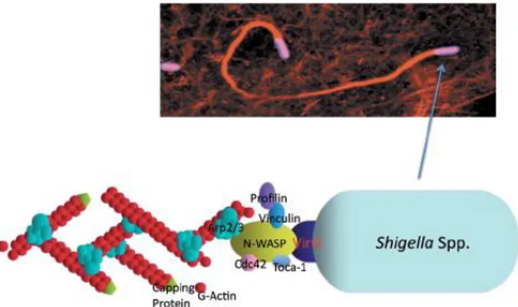
10 The ability of S. flexneri to move within the cytoplasm of invaded epithelial cells and to diffuse the infection to adjacent cells of the intestinal mucosa requires the expression and polar surface exposition of IcsA (VirG), a 110-kDa autotransporter protein encoded on the S. flexneri large virulence plasmid (Bernardini et al., 1989). Once translocated across the outer membrane (OM), IcsA exposes its N-terminal α-domain (the passenger domain) on the bacterial surface, forming a cap exclusively at the old bacterial pole (Goldberg et al., 1993, Goldberg et al., 1994). Exposed IcsA, by interacting with neural Wiskott-Aldrich syndrome protein (N-WASP) and vinculin, induces F-actin polymerization by activating host Arp2/3 complex, which induces polymerization of host globular actin into filamentous actin (Suzuki et al., 1998 , Cossart, 2000, Charles et al., 2001, Goldberg, 2001, May and Morona 2008, Heindl et al., 2010) (Fig. 2). Although the mechanism driving the polar localization of IcsA has not been fully elucidated, experimental evidence indicates that IcsA likely inserts directly at the old bacterial pole (Goldberg et al., 1993, Fukuda et al., 1995, Steinhauer et al., 1999).
Fig. 2. The actin-dependent Shigella motility. A confocal
immuno-fluorescence image of actin tails. Bottom, the machinery required for bacterial motility, VirG/IcsA, N-WASP, Arp2/3 complex, profilin, and Toca-1, accumulates at the old bacterial pole (Sasakawa 2010).
11 Furthermore, IcsA-mediated actin-polimerization is essential for S. flexneri to diffuse to adjacent cells via protrusion formation. Protrusions are membrane-bound cell extensions that are driven by the bacterium to propel itself into adjacent cells. Protrusions, which may extend tens of microns from the cellular surface, are characterized by the presence of a bacterium at its tip (Bernardini et al., 1989). By a process which likely resembles macropinocytosis, contact with the membrane of an adjacent cell is followed by uptake of the bacterium (Kadurugamuwa et al., 1991), leading to the spreading of the infection to neighbouring epithelial cells. Several host cell proteins have been implicated in protrusion-mediated Shigella cell-to-cell spreading, suggesting that a distinct set of actin regulatory factors likely interacts with motile bacteria after they contact the plasma membrane (Haglund and Welch 2011). Although actin polymerization and assembly are required for protrusion formation, the specific mechanism responsible for this phenomenon is poorly defined. It has been recently reported that actin nucleation in protrusion formation might be independent of the activity of the Arp2/3 complex and that protrusion formation and inter-cellular spreading depend on actin polymerization that requires the activation of the Diaphanous formin Dia (Heindl et al., 2010). Formins are a family of ubiquitous expressed proteins that, in contrast to the Arp2/3 complex, initiate de novo actin polymerization leading to cross-linking of actin polymers in parallel arrays (Heindl et al., 2010).
Apyrase (PhoN2) is an ATP-diphosphohydrolase encoded by phoN2 (apy), a gene located on a highly conserved region (the ospB-phoN2 operon) of
Shigella species and related enteroinvasive Escherichia coli (EIEC) strains
(Santapaola et al., 2002). ospB and phoN2 are co-transcribed as a 2 kb bicistronic, temperature-regulated mRNA from an upstream promoter that precedes ospB (Santapaola et al., 2002). OspB is a type III secretion system (T3SS) secreted effector and transcription of the ospB-phoN2 operon is regulated by the VirF/VirB cascade and by MxiE, in concert with IpgC (Buchrieser et al., 2000, Le Gall et al., 2005, Santapaola et al., 2006). Even if PhoN2 is not a secreted effector, phoN2 transcription is also up-regulated by MxiE when the T3SS system is activated, suggesting that PhoN2 may be relevant in post-invasion events and that both PhoN2 and OspB might be functionally related (Santapaola et al., 2006).
The purpose of this study was to evaluate the contribution of OspB and PhoN2 to the virulence of S. flexneri. It has been recently reported that OspB, once secreted into host cell, translocates into the nucleus where interacts with retinoblastoma protein thus participating at the remodeling of chromatin which leads to reduced inflammatory cytokines production and
12 to modulate the innate immune response (Zurawski et al., 2009). Moreover, it has been also shown that OspB, by activating the mitogen-activated extracellular kinase (MEK)/Erk1/2 pathway, leads to up-regulation of neutrophil attractant hepoxilin A3 (HXA3) which induces trans-epithelial migration of polymorphonuclear leukocytes (PMNs) and increases the inflammatory response (Kohler et al., 2002, Mumy et al., 2008, Zurawski et
al., 2009). Thus, it is conceivable that OspB may play a dual role depending
on the stage of infection. At an early stage, OspB appears to induce inflammation while at late stages (after its nuclear translocation) it appears to attenuate the inflammatory response.
In this study, we evaluated the role of OspB in the inflammatory response at early stages of infection. First, we confirmed that OspB is involved in the inflammatory response since an ospB null-mutant of the wild-type S.
flexneri 5a strain M90T failed to induce the levels of the inflammatory
response seen with wild-type, in the Serény test model of infection. Moreover, we found that OspB activates both Erk1/2 and p38 signaling pathways at early stages of infection. Since it is known that phosphorylation of Erk1/2 and p38 leads to the activation of complex pro-inflammatory pathways leading to PMNs migration, we hypothesized that OspB might play also a role in PMNs migration to the site of infection. Experiments are underway to unravel this point. A model is presented to explain this hypothesis.
PhoN2 is an ATP-diphosphohydrolase (apyrase) so far isolated only in
Shigella ed EIEC. Basing on its deduced biochemical structure, PhoN2 was
considered a periplasmic protein. Here we produced experimental evidence proving apyrase as a periplasmic protein of S. flexneri. Moreover, we have previously shown that PhoN2 is required, in a deoxynucleotide triphosphate-hydrolyzing activity-independent manner, for efficient inter-cellular spreading since a non polar phoN2 mutant of the wild-type S.
flexneri strain M90T presented altered polar IcsA exposition, aberrant
actin-based motility and a small plaque phenotype (Santapaola et al., 2006). The mechanism by which PhoN2 influences the polar exposition of IcsA has not been fully elucidated yet. However, since PhoN2 possesses an exposed N-terminal polyproline (43PPPP46) motif and proline-rich motifs have been reported to be involved in inter- and intra-molecular interactions, in protein folding and essential for virulence of various intracellular pathogens (Neibuhr et al., 1997, Suzuki and Sasakawa, 2001, Babu et al., 2002, Ansai et al., 2002, Makde et al., 2007), we hypothesized that PhoN2 might directly interact with IcsA or indirectly with other unknown
13 accessory outer membrane (OM) proteins necessary in assisting the correct polar exposition of IcsA (Santapaola et al., 2006).
The data presented in this work are based on the serendipitous discovery that the PhoN2 periplasmic pool localizes at either one (the old pole) or at both poles of S. flexneri. Here we analyzed the role of definite PhoN2 regions on PhoN2 polar localization, on expression of apyrase activity and protein stability. Polar localization of PhoN2 occurred also when PhoN2 was expressed in a E. coli K-12 genetic background, indicating a conserved mechanism of PhoN2 polar localization in both species. Analysis of deletion and point mutations encompassing the phoN2 DNA region encoding the N-terminal polyproline PPPP stretch, allowed us to demonstrate the pivotal role of this motif on the polar localization of PhoN2, on its stability and on the expression of its catalytic activity. PhoN2 is a member of the class A of nonspecific acid phosphohydrolases (A-NSAPs) (Rossolini et al., 1998). A-NSAPs share as common features an N-terminal PPPP motif and conserved amino acid residues over their C-terminal domain (Rossolini et al., 1998, Ansai et al., 2002, Babu et al., 2002, Sarli et al., 2005) (Fig. 3).
Fig. 3. The 3D structure of S. flexneri PhoN2 (B)obtained by threading using Swiss-Model with E. blattae EBPase (A) as PDB template (1D2T).
The EBPasi is shown as a monomer complexed with sulfate. Residues from the acitve site, conserved in both structures, are represented as ball and stick structure in yellow. The dimer and trimer contact residues are highlighted in green in A. The quite different sequences implicated in substrate binding are shown in blue in A and in cyan in B. the poly-proline region is colored in red in B. Red arrows indicate -helices 1 and 2. Adapted from Babu et al., 2002.
14 Finally, we showed that the PhoN2 PPPP motif, along with the Y155 residue, is involved in the structural conformation of PhoN2, leading to the proper folding of the protein, in its stability, and consequently in its polar localization and in the expression of its catalytic activity.
To determine possible interactors of PhoN2 needed for the correct delivery of IcsA at the old bacterial pole and for PhoN2 polar localization, two-hybrid technology and cross-linking experiments were conducted and OmpA was found as a strong interaction partner of PhoN2. OmpA is a 35 kDa monomeric protein embedded in the bacterial OM as a β-barrel protein, highly conserved among Gram-negative bacteria (Krishnan and Prasadarao 2012). OmpA is thought to play a pivotal role, along with other bacterial components, in the structural integrity of the OM and it has been reported to be an important virulence factor of several human pathogens (Smith et al., 2007, Reusch 2012, Krishnan and Prasadarao 2012). Furthermore, in an experimental animal model, OmpA of S. flexneri 2a has been identified as a novel molecule coordinating the innate and adaptive immune responses, strongly indicating that OmpA may also represent a promising antigen in vaccine development (Pore et al., 2009, Pore et al., 2012). Since, in spite of its importance, to date no reports have documented the role of OmpA on the virulence of S. flexneri, we decided to carry out experiments addressed to elucidate this point. Here we demonstrated that OmpA plays a pivotal role in the virulence of S. flexneri, since we found that OmpA is absolutely required for proper IcsA localization and exposition on the OM, plaque and protrusion formation. These results were published in PLoS One 2012 7:49626.
Basing on these results, we propose a model to explain how PhoN2, by interacting with OmpA, participates to the formation of the classical IcsA caps in S. flexneri.
15
Results
OspB is involved in the modulation of host inflammatory response
In a previous study we have shown that the S. flexneri ospB gene is a virulence-associated gene encoding a T3SS-secreted effector and that its expression is under the control of the VirF/VirB and MxiE regulatory network (Santapaola et al., 2006). Although the precise mechanism of OspB has not been fully elucidated yet, it has been recently reported that OspB might play a role in the host inflammatory response, the hallmark of bacillary dysentery (Zurawski et al., 2009). To confirm whether OspB is involved in S. flexneri induction of the inflammatory response, we performed Serény tests on Guinea pigs (Serény, 1957). Animals (three for each group) were infected with the wild-type S. flexneri 5a strain M90T, with its isogenic ospB deletion derivative strain HND201, with HND201 complemented with plasmid pOspB and with the non-invasive virB-null mutant HND53 (Table 1) included in the experiment as a negative control (Santapaola et al., 2006). Exponentially-growing bacterial suspensions were used to infect each animal. Briefly, 30 μl of each bacterial suspension, containing about 5.0 × 10⁸ bacteria, were used to infect one eye of each animal and symptoms were monitored daily for four days. The degree of keratoconjunctivitis was ranked on the basis of time of development, severity, and rate of clearance of symptoms. The results obtained (Table 2) clearly indicated a dramatic reduction in the intensity of keratoconjunctivitis and a delayed onset of symptoms for guinea pigs infected with the ospB mutant strain HND201, compared to parental strain. The observed delay in the emergence of symptoms of keratoconjunctivitis and inflammation was indeed due to the lack of OspB since complementation of HND201 with plasmid pOspB almost completely restored the inflammatory response seen with parental strain. As expected, no inflammation was observed when eyes were infected with the non-invasive virB mutant (strain HND53).
These results clearly indicated that, in this model of infection, OspB is involved in modulating the severity of the S. flexneri inflammatory response.
16
Table 1. Bacterial/yeast strains and plasmids used in this study.
Strain or plasmid
Relevant genotype or characteristic(s) Source or ref. strains
M90T wild-type S. flexneri serotype 5a Sansonetti et
al.1982 HND115 M90T ΔphoN2;susceptible Santapaola et al.2006 HND201 M90T ΔospB;susceptible Santapaola et al.2006 HND53 M90T ΔvirB;susceptible Santapaola et al.2006
HNDHA10 M90T phoN2::HA;susceptible This thesis
HND549 M90T ospB::3×FLAG;susceptible Santapaola et
al.2006
HND92 M90T ΔompA; Kmr This thesis
HND93 HND115 ΔompA; Kmr This thesis
SC560 M90T ΔicsA::Ω; Smr d’Hauteville and
Sansonetti 1992
BS176 M90T 220-kb pINV cured; Smr Sansonetti et
al.1981
DH10b E.coli K-12 Gibco BRL
XL1-blue E.coli K-12 Stratagene
ME9062 E.coli K-12 BW25113 Keio collection
JW0940 E.coli K-12 ME9062 ΔompA; Kmr
AH109 S.cerevisiae Clonthec
Plasmids
pGEM-T Cloning vector, Apr Promega
pSU315 Plasmid carrying HA epitope and Km
cassette; Kmr
Uzzau et al. 2001
pKD46 λRed helper plasmid; Apr Datsenko and
Wanner 2000
pCP20 FLP helper plasmid; Apr Cmr
pBAD28 Arabinose-inducibile expression
vector; Apr Cmr
Guzman et al. 1995
pACYC184 Low copy number cloning vector; Tcr Cmr
Fermentas
pOspB pACYC184 ospB; Tcr This thesis
pOspBF pACYC184 ospB::3xFLAG; Tcr This thesis
17
pOmpA pACYC184 ompA; Tcr This thesis
pAAAOmpA pOmpA derivative encoding
183AAAAA187 substitution
This thesis
pHND10 pBAD28 phoN2::HA; Apr Cmr This thesis
pHND11Δ79-223 pHND10 derivative carrying 144 nt
in-frame deletion 79-223
This thesis pHND23SPPP pHND10 encoding P43S substitution This thesis
pHND14PSPP pHND10 encoding P44S substitution This thesis
pHND15PPSP pHND10 encoding P45S substitution This thesis
pHND16PPPS pHND10 encoding P46S substitution This thesis
pHND19R192P pHND10 encoding R192P
substitution
This thesis
pHND21Y155A pHND10 encoding Y155A
substitution
This thesis
pGBKT7 Cloning vector; TRP1; Kmr Clontech
pGBKT7/phoN2 pGBKT7 carrying GAL4 DNA-BD fused with phoN2; TRP1; Kmr
This thesis pGADT7-Rec SmaI linearized coning vector used to
clone DNA library of M90T; LEU2, Apr
18 30 μl of exponentially-growing bacterial suspensions (approximately 5.0 × 10⁸ bacteria) were used to infect one eye of guinea pigs. Symptoms were monitored daily for four days. The degree of keratoconjunctivitis was ranked on the basis of time of development, severity, and rate of clearance of symptoms, if any, with the following scores: 0, no reaction or mild irritation; 1, mild keratoconjunctivitis or late development and/or rapid clearing; 2, keratoconjunctivitis, but not purulent; 3, fully developed keratoconjunctivitis with purulence; 4, eyes as in 3, but unusually swollen with excessive purulence.
Expression of S. flexneri ospB gene in infected HeLa cell monolayers and OspB localization in the host cell
To find best experimental conditions to use for localize OspB within host cell compartments, we measured, by quantitative Real-Time PCR analysis,
ospB transcription in HeLa cells infected with parental M90T strain. Briefly, semi-confluent monolayers were infected with strain M90T and, at selected time points after infection, total mRNA was extracted and the relative expression of ospB was measured, 30 min to 180 min post-infection, as previously described (Nicoletti et al., 2008). As shown in Figure 4, the intracellular expression of ospB reaches a peak at 60 min post-infection, and decreased gradually over the time. Next, we evaluated the intracellular localization of OspB on HeLa cells. To this end,
semi-Table 2. Role of ospB in inflammation
Strain 24 h 48 h 72 h 96 h
WT 2 3/4 4 3
ospB 1 2 2 ~3
ospB/pOspB 1/2 3 3 2/3
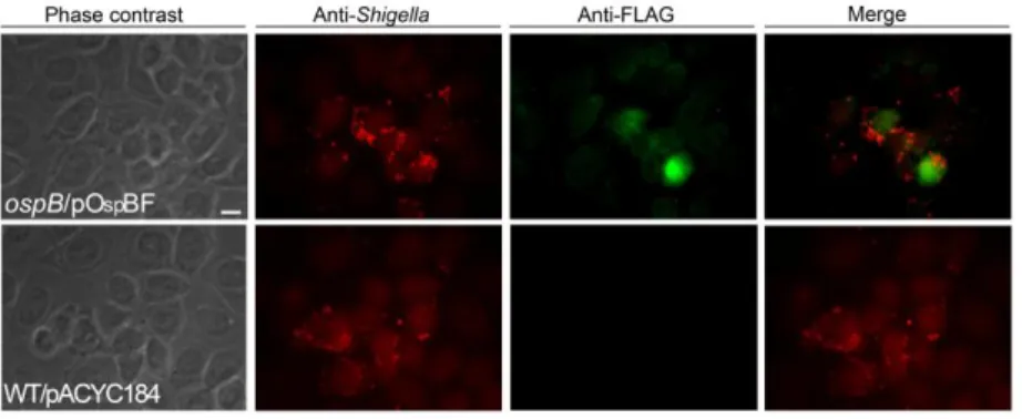
19 confluent HeLa cell monolayers were infected with the ospB mutant strain HND201 complemented with plasmid pOspBF (a pACYC184-based recombinant plasmid carrying the ospB gene fused at its C-terminus to the 3×FLAG tag; Table 1), and samples were analyzed by indirect immune-fluorescence. Interestingly, accumulation ofOspB within the host nucleus was observed already 30 min post-infection (Fig. 5) indicating that, once secreted into the cytosol, OspB rapidly moves to the nucleus. Remarkably, in silico analysis failed to evidence canonical nuclear localization signals (NLS) in OspB, indicating that OspB might directly interact with host proteins presenting canonical NLS to translocate within the nucleus.
Fig. 4. Expression of S. flexneri ospB gene during cell invasion.
Semi-confluent HeLa cell monolayers were infected with the wild-type S. flexneri M90T strain (MOI=150). At the indicated time points post-infection, total mRNA was extracted, cDNAs were synthesized and expression was measured by quantitative Real-Time PCR. Bars depict relative levels of
ospB mRNAs, normalized to the levels of the constitutively expressed
house-keeping gene rrsA (16S rRNA). Values represent mean±SD of three independent experiments, in duplicate.
20
Fig. 5. OspB localizes within host nucleus at early times of infection.
Semi-confluent HeLa cell monolayers were infected with the ospB mutant HND201 complemented with plasmid pOspBF (ospB/pOspBF) (Table 1), at a MOI of 150. At suitable intervals post-infection, cell monolayers were fixed and labeled with antibodies against the LPS of the S. flexneri serotype 5a strain M90T (red), and anti-FLAG (green). Images shown above were taken 30 min post-infection, recorded with a Leica camera and processed using Qwin software (Leica DMRE). Scale bar = 10 µm.
OspB activates the MAPK signalling pathways in Caco-2 cultured intestinal epithelial cells
Several pathogenic bacteria are able to modulate host MAPK signalling pathways to colonize and multiply within the susceptible host (Shan et al., 2007, Bhavsar et al., 2007). Accordingly with the timing of the release of OspB in infected host cells cytoplasm (data not shown), the role of OspB in the induction of MAPK signalling pathways was determined at early stages of infection. Semiconfluent monolayers of the colonic epithelial Caco-2 cell line were infected (MOI=150) with the wild-type, the ospB mutant strain HND201, and HND201 complemented with pOspB. At 5, 15, 30 and 60 min post-infection whole cell extracts were prepared and analyzed by Western blot by using polyclonal Erk1/2 and anti-phospho-p38 antibodies. Anti-total-Erk1/2 and anti-total-anti-phospho-p38 antibodies were used in order to detect the total level of Erk1/2 and p38 present in each sample as well as to ascertain that equal amounts of proteins were loaded in each well (Fig. 6A and B). The intensity of each band was quantified by densitometric analysis and expressed as the ratio between phosphorylated
21 and total protein. Interestingly, 5 min post-infection we found a remarkable reduction in the amounts of phosphorylated Erk1/2 in all samples analyzed. On the other hand, at 15 min time point onward, compared to cells infected with the wild-type strain, cells infected with the ospB mutant HND201 showed approximately 50% less phosphorylated Erk1/2 (Fig. 6C). Remarkably, a more dramatic reduction of the amount of phospho-p38 (about. 70%) was detected at 5 and 15 min post-infection in cells infected with the ospB mutant (Fig. 6D). Differently from pErk1/2, phospho-p38 levels returned to the values observed in cells infected with wild-type at 60 min post infection (Fig. 6). As expected, the ability to modulate phosphorylation of Erk1/2 and p38 at almost wild-type levels was restored when HND201 was complemented with plasmid pOspB (Fig. 6). These results indicated that OspB, during its translocation to the nucleus, someway targets cytosolic MAPKs, Erk1/2 and p38 inducing their activation at early stages of infection. Since it is known that phosphorylation of ERK1/2 and p38 leads to the activation of complex pro-inflammatory pathways leading to PMNs migration, we hypothesized that OspB might play also a role in PMNs migration to the site of infection.
22 F ig . 6 . O spB i nd uces E rk1 /2 a nd p3 8 ph o sph o ry la tio n. Fig u re sh o ws rep resen tativ e im ag e s o f W ester n b lo t an aly sis . C ac o -2 ce lls wer e in fec ted with wild -ty p e (W T ), th e o sp B m u tan t str ain HND2 0 1 ( o sp B ), a n d its co m p lem en ted -d er iv ativ e str ain ( o sp B /p Osp B ). At th e in d icate d tim e p o in ts p o st -in fec tio n , ce ll s wer e ly sed an d wh o le p ro tein e x tr ac ts s ep ar ated o n a 1 0 % SDS -PAGE . Af ter tr an sf er to PVDF m em b ran es, m em b ran es w er e p ro b ed with an ti -p h o sp h o -E rk 1 /2 a n d an ti -p h o sp h o -p 3 8 an tib o d ies (p an el A an d B , resp ec tiv ely ). M em b ran es wer e also p ro b ed with t h e co rr esp o n d in g an ti -to tal -E rk 1 /2 an d an ti -to tal -p38 an tib o d ies to ass u re th e lo ad in g o f eq u al am o u n ts o f p ro tein s (Pan els C an d D , resp ec tiv ely ). B ar s rep resen t th e m ea n s o f d en sito m etr ic an aly sis ( Im ag eJ so ftwar e ) o f th ree in d ep en d en t ex p er im en ts . Data ar e ex p ress ed a s ar b itra ry u n its . A ster is k s (* ) in d icate a statis tically sig n if ican t d if fer en ce co m p ar ed with to wild -ty p e (Stu d en t's t-test ; P v al u es < 0 .0 5 ). Un in fec ted C ac o -2 ce lls wer e u sed as c o n tr o l ( C tr l) .
23
Periplasmic PhoN2 (apyrase) polarly localizes in S. flexneri and E. coli cells
We have previously shown that PhoN2 (apyrase) is a virulence-associated factor required for efficient S. flexneri cell-to-cell spreading (Santapaola et
al., 2006). In spite of experimental and structural evidences indicating
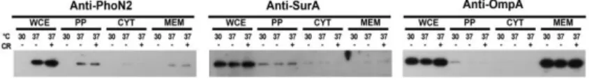
PhoN2 as a periplasmic protein (Bhargava et al., 1995, Babu et al., 2002), a conclusive demonstration has never been reported. To definitively settle this point, we analyzed protein extracts of different bacterial compartments (periplasm, cytosol and membrane fractions) of the wild-type strain M90T, grown at 30°C and at 37°C, in the presence of the Congo red dye in order to activate phoN2 MxiE-regulated transcription (Le Gall et al., 2005, Santapaola et al., 2006). Proteins were separated by SDS-PAGE and analyzed by Western blot using mouse polyclonal antibodies raised against PhoN2 (Santapaola et al., 2006). To assess contaminations in the fractions, the protein extracts were also challenged with antibodies against SurA and OmpA chosen as reporters of periplasmic and membrane proteins, respectively (Sklar et al., 2007, Smith et al., 2007). As shown in Figure 7, PhoN2 was found to be almost exclusively associated with the periplasmic fraction of bacteria grown at 37°C, in the presence or not of the Congo red dye. Accordingly, SurA and OmpA were found to be almost exclusively associated with the periplasmic and membrane fractions, respectively. As expected, phoN2 expression was repressed when bacteria were grown at the non-permissive temperature of 30°C (Le Gall et al., 2005; Santapaola et al., 2006).
Fig. 7. PhoN2 accumulates in the periplasmic compartment of S.flexneri. Whole cell extract (WCE), membrane, cytosolic and periplasmic
bacterial fractions of S. flexneri strain M90T were prepared from equal numbers of exponentially-grown bacteria. Bacteria were cultured at 30 and 37°C, and at 37°C in presence of CR, as described in Materials and Methods. Cell fractions were separated on a 12,5% SDS-PAGE. Following transfer of proteins to PVDF membrane, membranes were probed with anti-PhoN2 antibody and with anti-SurA, and anti-OmpA antibodies chosen as reporters of periplasmic and membrane proteins, respectively.
24 To trace PhoN2 within bacterial cells we constructed strain HNDHA10 by allelic exchange (Uzzau et al., 2001). Strain HNDHA10 is a derivative of the wild-type strain M90T carrying a C-terminal HA-tagged phoN2 gene (phoN2::HA) (Table 1). In this strain, the expression of the fused gene is under the control of its native promoter. The introduction of the HA-tag did not influence the delivery of PhoN2-HA into the periplasm space since PhoN2-HA was predominantly detected within the periplasmic compartment (data not show). Taken together, these results clearly confirmed that PhoN2 is a periplasmic protein and indicated that the introduction of a HA-tag at its C-terminus did not influence its periplasmic delivery. Next, the intracellular localization of PhoN2-HA was determined by indirect immune-fluorescence experiments using HNDHA10 and anti-HA monoclonal antibody. Unexpectedly, these experiments showed that PhoN2-HA displayed foci that were polarly localized (Fig. 8A). Fluorescence spots were detected at both bacterial poles in 54.8±5.8% and at one pole in 39.3±4.4% of immunostained bacteria (n=1.350). Remarkably, in bacteria displaying PhoN2-HA foci at both poles, the intensity of the fluorescence signal was significantly more intense at the old pole(80.5% of immunostained bacteria). To get further insight on the polar localization of PhoN2, plasmid pHND10, a pBAD28-based recombinant plasmid carrying the phoN2::HA fusion under the control of an L-arabinose-inducible promoter, was generated and introduced into HND115, a ΔphoN2 derivative of wild-type (Table 1) (Santapaola et al. 2006). Preliminarily, induction of phoN2::HA expression in HND115 carrying pHND10 was measured using a wide range of L-arabinose concentrations (from 0.002 to 0.2%). Real-Time PCR analysis showed that 0.016% of L-arabinose was the concentration that induced a level of phoN2::HA expression which more closely resembled that occurring in the wild-type strain grown in the presence of the Congo red dye, i.e. in conditions of MxiE-activated transcription (Fig. S1). Accordingly, unless otherwise indicated, 0.016% of L-arabinose was routinely used to induce phoN2::HA expression in the experiments described below. As expected, the phoN2 mutant harboring pHND10, grown in the presence or not of 0.016% of L-arabinose, was as efficient as the wild-type strain in invading HeLa cell monolayers and, only when grown in the presence of L-arabinose, pHND10 complemented HND115 for plaque size and catalytic activity (data not shown) (Santapaola et al., 2006). Immuno-fluorescence experiments showed that, as HNDHA10, also the phoN2 mutant harboring pHND10 displayed PhoN2-HA foci that were polarly localized both in exponentially-growing bacteria (91±6.5% of immunostained bacteriashowed PhoN2-HA
25 at bacterial poles or at one pole) (Fig. 8B) and in bacteria within infected HeLa cells (Fig.8D). Moreover, the intensity of the fluorescence spots displayed by HND115 (pHND10) induced with 0.016% of L-arabinose were apparently comparable to that of HNDHA10 where the expression of the fused gene was under the control of its native promoter. PhoN2-HA foci were visualized only upon induction with L-arabinose (data not shown).Western blot analysis confirmed the periplasmic localization of the fused protein encoded by pHND10 even if bands of lower intensity and corresponding to PhoN2-HA were also evidenced in the cytoplasmic and membrane fractions (Fig. 11).
In order to to get further insight of the mechanism by which PhoN2 polarly localizes, pHND10 was introduced into the E. coli K-12 strain DH10b and polar fluorescent PhoN2-HA foci were visualized only when bacteria were grown in the presence of the inducer L-arabinose (Fig. 8C, and data not shown). These results indicate that the mechanism driving the polar localization of PhoN2 is conserved among S. Flexner and E. coli.
26
Fig. 8. PhoN2 localizes polarly in both exponentially-growing S. flexneri and E. coli K-12 strains. Indirect immunofluorescence
experiments were carried out with HNDHA10 phoN2::HA, Panel A; HND115 (pHND10) cultured in the presence of 0.016% L-arabinose, Panel B and E. coli K-12 strain DH10b (pHND10) cultured in the presence of 0.016% L-arabinose, Panel C. PhoN2-HA was labelled with the anti-HA antibody (left column) and the fluorescence images were merged with phase contrast images (right column). Arrowheads indicate subpopulation of bacteria that display PhoN2-HA localized at bacterial poles. Bar = 2 µm. Panel D, Hela cells infected with HND115 (pHND10) strain, DAPI DNA staining (above) and DAPI and anti-HA merged fields (bottom). Boxs indicate subpopulation of bacteria that display PhoN2-HA localized at bacterial poles. Images are representative of three independent experiments.
27
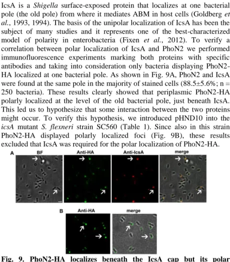
PhoN2 localizes beneath the IcsA cap
IcsA is a Shigella surface-exposed protein that localizes at one bacterial pole (the old pole) from where it mediates ABM in host cells (Goldberg et
al., 1993, 1994). The basis of the unipolar localization of IcsA has been the
subject of many studies and it represents one of the best-characterized model of polarity in enterobacteria (Fixen et al., 2012). To verify a correlation between polar localization of IcsA and PhoN2 we performed immunofluorescence experiments marking both proteins with specific antibodies and taking into consideration only bacteria displaying PhoN2-HA localized at one bacterial pole. As shown in Fig. 9A, PhoN2 and IcsA were found at the same pole in the majority of stained cells (88.5±5.6%; n = 250 bacteria). These results clearly showed that periplasmic PhoN2-HA polarly localized at the level of the old bacterial pole, just beneath IcsA. This led us to hypothesize that some interaction between the two proteins might occur. To verify this hypothesis, we introduced pHND10 into the
icsA mutant S. flexneri strain SC560 (Table 1). Since also in this strain
PhoN2-HA displayed polarly localized foci (Fig. 9B), these results excluded that IcsA was required for the polar localization of PhoN2-HA.
Fig. 9. PhoN2-HA localizes beneath the IcsA cap but its polar localization is independent by IcsA. Indirect immunofluorescence
experiments were carried out with HNDHA10 phoN2::HA (Panel A) and SC560 (pHND10) (Panel B) cultured in the presence of 0.016% L-arabinose. Images are representative of phase contrast (BF), anti-HA (green), anti-IcsA (red), and the merged fields (A); anti-HA (green) and merged fields in B. Arrowheads indicate subpopulation of bacteria that displayed PhoN2-HA and IcsA localized at bacterial pole Bar= 2 µm.
28
Characterization of PhoN2 domains involved in its polar localization
Functional and in silico analysis of PhoN2 have led to the identification of three relevant domains: i) an N-terminal 23 amino acid leader sequence, which is processed during its passage across the cytoplasmic membrane; ii) an N-terminal domain encompassing an exposed PPPP sequence (43PPPP46); and iii) a C-terminal wide domain encompassing the putative ATP-diphosphohydrolase catalytic site. We have previously reported that while PhoN2 was required for proper S. flexneri cell-to-cell spread, apyrase catalytic activity was not (Santapaola et al., 2006). To evaluate whether the catalytic activity of PhoN2 played a role in its polar localization, we introduced plasmid pHND19R192P [a pHND10-derivative plasmid carrying
the R192P amino acid substitution of PhoN2 known to suppress apyrase activity (Sarli et al., 2005; Santapaola et al., 2006)] (Table 1) into the
phoN2 mutant strain HND115. As revealed by Western blot analysis, the
R192P substituted protein was correctly delivered into the periplasmic compartment (Fig. 11). Since R192P substituted recombinant PhoN2-HA protein displayed polarly localized foci (Fig. 10B), this result clearly indicated that the deoxynucleotide triphosphate-hydrolyzing activity of PhoN2 is dispensable also for targeting PhoN2-HA to the bacterial poles. As expected, PhoN2-HA foci were detected only when bacteria were grown in the presence of L-arabinose (data not shown).
Next, a deletion of the fused gene which removed a DNA region encompassing the encoded PPPP motif (nucleotides 79-223) was generated in pHND10 (plasmid pHND11Δ79-223) (Table 1). When plasmid pHND11 Δ79-223 was introduced into the phoN2 mutant strain HND115, the Δ79-223
truncated HA-tagged protein, appeared as a diffuse signal (Fig. 10B). On average, in these experiments PhoN2-HA polarly localized foci were noticed in less than 3% of the bacterial population and in these cases the fluorescence signal was considerably reduced, compared to that displayed by parental pHND10. These results suggested that the polyproline domain could be important in the polar localization of PhoN2. Remarkably, pHND11Δ79-223 failed to express apyrase activity (Fig. 10C), indicating that
29
Fig.10 Characterization of PhoN2 domains involved in its polar localization and enzymatic activity
Panel A: in-frame deletion and amino acid substitution mutants of the
HA-tagged phoN2 gene. Amino acid residue 1 represents the first residue
of the leader peptide (dotted box), while numbers on the right indicate the last residue of the tagged HA epitope (white box). The position of the PPPP motif is indicated. Conserved D1, D2 and D3 domains corresponding to the putative catalytic site of PhoN2 (black box) (Babu et al., 2002; Sarli et al., 2005) and the substitution R192P, which inactivates apyrase activity, are indicated. Panel B: fluorescence microscopy, recombinant proteins were labeled with the anti-HA antibody (green) and fluorescence and phase contrast images were merged. Arrowheads indicate subpopulation of bacteria that display PhoN2-HA localized at bacterial poles. Panel C: ATP-hydrolysing activity of each recombinant protein calculated in comparison to the wild type protein. Bars represent the means of enzymatic activity of three independent experiments. Data are expressed as arbitrary units. Images are representative. Bar = 2 µm.
Site-directed mutagenesis were performed in order evaluate the role of the PPPP motif in the polar localization of PhoN2. To this end, a series of amino acid substitution within the PhoN2-HA PPPP motif carried by plasmid pHND10 were generated (see Materials and Methods for details). Four different derivatives of PhoN2-HA, carrying amino acid substitutions (P to S) of each of the P residues of the PPPP sequence, were generated and introduced separately into the phoN2 mutant strain HND115.
L-arabinose-30 induced HND115 carrying plasmids pHND23SPPP, pHND14PSPP,
pHND15PPSP, and pHND16PPPS (Table 1), were assayed for PhoN2 polar
localization, periplasmic delivery and for their ability to complement apyrase activity. As shown in Figure 11B, P to S substituted recombinant PhoN2-HA proteins encoded by pHND23SPPP, pHND14PSPP and
pHND15PPSP were correctly delivered into the periplasmic compartment,
although Western blot analysis showed that that encoded by pHND15PPSP
displayed a lower level of expression compared to the other two constructs (Fig. 11B). On the other hand, pHND16PPPS, which also showed low levels
of PhoN2-HA expression (Fig. 11B), was detectable only in the cytosolic fraction (Fig. 11B). Moreover, while pHND23SPPP, pHND14PSPP and
pHND15PPSP complemented HND115 for apyrase activity, (pHND15PPSP at
a lower-extent, compared to pHND10), pHND16PPPS failed to complement
(Fig. 11D). When the polar localization of the PhoN2-HA was analyzed (Fig. 11), substitution of the first (pHND23SPPP) and second (pHND14PSPP)
proline residues apparently did not influence the polar localization of PhoN2-HA recombinant proteins (respectively 92.8±5.6% and 94.1±6.1% of the cells displayed PhoN2-HA polarly-localized foci). On the other hand, substitution of the third proline residue (pHND15PPSP) induced a significant
reduction of the percent of bacteria presenting polarly localized PhoN2-HA foci (about 57.7±4.9 of fluorescent bacteria showed polar fluorescence spots while 43.4±2.9% of bacteria showed a diffuse signal; P values < 0.05) (Fig. 11C). Remarkably, no fluorescence signals or apyrase activity were observed when HND115 was complemented with plasmid pHND16PPPS.
31
F
ig
ure
32
Fig. 11. Characterization of the role of the PhoN2 PPPP motif and of amino acid residue Y155 in its periplasmic delivery, polar localization and enzymatic activity. Panel A: description of specific amino acid
substitutions introduced into PhoN2-HA. Amino acid residue 1 represents the first residue of the leader peptide (dotted box), while numbers on the right indicate the last residue of the tagged HA epitope (white box). The position of the PPPP motif is indicated. The conserved D1, D2 and D3 domains (black box) corresponding to the putative catalytic site of PhoN2 (Babu et al., 2002; Sarli et al., 2005) and the substitutions Y155A and R192P. Panel B: Whole cell extract (WCE), membrane, cytosolic and periplasmic bacterial fractions from equal numbers of exponentially-growing bacteria, cultured at 37°C, with 0.016% L-arabinose. Cell fractions were separated on a 12.5% SDS-PAGE. Following transfer PVDF membranes were probed with anti-PhoN2, anti-SurA and anti-OmpA antibodies. Panel C: fluorescence microscopy, recombinant proteins were labeled with anti-HA antibody (green) and fluorescence and phase contrast images were merged. Arrowheads indicate subpopulation of bacteria that display PhoN2-HA localized at bacterial poles Panel D: ATP-hydrolysing activity of each recombinant protein calculated in comparison to the wild type protein. Bars represent the means of enzymatic activity of three independent experiments. Data are expressed as arbitrary units. Images are representative. Bar= 2 µm.
33
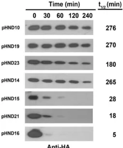
The PPPP motif controls PhoN2 stability
The finding that PhoN2-HA encoded by pHND15PPSP and pHND16PPPS
were expressed at lower levels, compared to that of pHND10, pHND23SPPP,
pHND14PSPP and pHND19R192P led us to consider whether the amino acid
substitution of the third and fourth proline residues might have affected PhoN2 stability. To verify this hypothesis, the intracellular stability of PhoN2-HA carrying the different amino acid substitutions was monitored after protein synthesis had been inhibited by the addition of 100 μg/ml of spectinomycin. Samples were removed at different time points and whole cell extracts were immunodetected by Western blot. Quantitative measurements were carried out to estimate the half-lives of each recombinant proteins. As seen in Figure 12, the PhoN2-HA proteins encoded by L-arabinose-induced phoN2 mutant carrying pHND10 (control), pHND23SPPP, pHND14PSPP and pHND19R192P were stable throughout the
duration of the experiment (half-lives of 276, 180, 265 and 270 min, respectively), while pHND15PPSP, and pHND16PPPS displayed an high
degree of protein instability already 30 min after protein synthesis had been inhibited (Fig. 12). We calculated half-lives of 28 and 5 min for proteins encoded by pHND15PPSP, and pHND16PPPS, respectively (Fig. 12). These
results clearly showed that P to S substitutions of the third and of the fourth proline residues dramatically affected PhoN2 stability.
Next, to test if some specific protease was responsible for the degradation of these recombinant protein, we examined the level of PhoN2-HA encoded amino acid substituted proteins in different genetic backgrounds. Noteworthy, no significant difference in the steady-state levels of PhoN2-HA was noticed when recombinant plasmids were introduced into the E.
coli K-12 strain DH10b (Fig. 5A), while high levels of PhoN2 instability
were detected when pHND15PPSP, and pHND16PPPS were introduced into
strain BS176, a virulence plasmid-cured derivative of the S. flexneri strain M90T (Table 1) (Fig. 5B). These results clearly indicated that the structural gene(s) encoding the protease(s) involved in PhoN2-HA protein degradation are S. flexneri-specific and excluded that were encoded by genes localized on the virulence plasmid of S. flexneri strain M90T.
34
Fig. 12. Assessment of PhoN2 stability. HND115 carrying the indicated
plasmids were grown to the mid-exponential phase, in the presence of 0.2% L-arabinose, and protein synthesis was inhibited by addition of Sp (100 μg ml-1). Equal amount of bacteria were removed at 0, 30, 60, 120 and 240 min after Sp was added and re-suspended in 1 × Laemmli buffer. Protein extract were subjected to Western blot analysis. Densitometric analysis (ImageJ software) of each band was performed and half-lives (t1/2 min) calculated
35
Fig. 13. A Shigella specific protease degrades PhoN2 mutant derivatives. Panel A: E. coli K-12 strain DH10b complemented with
pHND10PPPP, pHND15PPSP, and pHND16PPPS cultured with 0.2%
L-arabinose. Panel B: virulence plasmid-cured derivative of the S. flexneri strain BS176 complemented with pHND10PPPP, pHND15PPSP, and
pHND16PPPS cultured with 0.2% L-ara. Bacteria were grown to the
mid-exponential phase and protein synthesis was inhibited by the addition of Sp (100 μg ml-1). Equal amount of bacteria were removed at 0, 30, 60, 120 and 240 min and re-suspended in 1 × Laemmli buffer. Protein extract were subjected to Western blot analysis. Images are representative.
36
The polyproline PPPP motif indirectly influences S. flexneri virulence
Previous work has shown that deletion of the phoN2 gene led to altered actin comet tails and consequently to inefficient cell-to cell spreading and to a small plaque phenotype (Santapaola et al., 2006). The central bacterial mediator of actin based motility in S. flexneri is IcsA (Schoeder and Hilbi, 2008). Immunofluorescence experiments indicated that PhoN2 influenced IcsA exposition since, compared to the wild-type strain, the phoN2 mutant strain HND115 displayed a significant reduction of IcsA exposition. This reduction was due to the lack of PhoN2, since complementation with pHND10 restored almost parental IcsA caps (Fig. 14), only when bacteria were supplemented with L-arabinose (approximately 78.6±7.8% of immunostained bacteria showed proper IcsA caps). These results indicated that PhoN2 is involved in the complex process of correct export and exposition of IcsA on S. flexneri bacterial surface. pHND23SPPP,
pHND14PSPP, pHND15PPSP and pHND19R192P were also able to positively
complement, at almost parental level, IcsA exposition, while pHND16PPPS,
likely because the high level of instability of the encoded PhoN2-HA substituted protein (Fig. 12), did not. Overall, these results showed that apyrase activity is not required for proper IcsA exposition and indicated that the PPPP motif, by influencing PhoN2 protein stability, is at least indirectly involved in the virulence of S. flexneri.
37
Fig.14. Fluorescence microscopy of IcsA. Parental strain M90T, HND115
and HND115 complemented with recombinant plasmids pHND10PPPP,
pHND23SPPP, pHND14PSPP, pHND15PPSP, pHND16PPPS, and pHND19R192P.
Were cultured in the presence of 0.016% L-arabinose. IcsA was detected in exponentially-grown bacteria, fixed with paraformaldehyde and labeled with polyclonal anti-IcsA antibody. The arrowheads indicate subpopulations of IcsA-labeled bacteria. Bar = 2 µm.
38
The poly-proline region is necessary for PhoN2 3D structure
As outlined in the Introduction section, PhoN2 belongs to the family of the class A of the non-specific bacterial acid phosphatases (A-NSAPs) a group of phosphatases highly diffused among Gram-negative bacteria (Rossolini
et al.,1998). Among the relevant characteristics of this group of enzymes
there is the presence of an N-terminal PPPP motif which was found to be highly conserved among all A-NSAPs analysed so far. While no data are available for PhoN2, it has been recently reported that the invariant residue Tyr-154 (Tyr-155 in PhoN2) by binding to the distal Pro-40 residue of the PPPP motif of PhoN (an A-NSAP encoded by Salmonella Typhimurium) stabilizes the loop harbouring the active site residues (Fig. 15) (Ishikawa et
al., 2000, Babu et al., 2002, Makde et al., 2007).
Fig. 15. Structurally conserved domains between PhoN of Salmonella Typhimurium and PhoN2 of S. flexneri. The loop harbouring active site
residues (154YPSGH158) in PhoN from S. typhimurium (PhoNSt) is stabilized
by the interaction between the highly conserved Tyr-154 residue and the distal 40PPPP43 motif (shown in magenta). The sequence of PhoSt in those
critical motifs was aligned with PhoN2 from S. flexneri (PhoN2Sf) to
highlight highly conserved residues between primary sequences. Adapted from Makde et al., 2006.
39 On this basis, it was conceivable to hypothesize that the Tyr-155 residue of PhoN2 could exert a crucial structural role in protein conformational stability. To unravel this point, we generated pHN21Y155A, a recombinant
plasmid encoding the PhoN2-HA Y155A substitution (Table 1) (see Fig. 11 legend for details). pHN21Y155A was introduced into HND115 and stability
of the substituted recombinant protein was evaluated by Western blot, after protein synthesis has been inhibited by the addiction of 100 µg/ml of spectinomycin, as described above. As shown in Figure 12, the substituted protein encoded by pHND21Y155A displayed a high degree of protein
instability already 30 min after protein synthesis had been inhibited (half-live of 18 min). These results clearly showed that Tyr-155 along with the PPPP motif play a critical role in conformational stability of PhoN2 and indeed that the conformational stability is a key determinant of the proteolytic susceptibility of this protein. Interestingly, the inactivation of the catalytic domain does not influence the stability of the protein (pHND19R192P: half-live of 270 min).
Finally, the influence of the Y155A substitution on the expression of apyrase activity and on the polar localization of PhoN2 was also evaluated. Accordingly to the results obtained with the highly unstable PhoN2-HA recombinant protein encoded by pHND16PPPS, pHN21Y155A failed to
complement HND115 for apyrase activity and no polar PhoN2 foci were detected in immune-fluorescence experiments (no fluorescence signals were observed) (Fig. 11), indicating that proper PhoN2 folding is required for the polar localization of this protein. Taken together these results clearly indicated that residue Tyr-155, by regulating protein stability, positively influences apyrase activity and the ability of PhoN2 to localize at the old bacterial pole of S. flexneri.
PhoN2 binds to the OM protein A (OmpA)
The localization of PhoN2 at the old bacterial pole raises the possibility that it might interact with bacterial protein(s) which assist PhoN2 in the accumulation at polar bacterial sites. To find and to characterize these specific protein(s), co-purification experiments were conducted with C-terminal 3xFLAG- or 6-His-tagged PhoN2, using affinity chromatography resins. Bound resins, challenged with a clear lysate of the wild-type strain, failed to detect any interaction partner (data not shown). Interaction partners were then searched by the two-hybrid technique in yeast. By using pGBKT7/phoN2 as bait, the cDNA library of the wild-type strain fused in frame to the coding sequence of the GAL4-activating domain of plasmid
40 pGADT7-Rec (Table 1) was screened, as described in Materials and Methods. Saccharomyces cerevisiae AH109 competent cells were simultaneously transformed with the bait plasmid and the prey plasmid library and spread on selective plates. Plasmid DNA preparations of 16 independent transformants grown on minimal medium agar plates lacking leucine, tryptophan, histidine and adenine and expressing high-level of α-galactosidase, were used to transform E. coli DH10b competent cells selecting for kanamycin-resistance. The DNA inserts carried by independent prey plasmid DNA preparations were sequenced. Remarkably, the great majority of proteins encoded by the prey plasmids (13 out of the 16 examined) corresponded to the C-terminal domain of OmpA (Table 3). All OmpA inserts encompassed a common region composed of residues 189-273. The 189-273 residues represent the C-terminal domain of OmpA known to be exposed into the bacterial periplasm when the protein is in the eight-stranded β-barrel conformation (Koebnik and Kramer, 1995). These results were highly indicative of a PhoN2-OmpA interaction involving the periplasmic-exposed C-terminal domain of OmpA. As stated above, this interaction was not detected in co-purification experiments, most probably because the soluble form of OmpA was either not present in the bacterial clear lysate or not sufficient to be visualized by Comassie staining.
The sequence of the inserts of the three remaining prey plasmids (Table 3) encompassed amino acid residues of three different cytosolic S. flexneri proteins, namely Adh, AspRS and EF-2. The inserts encoded by these three plasmids were not analyzed further.
41
Table 3. Preys selected by the PhoN2 bait.
Prey Starta Enda Number of
clonesb Common regionc OmpA (348 aa) 189 276 5 189-273 185 304 2 43 275 2 188 299 1 51 273 1 72 277 1 186 284 1 Adh (336 aa) 129 218 1 AspRS (590 aa) 358 472 1 EF-2 (665 aa) 114 264 1
aAminoacid coordinates, with respect to the sequence of the native proteins,
of the first and last residue encoded by the insert in each prey plasmid.
bNumber of independent clones containing identical prey fragments. c
Coordinates of the common region carried by all preys corresponding to the same protein.
In vivo cross-linking experiments
To confirm the interaction between PhoN2 and OmpA, in vivo cross-linking experiments were conducted using formaldehyde (Fig. 16). Cross-linking was performed with the phoN2 mutant strain HND115 carrying pHND10, grown in the presence (Fig. 16 B and D) or not (Fig. 16 A and C) of 0.016% of L-arabinose (see Materials and Methods for details). Total protein extracts were separated by SDS-PAGE and analyzed by Western blot using monoclonal anti-HA (Fig. 7A and B) and polyclonal anti-OmpA antibodies (Fig. 16 C and D). Western blot of bacterial extracts, denatured at 95°C in Laemmli buffer prior to electrophoresis, detected PhoN2-HA (Fig. 16 B, lanes 4 and 6) and unfolded OmpA (Fig. 16 C and D, lanes 7, 9, 10 and 12) of the expected molecular size (28 and 35 kDa, respectively), while in samples heated at 37°C OmpA migrates as a folded 30 kDa protein (Fig. 16 C and D, lanes 8 and 11) (Schweizer et al., 1978). On the other hand, extracts of formaldehyde-treated bacteria, expressing both PhoN2-HA and OmpA, generated an additional molecular species when samples were heated at 37°C (Fig. 16 B and D, lanes 5 and 11). This new molecular species migrated with an apparent mass of 63 kDa, consistent with the molecular size corresponding to the sum of mature PhoN2-HA and OmpA.