UNIVERSITÀ DI PISA
Dipartimento di Farmacia
Corso di Laurea Specialistica in
Chimica e Tecnologia Farmaceutiche
“Arylthioamides and aryliminothioethers as new slow H
2S- releasing agents”
Candidato:
Mariagrazia Angelini
Relatore: Relatore:
Dott.ssa Sabrina Taliani Dott. Vincenzo Calderone
Dott.ssa Isabella Pugliesi
Index
1. Introduction
1
1.1
H
2S chemistry and biosynthesis 3
1.2
H
2S catabolism 7
1.3
Tissue distribution of H
2S-synthesizing enzymes 8
1.4
Mechanisms of H
2S biological actions 14
1.5
Physiological roles of H
2S 19
1.5.1
Central nervous system
1.5.2
Gastrointestinal system
1.5.3
Endocrine system
1.5.4
Cardiovascular system
1.6
H
2S donors 25
1.6.1
H
2S- non steroidal anti-inflammatory drugs
1.6.2
H
2S-Latanoprost
1.6.3
H
2S-Sildenafil
1.6.4
H
2S-Sartans
1.6.5
H
2S-releasing derivatives of L-DOPA
2. Introduction to Experimental Section
35
3. Biological Studies
46
4. Experimental Section
54
2
INTRODUCTION.
In the last few decades, investigation of the pathophysiological and pharmacological roles of ‘‘gasotransmitters’’ has represented a challenging research field, which is still widely unexplored. This research activity led to achieve useful knowledge about the biological importance of such endogenous compounds in many systems, with heavy implications for drug discovery1.
The discovery of the gaseous endogenous mediator nitric oxide (NO) and the understanding of its multiple roles in the physiological modulation of cardiovascular functions has been a “revolutionary” milestone, which has deeply influenced the development of cardiovascular pharmacology in the last decades2. The recognition of carbon monoxide (CO) soon followed as a second gaseous neurotransmitter that acts similarly to NO3. Nitric oxide and carbon monoxide activates soluble guanylate cyclase and elevates cGMP in target tissues, which dilates blood vessels⁴. Presently, a third candidate is emerging as an important subject of great scientific interest: a body of recent findings indicates that hydrogen sulphide (H₂S, another gas previously regarded as a toxic agent) is an endogenous compound produced in substantial amounts by mammalian tissues, which exerts a variety of physiological effects in different systems, particularly in the cardiovascular system1.
Gasotransmitters share some common properties: • easy diffusion across biological membranes; • fine-tuning of their endogenous production;
• capacity to regulate their biological functions at physiological concentration; • presence of specific pharmacological targets;
• short half-life;
• potential toxicity at higher concentrations⁵.
Particularly, hydrogen sulphide (H₂S) is a colourless, flammable, watersoluble gas with the characteristic smell of rotten eggs. The toxicology of high concentrations of H2S as an
environmental pollutant has been studied extensively. Since it was first discovered to be synthesized in mammalian and human tissues, it has attracted considerable interest in a
3 relatively short period of time as an endogenous gaseous mediator and potential pharmacological and therapeutic tool. Studies in animals and humans have shown H₂S to be involved in diverse physiological and pathophysiological processes, such as regulation of blood pressure, inflammation, neurodegenerative diseases and metabolic disorders, including obesity and diabetes ⁸.
1.1 H₂S CHEMISTRY AND BIOSYNTHESIS.
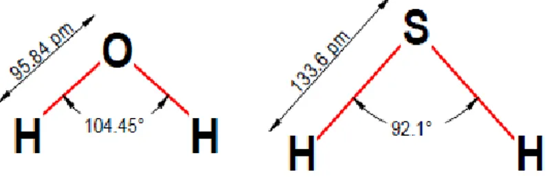
The structure of H₂S is very similar to the structure of water, because of the electronegativity of sulfur and oxygen.
Figure 1 - Similarity between H₂O and H₂S.
These molecules have both polar characteristics, but H₂S has a lower polarity and intermolecular forces than water.
In aqueous solution, H₂S is weakly acidic (pKa = 6.76 at 37 ◦C) and dissociates to form two dissociation states: HS- (hydrosulfide anion) (pKa = 7.04) and S2-(sulfide anion) (pKa = 11.96), according to the following sequential reactions:
H
2S
Ka1H
++ HS
- Ka2H
++ S
2-At pH 7.4, approximately 18.5% of the total sulfide exists as the undissociated acid and 81.5% as HS‾ . It is currently not known whether the biological effects of H₂S are mediated directly by H₂S itself or by derived species that will also exist at physiological pH, predominantly HS- but also S2-. It is unlikely S2- will play a significant role, since it will only be present at a high pH.
4 As a varied mixture of these species will always exist under physiological conditions irrespective of the source of H₂S, it is prudent to use the term ‘H₂S’ to encompass the sum of these species present under physiological conditions ⁸.
Due to its high lipophilicity, H₂S freely crosses the biological membranes and penetrates all types of cells; this property confers to H₂S a high biological potential. HS-, which is the prevalent sulphide species, acts as a nucleophile and readily binds to metal centers of biological molecules, such as the hemoglobin oxygen-binding site. This chemical behaviour, together with other kinds of catabolic outcome, may lead to a substantial lowering of sulphide concentration in the body1. Endogenous H₂S can be produced in mammalian tissues by both enzymatic and nonenzymatic pathways. The nonenzymatic way, although less important, proceeds via the nonenzymatic reduction of elemental sulphur to H₂S using reducing equivalents obtained from the oxidation of glucose.
Lactate is the main product of glucose metabolism, as summarized in the following reaction:
2C
6H
12O
6+ 6S
0+ 2H
2O
3C
3H
6O
3+ 6H
2S + 3CO
2Nevertheless, from the experimental data, it emerges that for every 2 molecules of glucose consumed there are 6 H₂S and 2 CO₂ produced, and this lack of one CO₂ molecule is possibly correlated with pyruvate excretion. Although glucose best supports H₂S production, other substrates also are effective. For example, the electron carriers NADH and NADPH are likely to be involved in this process. In fact, NADH, NADPH, and glutathione (GSH) have been found to stimulate H₂S production in human erythrocytic cellular lysates. Probably, GSH is directly responsible for most H₂S production. In turn, the resulting oxidized glutathione (GSSG) is reduced by NAD(P)H and used again.
With regard to the enzymatic route, L-cysteine is a substrate for the enzymatic production of H₂S, and this process is ensured by either of the two pyridoxal-5'-phosphate-dependent enzymes: cystathionine β-synthase (CBS) and cystathionine γ-lyase (CSE)1. In mammalian tissues, H₂S is produced through at least four different biosynthetic pathways:
5 ➢ in the first biosynthetic route, CBS hydrolyzes L-cysteine with formation of equimolar amounts of L-serine and H₂S (figure 2).
L-Cysteine
CBS
Vit B6
L-Serine + H
2S
Figure 2 - Biosynthesis of H₂S: first pathway.
➢ In the second pathway, (figure 3) two L-cysteine molecules react to form L-cystine (L-cysteine dimer), which is broken down to thio(L-cysteine, pyruvate, and NH₃ through a CSE-mediated reaction. In turn, thiocysteine undergoes two different processes:
• a nonenzymatic process, resulting in the formation of L-cysteine and H₂S,
• a CSE-dependent reaction with a thiol compound R-SH (such as cysteine or glutathione), resulting in the production of H₂S and CysS-R.
2 L- Cysteine
L-Cysteine
CSE
Vit B6
Thiocysteine + Pyruvate + NH
3L- Cysteine + H
2S
non enzimatically
+
RSH
CysSR + H
2S
dimerization
6 ➢ The third pathway (figure 4) involves the participation of the enzyme cysteine aminotransferase (CAT), which catalyzes a reaction between L-cysteine and α- ketoglutarate, leading to the formation of 3-mercaptopyruvate and L-glutamate; 3- mercaptopyruvate can be :
• desulphurated by 3-mercaptopyruvate sulphurtransferase (MPST) to yield pyruvate and H₂S ,
• alternatively, when sulphite ions (SO₃2-) are available, 3-mercaptopyruvate can be converted to pyruvate and thiosulphate (S₂O₃2-), which in turn reacts with reduced GSH to produce H₂S , SO₃2-, and oxidized glutathione (GSSG).
L-Cysteine +
- Ketoglutarate
CATViT B6
3-mercaptopyruvate + L-glutamate
+
SO
32-Pyruvate + H
2S
Thiosulphate + Pyruvate
H
2S + H
2SO
3+ GSSG
GSH CAT MPST7 ➢ In the fourth enzimatic pathway, L-cysteine and sulphite are converted to L-cysteate and H₂S by cysteine lyase (figure 5)1.
L-Cysteine + SO
32- Cysteine lyaseVit B6
L-Cysteate + H
2S
Figure 5 - Biosynthesis of H₂S: fourth pathway
1.2 H₂S CATABOLISM.
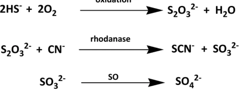
H₂S shows a relatively short half-life (lower than 30 minutes), and several pathways responsible for H₂S “destruction” have been described2. It is noteworthy that H₂S is a reducing agent that can be easily oxidized by several circulation oxidants. However, one of the main pathways of H₂S catabolism operates in mitochondria. In particular, it has been recently observed that rat liver mitochondria can efficiently oxidize H₂S. This oxidative process is prevented by inhibitors of complexes III and IV of the respiratory chain, such as myxothiazole and cyanide, respectively. The oxidation mechanism proceeds through several enzymatic steps, mediated by quinone oxidoreductase, S-dioxigenase, and-transferase and, overall, leads to the formation of thiosulphate. Thiosulphate is further biotransformed by rhodanase to sulphite with a reaction that requires also the presence of cyanide, which in turn is converted to thiocyanate. Sulphite is finally oxidized to sulphate by sulphite oxidase.
Although inorganic sulphate is the predominant stable end-product of H₂S catabolism, it cannot be taken as a reliable bio-marker for an accurate quantitative estimation of H₂S production in mammalian blood. In fact, sulphate ions can also be generated by other sources, such as the direct oxidation of cysteine by cysteine dioxygenase and the oxidation of sulphites produced by other sources5 (figure 6).
In mitochondria:
2HS
-+ 2O
2 oxidationS
2O
32-+ H
2O
S
2O
32-+ CN
- rhodanaseSCN
-+ SO
32-SO
32- SOSO
48 Other mechanisms are also known including the interaction between H2S and
methemoglobin that leads to sulphemoglobin, which is considered a possible bio-marker of plasmatic H₂S (figure 7).
In blood:
H
2S + methemoglobin
sulphemoglobin
Figure 7 - H₂S catabolism in blood.
An additional alternative pathway which operates only in the cell cytosol and involves smaller amounts of H₂S, is the methylation of H₂S by thiol S-methyltransferase (TSMT) to yield methanethiol and then dimethylsulphide (figure 8). Other minor oxidative routes of H₂S to sulphite also have been found in activated neutrophils2.
In cytosol:
H
2S
TMSTCH
3-SH
TMSTCH
3-S-CH
3Figure 8 - H₂S catabolism in cytosol.
1.3 TISSUE DISTRIBUTION OF H2S-SYNTHESIZING ENZYMES.
It been widely suggested that the expression of CSE, CBS, MPST and CAT in rodents and humans showed a marked degree of tissue specificity. However, as more researchers investigate the emerging role of H₂S in their particular system, this simple and convenient distinction is no longer as clear as once thought. Up until recently, the current literature consensus was that CBS was the predominant source of H₂S in the brain and nervous tissue, highly concentrated in cerebellar Purkinje and hippocampal neurons, and that in the vasculature (e.g. smooth muscle and endothelium) CSE was the major source of H₂S. However, it has been known for some time that neuronal tissue clearly contains CSE and vascular tissue contains CSE and CBS, and the previous assumption on distinct tissue distribution is not clear. Furthermore, CBS has been shown to be preferentially expressed in radial glia/astrocytes of adult and developing mouse brain, but is not present in neurons, and at least the rodent macrovasculature has an additional pathway for synthesizing H₂S via MPST/CAT8.
9 However, the distributions of CSE and CBS are much more widespread, highlighting the importance of H₂S synthesis in a multitude of tissues. The majority of studies have been conducted using animal tissues and it is assumed, but not yet demonstrated, that the distribution of CSE, CBS, CAT and MPST will be similar in humans. H₂S synthesis from CAT/MPST has only been demonstrated in animal tissue. Cysteine lyase has only been shown to be present in non-mammalian species (Table 1)8.
Table 1. Some examples of the tissue distribution of H₂S-synthesizing enzymes.
Enzyme Species Tissue Distribution (localization)
CSE Human Brain Astrocytes [163,188], pre-central
cortex [163],
mRNA expression and catalytic activity in cerebellar and granule and Perkinje cells, pyramidal neurons of CA3 and granule cell layer of dentate gyrus, and reticular neurons in midbrain Lens Epithelium, cortex and nucleus; CBS
more abundant in the epithelium of younger individuals (17–21 years) compared with older individuals; CBS is more abundant in the older age group (63–66 years)
Joint cells Articular chondrocytes, synoviocytes and trabecular bone-derived mesenchymal cells
Vasculature Internal mammary artery Lung Pulmonary artery smooth muscle Intrauterine tissue Chorion, amnion, myometrium and
placenta
Mouse Brain Purkinje cells (cell bodies and
neuronal processes),hippocampus (cell bodies and neuronal processes), optic nerve, cerebral cortex, striatum, thalamus and spinal cord, neuroblasts (early stages of development), hippocampal dentate gyrus, cerebellar astrocytes and Berman glia, cerebellar and granule and Purkinje cells, pyramidal neurons of CA2 and CA3 subfields of the hippocampus, reticular neurons of the midbrain and granule cell layer of dentate gyrus Pancreas Acinar cells, exocrine cells and iselets Ovary Ovary follicular cells (cell bodies)
10
Pig Vasculature Endothelial cells
Lens Epithelial layer, outer cortex, inner cortex, and nucleus; relative concentration ratio of 3:1:1:1 respectively
Rabbit Reproductive organs Vaginal and clitoral cavernosal smooth muscle
Rat Brain Hippocampus; CBS highly expressed in
the hippocampus and cerebellum compared with the cortex and brain stem
Liver Induced expression and activity in during acute endotoxaemia
Kidney Induced expression and activity in during acute endotoxaemia
Gastrointestinal tract
Liver, stomach, duodenum, jejunum, ileum, colon (Western blotting); diffuse immunostaining in the colon and surrounding blood vessels Lung Airway and vascular smooth muscle Intrauterine tissue Uterus, placenta and fetal membrane
CBS Human Colon Low basal level of exoression
Brain Neurons in cerebral cortex, cerebellum (Purkinje cells), hippocampus (dentate gyrus and CA3 pyramidal neurons) and mid-brain (reticular neurons)
Joint cells Articular chondrocytes, synoviocytes and trabecular bone-derived
mesenchymal cells; cytokine and LPS-inducible expression and activity of CSE
Eye Lens
Intrauterine tissue Chorion, amnion, myometrium and placenta
Rabbit Genitals Vaginal and clitoral cavernosal smooth muscle
Eye Lens
Guinea-pig Gastrointestinal tract
Ileum
Rat Blood vessels Portal vein, thoracic aorta, and pulmonary, mesenteric and tail arteries
Heart Myocardium
Gastrointestinal tract
Stomach, duodenum, jejunum, ileum and colon; in the colon, CBS immunostaining was primarily localized in muscularis mucosa, submucosa and lamina propria; no immunostaining in crypt, goblet or
11 epithelial cells
Brain Cerebellar, hippocampus
Intrauterine tissue Uterus, placenta and fetal membrane
Pig Eye Lens: mRNA and CSE activity
detected; activity and mRNA expression decreased with age Mouse Pancreas Acinar cells and exocrine cells
Liver, brain and colon
CBS immunoreactivity detected in hippocampal neuronal bodies and neocortical cell bodies
CAT Rat Brain Olfactory bulb and cerebellum, and
cerebral cortex
Vasculature Thoracic aorta (cystol and mitochondrial distribution in the endothelium)
MPST Mouse Brain Neurons of mitral cell layers,
glomerular and external plexiform layers of olfactory bulb, spinal cord (large neurons), cerebral cortex, Purkinje cell somata, proximal dendrites, pons (pontine nuclei) and hippocampus (CA1 and CA3 pyramidal cells)
Rat Vasculature and liver Endothelium and smooth muscle of the thoracic aorta
Liver, kidney and brain
Cytoplasm and mitochondrial expression in proximal tubular epithelial cells in kidney; pericentral hepatocytes in the liver; perinuclear area of myocardial cells, and glial cells in brain
Human U373 astrocytoma
Cells
MPST activity only
Uterus Myometrium and leiomyomas Cysteine lyase Amphibians,
repltiles and fish
Muscle Species of batrachian, Caudata, Nothobranchiidae, Neoselachii, Rivulus and Chamaeleo
Birds Muscle Gallus gallus
In humans, the gene for CSE is located on chromosome 1. CSE is a member of the
-family of PLP dependent enzymes and is a 405-amino-acid protein consisting of a tetramer formed by two homodimers. The crystal structure of human CSE in the apo form and in complex with PLP (pyridoxal phosphate) has been determined recently (figure 9)11. CSE catalyses the α,-carbon elimination of cystathionine to produce cysteine, α-oxobutyrate and ammonia. Additionally CSE may catalyse the β-elimination of cysteine
12 (cysteine disulfide) via the formation of thiocysteine, which then decomposes nonenzymatically to H₂S. However, in the presence of physiological concentrations of cysteine(~100 μM), homocysteine (10 μM) or cystathionine (5 μM), although the catalytic-centre activity number for CSE-mediated cystathionine cleavage was 5-fold greater than for cysteine and 12-fold higher than for homocysteine, the CSE-catalysed
α,-elimination of cysteine was the predominant source of H₂S accounting for ~70% of the H₂S produced, whereas the α,-elimination of homocysteine accounted for ~29% of
the measured H₂S8.
Figure 9 - Oligomeric states of hCSE
At least two CSE mRNA splice variants have been demonstrated producing long (CSE-l) and truncated (CSE-s) CSE proteins. Although CSE-s has been suggested to be inactive, the experiments to determine this only measured cysteine accumulation from added cystathionine as an index of enzymatic activity but did not examine H₂S production. As cysteine will also serve as a preferential substrate for CSE, the lack of accumulation of cysteine from CSE-s suggests it was probably consumed in the generation H₂S. However, the precise role of these splice variants in regulating CSE activity in terms of H₂S production requires further attention and may offer therapeutic potential for controlling endogenous H₂S synthesis8.
13 The human CBS gene is located on chromosome 21. CBS is a homotetramer consisting of 551-aminoacid subunits which bind two cofactors (haem and PLP) and two substrates (homocysteine and serine). To generate H₂S, human CBS can use either cysteine, forming lanthionine, or cysteine plus homocysteine, forming cystathionine. This latter reaction is predicted to predominate under physiological conditions and account for ~96% of the total H₂S generated from CBS, whereas the reactions in the absence of homocysteine represent up to 2.6% of the total H₂S. Although the CBS gene encodes several mRNAs, the functional product of these mRNA isoforms in terms of H₂S synthesis have yet to be examined (figure 10)12
.
Figure 10 – Modular organization of human cystathionine β-synthase and structure of the truncated
enzyme. (A)Schematic depiction of the modular organization of human cystathionine -synthase. The boundaries for the various domains are indicated as are the two CBS domains in the regulatory C-terminal region. (B) The structure of dimeric human cystathionine β-synthase lacking the C-terminal regulatory domain.
14 In contrast with CSE and CBS, very little information is currently available with regards to human MPST. It is a ~33 kDa monomeric or disulfide-linked dimeric protein containing two rhodanese domains. It is located in the cytoplasm and mitochondria, and the human MPST gene is located on chromosome 22. At least two splice variants of human MPST are present, but as with CSE and CBS, their regulation and role in H₂S synthesis are not understood. Although originally described in 1954, the formation of H₂S from MPST has gained renewed interest and has so far been confined to rodent macrovascular endothelium and brain homogenates, where H₂S is generated through 3-mercaptopyruvate, α-oxoglutarate and cysteine. Unravelling the relative contribution of MPST to tissue H₂S production will be complex since there are currently no available MPST inhibitors. The inhibitors that have been used in isolated cell or tissue homogenates would not exhibit any degree of specificity to be useful in cell culture or in vivo.
The work on cysteine lyase has exclusively focused on fish, amphibians and birds; it is not certain whether this enzyme is present in or is capable of synthesizing H₂S in human (or mammalian) cells and, as a PLP-dependent enzyme, its activity is also likely to be inhibited by commonly used inhibitors of CSE and CBS8.
1.4 MECHANISMS OF H₂S BIOLOGICAL ACTIONS.
Relatively high concentrations of H₂S (> 50 μM) affect the activity of the respiratory chain, causing a non-competitive complete inhibition of the complex IV, the cytochrome oxidase.
However, more recent evidence has demonstrated that at lower concentrations (<20 μM) H2S can be a substrate able to donate electrons to the electron transport chain at the
level of ubiquinone.
These electrons follow the respiratory chain, passing to complex III, cytochrome oxidase, and finally to oxygen. This oxidation of sulphide is catalyzed by an enzyme, which has been found in the inner mitochondrial membrane: the sulphide-quino-oxidoreductase⁵. These new findings have demonstrated that, actually, low concentration of H₂S stimulates oxidative phosphorylation and increases ATP biosynthesis. This dual aspect of H₂S
15 suggests the existence of a threshold level of the gas, able to discriminate between a beneficial effect and a series of cytotoxic events that can lead to cell death1 (figure 11).
Figure 11 - Electron transport chain.
In line with the well-known chemical behavior of inorganic and organic sulphites, the first mechanistic explanation of the biological activity of H₂S was ascribed to its interaction with redox systems. Indeed, H₂S reacts with at least four different reactive species:
• superoxide radical anion • hydrogen peroxide • peroxynitrite • hypochlorite.
These compounds are highly reactive and their neutralization by H₂S results in the protection of proteins and lipids from damage induced by such aggressive molecules2. A non-specific cytoprotective role for H₂S resides also in:
• suppression of ROS production,
• reduction of up-regulated cleaved caspase-3 expression, • prevention of GSH fall,
16 • reduction of some events associated with oxidative and proteolytic stresses, such as nitration of tyrosine residues and myocardial matrix metalloproteinases activation,
• as observed in cultured neuroblastoma cells, protection from oxidative stress by two mechanisms: increase of the production of GSH by enhancing cystine/cysteine transporters and redistribution of GSH to mitochondria5.
With this aspecific mechanism of action, H2S can also exerts its effects through specific
molecular targets. In particular, in many systems, the effect of H2S is mediated by
ATPsensitive potassium channels (KATP). This conclusion is mostly based on the
observation that many effects of H2S are mimicked by KATP openers such as pinacidil or
diazoxide and abolished by their inhibitors (sulfonylurea derivatives) such as glibenclamide⁷.
KATP channels are almost ubiquitous and their role is particularly important in the
regulation of biological functions in several districts such as pancreatic beta cells, neurons, myocardial, skeletal and smooth muscle cells. Therefore, H2S is coupled to a
transduction mechanism that accounts for many important biological effects, and this pharmacodynamic feature discloses attractive perspectives for the potential pharmacotherapeutic usefulness ofdrugs acting on the H2S pathway2.
KATP channels are regarded as an efficient biological mechanism able to link the metabolic
status of the cells with their excitability, because the intracellular levels of ATP and ADP are the key factors determining channel inhibition and activation, respectively. In particular, under conditions of satisfactory phosphorylation potential, high levels of ATP are the prevalent inhibitory factors on channel activity. By contrast, under conditions of reduced energetic metabolism, linked to a rise in ADP and a decrease in the ATP/ADP ratio, the channel is activated and ensures an outward flow of potassi um ions, with consequent membrane hyperpolarization. Such a peculiar mechanism of metabolism-dependent activation/inactivation makes the KATP channel a pivotal factor deeply involved
in the regulation of several biological functions, including heart activity, smooth muscle tone, insulin secretion, neurotransmitter release1.
The KATP channel exhibits a hetero-octameric architecture. It is composed of the
17 • Kir: pore-forming proteins, belonging to the family of inward rectifying potassium
channels;
• SUR (sulphonylurea receptor): regulatory proteins, which can be viewed as sensors of ATP/ADP ratio (figure 12)13.
Figure 12 - Molecular structure of the KATP channel. (A) Schematic representation of the transmembrane
topology of a single SURx (left) or Kir6.x (right) subunit. (B) Schematic representation of the octameric KATP
channel complex viewed in cross section. (C) Model of how SUR1 and Kir6.2 might assemble to form the KATP
channel.
In particular, KATP channels are formed by four Kir proteins belonging to the subfamily 6
(Kir6), and each of them is associated with a SUR subunit.
Two subtypes of Kir6 proteins (classified as Kir6.1 and Kir6.2) and three subtypes of SUR (SUR1 and the splicing variants SUR2A and SUR2B) are known to be involved in the structure of KATP channels. Different tissues express KATP channels formed by different
combinations of Kir6-SUR subunits:
• pancreatic channels are composed of the combination of Kir6.2 and SUR1; • cardiac channels result from the association of Kir6.2 and SUR2A;
18 • vascular smooth muscle expresses KATP channels formed by Kir6.1 and SUR2B;
• nonvascular smooth muscle channels are a combination of Kir6.2 and SUR2B2.
The KATP channel displays a high level of structural complexity, because it encompasses 96
transmembrane domains and 12 nucleotide binding sites. Different classes of drugs (or endogenous compounds), acting as KATP-activators or KATP-blockers exert their effects
through molecular interactions with different action sites1.
The molecular interaction between H2S and KATP channels is still poorly understood.
Given the mechanism of activation/inactivation of KATP channels, it could be hypothesized
that the H2S-induced activation of these channels can be due to its ability to cause
metabolic inhibition and consequent decrease of the intracellular ATP concentration. In line with this hypothesis, it has been reported that H2S decreased ATP levels in aortic
rings, and H2S-mediated vasorelaxation is associated with this metabolic effect2. Recent
findings, based on site-directed point mutation experiments, have provided a first demonstration of a possible direct effect. Through the heterologous expression of Kir6.1 and SUR1 subunits in the HEK-293 cell line, it has been shown that H2S-induced KATP
channel activation obligatorily requires the coexpression of both the subunits, while H2S is
ineffective when only the Kir6.1 subunit is expressed. Accordingly, the site of interaction between H2S and the KATP channel is located in the SUR subunit, and in particular in its
extracellular N-terminus5.
Another type of potassium channel seems to be a possible target for H2S action in several
systems: the calcium-activated potassium channels (KCa). An action of H2S on potassium
transport across the rat distal colon has been demonstrated, and in particular when applied from the serosal side, both glibenclamide (a blocker of KATP channels) and
tetrapentylammonium (a blocker of KCa channels) suppressed H2S-induced secretion of
chloride ions, suggesting that different types of potassium channels are stimulated by H2S
and that H2S induces colonic ion secretion by stimulation of apical as well as basolateral
epithelial potassium channels1. Three subtypes of KCa are classified :
• SK (small conductance, 2–25 pS)
• IK (intermediate conductance, 25–100 pS) • BK (big conductance, 100–300 pS) (figure 13)14
19 Of them, the BK channels are the most investigated and promising ones, thanks to the influence exerted on membrane potential due to their high conductance. In fact BK channels possess a unique property of a double mechanism of activation: they are regulated by intracellular calcium concentration and membrane potential, acting in an independent but synergistic manner1.
Figure 13 - BK channel structure. (a) Membrane topology of the Slo1 subunit of BK channels, which
highlights the S0 segment, voltage sensing domain (VSD; S1–S4 segments), pore gate domain (PGD; S5, P and S6 segments) and cytosolic domain (RCK1 and RCK2). (b) Sketch of a functional BK channel where two opposing subunits are shown for clarity. (c) A homology model of the BK channel. (Left) Top view as seen from the extracellular side. (Right) Side view14.
BK channels have an important biological role, because they are involved in a plethora of physiological functions, such as the control of vascular tone, hormone secretion, release of neurotransmitters and electrical activity of cells.
20 Recently, an H2S preconditioning effect, preventing postischemic mitochondrial
dysfunction in rat intestine, has been described and attributed to a BK channel dependent mechanism15. Recent experimental findings, obtained with the patch clamp technique on rat GH3 pituitary tumour cells, showed that H2S enhances BK outward currents, inducing
a reversible increase in channel opening probability in a voltage-dependent but calcium-independent manner16. The above findings on BK channels strongly suggest that H2S is an
activator of this type of potassium channel. However, a recent study reporting that BK channels expressed in HEK293 cells are inhibited by H2S and activated by CO, must be
mentioned17. Furthermore, a role for H2S as an activator of SK channels has been
suggested in dorsal raphe serotoninergic neurons, where the possible neuroprotective action of H2S seems to be mediated through the activation of this type of potassium
channel1.
1.5 PHYSIOLOGICAL ROLES OF H2S.
As an almost ubiquitous bioactive molecule, H2S exerts important regulatory effects in
several biological systems.
1.5.1 Central nervous system.
In the central nervous system, a high expression of CBS in the rat hippocampus and cerebellum was first observed by Abe and Kimura in 1996, who could also prove the production of H2S in brain homogenates18. These early findings were strengthened by the
evidence that CBS inhibitors reduced H2S concentrations in the brain; as well, brain levels
of H2S were reduced also in CBS-deficient mice. In central nervous system neurons, H2S
enhances cAMP production, thus leading to an increased sensitivity of NMDA (N-Methyl-D-Aspartate) receptors to glutamate. This sensitization of NMDA receptors, elicited by quite high concentrations of H2S (50–160 mM), contributes to the induction of
hippocampal long-term potentiation, a process of synaptic plasticity involved in the mechanisms of learning and memory2. Increased production of cAMP activates protein kinase A which regulates brain function through intracellular protein phosphorylation. This is not the only intracellular signalling in which H2S seems to be involved. In fact, H2S
21 may enhance reducing activity and protect neurons against oxidative stress via activation of upstream receptor tyrosine kinase. Finally, recent findings reported that H2S inhibits
lipopolysaccharide(LPS)-induced NO production in microglia via inhibition of p38-MAPK and that MAPKs regulate cellular activities, such as apoptosis, differentiation, metabolism.
These data suggest a role for H2S in the treatment of cerebral ischemia and
neuroinflammatory diseases1. Moreover, H2S enhances Ca2+ concentration in glial cells
and promotes cytosolic Ca2+ waves in astrocytes-mediating glial signal transduction. Antioxidant actions and activation of KATP channels are responsible for the H2S induced
protection of human cultured neuron cells from oxidative stress induced by peroxynitrite, and this effect opens up a promising perspective for the possible role of H2S in protection
from neurodegenerative disease. Actually, a dramatic decrease of CBS activity and consequent drastic fall in H2S levels (about 50%) have been detected in the brains of
patients affected by Alzheimer’s disease2.
1.5.2.Gastrointestinal system.
H2S has been indicated as a possible important mediator of gastrointestinal motility.
Indeed, CSE and CBS have been detected in rat gut. Furthermore, the administration of exogenous H2S causes dose-dependent relaxation of the ileum and colon smooth muscle.
Such a relaxant action is fully antagonized by glibenclamide, indicating the involvement of KATP channels in the mechanism of action. Together with its relaxant effects, it is
noteworthy that H2S can exert antinociceptive effects, as demonstrated by a H2
S-mediated reduction of pain induced by colorectal distension in both healthy rats and those with colitis. Again, these effects were prevented by KATP-blockers, but they were
independent of the relaxant activity of H2S on smooth muscle. Furthermore, the
administration of exogenous H2S was found to stimulate chloride secretion in both guinea
pig and human colon. Concerning the gastric system, H2S plays a significant role as a
protective factor against mucosal injury, and both CSE and CBS have been found in the gastric mucosa.
22 However, CSE seems to be the main source of this mediator, because the production of H2S by the gastric mucosa does not require calmodulin, which is an essential cofactor for
CBS, but it is inhibited by propargylglycine (PAG), an irreversible inhibitor of CSE. H2S is
likely to be a significant regulator of gastric mucosal blood flow. In particular, flow-metric measurements showed that the administration of H2S could cause a significant increase
of gastric mucosal blood flow and the H2S-induced gastric vasodilatation is antagonized by
glibencamide, clearly indicating the involvement of KATP channels1.
1.5.3 Endocrine system.
The crucial role of KATP channels in controlling insulin secretion from pancreatic β-cells is
well known. As well, the hypoglycemic responses induced by KATP-blockers (i.e.
antidiabetic sulphonylureas) are usefully employed in the pharmacological management of type 2 diabetes. Of note, the pharmacological activation of KATP channels has been
more recently indicated as a possible strategy for protecting pancreatic β-cells in patients with metabolic syndrome and preventing their apoptotic death, which is the main cause of the progression of metabolic syndrome toward type 2 diabetes9. H2S is likely to be
involved in controlling insulin secretion, because relatively large amounts of both CSE and CBS have been found in the pancreas. In accordance with the stimulant activity of H2S on
KATP channels, propargylglycine (PAG, inhibitor of CSE) increases insulin release from rat
insulinoma cells, the exogenous application of H2S reduces the glucose-induced insulin
release in these cells and in isolated mouse pancreatic islets. Furthermore, CSE and CBS expression is upregulated in the pancreas of streptozotocin diabetic animals2. On the other hand, it has been recently shown that high glucose, which is per se a proapoptotic stimulus for β-cells, increases CSE expression and H2S formation in β-cells from
normoglycemic animals. Exogenous H2S inhibits high glucose-induced apoptosis of β-cells;
thus, a glucose-induced enhanced production of endogenous H2S can be viewed as a
protective mechanism which preserves β-cells from glucotoxicity and attenuates the apoptotic death of pancreatic β-cells elicited by high glucose. Another recent study reported that the enzymatic biosynthesis of H2S is impaired in a mouse model of type 1
23 of vascular reactivity. Owing to the close correlation between diabetes and its cardiovascular complications and the pivotal regulatory roles played by H2S at the
cardiovascular level, these findings suggest that a deficit of H2S can be a relevant
pathogenic step in the progression of diabetes-associated cardiovascular diseases1.
1.5.4 Cardiovascular system
The relevant effects played by physiological levels of H2S in heart and blood vessels clearly
highlight its pivotal role in regulating cardiovascular homeostasis. In particular, H2S
exhibits many of the positive effects of NO, but it is not a source of toxic metabolites. Furthermore, the role of H2S in cardiovascular homeostasis seems to be more relevant,
when the NO-mediated control is impaired, such as in the case of endothelial dysfunction5.
H2S evokes relaxing responses in the vascular smooth muscle, and this action has been
observed in large vessels, such as the rat thoracic aorta and portal vein, as well as (and with higher potency) in peripheral resistance vessels, which play a more significant role than large conduit arteries in the regulation of vascular resistance and blood pressure. The vasomotor effects of H2S are mimicked by L-cysteine and the vasorelaxing activity of
L-cysteine is abolished by the CSE inhibitor PAG, thus proving that L-cysteine acts as a H2S
source. Consistently, genetic deletion of CSE in mice markedly reduces H2S levels in the
serum, heart, and aorta, and mutant mice lacking CSE develop marked hypertension and a decreased endothelium dependent vasorelaxant effect. It is currently acknowledged that H2S relaxes blood vessels mainly (not exclusively) by opening the KATP channels of
vascular smooth muscle cells. Electrophysiological studies have also demonstrated that H2S induces KATP-dependent currents and induces membrane hyperpolarization of
isolated vascular smooth muscle cells1. The potential involvement of further mechanisms in the vascular effects of H2S has been hypothesized, but poorly investigated. Although
H2S induced vasorelaxation can be attributed to direct effects on smooth muscle cells,
inhibitors of endothelial NO-synthase or endothelial removal attenuate the H2S induced
vasorelaxing activity, suggesting that endothelial NO can partially contribute. This ancillary endothelium-dependent effect is not linked to KATP channels, suggesting
24 involvement of ion channels other than the KATP. It has been observed that NO-releasing
drugs activate CSE and thus increase H2S production, through a cGMP-mediated pathway.
Furthermore these agents have been found to cause a rise in CSE mRNA and protein expression in cultured vascular smooth muscle cells. Consistently, the expression and activity of CSE in the cardiovascular system are reduced after the chronic inhibition of NO biosyntesis, with the consequent reduction of the circulating level of H2S. It has been also
proposed that H2S acts has a scavenger of the excessive amounts of NO produced during
inflammation. In addition, some experimental studies have suggested that H2S
downregulates the L-arginine/ NO-synthase/NO pathway in vessels and inhibited endothelial NO synthase5.
In agreement with its vascular effects, deficiency of endogenous H2S contributes to the
pathogenesis of hypertension. Indeed CSE expression/activity are lower in spontaneously hypertensive rats (SHR). In addition, chronic administration of NaHS lowers blood pressure in SHR but not in normotensive rats, while the administration of CSE inhibitors reduces plasma H2S concentration and elevates blood pressure in normotensive rats but
not in SHR. These findings indicate that H2S is involved in the regulation of vascular tone
under baseline condition, and that H2S production is reduced in hypertension1.
Together with the vasorelaxing activity, H2S is endowed with a wide range of additional
biological roles. For example, H2S inhibits platelet aggregation/adhesion induced by ADP,
collagen, epinephrine, arachidonic acid, thromboxane mimetic U46619, and thrombin. Such an effect does not seem to be related to the cAMP/cGMP generation, NO release, or potassium-channel opening. H2S attenuates vascular remodeling in experimental models
of hypertension, downregulates mitogen-activated protein kinases, and suppresses endothelin-induced proliferation of rat aortic smooth muscle cells, thus effectively reducing the progression of atherosclerotic lesions2. NaHS could increase H2S plasma
levels, decrease the atherosclerotic plaque size and reduce plasma and aortic tissue ICAM-1 levels. All these beneficial effects were reversed by the administration of the CSE inhibitor, PAG.
There are controversial findings about the influence of H2S on vascular inflammatory
pro-25
inflammatory effects on vascular smooth muscle cells have been also reported5. At present, it is widely recognized that the myocardium subjected to episodes of
sublethal ischemia becomes less sensitive to subsequent more severe ischemic insults. This phenomenon is known as ‘‘ischemic preconditioning’’ (IPC), and such a protective effect against myocardial ischemia/reperfusion injury is mostly due to activation of the cardiac KATP channels, with particular regard for those expressed in the mitochondrial
inner membrane. In this respect, the development of innovative activators of mitochondrial KATP has been a challenging scientific issue in the last few years.
Experimental evidence has shown that the blockade of endogenous H2S production by
PAG reduces the protective effect of IPC. Therefore, endogenous H2S is likely to play a
role as a mediator of IPC and, consistently, protective effects of exogenous H2S against
myocardial ischemia/reperfusion injury (i.e. the pharmacological preconditioning) have been shown both in isolated perfused rat heart and experimental models of acute infarct. These antiischemic effects are abolished by KATP channel blockers1.
1.6 H2S DONORS.
Based on current knowledge about the pathophysiological actions of endogenous H2S in
many systems, with particular regard to the cardiovascular system, the pharmacological modulation of such an important gaseous mediator is becoming a challenging field of research in drug discovery. Exogenous compounds, which behave as sources of exogenous H2S, are viewed as powerful tools for basic studies and innovative
pharmacotherapeutic agents for a variety of cardiovascular diseases.
The administration of gaseous H2S is greatly limited by the difficulty to ensure an accurate
posologic control and the risk of overdose (with dramatic consequences due to H2S
toxicity). For these reasons, the use of H2S-releasing compounds seems to be the most
convenient and compelling strategy1.
NaHS is the prototypical example of H2S-generating agent and is a rapid H2S-donor for
experimental purposes. However, this salt is not appropriate for clinical uses, since quick release of H2S may cause adverse effects. Calcium sulfide (CaS) has been recently
26 proposed as a possible alternative to NaHS, but it should be noted that the rate and mechanism of H2S release from these two inorganic salts are almost equivalent.
Ideal H2S-donors for therapeutic purposes should generate H2S with slow releasing rates.
This pharmacological feature is exhibited by some natural derivatives present in several plants such as garlic (Allium sativum L)5. In particular, diallyl polysulphides (such as diallyl disulphide and diallyl trisulphide) release H2S, with a relatively slow mechanism which
requires the cooperation of endogenous thiols (such as reduced glutathione GSH). Of course the mechanism of H2S-release requires also the efficiency of the metabolic
pathway ensuring the maintenance of adeguate levels of reduced GSH (figure 14)2.
S S Diallyl disulphide S S S Diallyl trisulphide
Figure 14 - H2S-releasing organic polysulphide from garlic.
Besides the above mentioned organic polysulphide of natural origin, which can represent useful templates for the design of novel synthetic compounds endowed with well-calibrated H2S-releasing properties, some original synthetic H2S-releasing agents are
beginning to be reported in literature. Among them the phosphinodithioate derivative GYY4137, (morpholin-4-ium 4-methoxyphenyl-morpholino-phosphinodithioate) represent an interesting example (figure 15)2.
O
+H
2N
P
S
S
-N
O
MeO
27 This compound behaves as a slow H2S-donor, able to release H2S spontaneously in an
aqueous solution at physiological pH and temperature. The release of H2S from GYY4137
is influenced by pH and temperature, because it was enhanced by strong acidic pH and reduced by low temperatures1. When administered intravenously in vivo, it was proven to be able to maintain its characteristics of slow H2S-donor and to cause significant
antihypertensive affect in animal models of hypertension2. Further studies have shown that GYY4137 can promote antiinflammatory effects on experimental models of endotoxic shock. In particular, the administration of this H2S releasing compound to rats
pretreated with LPS limited the LPS-induced hypotensive response. As well, GYY4137 attenuated the LPS-induced rise of circulating proinflammatory cytokines (TNFa, IL-1b, and IL-6) and reduced the expression of iNOS and type 2 cyclooxigenase. A reduced activity of lung myeloperoxidase and an increased blood level of the anti-inflammatory cytokine IL-10 were also produced by GYY4137 in the LPS-treated animals1.
It is well known that the medical management of complex diseases is often required to act simultaneously on different and complementary pharmacological targets. On one hand, this problem can be resolved by the use of ‘‘pharmacological cocktails,’’ composed of more drugs possessing different mechanisms of action. On the other hand, it has been observed that the casual, unintentional, and unpredictable presence of ancillary mechanisms in certain drugs can result in their improved therapeutic effectiveness, making these ‘‘poorly selective’’ compounds (‘‘dirty drugs’’) more satisfactory than the highly selective ones.
Thus, this evidence has been taken as a strong argument against the ‘‘one-drug-one-target’’ paradigm, leading to a quite revolutionary approach in medicinal chemistry: the intentional attempt for ‘‘playing dirty’’10. Cardiovascular homeostasis, and in particular the physiological regulation of the hemodynamic function, are ensured by a number of complex pathways. Therefore, it is not surprising that the field of cardiovascular pharmacology has been (and is still) regarded as an ideal topic for the discovery of multitarget compounds. This research has produced an impressive number of interesting molecules which act simultaneously on two or more relevant targets. In several aspects H2S has been viewed as ‘‘the new NO’’. Indeed, these two gaseous mediators act through
28 different mechanisms of action but share a variety of ‘‘final’’ effects, such as vasorelaxation, antiplatelet activity, and cardioprotective pharmacological preconditioning.
Thus, it is not surprising that several concepts, which have been previously used by medicinal chemistry for improving well-known drugs through the development of ‘‘NO-hybrids,’’are presently being translated to the design of ‘‘H2S hybrids”1.
1.6.1 H2S-non steroidal anti-inflammatory drugs
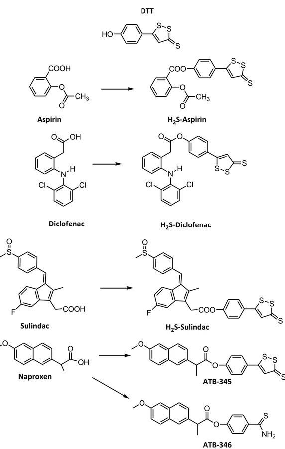
The first and most studied examples of NO-releasing drugs have been the NO-releasing nonsteroidal anti-inflammatory drugs (NSAIDs), which have been generally obtained by the conjugation of the NSAID of interest with different types of suitable NO-donor moieties. These hybrids were originally designed in order to maintain the original anti-inflammatory activity and to gain a NO-mediated reduction in the potential of gastric injury, through an improvement of mucosal blood flow, stimulation of mucus production, and inhibition of leukocyte adhesion. Moreover, the NO-mediated effects enhanced the antiplatelet effects of aspirin and, more in general, of NSAIDs. This general concept has been applied also to the design of H2S-releasing NSAIDs obtained through the conjugation
of the ‘‘parent’’ NSAIDs with a dithiolethione moiety [5-(4-hydroxyphenyl)-3H-1,2-dithiole-3-thione] (DTT), which is currently the most widely used H2S-releasing moiety for
synthesizing pharmacological hybrids. These novel derivatives mantained the capability to inhibit cyclooxigenase activity, while producing a significant increase in plasmatic H2S
29 S S S HO DTT COOH O CH3 O Aspirin COO O CH3 O S S S H2S-Aspirin O OH N H Cl Cl O O N H Cl Cl S S S Diclofenac H2S-Diclofenac S COOH F O S COO F O S S S Sulindac H2S-Sulindac O O OH O O O S S S O O O S NH2 Naproxen ATB-345 ATB-346
Figure 16 - Chemical structures of aspirin, diclofenac, sulindac, and naproxen, and the related H2S-releasing hybrids.
30 1.6.2 H2S-Latanoprost
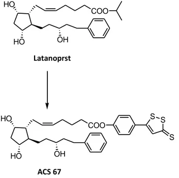
Latanoprost is a prostaglandin derivative used for the pharmacological management of glaucoma, because of its ability to lower intraocular pressure. Since appropriate concentration of H2S protect neurons from oxidative stress and from cell damage due to
the beta-amyloid protein, a H2S-releasing derivative of latanoprost (ACS-67) has been
projected in order to improve the pharmacological profile of the prostaglandin. This H2
S-latanoprost hybrid has been synthesized through the usual conjugation of the parent prostanoid with DTT. The effectiveness of ACS-67 in reducing intraocular pressure was equivalent to that of latanoprost. As regards the further activities of the DTT portion, it has been found that ACS-67 reduced the oxidative damage induced by H2O2 in the
transformed neuronal precursor cell line RGC-5. In studies performed on this cell line, the hybrid compound increased the GSH levels and enhanced levels of GSH were also observed in the aqueous humor of rabbit eyes after a topical application of ACS-67. All these additional effects were not shown by the parent drug latanoprost5 (figure 17).
HO HO COO OH Latanoprst HO HO COO OH S S S ACS 67
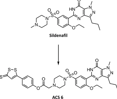
31 1.6.3 H2S-sildenafil
Sildenafil, the pioneer drug in the oral therapy of erectile dysfunction, is a selective inhibitor of phosphodiesterase type 5, particularly expressed in penile smooth muscle. Sildenafil has also been indicated as a potentially useful drug against acute respiratory distress syndrome, a lung pathology closely associated with progressive worsening of PH. The beneficial effects of sildenafil have been ascribed to the increment of cGMP levels, with consequent increase in the activity of cGMP-dependent protein kinases. Among the different heterogeneous factors that can play a pathogenic role in this disease, oxidative stress resulting from NADPH-oxidase hyperactivity and superoxide formation in pulmonary vessels is viewed as a critical step. Because such an oxidative stress can be counteracted by H2S, an H2S-releasing derivative of sildenafil (ACS6) has been synthesized
through the usual conjugation with DTT, in order to obtain a potentially more effective therapeutic tool for the management of acute respiratory distress syndrome. On the other hand, H2S has been shown to elicit relaxing effects on smooth muscle cells of the
human corpus cavernosum and to play a role in the physiological control of erectile function. Consistently, ACS6 has been proven to be more effective than the parent drug sildenafil in producing relaxing effects on rabbit isolated corpus cavernosum1 (Figure 18).
N N S O O O N HN O N N Sildenafil N N S O O O N HN O N N O O S S S ACS 6
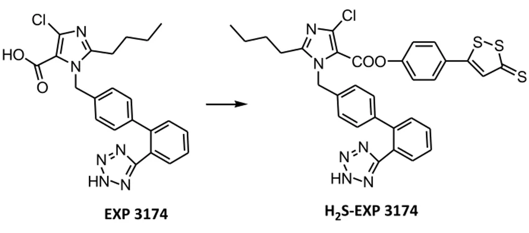
32 1.6.4 H2S-sartans
Sartans are widely used for the pharmacological management of hypertension. They act on the last step of the renin-angiotensin proteolytic cascade through the antagonism on the AT1 receptor for angiotensin II. This effective pharmacodynamic mechanism prevents all the short and long-lasting pro-hypertensive effects of angiotensin II, which are mainly mediated by AT1-receptors. Nevertheless, sartans are not endowed with significant direct relaxing effects on vascular smooth muscle or antiplatelet and antiischemic cardioprotective properties. Such additional pharmacological activities could be useful complementary features for strengthening the antihypertensive effects and/or reduce some dangerous risk factors often associated with hypertension. These considerations have been taken as a rational basis for the development of NO-sartans which were synthesized in order to confer additional NO-mediated but bradykinin-independent useful properties to sartans. Currently, the ‘‘translation strategy’’ has led again to apply the above concepts for the design of H2S-releasing sartans, which have been obtained by
conjugation with DTT.
As already observed for NO-sartans, it is reasonable to expect that, also in the case of H2S-sartans, the antihypertensive effects due to AT1 receptor antagonism may be
enriched with additional H2S-mediated beneficial properties, such as a direct vasorelaxing
effect, antiplatelet activity, and cardioprotective effect (Figure 19)2 .
N N HN N N N Cl HO O N N HN N N N Cl COO S S S EXP 3174 H2S-EXP 3174
Figure 19 - Representative example of H2S-sartan. Chemical structure of EXP 3174 and of the corresponding H2S-releasing hybrid.
33 1.6.5 H2S-releasing derivatives of L-DOPA
Parkinson’s disease is a progressive neurodegenerative disorder which almost selectively leads to the loss of dopaminergic nigrostriatal neurons. The pharmacotherapy of Parkinson’s disease is aimed at compensating the reduced striatal levels of the neurotransmitter dopamine; currently, the aminoacid L-DOPA (precursor of dopamine) is the most frequently used pharmacological tool. Of course, L-DOPA is considered as a symptomatic treatment, but it cannot arrest the neurodegenerative processes. Furthermore, L-DOPA is very effective only in the first years of therapy, while in the long-term treatment, ineffectiveness of L-DOPA and severe side-effects (such as L-DOPA-induced diskinesias) are found. Among the different (and still poorly clarified) etiopathological factors of Parkinson’s disease, oxidative stress and neuroinflammation are likely to play a fundamental role in the progressive death of nigrostriatal dopaminergic neurons. As well, the antiinflammatory and neuroprotective effects of endogenous H2S in human astrocytes are known, indicating a great therapeutic potential
of H2S in neurodegenerative pathologies. Therefore, some interesting H2S-releasing
hybrid derivatives have been developed. From a chemical point of view, such compounds exhibit some elements of novelty, because they are obtained by the conjugation through an amide function of the methyl ester of L-DOPA with the usual DTT-like moieties, but also with original H2S-releasing portions, such as an allyl-disulphide chain and a cyclic
1,3-dithiol-2-thione moiety. As regards the pharmacological aspects, these hybrid compounds underwent demethylation and a slower hydrolysis of the amidic bond, and were able to ensure a significant increase of brain L-DOPA. Furthermore, the H2S-releasing hybrids
increased the intracellular levels of H2S in human astrocytes, improved the levels of
reduced glutathione, and exhibited a selective inhibition of type B monoamino oxidase (responsible for dopamine breakdown in the brain). Furthermore, they reduced the neuronal injury in an experimental model of glial-mediated neurotoxicity with a significant decrease in the release of proinflammatory factors (such as IL-6, TNFa, and NO) from human microglia and astrocytes1 (Figure 20).
34 COOCH3 HN HO HO R
L- DOPA methyl ester
S S O S ACS 85 S S S O ACS 83 S S O ACS 86 S S S O O ACS 84
Figure 20 - Chemical structure of L-DOPA methyl ester and of four different kinds of H2S-releasing moieties, used for synthesizing the H2S-releasing hybrids of the anti-Parkinson drug.