Peripheral Benzodiazepine Receptor: Characterization in
Human T-Lymphoma Jurkat Cells
Barbara Costa, Alessandra Salvetti, Leonardo Rossi, Francesca Spinetti, Annalisa Lena,
Beatrice Chelli, Mariarosa Rechichi, Eleonora Da Pozzo, Vittorio Gremigni, and
Claudia Martini
Department of Psychiatry, Neurobiology, Pharmacology and Biotechnology, University of Pisa, Pisa, Italy (F.S., B.C., E.D.P., C.M.); and Department of Human Morphology and Applied Biology, University of Pisa, Pisa, Italy (B.C., A.S., L.R., A.L., M.R., V.G.)
Received June 9, 2005; accepted September 27, 2005
ABSTRACT
Peripheral benzodiazepine receptor (PBR) has been considered a promising drug target for cancer therapy, and several ligands have been developed for this purpose. Human T-lymphoma Jurkat cells have been considered as lacking PBR and are often used as negative control to prove the specificity of PBR ligands effects. It is surprising that we evidenced PBR protein expres-sion in this cell line by means of Western blotting and immu-nocytochemistry assays using specific anti-PBR antibodies. PBR intracellular localization was evidenced in mitochondria and nuclei, as demonstrated by confocal and electron micros-copy. The binding of the [3H]4⬘-chloro derivative of diazepam [3 H]7-chloro-5-(4-chlorophenyl)-1,3-dihydro-1-methyl-2H-1,4-benzodiazepin-2-one (Ro5-4864) and the isoquinoline carbox-amide derivative [3 H]1-(2-chlorophenyl)-N-methyl-N-(1-methyl-propyl)-3 isoquinolinecarboxamide (PK11195) evidenced a single class of binding sites with an unusual affinity constant
(Kd) of 1.77 ⫾ 0.30 and 2.20 ⫾ 0.20 M, respectively. The pharmacological profile of the classic ligands showed that PK11195 was the most potent inhibitor in the radioligand bind-ing assays followed by Ro5-4864 and diazepam, whereas clon-azepam, a specific ligand for the central-type receptor, showed a Ki⬎1.0 ⫻ 10⫺4M. By a combined strategy of reverse tran-scriptase-polymerase chain reaction and Southern blot exper-iments, we succeeded in isolating and cloning the full-length Jurkat PBR cDNA, called JuPBR. The JuPBR gene showed two single-nucleotide polymorphisms resulting in the two substitu-tions, Ala147 3 threonine and His162 3 arginine, of PBR amino acidic sequence. In conclusion, for the first time, we demonstrated PBR expression in Jurkat cells: the protein bound classic PBR ligands with micromolar affinity constants and presented a modified amino acidic sequence consequent to the detection of two gene polymorphisms.
The peripheral benzodiazepine receptor (PBR), originally discovered as an alternative binding site for the benzodiaz-epine diazepam (Valium; Hoffman-La Roche, Nutley, NJ), forms a unique class of receptors that are pharmacologically and functionally different from the central-type receptor (Gavish et al., 1999). At a subcellular level, it has been found to be primarily localized in mitochondria at the contact sites between the outer and the inner membranes, in which it seems to take part in the formation of a heteromeric receptor
complex with other proteins, including the 30-kDa adenine nucleotide transporter and the 34-kDa voltage-dependent anion channel (McEnery et al., 1992). This mitochondrial multiproteic complex has been suggested to constitute the mitochondrial permeability transition (MPT) pore, whose ex-tensive and prolonged opening can cause the dissipation of the transmembrane mitochondrial potential leading to the release of proapoptotic intermembrane proteins. So in the machinery leading to apoptosis, MPT pore modulation has been suggested as a critical event in the regulation of pro-cesses underlying cellular survival/death (Casellas et al., 2002). Numerous findings have suggested that PBR ligands, including the benzodiazepine Ro5-4864 and the isoquinoline carboxamide derivative PK11195 (Braestrup and Squires, 1977; Le Fur et al., 1983), may act as potential therapeutic
This work was supported by a grant from Ministero dell’Universita` e della Ricerca Scientifica e Tecnologica-Italy (Cofinanaziamento programmi di ricerca di interesse nazionale, 2002) (to M.C. and G.V.).
C.B. and S.A. contributed equally to this work.
Article, publication date, and citation information can be found at http://molpharm.aspetjournals.org.
doi:10.1124/mol.105.015289.
ABBREVIATIONS: PBR, peripheral benzodiazepine receptor; PK11195, 1-(2-chlorophenyl)-N-methyl-N-(1-methyl-propyl)-3-isoquinolinecarbox-amide; Ro5-4864, 7-chloro-5-(4-chlorophenyl)-1,3-dihydro-1-methyl-2H-1,4benzodiazepin-2-one; RT-PCR, reverse transcriptase-polymerase chain reaction; MPT, mitochondrial permeability transition; DAPI, 4,6-diamidino-2-phenylindole; PCR, polymerase chain reaction; PBS, phos-phate-buffered saline; DIG, digoxigenin.
MOLECULARPHARMACOLOGY Vol. 69, No. 1
Copyright © 2006 The American Society for Pharmacology and Experimental Therapeutics 15289/3068164
Mol Pharmacol 69:37–44, 2006 Printed in U.S.A.
agents useful in the management of a large spectrum of diseases, including cancer, through the modulation of the MPT-pore activity (Hirsch et al., 1998; Decaudin et al., 2002; Chelli et al., 2004, 2005).
The PBR full-length cDNA was originally cloned from the rat adrenal (781 bp). Since then, human, bovine, and mouse PBR cDNAs also have been cloned (Gavish et al., 1999), and their nucleotide sequences have been found to be signifi-cantly conserved. The human PBR gene promoter is known to contain Sp1-binding sites (Lin et al., 1993), which is consis-tent with housekeeping gene family. Indeed, ubiquitous lo-calization of the gene product has been observed in mamma-lian tissues (Gavish et al., 1999), including cells from the hematopoietic system (Canat et al., 1993; Carayon et al., 1996). Moreover, it has been demonstrated that PBR is abun-dantly expressed in a wide variety of malignant cells, even being the hallmark of cancerogenesis in some tissues (e.g., gliomas) (Miettinen et al., 1995). PBR expression level has been also suggested to be a clinically relevant prognosis factor in cancer (Hardwick et al., 1999, 2001). Among lym-phoma cell lines (Alexander et al., 1992; Canat et al., 1993) and among myeloid/lymphoid cells from patients with leuke-mia (Carayon et al., 1996), a wide range of PBR expression levels have been evidenced. A peculiar condition is repre-sented by the cell line Jurkat, deriving from a patient af-fected by acute lymphoblastic leukemia, in which the pres-ence of PBR mRNA and protein is still debated. Indeed, several authors have failed to detect PBR expression (Canat et al., 1993; Carayon et al., 1996; Hans et al., 2005). Never-theless, in the Jurkat cell line, PBR-selective ligands have been demonstrated to induce the same effects observed in other PBR-expressing cells (Zisterer et al., 2000). In addition, Decaudin and coworkers (2002) have succeeded in demon-strating detectable PBR mRNA levels in the Fas-resistant Jurkat T-cell line.
Within a general project in which we demonstrated the proapoptotic effects of classic and new synthesized PBR li-gands in the C6 glioma cell line (Chelli et al., 2004, 2005), we found that PBR ligands induced cell death also in Jurkat cells, used as control (unpublished data). These findings prompted us to investigate the expression and the cellular localization of PBR in this cell line. Here, we report that Jurkat cells expressed PBR with unusual affinity constants for PBR ligands. Moreover, we demonstrated that the Jurkat PBR gene contains two point mutations reported previously in other human tumoral cell lines (Hardwick et al., 1999) and in normal cells (Kurumaji et al., 2001).
Materials and Methods
Materials. The human T-lymphoma cell line Jurkat was obtained from Interlab Cell Line Collection (http://www.biotech.ist.unige.it/ interlab/cldb.html). Rat C6 glioma cells were kindly gifted by Dr. Damir Janigro (Cleveland Clinic Foundation, Cleveland, OH). Cell-culture media and fetal bovine serum were from Cambrex Bio Sci-ence Walkersville, Inc. (Walkersville, MD). PBR polyclonal primary antibodies were obtained from Santa Cruz Biotechnology (Santa Cruz, CA) and Trevigen (Gaithersburg, MD). Cytochrome c primary antibody and rhodamine-conjugate anti-rabbit secondary antibody were purchased by Santa Cruz Biotechnology. Horseradish peroxi-dase-conjugated secondary antibodies, and nonfat dry milk were from Bio-Rad (Hercules, CA). Fluorescein isothiocyanate-conjugated anti-mouse secondary antibody was from Invitrogen (Carlsbad, CA).
[3H]PK11195 (specific activity, 83.5 Ci/mmol) and [3H]Ro5-4864
(specific activity, 83.5 Ci/mmol) were obtained from PerkinElmer Life and Analytical Sciences (Boston, MA); PK11195, Ro5-4864, di-azepam, clondi-azepam, and DAPI were from Sigma/RBI (Natick, MA). All other reagents were from standard commercial sources.
Cell Culture and Membrane Preparations. The human T-lymphoma cell line Jurkat (T cell derived from a patient with acute lymphoblastic leukemia) was maintained in RPMI 1640 medium supplemented with 10% fetal bovine serum, 2 mML-glutamine, 100 U/ml penicillin, and 200g/ml streptomycin in a humidified atmo-sphere of 95% air/5% CO2at 37°C. Nuclear membranes from Jurkat
cells were prepared as described previously (Zamzami et al., 1996). Mitochondrial membranes from Jurkat cells were prepared as de-scribed previously with minor modifications (Miccoli et al., 1999). In brief, cells were collected by centrifugation at 1000g for 5 min. The cell pellet was homogenized in 20 volumes of ice-cold buffer (0.32 M sucrose, 1 mM EDTA, 10 mM Tris-HCl, pH 7.4, containing 160g/ml benzamidine, 200g/ml bacitracin, and 20 g/ml trypsin inhibitor) by 70 strokes of a tight-fitting glass pestle in a Potter Elvehjem glass homogenizer and centrifuged at 1500g for 5 min at 4°C. Supernatant was centrifuged at 12,000g for 10 min at 4°C. The resulting mito-chondria pellet, suspended in 20 volumes of 50 mM Tris-HCl, pH 7.4, buffer and homogenized by Ultraturrax, was centrifuged at 48,000g for 20 min at 4°C. Protein concentration was estimated by the method described by Lowry et al. (1951) using bovine serum albumin as standard.
Cloning and Sequencing of PBR Gene. The full-length cDNA for the PBR gene was obtained from Jurkat cells RNA by RT-PCR. Total RNA was isolated using NucleoSpin Kit reagents, according to the manufacturer’s instructions. The first-strand cDNA was ob-tained from 1g of total RNA using Superscript II RNase H-reverse transcriptase (Invitrogen) and oligo(dT)15–18. Amplification of the full-length PBR cDNA was obtained with the sequence-specific prim-ers (PBR forward, 5⬘-ACAGCAGCTGCAGCAGCC-3⬘ and PBR re-verse, 5⬘-ACGGCCACCACATCACAAG-3⬘) designed on the basis of human PBR sequence (GenBank accession number BC001110) by using Advantage 2 PCR enzyme system (BD Biosciences, San Jose, CA). After initial denaturation of cDNA at 95°C for 2 min, 35 cycles of PCR reaction were performed as follows: 95°C for 50 s, 54°C for 45 s, and 72°C for 1 min. The last cycle was extended to 10 min at 72°C. Water instead of cDNA was amplified as a negative control. Some of the amplification reaction products were resolved by agarose gel electrophoresis, transferred to a nylon membrane (Hybond N⫹; GE Healthcare, Little Chalfont, Buckinghamshire, UK), and probed with an internal PBR-specific DIG-labeled oligonucleotide (5 ⬘-CG-GCTCCTACCTGGTCTG-3⬘) according to the manufacturer’s in-structions (Roche Applied Science, Indianapolis, IN). Chemilumines-cent detection was performed with a DIG luminesChemilumines-cent detection kit (Roche Applied Science). The remaining PBR amplification reaction product was resolved by electrophoresis, and the PCR product cor-responding to the band hybridized by the internal DIG-labeled oli-gonucleotide was gel-purified and cloned in pGEMT-Easy vector (Promega, Madison, WI). Several clones were sequenced by auto-mated fluorescent cycle sequencing (Applied Biosystems, Foster City, CA).
Western Blot Analysis. Jurkat and lymphocyte cell protein sam-ples (30g) were separated by SDS-polyacrylamide gel electrophore-sis and transferred onto nitrocellulose membranes. The membranes were blocked for several hours in 5% blotting-grade blocker nonfat dry milk and incubated overnight with a 1:1000 dilution of anti-PBR (FL-169; Santa Cruz Biotechnology, developed against the full-length PBR of human origin) or 1:1000 of anti-PBR (Trevigen, de-veloped against an internal sequence of PBR protein consisting of amino acids 71–88) in 1% blotting-grade blocker nonfat dry milk. After several washes in Tris-buffered saline (10 mM Tris-HCl, pH 8, and 150 mM NaCl) containing 0.05% Tween 20, antibody binding was detected using anti-rabbit horseradish peroxidase-conjugated secondary antibodies (1:100,000). Cross-reactivity was detected
us-ing the SuperSignal West Dura Extended Duration Substrate (Pierce Biotechnology, Inc., Rockford, IL). Protein concentration was assayed using a Bio-Rad assay system. In some cases, the specificity of the bands recognized by the anti-PBR antibody was demonstrated using preabsorbed antibody, prepared by incubating overnight at 4°C the anti-PBR antibody (Trevigen) with a 5-fold (by weight) excess of recombinant full-length PBR protein in a small volume of phosphate-buffered saline (PBS). In parallel, the same amount of anti-PBR antibody was incubated overnight at 4°C in PBS alone.
Immunocytochemistry and Fluorescence Microscopy. Jur-kat cells were collected by centrifugation, washed in PBS, and fixed with 4% paraformaldehyde at room temperature for 30 min. A thin-layer cell preparation was obtained on poly(D-lysine)-coated glass slides by using a cytospin cytocentrifuge. Cells were blocked in PBS supplemented with 0.1% Triton X-100 and 0.5% bovine serum albu-min and incubated for 1 h at room temperature in blocking solution containing 1:50 PBR antibody and 1:50 cytochrome c anti-body (both from Santa Cruz Biotechnology) for fluorescence micros-copy, or with 1:200 anti-PBR antibody (Trevigen) and 20g/ml DAPI for confocal microscopy. After extensive washes in blocking solution, cells were incubated for 1 h at room temperature with a 1:200 dilution of fluorescein isothiocyanate-conjugated anti-mouse second-ary antibody and 1:200 dilution of rhodamine-conjugated anti-rabbit secondary antibody in blocking solution for fluorescence microscopy, or with 1:200 dilution of rhodamine-conjugated anti-rabbit second-ary antibody in blocking solution for confocal microscopy. Cells were washed in PBS and mounted for microscope analysis. The analysis were performed using an Axioplan (Carl Zeiss Inc., Thornwood, NJ) epifluorescence microscope and a TCS-SP laser scanning confocal microscope (Leica Microsystems, Mannheim, Germany). A confocal series was collected every 0.15m. Each confocal image shown here corresponded to the middle image from a 66-section series. Negative controls were performed, omitting the primary antibodies.
Immunogold Electron Microscopy. Jurkat cells were pro-cessed for electron microscopy immunocytochemistry as described previously (Trincavelli et al., 2002) using a rabbit polyclonal anti-PBR antibody (Trevigen) diluted 1:10 in PBS containing 0.1% gelatin and 0.5% bovine serum albumin. Negative controls were performed in the absence of anti-PBR antibody. Ultrathin sections were exam-ined using a 100 SX electron microscope (JEOL, Tokyo, Japan).
Radioligand Binding Assays. For equilibrium binding param-eters determination, [3H]PK11195 and [3H]Ro5-4864 were used.
[3H]PK11195 (specific activity, 0.44 Ci/mmol) binding assays were
conducted in a final volume of 500l of 50 mM Tris-HCl, pH 7.4, buffer containing membranes (50g of protein) and 100 nM to 10 M concentration of [3H]PK11195. Nonspecific binding was determined
in the presence of 100 M concentration of unlabeled PK11195. Samples were incubated in triplicate for 90 min at 0°C. In parallel,
[3H]PK11195 Scatchard analyses were carried out on human
lym-phocyte and C6 glioma cell membranes that were prepared as de-scribed for Jurkat cells. C6 glioma cells were cultured as dede-scribed by Chelli et al. (2005).
[3H]Ro5-4864 (specific activity, 0.42 Ci/mmol) binding assays were
performed in a final volume of 500l of 118 mM NaCl, 4.8 mM KCl, 1.2 mM CaCl2, 1.2 mM MgCl2, and 15 mM Tris/HCl, pH 7.4 buffer
containing membranes (50 g of protein) and 100 nM to 10 M concentration of [3H]Ro5-4864. Nonspecific binding was determined
in the presence of 100 M concentration of unlabeled Ro5-4864. Samples were incubated in triplicate for 60 min at 4°C. Incubations were stopped by centrifugation of the samples for 2 min at 13,000g. Pellets were quickly washed three times with 1 ml of ice-cold binding assay buffer. Radioactivity in the pellets was counted after the ad-dition of 1 ml of 0.2 N NaOH and 0.19 ml of 1 N acetic acid. Bound radioactivity was measured in a scintillation counter (TopCount; PerkinElmer Life and Analytical Sciences) using scintillation liquid (65% counting efficiency). Incubation of samples was performed in safe-lock tubes (Eppendorf AG, Hamburg, Germany). In competition experiments, membranes (50g of protein) were incubated with 1.5 M [3H]PK11195 or 1.5M [3H]Ro5-4864 in binding assay buffer
and increasing concentrations of Ro5-4864, PK11195, diazepam, and clonazepam.
The equilibrium binding of [3H]PK11195 was also measured using
homologous binding displacement, whose theoretical validity has been discussed elsewhere (Rovati, 1998; Bylund and Murrin, 2000). Jurkat membranes (150g) were incubated with a fixed concentra-tion of labeled ligand (0.5M; specific activity, 1.33 Ci/mmol) and increasing concentrations of unlabeled ligand up to 100M. Binding equilibrium was reached after a 90-min incubation at 0°C. Samples were filtered rapidly under vacuum through GF/C glass fiber filters to separate bound and unbound ligand. After being washed four times with 3 ml of assay buffer, radioactivity trapped on the filter was measured by liquid scintillation counting. Nonspecific binding was measured in the presence of excess PK11195 (100M).
Data Analysis. Saturation experiments of [3H]PK11195 and
[3H]Ro5-4864 were analyzed with the EBDA and the LIGAND
com-puter programs (Biosoft-Elsevier, Cambridge, England) (Munson and Rodbard, 1980; McPherson, 1985). Single- and multiple-site models were statistically compared with determine the best fit, and differences between models were tested by comparing the residual variance using a partial F test and a significance level of P⬍ 0.05. Displacement curves were analyzed and fitted using Prism software (version 3.0; GraphPad Software Inc., San Diego, CA). Derived IC50
values obtained from displacement curves were converted to Ki
val-ues by the Cheng and Prusoff equation (Cheng and Prusoff, 1973). Values represent the means⫾ S.E.M. of at least three independent experiments. Saturation data were also fitted with GraphPad Prism.
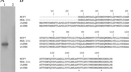
Fig. 1. PBR cDNA isolation in Jurkat cells. A, Southern blot of PBR amplification products. Lane 1, Jurkat cDNA amplified with PBR-specific primers. An autoradiographic band of approximately 600 base pairs was visible. Lane 2, negative RT-PCR control. B, comparison of the deduced amino acid sequence of Jurkat PBR (JuPBR) with those of other human PBR protein isoforms: MCF-7, human breast cancer cell line accession number AAC31173.1; MDA-231, human breast cancer cell line (Hardwick et al., 1999); wt-PBR, mammalian gene collection program accession num-ber. AAH01110. Asterisks indicate the amino acid substi-tutions.
Results
Detection of PBR Transcript in Jurkat Cells. By using a combined strategy of RT-PCR and Southern blot experi-ments, we succeeded for the first time in the isolation of a full-length cDNA clone, called JuPBR, encoding for PBR in Jurkat cells (GenBank accession number AY998017; Fig.
1A). JuPBR is 593 base pairs long and contains an open reading frame encoding for a 169 amino acid putative PBR protein, with an estimated molecular mass of 18 kDa. Se-quence comparison indicates that JuPBR shares high nucle-otide identity with the other previously isolated human PBR genes. However, JuPBR shows a point mutation at base 526, which changes alanine (ACG) to threonine (GCG), and a point mutation at base 572, which changes arginine (CGT) to histidine (CAT) (Fig. 1B). The same variations were reported in the PBR gene isolated from other human tumoral cell lines (Hardwick et al., 1999) and, with high frequency, in genomic DNA of normal cells (Kurumaji et al., 2001).
Detection and Subcellular Localization of PBR Pro-tein in Jurkat Cells. Using commercially available PBR polyclonal antibodies, PBR protein was identified in Jurkat cells.
Western blot analysis on total proteins obtained from Ju-rkat and lymphocytes cells, as control, was performed. Two different commercial polyclonal anti-PBR antibodies recog-nized the typical 18-kDa PBR protein in both Jurkat and lymphocyte cells. In addition, a 36-kDa protein was also detected, suggesting the presence of polymers of PBR in these cells (Fig. 2A). Immunoreactivities were specific, as demonstrated by preabsorbing the anti-PBR antibody with the recombinant full-length PBR protein (Fig. 2B).
Fig. 2. Western blot analysis of PBR protein. A, PBR polyclonal antibody (anti-PBR) (Trevigen) recognizes two proteins of approximately 18 and 36 kDa in both Jurkat (J) and lymphocyte (L) cells. B, specificity of the immunoreactivities seen in A, examined by preabsorbing anti-PBR with the recombinant full-length PBR protein.
Fig. 3. Subcellular distribution of PBR protein in Jurkat cells. A, distribution of PBR as visualized by means of an anti-PBR antibody by using fluorescent microscopy. B, sub-cellular distribution of mitochondria as revealed by means of an anti-cytochrome c antibody and fluorescent micros-copy. C, merged A and B demonstrate that although PBR protein is accumulated in mitochondria, a widespread sig-nal is also observable in other subcellular compartments. Scale bar (A–C), 10m. D–F, confocal fluorescence images obtained from immunocytochemistry preparations. D, dis-tribution of PBR as revealed by use of anti-PBR antibody. E, the section depicted in D, stained with the nuclear dye DAPI; F, merged D and E demonstrate the nuclear local-ization of JuPBR. Scale bar (D–F), 5m. G, electron mi-croscope immunocytochemistry. Large clusters of gold par-ticles (arrows) are visible on the nucleus and cytoplasm of Jurkat cells. m, mitochondrion; N, nucleus. Scale bar, 1.5m.
Immunocytochemistry analysis revealed that the PBR pro-tein seemed distributed all over the cytoplasm and at a nuclear level (Fig. 3). The use of the specific mitochondrial marker anti-cytochrome c antibody (Fig. 3B) evidenced that the pattern of the subcellular distribution of mitochondria partially overlapped the pattern of PBR staining (Fig. 3C). No signal was detected in negative controls carried out in the same experimental conditions, except that no primary anti-body was added. By performing immunohistochemistry using anti-PBR antibody in combination with the specific nuclear dye DAPI and subsequent confocal microscopy, nuclear local-ization of PBR was demonstrated (Fig. 3, D–F). Mitochon-drial and nuclear PBR localization was also confirmed by immunogold electron microscopy (Fig. 3G).
[3H]PK11195 Binding Characterization. As a first step, we used Jurkat cell mitochondrial membranes for ra-dioligand binding assays using nanomolar concentrations of [3H]PK11195 as described in several cellular systems (Alex-ander et al., 1992; Canat et al., 1993, Hardwick et al., 1999). In these conditions, no [3H]PK11195-specific binding was detected. The presence of PBR protein evidenced using the specific anti-PBR antibody, as described above, prompted us to study a potentially lower [3H]PK11195 binding affinity constant in this cell line. Binding at higher ligand concentra-tions (⬎100 nM) was investigated in Jurkat cell membranes using radioligand diluted with nonradioactive ligand for lowering its specific activity (Bylund and Murrin, 2000). Moreover, bound and unbound radiolabeled ligands were separated by the centrifugation procedure according to low-affinity receptor binding assays (Kdvalues of approximately 10⫺7 M or 10⫺6 M) (Bennett and Yamamura, 1985). The conditions necessary for the [3H]PK11195 binding were ini-tially studied to optimize yield. In mitochondrial membrane preparations, the specific binding of [3H]PK11195 increased linearly with increasing protein concentrations in the range 20 to 150g of protein (data not shown). To study the satu-rability of specific [3H]PK11195 binding to Jurkat mitochon-drial membranes, aliquots of membrane preparations were incubated with increasing concentrations of [3H]PK11195 (100 nM to 10 M). Specific binding of [3H]PK11195 was saturable, whereas nonspecific [3H]PK11195 binding in-creased linearly (data not shown). Scatchard analysis of the saturation data yielded a straight-line plot, compatible with the existence of a single population of binding sites (F test, P⬍ 0.05) (Fig. 4). In mitochondrial membranes, the Kdvalue for [3H]PK11195 was 2.20 ⫾ 0.20 M, and the maximum amount of specifically bound ligand (Bmax) was 706 ⫾ 75 pmol/mg of protein.
The low-affinity [3H]PK11195 binding site was investi-gated using nonradioactive saturation experiments too. Scat-chard analysis of these data indicated the existence of one class of binding sites because the one-site model yielded a significantly better fit (F test, P⬍ 0.05) than fitting experi-mental data to a two-site model. The best fit estimated of the Kd value (9.90 ⫾ 1 M) for this low-affinity [
3H]PK11195 binding site was comparable with that obtained by “hot” saturation experiments (Fig. 5).
In nuclear membranes, Scatchard analysis of [3H]PK11195 saturation data evidenced a single population of binding sites with a Kd value of 1.10 ⫾ 0.10 M, comparable with that obtained in mitochondrial membranes. The Bmaxvalue (27⫾ 3 pmol/mg of protein) resulted approximately 26 times lower
than that found in mitochondrial extracts, suggesting a sig-nificant lesser expression level of PBR protein in this cell compartment. Competition experiments with specific and se-lective PBR ligands were performed to test the pharmacolog-ical properties of binding sites for [3H]PK11195 in Jurkat cell membranes. Displacement studies of [3H]PK11195 specifi-cally bound to Jurkat cell membranes were performed by incubating aliquots of membranes with [3H]PK11195 and 9 to 10 different concentrations of a ligand, as described under Materials and Methods. Analysis of displacement data fitted using the Prism software revealed that both the selective PBR ligands PK11195 and Ro5-4864 and the specific PBR ligand diazepam displaced specific [3H]PK11195 binding in a concentration-dependent manner. Ki values of ligands are shown in Table 1. The binding of radioligand was effectively displaced by PBR ligands PK11195, Ro5-4864, and diazepam at micromolar concentrations; in contrast, clonazepam, a se-lective ligand for the central benzodiazepine receptor, was not effective up to 10⫺4M.
To clarify whether the unusual [3H]PK11195 low-affinity binding site evidenced in Jurkat cells was also present in cells showing [3H]PK11195 high affinity, we performed on human lymphocyte and C6 glioma mitochondrial membranes
Fig. 4. Saturation curve and Scatchard plot (inset) of specific [3H]PK11195 binding to mitochondrial Jurkat cell membranes.
Mitochon-drial membranes were incubated with increasing concentrations of [3H]PK11195 ranging from 100 nM to 10M. Nonspecific binding was
measured in the presence of 100M PK11195. Data are from a single experiment carried out in triplicate. The Kdand Bmaxvalues were 2.05
M and 662 pmol/mg of protein, respectively. Three such experiments yielded similar results.
Fig. 5. Homologous displacement of [3
H]PK11195 in Jurkat cell mito-chondria membranes. Bound radioactivity is expressed as a percentage of specific binding in the absence of competitor. EBDA/ligand analysis in-dicated a single class of binding sites with an estimated Kd⫽ 9.90 ⫾ 1
M. Data points represent the means (⫾ S.E.M.) of triplicate determina-tions pooled from three independent experiments.
[3H]PK11195 saturation analysis. For this purpose, we used the same experimental conditions described for Jurkat cells. Data reported in Table 2 showed that [3H]PK11195 binding parameters were similar in all analyzed cells.
[3H]Ro5-4864 Binding Characterization. Radioligand binding assays, performed as described previously (Mak and Barnes, 1989), with nanomolar concentration of [3 H]Ro5-4864 did not evidence specific binding of radioligands in Jurkat cell membranes. Thus we, used the above-mentioned experimental approach for extending the [3H]Ro5-4864 con-centrations range. Binding assays demonstrated specific, sat-urable binding of [3H]Ro5-4864 to human Jurkat cell mem-branes. Scatchard analysis of [3H]Ro5-4864 saturation curve revealed a single class of binding sites with a dissociation constant (Kd) of 1.77⫾ 0.3 M and a maximal binding ca-pacity (Bmax) of 200⫾ 21 pmol/mg of protein. Scatchard plots of the binding data were linear, consistent with the interpre-tation that a homogeneous population of binding sites was present in membranes of Jurkat cells (Fig. 6).
The pharmacological specificity of the binding of [3 H[Ro5-4864 was studied in the Jurkat cell membranes using various displacing drugs. Displacement curves were obtained in the presence of 1.5M [3H]Ro5-4864: the estimated K
ivalues for each competing ligand were calculated from these data and are presented in Table 1. In Jurkat cell membrane prepara-tions, the tritiated ligand was effectively displaced by PK11195 and Ro5-4864. Diazepam was less effective, whereas clonazepam, at the concentration of 100 M, was able to displace 65% of [3H]Ro5-4864 binding from its binding site.
Discussion
In the present report, we demonstrate that PBR is ex-pressed in the human T-lymphoma Jurkat cell line and that
Jurkat PBR protein presents benzodiazepine and isoquino-line carboxamide ligand low-affinity binding sites.
In Jurkat cells PBR expression is still debated. By using RT-PCR, no PBR transcript was shown previously by Canat et al. (1993) and Hans et al. (2005). On the contrary, some evidences have demonstrated that PBR mRNA is expressed in human Fas-resistant T-cell line Jurkat (Decaudin et al., 2002). By using a combined strategy of RT-PCR followed by Southern blot hybridization, we succeeded in the isolation and cloning of the Jurkat PBR full-length cDNA (JuPBR). PBR mRNA detection required a highly sensitive experimen-tal strategy, suggesting that Jurkat cells expressed PBR mRNA at lower level than other tumoral cell lines. Compared with the human GenBank PBR sequence, JuPBR showed two point mutations resulting in the change of alanine to threonine (A147T) and of histidine to arginine (H162R) lo-calized in the fifth putative transmembrane region and in the carboxyl-terminal region of the PBR, respectively. The mis-sense variants (A147T; H162R) have been found in other tumoral cell lines, such as MDA-231 (Hardwick et al., 1999) and, with high frequency, in genomic DNA of normal cells (Kurumaji et al., 2001), suggesting that they can be the consequence of single nucleotide polymorphisms of human PBR gene. It is not known whether these mutations have any significant effect on PBR structure and function. Molecular modeling of the receptor indicates that the first residue (A147T) lies within the cholesterol entry region of the recep-tor. However, this mutation does not seem to alter the ability of cholesterol to move through PBR because cholesterol is incorporated into MDA-231 nuclei (Hardwick et al., 1999). The second mutation (H162R) is a neutral mutation, given that both histidine and arginine are basic amino acids, and it is unlikely to have any significant effect on PBR function.
The PBR amino acidic substitution H162R may explain why some authors have not detected protein in Jurkat cells by using the monoclonal anti-PBR 8D7 antibody (Carayon et al., 1996; Hans et al., 2005), which recognized the human PBR C-terminal region that contains His162 instead of arginine.
In our experimental conditions, PBR expression was evi-denced in Jurkat cells by using two different polyclonal
anti-Fig. 6. Saturation curve and Scatchard plot (inset) of specific [3
H]Ro5-4864 binding to mitochondrial Jurkat cell membranes. Mitochondrial membranes were incubated with increasing concentrations of [3
H]Ro5-4864 ranging from 100 nM to 10M. Nonspecific binding was measured in the presence of 100M Ro5-4864. Data are from a single experiment carried out in triplicate. The Kdand Bmaxvalues were 1.05M and 210
pmol/mg of protein, respectively. Three such experiments yielded similar results.
TABLE 1
Pharmacological characterization of关3H兴PK11195 and 关3H兴Ro5-4864
binding to Jurkat cell line membranes
Jurkat cells membranes were incubated with 1.5M 关3H兴PK11195 or 关3H兴Ro5-4864 and increasing drug concentrations. The IC50values were calculated, and the Ki inhibitory constant was derived (Cheng and Prusoff, 1973). Each value represents the mean⫾ S.E.M. of three experiments, each performed in triplicate.
Compound Kior Percentage of Inhibition vs关3H兴PK11195 vs关3H兴Ro5-4864 M PK11195 1.90⫾ 0.20 3.68⫾ 0.35 Ro5-4864 32.3⫾ 4.00 8.74⫾ 0.79 Diazepam 79.0⫾ 8.00 18.4⫾ 1.90 Clonazepam No displacement up to 10⫺4M 35 ⫾ 4% at 10⫺4M TABLE 2
关3H兴PK11195 binding parameters in Jurkat cells, lymphocytes, and C6
glioma cells
Lymphocytes, Jurkat, and C6 cell membranes were incubated with increasing con-centrations of关3H兴PK11195 (100 nM to 10M; specific activity, 0.44 Ci/mmol) as described under Materials and Methods. Each value represents the mean⫾ S.E.M. of three experiments, each performed in triplicate.
Cell Type Kd Bmax
M pmol/mg protein
Jurkat 2.20⫾ 0.20 706⫾ 75
Lymphocytes 1.80⫾ 0.20 726⫾ 83
PBR antibodies that specifically recognized the 18-kDa reac-tive band. The data obtained by Western blot showed two different bands at 18 and 36 kDa accordingly, as reported previously in other cell lines (Delavoie et al., 2003; Corsi et al., 2005). Immunolocalization experiments provide a further demonstration of the presence of PBR protein in Jurkat cells. We evidenced PBR in mitochondria in line with other studies (Gavish et al., 1999) and in nuclei, as demonstrated previ-ously in other tumoral cell lines (Hardwick et al., 1999; Brown et al., 2000; Delavoie et al., 2003; Corsi et al., 2005). Emerging data have indicated that different PBR subcel-lular localization is frequently associated with cell malig-nancy. In particular, in the human aggressive phenotype of breast tumor, the PBR protein is primarily expressed in and around the nucleus (Hardwick et al., 1999). A peculiar asso-ciation between PBR nuclear localization and cell malig-nance has also been evidenced in human gliomas (Brown et al., 2000). Further studies are necessary for understanding the association of Jurkat PBR subcellular localization and cell malignancy.
Jurkat cell line membranes were used in quantitative ra-dioligand binding assays using the specific and selective PBR radioligands [3H]PK11195 and [3H]Ro5-4864. According to literature data (Canat et al., 1993), no specific [3H]PK11195 binding was detected using nanomolar radioligand concen-trations. However, the presence of receptor protein revealed by immunoblotting, transmission electron microscopy, and confocal microscopy analysis prompted us to investigate the possibility of more low-radioligand equilibrium binding pa-rameters.
For this aim, we covered a higher radioligand concen-tration range (100 nM to 10M) according to the general guidelines for receptor saturation experiments (Bylund and Murrin, 2000). Scatchard analysis of the saturation [3H]PK11195 binding data yielded a K
dvalue approximately 200 times lower than affinity constants derived from satura-tion studies performed on membranes of human lymphocytes and other leukemia cells (Canat et al., 1993).
A comparable low-affinity [3H]PK11195 K
dvalue was also evidenced by using nonradioactive saturation experiments.
PBR nanomolar affinity constants have been detected in leukemia cell lines such as CCRF-CEM, Raji, and K562, whereas no binding has been evidenced using up to 20 nM PBR radioligand concentration in Molt-4B, suggesting that the absence of the high-affinity PK11195 binding parameter may not be exclusive of the Jurkat cell line (Alexander et al., 1992). Using micromolar radioligand concentration in this article, we demonstrated the presence of a low-affinity PK11195 binding site in normal immune system cells like lymphocytes and in a cell line of solid tumors like C6 glioma cells, in which a high-affinity PK11195 binding site has been reported (Alexander et al., 1992; Zisterer et al., 1998). These data suggested that the low affinity of PK11195 binding sites evidenced in Jurkat cells corresponded to a second popula-tion of binding sites, usually present in other cells.
In the Jurkat cell line, competition curve data with specific and selective PBR ligands PK11195, Ro5-4864, diazepam, or the central-type receptor-specific ligand clonazepam allowed us to derive Kivalues for specific PBR ligands (listed in Table 1), whereas clonazepam was not effective up to 10⫺4 M. Although the binding affinity observed for each of these li-gands ranged over several orders of magnitude compared
with that estimated in human lymphocytes and in other human cell types (Parola et al., 1993), the characteristic rank order of ligand binding potency (PK11195 ⬎ Ro5-4864 ⬎ diazepam clonazepam) was typical of PBR.
In the Jurkat cell line, [3H]Ro5-4864 did not show specific binding at radioligand nanomolar concentrations. Scat-chard analysis of [3H]Ro5-4864 saturation curve, performed with experimental approaches as described above for [3H]PK11195 binding assay, showed the presence of a single population of low-affinity binding sites with a dissociation constant of 1.77⫾ 0.3M. Unlike PK11195 binding proper-ties, that usually demonstrated a high-affinity binding in tissues from many species, Ro5-4864 binding varied depend-ing on species and tissue. In contrast to rodent PBR (Kd ⫽ 1–10 nM), binding of Ro5-4864 to PBR of most human tissues occurred only with low affinity (Kd approximately 1 M) (Awad and Gavish, 1991), except for human lymphocytes and platelets and the human breast cancer cell line BT-20 (Kd⫽ 8.5 nM) (Beinlich et al., 1999). In accordance with our data, in HL-60 acute promyelocytic leukemia cell membranes, a second class of benzodiazepine binding sites with lower bind-ing affinity has been detected (Kdvalue of the benzodiazepine drug [3H]diazepam, 28.6M; B
max, 199 pmol/mg of protein) (Ishiguro et al., 1987). In Jurkat cell membranes, competition experiments of [3H]Ro5-4864 binding with various ligands displayed the typical PBR order of potency: PK11195⬎ Ro5-4864⬎ diazepam clonazepam.
The first conclusion we can draw is that Jurkat cells con-tain PBR, which presents a micromolar affinity constant for classic ligands, and these cells cannot be used as control in an assay in which high ligand concentrations are used. The hypothesis that PBR classic high-affinity binding site could be changed in Jurkat cells as result of the two amino acidic substitutions has been discarded because human breast can-cer cell lines MCF-7 and MD-231, with the same missense variants (Hardwick et al., 1999), bind the isoquinoline car-boxamide derivative with high-affinity ([3H]PK11195 K
d val-ues, respectively, of 2.9 (Carmel et al., 1999) and 7.8 nM (Hardwick et al., 1999). Because PBR is associated with other proteins to constitute the mitochondrial pore, it can be hy-pothesized that in these tumoral cell lines, a different PBR-proteins stechiometric ratio might exist, resulting in a con-formational receptor change for high-affinity binding sites. The presence of the PBR low-affinity binding site can justify micromolar PBR ligand concentration required to obtain a functional effect mediated by PBR as apoptosis induction or facilitation.
Acknowledgments
We thank Drs. Ranieri Bizzarri and Teresa Locci for technical assistance in the confocal microscopy studies and Serena Spinelli for the use of cytospin cytocentrifuge.
References
Alexander BE, Roller E, and Klotz U (1992) Characterization of peripheral-type benzodiazepine binding sites on human lymphocytes and lymphoma cell lines and their role in cell growth. Biochem Pharmacol 44:269 –274.
Awad M and Gavish M (1991) Peripheral-type benzodiazepine receptors in human cerebral cortex, kidney and colon. Life Sci 49:1155–1161.
Beinlich A, Strohmeier R, Kaufmann M, and Kuhl H (1999) Specific binding of benzodiazepines to human breast cancer cell lines. Life Sci 65:2099 –2108. Bennett JP and Yamamura HI (1985) Neurotransmitter, hormone, or drug receptor
binding methods, in Neurotransmitter Receptor Binding, 2nd ed (Yamamura HI ed) pp 61– 89, Raven Press, Philadelphia.
characterized by high-affinity (3H)diazepam binding. Proc Natl Acad Sci USA 74:3805–3809.
Brown RC, Degenhardt B, Kotoula M, and Papadopoulous V (2000) Location-dependent role of the human glioma cell peripheral-type benzodiazepine receptor in proliferation and steroid biosynthesis. Cancer Lett 156:125–132.
Bylund DB and Murrin LC (2000) Radioligand saturation binding experiments over large concentration ranges. Life Sci 67:2897–28911.
Canat X, Carayon P, Bouaboula M, Cahard D, Shire D, Roque C, Le Fur G, and Casellas P (1993) Distribution profile and properties of peripheral-type benzodi-azepine receptors on human hemopoietic cells. Life Sci 52:107–118.
Carayon P, Portier M, Dussossoy D, Bord A, Petitpretre G, Canat X, Le Fur G, and Casellas P (1996) Involvement of peripheral benzodiazepine receptors in the pro-tection of hematopoietic cells against oxygen radical damage. Blood 87:3170 –3178. Carmel I, Fares FA, Leschiner S, Scherubl H, Weisinger G, and Gavish M (1999) Peripheral-type benzodiazepine receptors in the regulation of proliferation of MCF-7 human breast carcinoma cell line. Biochem Pharmacol 58:273–278. Casellas P, Galiegue S, and Basile AS (2002) Peripheral benzodiazepine receptors
and mitochondrial function. Neurochem Int 40:475– 486.
Chelli B, Lena A, Vanacore R, Pozzo ED, Costa B, Rossi L, Salvetti A, Scatena F, Ceruti S, Abbracchio MP, et al. (2004) Peripheral benzodiazepine receptor ligands: mitochondrial transmembrane potential depolarization and apoptosis induction in rat C6 glioma cells. Biochem Pharmacol 68:125–134.
Chelli B, Rossi L, Da Pozzo E, Costa B, Spinetti F, Rechichi M, Salvetti A, Lena A, Simorini F, Vanacore R, et al. (2005) PIGA (N,N-di-n-butyl-5-chloro-2-(4-chlorophenyl)indol-3-ylglyoxylamide), a new mithocondrial benzodiazepine recep-tor ligand, induces apoptosis in C6 glioma cells. Chem Biochem 6:1– 8. Cheng YC and Prusoff WH (1973) Relationship between the inhibition constant (Ki)
and the concentration of the inhibitor which causes 50% inhibition (IC50) of an enzymatic reaction. Biochem Pharmacol 22:3099 –3108.
Corsi L, Geminiani E, Avallone R, and Baraldi M (2005) Nuclear location-dependent role of peripheral benzodiazepine receptor (PBR) in hepatic tumoral cell lines proliferation. Life Sci 76:2523–2533.
Decaudin D, Castedo M, Nemati F, Beurdeley-Thomas A, De Pinieux G, Caron A, Pouillart P, Wijdenes J, Rouillard D, Kroemer G, et al. (2002) Peripheral benzo-diazepine receptor ligands reverse apoptosis resistance of cancer cells in vitro and in vivo. Cancer Res 62:1388 –1393.
Delavoie F, Li H, Hardwick M, Robert JC, Giatzakis C, Peranzi G, Yao ZX, Maccario J, Lacapere JJ, and Papadopoulos V (2003) In vivo and in vitro peripheral-type benzodiazepine receptor polymerization: functional significance in drug ligand and cholesterol binding. Biochemistry 42:4506 – 4519.
Gavish M, Bachman I, Shoukrun R, Katz Y, Veenman L, Weisinger G, and Weizman A (1999) Enigma of the peripheral benzodiazepine receptor. Pharmacol Rev 51: 629 – 650.
Hans G, Wislet-Gendebien S, Lallemend F, Robe P, Rogister B, Belachew S, Nguyen L, Malgrange B, Moonen G, and Rigo JM (2005) Peripheral benzodiazepine recep-tor (PBR) ligand cytotoxicity unrelated to PBR expression. Biochem Pharmacol
69:819 – 830.
Hardwick M, Fertikh D, Culty M, Li H, Vidic B, and Papadopoulos V (1999) Periph-eral-type benzodiazepine receptor (PBR) in human breast cancer: correlation of breast cancer cell aggressive phenotype with PBR expression, nuclear localization and PBR-mediated cell proliferation and nuclear transport of cholesterol. Cancer
Res 59:831– 842.
Hardwick M, Rone J, Han Z, Haddad B, and Papadopoulos V (2001) Peripheral-type benzodiazepine receptor levels correlate with the ability of human breast cancer MDA-MB-231 cell line to grow in SCID mice. Int J Cancer 94:322–327. Hirsch T, Decaudin D, Susin SA, Marchetti P, Larochette N, Resche-Rigon M, and
Kroemer G (1998) PK11195, a ligand of the mitochondrial benzodiazepine recep-tor, facilitates the induction of apoptosis and reverses Bcl-2-mediated cytoprotec-tion. Exp Cell Res 241:426 – 434.
Ishiguro K, Taft WC, DeLorenzo RJ, and Sartorelli AC (1987) The role of
benzodi-azepine receptors in the induction of differentiation of HL-60 leukemia cells by benzodiazepines and purines. J Cell Physiol 131:226 –234.
Kurumaji A, Nomoto H, Yamada K, Yoshikawa T, and Toru M (2001) No association of two missense variations of the benzodiazepine receptor (peripheral) gene and mood disorders in a Japanese sample. Am J Med Genet 105:172–175.
Le Fur G, Guilloux F, Rufat P, Benavides J, Uzan A, Renault C, Dubroeucq MC, and Gueremy C (1983) Peripheral benzodiazepine binding sites: effect of PK 11195, 1-(2-chlorophenyl)-n-methyl-(1-methylpropyl)-3 isoquinolinecarboxamide: II. In vivo studies. Life Sci 32:1849 –1856.
Lin D, Chang YJ, Strauss JF, and Miller WL (1993) The human peripheral benzo-diazepine receptor gene: cloning and characterization of alternative splicing in normal tissues and in a patient with congenital lipoid adrenal hyperplasia.
Genomics 18:643– 650.
Lowry OH, Rosebrough NJ, Farr AL, and Randall RJ (1951) Protein measurement with the Folin phenol reagent. J Biol Chem 193:265–275.
Mak JC and Barnes PJ (1989) Peripheral type benzodiazepine receptors in human and guinea pig lung: characterization and autodiographic mapping. J Pharmacol
Exp Ther 252:880 – 885.
McEnery MW, Snowman AM, Trifiletti RR, and Snyder SH (1992) Isolation of the mitochondrial benzodiazepine receptor: association with the voltage-dependent anion channel and the adenine nucleotide carrier. Proc Natl Acad Sci USA 89: 3170 –3174.
McPherson GA (1985) Analysis of radioligand binding experiments. A collection of computer programs for the IBM PC. J Pharmacol Methods 14:213–228. Miccoli L, Oudard S, Beurdeley-Thomas A, Dutrillaux B, and Poupon MF (1999)
Effect of 1-(2 chlorophenyl)-N-methyl-N-(1-methylpropyl)-3-isoquinoline carbox-amide (PK11195), a specific ligand of the peripheral benzodiazepine receptor, on the lipid fluidity of mitochondria in human glioma cells. Biochem Pharmacol
58:715–721.
Miettinen H, Kononen J, Haapasalo H, Helen P, Sallinen P, Harjuntausta T, Helin H, and Alho H (1995) Expression of peripheral-type benzodiazepine receptor and diazepam binding inhibitor in human astrocytomas: relationship to cell prolifera-tion. Cancer Res 55:2691–2695.
Munson PJ and Rodbard D (1980) Ligand: a versatile computerized approach for characterization of ligand-binding systems. Anal Biochem 107:220 –239. Parola AL, Yamamura HI, and Laird HE 3rd (1993) Peripheral-type benzodiazepine
receptors. Life Sci 52:1329 –1342.
Rovati GE (1998) Ligand-binding studies: old beliefs and new strategies. Trends
Pharmacol Sci 19:365–369.
Trincavelli ML, Tuscano D, Marroni M, Falleni A, Gremigni V, Ceruti S, Abbracchio MP, Jacobson KA, Cattabeni F, and Martini C (2002) A3 adenosine receptors in human astrocytoma cells: agonist-mediated desensitization, internalization and down-regulation. Mol Pharmacol 62:1373–1384.
Zamzami N, Susin SA, Marchetti P, Hirsch T, Gomez-Monterrey I, Castedo M, and Kroemer G (1996) Mitochondrial control of nuclear apoptosis. J Exp Med 183: 1533–1544.
Zisterer DM, Campiani G, Nacci V, and Williams DC (2000) Pyrrolo-1,5-benzoxazepines induce apoptosis in HL-60, Jurkat and Hut-78 cells: a new class of apoptotic agents. J Pharmacol Exp Ther 293:48 –59.
Zisterer DM, Hance N, Campiani G, Garofalo A, Nacci V, and Williams DC (1998) Antiproliferative action of pyrrolobenzoxazepine derivatives in cultured cells: ab-sence of correlation with binding to the peripheral-type benzodiazepine binding site. Biochem Pharmacol 55:397– 403.
Address correspondence to: Dr. Martini Claudia, Department of Psychia-try, Neurobiology, Pharmacology and Biotechnology, University of Pisa, via Bonanno, 6-56126 Pisa, Italy. E-mail: [email protected]

![Fig. 4. Saturation curve and Scatchard plot (inset) of specific [ 3 H]PK11195 binding to mitochondrial Jurkat cell membranes](https://thumb-eu.123doks.com/thumbv2/123dokorg/7329699.90642/5.877.501.773.484.660/saturation-curve-scatchard-specific-binding-mitochondrial-jurkat-membranes.webp)
![Fig. 6. Saturation curve and Scatchard plot (inset) of specific [ 3 H]Ro5-](https://thumb-eu.123doks.com/thumbv2/123dokorg/7329699.90642/6.877.55.432.770.896/fig-saturation-curve-scatchard-plot-inset-specific-ro.webp)
