1 Introduction
Leishmaniasis is a parasitic disease caused by an obligate intramacrophage protozoan of the genus Leishmania, which is transmitted by female sandflies of the genus Phlebotomus. It is a disease with a worldwide distribution, especially in many tropical and sub-tropical countries, affecting both humans and animals. Its incidence is increasing in non-endemic areas due to changing patterns of international travel and population migration. It is classified on the basis of symptomatology as cutaneous, visceral, muco-cutaneous or diffused cutaneous forms. At present, there are 12 million new cases of leishmaniasis in the world per year [1].
Current chemotherapy strategies have several disadvantages, including problems of low efficacy, severe toxic side effects and drug resistance [2]. There are two principal treatments against the several forms of leishmaniasis: the first-choice treatment is pentavalent antimonials, which are potentially toxic and often ineffective, typically with therapeutic failure, undesirable side effects and slow healing of lesions [3,4]; the second-line compounds generally include pentamidine and amphotericin B, which may be very toxic and in some areas are ineffective [5]. Newer therapies, such as the lipid formulations of amphotericin B, are extremely expensive, which is an important factor since, in general, leishmaniasis mainly affects those in the developing world [2]. These pharmaceutical problems point towards the need to develop new antileishmanial drugs.
Interest in marine organisms as a potential and promising source of pharmaceutical agents has increased during the last few years [6-8]. Particularly, seaweeds are being considered as a source of bioactive metabolites characterized by a broad spectrum of biological activities [9]. Compounds with cytostatic, antiviral, anti-helminthic, antifungal and antibacterial activities have been detected in green, brown and red algae [10-22]. Among red algae, Asparagopsis species (Bonnemaisoniales, Rhodophyta) are reported to have strong antifungal, antibacterial and DOI 10.1515/biol-2015-0050
Received June 9, 2014; accepted November 20, 2014
Abstract: Leishmaniasis is a disease with a worldwide distribution affecting both humans and animals. There is a need to identify and develop new drugs for the treatment of leishmaniasis. This study showed that crude ethanolic extracts of the red alga Asparagopsis taxiformis have a powerful effect against L. infantum, the prevalent species of the genus Leishmania in the Mediterranean basin. L. infantum demonstrated decreased vitality with increasing concentration of the algal extracts. At a concentration of 40 µg/mL, the extracts achieved 100% mortality of the parasite and the LD50 value was 25 µg/mL for promastigotes and 9 µg/mL for amastigotes. Algal extracts caused morphological alterations and apoptosis in Leishmania cells. The potential cytotoxic action of crude extracts was investigated by a MTT viability assay on DH82 and Vero cell lines but there was no cytotoxic effect. The potential of red alga A. taxiformis metabolites as anti-leishmanial agents merits further pharmacological investigation. Keywords: Antimicrobials; Asparagopsis taxiformis; Leishmania infantum; Mediterranean basin; natural drugs
Research Article
Open Access
Fabrizio Vitale
#, Giuseppa Genovese
#, Federica Bruno, Germano Castelli, Maria Piazza,
Antonella Migliazzo, Simona Armeli Minicante*, Antonio Manghisi, Marina Morabito
Effectiveness of red alga Asparagopsis
taxiformis extracts against Leishmania infantum
*Corresponding author Simona Armeli Minicante, Department of
Environmental Sciences, Informatics and Statistics, University Ca’ Foscari Venezia, Dorsoduro 2137, 30123 Venezia, Italy, E-mail: simona. [email protected]
Fabrizio Vitale, Federica Bruno, Germano Castelli, Maria Piazza,
An-tonella Migliazzo Centro di Referenza Nazionale per le Leishmaniosi (C.Re.Na.L.), Istituto Zooprofilattico Sperimentale della Sicilia, Viale Gino Marinuzzi 3, 90129 Palermo, Italy
Giuseppa Genovese, Antonio Manghisi, Marina Morabito,
Depart-ment of Biological and EnvironDepart-mental Sciences (Botany), University of Messina, Salita Sperone 31, 98166 Messina, Italy
2.3 Cell sensitivity assays: the MTT assay
The potential cytotoxic action of the crude extracts was checked by the MTT viability assay on DH82 and Vero cell lines [31]. The Vero cell line (CCL-81) was obtained from ATCC (American Type Culture Collection, Manassas, VA, USA). The cell line was cultured in Eagle’s minimum essential medium (MEM, Gibco, Grand Island, NY, USA), supplemented with 10% fetal bovine serum (FBS, Gibco), penicillin (100 U/mL) and streptomycin (100 mg/mL). DH82 cells (ATCC CRL- 10389) were propagated in MEM with non-essential amino acids, 2 mM L-glutamine, and 10% FBS. Cells were incubated at 37°C in 5% CO2 and passaged semi-weekly.These cells lines were susceptible to intracellular growth of Leishmania, proving to be good models for further studies of the necessary conditions for the conversion of promastigotes to amastigotes [32]. The Vero cell line, a non-phagocytic cell, supports the intracellular mechanism of Leishmania chagasi [32]. Pessotti et al. [32] demonstrated that L. chagasi promastigotes could interact, transform to amastigote forms, and multiply in non-phagocytic cells, proving to be a new model to study the intracellular cycle of this protozoan.
In each experiment, exponentially growing cells were plated in 100 μL aliquots of growth medium into 96-well plates at 105 cells per well, respectively, and incubated for 24 h. For loading of the drug, the cells (in 96-well plates) were incubated with algal crude extracts at concentration of 5, 10, 20, 30, 40 μg/mL, and without algal extract as a control. Ten replicates were performed for each concentration. After 48 h incubation, the MTT labeling reagent (final concentration, 5 mg/mL) was added to each including a blank; and after a 4-h incubation at 26°C, 200 μL DMSO was added to dissolve the formazan crystals and obtain a homogeneous blue solution suitable for measurement of the absorbance with an enzyme-linked immunosorbent assay plate reader (wavelength, 570 nm). The absorbance was measured using a microplate reader Spectrostar Nano (BMG LabTech) at 570 nm. The conversion of MTT to the formazan product by the mitochondrial electron transport chain is an indicator of cell viability, and a decrease in the amount of MTT converted indicates toxicity to the cell. The percentage of surviving cells versus the number of surviving control cells was assessed by the formula = [(absorbance sample – absorbance blank) / (absorbance control – absorbance blank)] × 100.
antiprotozoal properties [23-29]. The present research deals with the evaluation of extracts of the red alga A. taxiformis against L. infantum.
2 Experimental Procedures
2.1 Asparagopsis taxiformis extracts
Plants of A. taxiformis were collected from the Straits of Messina (Sicily, Italy), (38°12’02.10” N; 15°33’34.46” E) in February 2011. Samples were identified by DNA barcoding and had identical COI-5’ sequences with an isolate previously deposited in GenBank [ID: JN642177, 29].
Thalli were manually cleaned and stored at -20°C. Crude extracts were obtained with ethanol at room temperature and dried by Rotavapor® at low temperature (35°C) to prevent volatile compounds from evaporation.
2.2 Antileishmanial bioassays
Leishmania infantum promastigotes (IPT1/MON1, received from the Higher Institute of Health - Rome, Italy) were treated in phosphate buffered saline (PBS) and cultured at 25°C and at pH 7.2, in RPMI-PY medium, which consisted of RPMI 1640 (Sigma R0883) supplemented with equal volume of Pepton-yeast medium [30], 10% fetal bovine serum (FBS), 1% glutamine, 250 μg/mL gentamicin and 500 μg/mL of 5-fluorocytosine.
Temperature, differentiation time, and acidification of the medium were used as variables for preconditioning of the promastigote cultures of L. infatum. The influence of temperature was evaluated by incubating the promastigotes from 25°C at 37°C. The conditioning time for the promastigotes varied from 24 h to 72 h and the pH was lowered with 1 N HCl to 5.4 to obtain amastigote parasites.
Flasks containing 5 mL of culture medium were inoculated with 4 × 10⁶/mL promastigotes or amastigotes, and respectively treated with serial dilutions of the crude extracts of A. taxiformis (5, 10, 20, 30 and 40 μg/mL). After 48 hours treatment at 24°C, percentage of vitality of L. infantum promastigotes and amastigotes was observed by counting in a Burker hemocytometer and staining with May-Grünwald-Giemsa. All tests were performed in triplicate and compared with a negative control. The results were expressed as the median lethal dose (LD50), i.e., the drug concentration that decreases the rate of cell vitality by 50%, calculated by regression analysis.
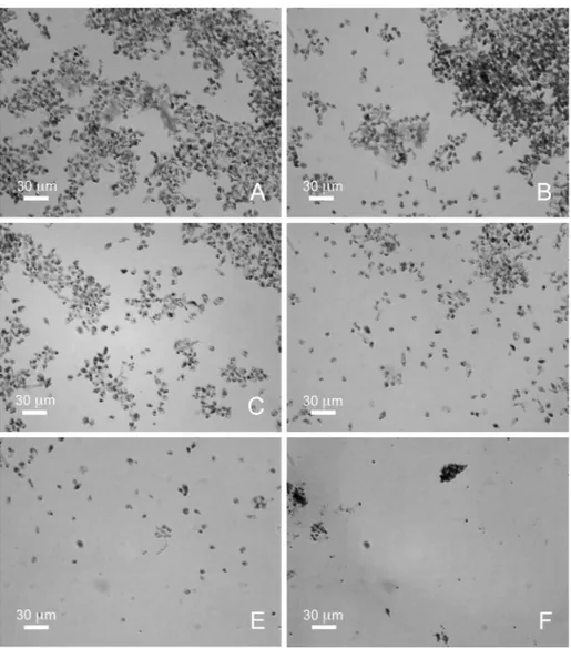
The leishmanicidal effect of A. taxiformis was further verified by evaluating the morphological characteristics of promastigotes and amastigotes of L. infantum after staining with May-Grünwald-Giemsa, with 48 hours of treatment (Figures 2, 3). At a concentration of 5 μg/mL of algal extracts (Figure 2B), the cultures showed a clear decrease in the number of promastigotes compared with the control (Figure 2A) and, at a concentration of 20 μg/mL (Figure 2D), they were characterized by the presence of abnormal and roundish forms. In cultures with 30 μg/mL of algal extracts (Figure 2E), L. infantum cells were aggregates, rounded and without flagella. Finally, in L. infantum cultures with 40 μg/mL of algal extracts (Figure 2F) there were no intact forms of protozoa but only apoptotic bodies. At concentrations of 5, 10, 20, 30 μg/mL (Figures 3B, 3C, 3D, 3E), the cultures showed a clear decrease in the number of amastigotes compared with the control (Figure 3A). Finally, in L. infantum amastigotes cultures with 40 μg/mL of algal extract (Figure 3F) there were no intact forms of amastigotes but only apoptotic bodies.
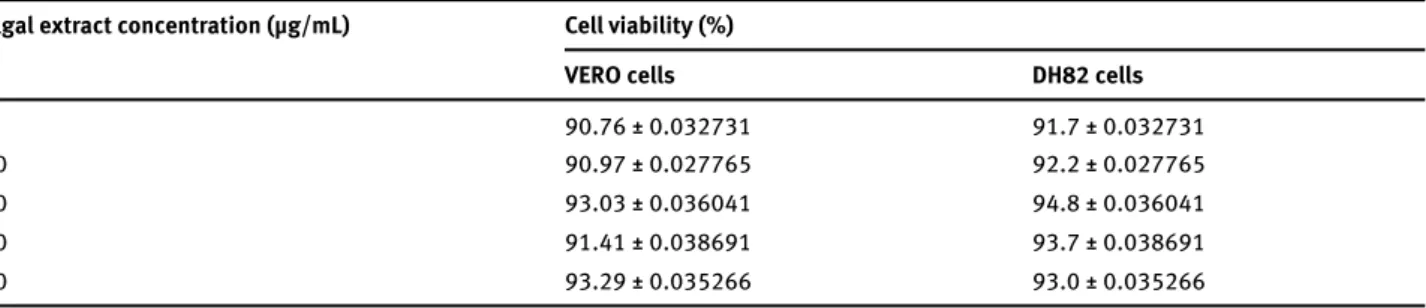
The cytotoxicity test (MTT assay) showed a viability of 91.7-94.8% of DH82 cells and of 88.55-93.29% of VERO cells in tested conditions (Table 1). Therefore, algal extracts did not have a cytotoxic effect on Vero and DH82 cells at tested concentrations.
2.4 Statistical analysis
All experiments were performed by two observers in three replicates and repeated with three new batches of parasites. The mean and standard error were determined. The differences between the mean values obtained for experimental groups were evaluated by Student’s t test. P-values of 0.05 or less were considered significant. The LD50 values were calculated using the GraphPad Prism(®) 5 (Version 5.01, GraphPad Software, Inc., USA).
3 Results
The viability of Leishmania infantum promastigotes treated with serial dilutions of Asparagopsis taxiformis extracts is shown in Figure 1A. Cultures of Leishmania with a concentration of 5 µg/mL of algal extract showed 95% vitality, and the percentage of viable Leishmania decreased proportionally with increasing concentration of the algal extract, reaching 100% mortality of the parasite at 40 µg/mL. From the results observed, the value of LD50 of algal extract obtained is equal to 25 µg/mL. The viability of L. infantum amastigotes treated with serial dilutions of A. taxiformis extracts is shown in Figure 1B. Also in this case, the vitality of Leishmania decreased proportionally with increasing concentration of A. taxiformis extracts. From observed results, the value of LD50 of algal extract is equal to 9 µg/mL.
Figure 1. The viability of for L. infantum promastigotes (A) and amastigotes (B) treated with serial diluitions of A. taxiformis extracts. The LD50
against promastigotes is 25 μg/mL and against amastigotes is 9 μg/mL. The percentage of viable Leishmania was calculated by defining the viability of cells without A. taxiformis extracts treatment as 100%. Data are the results of three independent experiments and are presented as the mean ± SD and the data obtained are statistically significant at p < 0.001.
Figure 2. Cultured promastigotes of L. infantum stained with May-Grünwald-Giemsa. (A) cultures without algal extracts; (B) cultures with
5 μg/mL of algal extracts; (C) cultures with 10 μg/mL of algal extracts; (D) cultures with 20 μg/mL of algal extracts; (E) cultures with 30 μg/mL of algal extracts; (F) cultures with 40 μg/mL of algal extracts. Scale bar = 30 μm.
Table 1. Viability of VERO cells and DH82 cells incubated with algal extract. Algal extract concentration (μg/mL) Cell viability (%)
VERO cells DH82 cells
5 90.76 ± 0.032731 91.7 ± 0.032731 10 90.97 ± 0.027765 92.2 ± 0.027765 20 93.03 ± 0.036041 94.8 ± 0.036041 30 91.41 ± 0.038691 93.7 ± 0.038691 40 93.29 ± 0.035266 93.0 ± 0.035266
4 Discussion
Antileishmanial effects had already been reported for extracts from the red alga Asparagopsis taxiformis [25]. The present work extends these findings by showing that ethanolic crude extracts have a powerful action
against Leishmania infantum, the prevalent species in the Mediterranean basin. In addition, the algal extracts did not show a cytotoxic effect on immortalized Vero and DH82 cell lines. The treatment of leishmaniasis has been the subject of numerous studies and interest in this disease is very high [33]. Our evaluation of the
protocols for the treatment of leishmaniasis and new drugs with leishmanicidal and immunomodulatory activity are needed, in order to achieve total elimination of the parasite. At the same time, it is necessary to develop economical drugs in order to decrease the gap with developing countries, where the impact of leishmaniasis is far greater.
Among marine organisms, seaweeds produce a diverse array of novel secondary metabolites as a chemical defense strategy [35] and these are characterized by a broad spectrum of biological activities [6,10,11,13-15,17,19,20,36].
The study area in Sicily, in the heart of the Mediterranean basin, is an ideal habitat for endemic leishmaniasis infections in both dogs and humans. In this paper, we highlighted the important potentialities of red antiprotozoal activity of A. taxiformis, collected from the
Straits of Messina (Sicily), showed that ethanolic extract had antileishmanial activity characterized by a trend dose-effect.
Dogs are the main reservoir for diffusion of the visceral form of leishmaniasis, which for this reason is widespread in the human population [34]. At present, it is an incurable and complex zoonosis, whose true incidence is not known and for ethical reasons drastic measures (e.g. animal culling) are not proposed. Furthermore, there is a large difference between immunocompetent patients, who heal completely after treatment, and patients in which the condition recurs regularly regardless of the drug used. Current drugs decrease the parasitic charge and promote cell-mediated immune response, but fail to eradicate the disease. Therefore, the development of new therapeutic
Figure 3. Cultured amastigotes of L. infantum stained with May-Grünwald-Giemsa. (A) cultures without algal extracts; (B) cultures with
5 μg/mL of algal extracts; (C) cultures with 10 μg/mL of algal extracts; (D) cultures with 20 μg/mL of algal extracts; (E) cultures with 30 μg/mL of algal extracts; (F) cultures with 40 μg/mL of algal extracts. Scale bar = 30 μm.
[13] Chanda S., Dave R., Kaneria M., Nagani K., Seaweeds: A novel, untapped source of drugs from sea to combat Infectious diseases, In: Current Research, Technology and Education Topics in Applied Microbiology and Microbial Biotechnology, Méndez-Vilas A. (Ed.), Formatex, 2010.
[14] Cumashi A., Ushakova N.A., Preobrazhenskaya M.E., D’incecco A., Piccoli A., Totani L., et al., A comparative study of the anti-inflammatory, anticoagulant, antiangiogenic, and antiadhesive activities of nine different fucoidans from brown seaweeds, Glycobiology, 2007, 17, 541–552.
[15] Dhargalkar V.K., Verlecar X.N., Southern Ocean seaweeds: a resource for exploration in food and drugs, Aquaculture, 2009, 287, 229–242.
[16] Gonzalez Del Val A., et al., Screening of antimicrobial activities in red, green and brown macroalgae from Gran Canaria (Canary Islands, Spain), Int. Microbiol., 2001, 4, 35-40.
[17] Kamenarska Z., Serkedjieva J., Najdenski H., Stefanov K., Tsvetkova I., Dimitrova-Konaklieva S., et al., Antibacterial, antiviral, and cytotoxic activities of some red and brown seaweeds from the Black Sea, Bot. Mar., 2009, 52, 80-86. [18] Shanmughapriya S., Manilal A., Sujith S., Selvin J., Kiran G.,
Natarajaseenivasan K., Antimicrobial activity of seaweeds extracts against multiresistant pathogens, Ann. Microbiol., 2008, 58, 535-541.
[19] Smit A.J., Medicinal and pharmaceutical uses of seaweed natural products: A review, J. Appl. Phycol., 2004, 16. [20] Talarico L.B., et al., Anti-herpes simplex virus activity of
sulfated galactans from the red seaweeds Gymnogongrus
griffithsiae and Cryptonemia crenulata, Int. J. Biol. Macromol.,
2004, 34, 63-71.
[21] Vallinayagam K., Arumugam R., Ragupathi Raja Kannan R., Thirumaran G., Anantharaman P., Antibacterial Activity of Some Selected Seaweeds from Pudumadam Coastal Regions, Glob. J. Pharmacol., 2009, 3, 50-52.
[22] Vonthron-Sénécheau C., et al., Antiprotozoal activities of organic extracts from french marine seaweeds, Mar. Drugs, 2011, 9, 922-933.
[23] Bansemir A., Blume M., Schröder S., Lindequist U., Screening of cultivated seaweeds for antibacterial activity against fish pathogenic bacteria, Aquaculture, 2006, 252, 79-84. [24] Burreson B.J., Moore R.E., Roller P., Haloforms in the essential
oil of the alga Asparagopsis taxiformis (Rhodophyta), Tetrahedron Lett., 1975, 473-476.
[25] Genovese G., Tedone L., Hamann M.T., Morabito M., The Mediterranean Red Alga Asparagopsis: A Source of Compounds against Leishmania, Mar. Drugs, 2009, 7, 361-366.
[26] Jiao G., Yu G., Zhang J., Ewart H.S., Chemical Structures and Bioactivities of Sulfated Polysaccharides from Marine Algae, Mar. Drugs, 2011, 9, 196-223.
[27] Mcconnell O., Fenical W., Halogen chemistry of the red alga
Asparagopsis, Phytochemistry, 1977, 16, 367-374.
[28] Salvador N., Gomez Garreta A., Lavelli L., Ribera M.A., Antimi-crobial activity of Iberian macroalgae, Sci. Mar., 2007, 71, 101-113.
[29] Genovese G., Faggio C., Gugliandolo C., Torre A., Spanò A., Morabito M., et al., In vitro evaluation of antibacterial activity of Asparagopsis taxiformis from the Straits of Messina against pathogens relevant in aquaculture, Mar. Environ. Res., 2012, 73, 1-6.
alga A. taxiformis metabolites as anti-leishmanial agents which merit further pharmacological investigations. In particular, the purification of the active principle could be interesting, although it is not possible to exclude the possibility that several molecules contribute to the antileishmanial activity in a synergic mechanism.
Acknowledgements: This study was supported by grants to F.V. (IZS SI 13/2012 RC) and G.G. (INNOVAQUA PON02_00451_3362185).
Conflict of interest: Authors declare nothing to disclose.
References
[1] Anonymous, WHO - Leishmaniasis (http://www.who.int/ leishmaniasis/en/), 2014.
[2] Meena A.K., Kandale A., Nigam S., Panda P., Singh B., Rao M.M., Review on Marine organisms with antileishmanial activity, J. Pharm. Res., 2010, 3, 818-821.
[3] Croft S.L., Recent development in the chemotherapy of leishmaniasis, Trends Pharmacol. Sci., 1988, 9, 376-381. [4] Ribeiro A.L., Drummond J.B., Volpini A.C., Andrade A.C., Passos
V.M., Electocardiographic changes during low-dose, short term therapy of cutaneous leishmaniasis with pentavalent antimonial meglumine, Braz. J. Med. Biol. Res., 1999, 32, 297-301.
[5] Berma J.D., Treatment of new world cutaneous and mucosal leishmaniasis, Clin. Dermatol., 1996, 14, 519-522.
[6] Mayer A.M.S., Rodríguez A.D., Berlinck R.G.S., Fusetani N., Marine pharmacology in 2007–8: Marine compounds with antibacterial, anticoagulant, antifungal, anti-inflammatory, antimalarial, antiprotozoal, antituberculosis, and antiviral activities; affecting the immune and nervous system, and other miscellaneous mechanisms of action, Comp. Biochem. Phys. C, 2011, 153, 191-222.
[7] Newman D.J., Cragg G.M., Snader K.M., Natural products as sources of new drugs over the period 1981-2002, J. Nat. Prod., 2003, 66, 1022-1037.
[8] Blunt J.W., et al., Marine natural products, Nat. Prod. Rep., 2008, 25, 35-94.
[9] Fouladvand M., Barazesh A., Farokhzad F., Malekizadeh H., Sartavi K., Evaluation of in vitro anti-Leishmanial activity of some brown, green and red algae from the Persian Gulf, Eur. Rev. Med. Pharmacol. Sci., 2011, 15, 597.
[10] Allmendinger A., Spavieri J., Kaiser M., Casey R., Hingley-Wilson S., Lalvani A., et al., Antiprotozoal, antimycobacterial and cytotoxic potential of twenty-three British and Irish red algae, Phytother. Res., 2011, 24, 1099-1103.
[11] Ballesteros E., Martin D., Uriz M.J., Biological Activity of Extracts from Some Mediterranean Macrophytes, Bot. Mar., 1992, 35, 481-485.
[12] Bouhlal R., Riadi H., Martínez J., Bourgougnon N., The antibacterial potential of the seaweeds (Rhodophyceae) of the Strait of Gibraltar and the Mediterranean Coast of Morocco, Afr. J. Biotechnol., 2010, 9, 6365-6372.
[33] Ashford R.W., The leishmaniases as emerging and reemerging zoonoses, Int. J. Parasitol., 2000, 30, 1269-1281.
[34] Courtenay O., Quinnell R.J., Garcez L.M., Shaw J.J., Dye C., Infectiousness in a cohort of Brazilian dogs: why culling fails to control visceral leishmaniasis in areas of high transmission, J. Infect. Dis., 2002, 186, 1314-1320.
[35] McClintock J.B., Baker J.B., Marine Chemical Ecology, CRC Press, Boca Raton, Florida, 2001.
[36] Ioannou E., Vagias C., Roussis V., Bioactive metabolites from marine algae, Bio. Environ., 2010, 26, 68-72.
[30] Limoncu M.E., Balcioglu I., Yereli K., Ozbel Y., Ozbilgin A., A new experimental in vitro culture medium for cultivation of
Leishmania species, J. Clin. Microbiol., 1997, 35, 2430-2431.
[31] Carmichael J., Degraff W.G., Gazdar A.F., Minna J.D., Mitchell J.B., Evaluation of a tetrazolium-based semiautomated colorimetric assay: assessment of chemosensitivity testing, Cancer Res., 1987, 47, 936–942.
[32] Pessotti J.H., Zaverucha Do Valle T., Corte-Real S., Gonçalve Da Costa S.C., Interaction of Leishmania (L.) chagasi with the Vero cell line, Parasite, 2004, 11, 99-102.