UNIVERSITÀ DEGLI STUDI DI MESSINA
DIPARTIMENTO DI SCIENZE CHIMICHE, BIOLOGICHE,
FARMACEUTICHE ED AMBIENTALI
DOTTORATO DI RICERCA IN
BIOLOGIA APPLICATA E MEDICINA SPERIMENTALE
XXIX CICLO
Anti-oxidant and anti-inflammatory
activities of a flavonoid-rich extract from
Citrus bergamia Risso et Poiteau juice in
both cell-free and in vitro models
T
ESI DID
OTTORATO:
D
OTT.
SSAS
ANTAC
IRMI
T
UTOR:
P
ROF.
M
ICHELEN
AVARRAC
OORDINATORE DELC
ORSO DID
OTTORATOC
HIAR.
MOP
ROF.
S
ALVATOREC
UZZOCREAInflammation consists in a series of biological reactions induced by the alteration of tissue homeostasis occurring in response to biological, chemical or physical agents in the organism. Oxidative stress is defined as an imbalance between production and elimination of free radicals and reactive metabolites (oxidants). This is a natural physiological process where the presence of ROS overpowers the cellular radical scavenging ability, thus creating an imbalance in the oxidative status between the oxidants and the anti-oxidants.
Inflammation and oxidative stress are closely related pathophysiological events that are strongly linked each other. One of them may appear before or after the other, but when one emerges the other one is most likely to follow, and then both of them take part in the pathogenesis of many disorders.
In recent years, there has been an extraordinary increase in the number of studies on anti-oxidant properties of various phytochemicals, able to counteract reactive species (RS) overproduction. Among these, flavonoids have been extensively studied mainly for their anti-oxidant property which ameliorates many inflammatory diseases and was linked to the maintenance of good health. Citrus fruits and their juices are the main food sources of flavonoids which exert protective effects against numerous degenerative processes. In the last decade a number of studies have investigated the anti-oxidant and anti-inflammatory effects of each single Citrus flavonoid as pure compounds. However, few studies have focused on the pharmacological activity of
Citrus juices and extracts and its molecular mechanisms underlying their potential
extract from Citrus bergamia juices (BJe) and its effect against inflammatory processes. First, we tested the anti-oxidant properties of BJe in cell-free experimental models by ORAC, DPPH, Folin-Ciocalteu and Reducing Power assays, proving its anti-oxidant
activity. Then, we assayed its ability to prevent the cytotoxic effects induced by H2O2 or
Fe2(SO4)3. Our results provided evidences that BJe reduces cell death, generation of
ROS and membrane lipid peroxidation, improve mitochondrial functionality and
prevents DNA-oxidative damage in A549 cells incubated with H2O2. Moreover, BJe is
able to both induce catalase expression and increase its activity. Furthermore,
evidences, that BJe prevents the cytotoxic effects by Fe2(SO4)3, have suggested that
this extract could possess chelating properties. This hypothesis was confirmed by measuring the presence of redox-active iron in the cells pre-treated with the extract and then exposed to the metal.
In the light of these observations, we wondered whether BJe may be effective against inflammatory processes. To this aim, we used THP-1 monocytes to investigate the mechanisms underlying the beneficial potential of BJe against two different models of
inflammation in which the THP-1 cells were exposed to LPS or amyloid-beta1–42
(Aβ1−42). Exposure of THP-1 cells to BJe inhibited both gene expression and secretion of
LPS-induced pro-inflammatory cytokines (IL-6, IL-1β, TNF-α) by a mechanism involving
the inhibition of NF-ĸB activation. In addition, BJe treatment reversed the
LPS-enhanced acetylation of p65 in THP-1 cells. Furthermore, increasing concentrations of Sirtinol were able to suppress the inhibitory effect of BJe via p65 acetylation, underscoring that NF-ĸB-mediated inflammatory cytokine production may be directly linked to SIRT1 activity.
of IL-6 and IL-1β in THP-1 cells and increased the phosphorylation of ERK 1/2 as well as
p46 and p54 members of JNK family. Moreover, Aβ1−42 raised AP-1 DNA binding activity
in THP-1-treated cells. Interestingly, all these effects were reduced in the presence of BJe.
In conclusion, our studies provided evidences that BJe may be used in preventing oxidative cell injury suggesting a promising role as a natural drug against inflammatory processes. In addition, the complex mixture of phytochemicals present in the whole extract acts better than the single constituent. This is because all molecules present in a phytocomplex can modulate simultaneously different targets of action in both human cells and microorganisms, leading to a pool of pharmacological effects contributing together to improve patient’s health.
Key words: oxidative stress, inflammation, bergamot juice extract, flavonoids,
1.
INTRODUCTION
1.1. The inflammation pag. 1
1.2. The radical species pag. 2
1.3. Oxidative stress and inflammation pag. 3
1.4. Natural products for the treatment of inflammation pag. 4
1.5. The Citrus bergamia Risso et Poiteau pag. 5
2.
AIM OF THE RESEARCH
pag. 83.
MATERIALS AND METHODS
3.1. Flavonoid-rich extract from bergamot juice pag. 9
3.2. Chemical characterization of BJe pag. 9
3.3. Evaluation of the anti-oxidant capacity in cell-free models pag. 10
3.3.1. Folin-Ciocalteu assay pag. 10
3.3.2. Quenching of the stable 2,2-Diphenylpicrylhydrazyl (DPPH)
Radical assay
pag. 10
3.3.3. Oxygen radical absorbance capacity (ORAC) assay pag. 10
3.3.4. Reducing Power assay pag. 11
3.4. Evaluation of the anti-oxidant activity of BJe in in vitro models
pag. 12
3.4.1. Cell culture and treatment pag. 12
3.4.2. Cytofluorimetric analyses pag. 12
3.4.2.1. DCF-DA staining assay pag. 13
3.4.2.2. DPP staining assay pag. 13
3.4.2.3. R123 staining assay pag. 13
3.4.2.4. 8-oxo-dG assay pag. 14
3.4.2.5. PI supravital staining assay pag. 14
3.4.3. Comet assay pag. 15
3.4.4. ROS and Δ m determinations by confocal microscopy
observations
pag. 16
3.4.5. Statistical analysis pag. 16
3.5. Evaluation of the chelating property of BJe in in vitro models pag. 17
3.5.1. Cell culture and treatment pag. 17
3.5.2. Cell viability assay pag. 17
3.5.3. Analyses of oxidative stress markers pag. 18
3.5.4. Comet assay pag. 18
3.5.5. Analysis of intracellular free iron pag. 19
3.5.6. Fluorimetric abiotic assay pag. 19
3.5.7. Enzymatic activity and mRNA level of the antioxidant catalase
enzyme
3.6. Assessment of the anti-inflammatory effect of BJe against LPS in an in vitro models
pag. 22
3.6.1. Cell culture and treatment pag. 22
3.6.2. Cell viability assay pag. 23
3.6.3. Real-Time PCR pag. 23
3.6.4. Evaluation of cytokine secretion by ELISA pag. 24
3.6.5. Electrophoretic mobility shift assay pag. 24
3.6.6. Immunoprecipitation and immunoblotting analyses pag. 25
3.6.7. Statistical analysis pag. 26
3.7. Employment of an in vitro model to assess the capability of BJe to prevent the neuro-inflammatory action of β-amyloid
pag. 27
3.7.1. Cell culture and treatment pag. 27
3.7.2. Cell transfection using AP1 ODNs pag. 28
3.7.3. Analysis of cytokine expression and secretion pag. 28
3.7.4. Cell viability assays pag. 29
3.7.5. Measurement of intracellular reactive oxygen species pag. 29
3.7.6. Analysis of MAPK expression pag. 30
3.7.7. Assessment of transcription factor activation pag. 30
3.7.8. Statistical analysis pag. 31
4.
RESULTS
4.1. Flavonoid composition of BJe pag. 32
4.2. Anti-oxidant capacity in cell-free models pag. 36 4.3. Protective effect of BJe from H2O2-induced oxidative stress in
A549 cells
pag. 37
4.3.1. BJe prevents the H2O2-induced increase of ROS pag. 37
4.3.2. BJe reduces the H2O2-caused cell death pag. 39
4.3.3. BJe counteracts lipid peroxidation induced by H2O2 in A549
cells
pag. 40
4.3.4. BJe attenuates the fall in Δ m induced by H2O2 pag. 40
4.3.5. BJe decreases oxidative DNA damage induced by H2O2 pag. 42
4.4. Protective role of BJe in Fe3+-induced oxidative stress in A549 cells
pag. 44
4.4.1. Effects of BJe on cell viability in presence of Fe3+ pag. 44
4.4.2. BJe reduces the Fe3+-induced increase of ROS pag. 45
4.4.3. BJe counteracts lipid peroxidation induced by Fe3+ pag. 45
4.4.4. BJe reduces the fall in Δ m caused by Fe3+ pag. 46
4.4.5. BJe decreases oxidative DNA damage exerted by Fe3+ pag. 47
4.4.6. Chelating activity of BJe pag. 48
4.5.1. Cell viability assay in presence of LPS pag. 51
4.5.2. Expression of LPS-induced pro-inflammatory cytokines in
presence of BJe
pag. 52
4.5.3. BJe treatment reduces the release of cytokines caused by LPS pag. 53
4.5.4. Inhibition of LPS-induced NF-kB activation by BJe pag. 55
4.5.5. BJe treatment reverts LPS-enhanced acetylation of p65
through the involvement of SIRT1
pag. 56
4.6. BJe attenuates β-amyloid-induced pro-inflammatory activation of THP-1 cells
pag. 58
4.6.1. Aβ1−42 causes an increase of pro-inflammatory cytokines gene
expression
pag. 58
4.6.2. Effect of Aβ1-42 and BJe on THP-1 cell viability and ROS
production
pag. 60
4.6.3. BJe reduces IL-1β and IL-6 gene expression and secretion in
Aβ1-42-treated cells
pag. 61
4.6.4. BJe decreases the phosphorylation of MAPK caused by Aβ1-42 pag. 63
4.6.5. BJe determines a decrease of AP-1 DNA binding activity in Aβ
1-42-treated THP-1 cells
pag. 65
4.6.6. Inhibition of AP-1 DNA binding activity by AP-1 ODN attenuates
Aβ1−42-nduced up-regulation and secretion of cytokines in
THP-1 cells
pag. 66
5. DISCUSSION pag. 68
6. CONCLUDING REMARKS pag. 80
1
1.INTRODUCTION
1.1. The inflammation
Inflammation consists in a series of biological reactions induced by the alteration of tissue homeostasis occurring in response to biological, chemical, or physical agents in the organism (Medzhitov et al., 2008). The classical key features of inflammation are redness, warmth, swelling and pain. Inflammation can be either acute or chronic, depending on the type of stimulus and the effectiveness of the inflammatory process resolution. Acute inflammation begins quickly and persists few hours or few days. It is characterized by the exudation of fluid and plasma proteins as well as leukocyte migration (mainly neutrophils). When the immune system successfully eliminates damaging agents in acute inflammation, the reaction disappears. However, if the inflammatory response fails to remove its cause, a chronic phase occurs. Inflammation cascades can lead to the development of chronic diseases such as asthma, rheumatoid arthritis, multiple sclerosis, inflammatory bowel disease and psoriasis. Chronic inflammation is associated with the presence of lymphocytes and macrophages, vascular proliferation, fibrosis, and tissue destruction. Experimental and clinical studies together with epidemiological observations have identified both chronic infections and inflammation as major risk factors for various types of cancer. It has been estimated that the underlying infections and inflammatory reactions are linked to 15-20% of all cancer deaths (Mantovani et al., 2008). The inflammatory response is characterized by coordinated activation of various signaling pathways that regulate expression of both pro- and anti-inflammatory mediators in cells of inflamed tissue as well as leukocytes recruited from the blood. The nuclear transcription factor κB (NF-ĸB) is the master
2
regulator of the inflammatory response, that drives the activation of genes associated with the transcription of inflammatory mediators, such as interleukins, tumor necrosis factor (TNF) and prostaglandins (PGs), as well as inflammatory enzymes, like inducible nitric oxide synthase (iNOS) and cyclooxygenases (COXs).
1.2. The radical species
Radical species (RS), including reactive oxygen species (ROS) and reactive nitrogen species (RNS), are highly reactive molecules physiologically produced in a tightly-regulated manner and in small quantity during the cellular metabolism in aerobic organisms. They are involved in cell homeostasis and control several cellular functions such as signal transduction and gene expression (Kumar and Pandey, 2015). In physiological condition, ROS and RNS are removed quickly by the endogenous anti-oxidant systems such as glutathione peroxidase (GSH-Px), superoxide dismutase (SOD) and catalase (CAT), as well as exogenous anti-oxidant introduced by diet, like vitamin C and E, carotenoids and polyphenols. Imbalance between generation and elimination of RS, due to their overproduction or reduced metabolization can lead to the damage of important cellular and molecular structures such as DNA, proteins, and lipids (Duračková et al., 2010). This can cause pathological conditions (apoptosis, necrosis, uncontrolled cell proteolysis, oxidative DNA damage, lipid peroxidation, etc.) which govern a wide array of diverse disorders (Beckman and Ames, 1998; Golden et al., 2002). Under pathological inflammatory status, there may be exaggerated generation of RS that can diffuse out of the cells, inducing localized oxidative stress and tissue injury (Fialkow et al., 2007). Moreover, the activated phagocytic cells produce large amounts of ROS and RNS. Once generated, they can further generate other RS, leading
3
to extensive damage. At the onset of inflammation, the infection or tissue damage is sensed by pattern recognition receptors like toll-like receptors (TLR), NOD-like receptors (NLR), and the receptor for advanced glycation end products (RAGE). The stimulation of these receptors upon binding with specific molecules leads to the activation of transcription factors such as nuclear factor-ĸB (NF-ĸB) and activating protein-1 (AP-1), that in turn induce pro-inflammatory gene expression, exert antimicrobial functions and recruit additional immune cells (Tabas et al., 2015; Bierhaus et al., 2005). It has been demonstrated that RS (i.e., hydrogen peroxide) can induce inflammation through activation of these same transcription factors (Oliveira-Marques et al., 2009; Vollgraf et al., 1999; Kang et al., 2011).
1.3. Oxidative stress and inflammation
Oxidative stress is defined as an imbalance between production and elimination of free radicals and reactive metabolites (oxidants). This is a natural physiological process where the presence of ROS overpowers the cellular radical scavenging ability, thus creating an imbalance between the oxidants and the antioxidants.
Inflammation and oxidative stress are closely related pathophysiological events that are strongly linked with each other. One of them may appear before or after the other, but when one emerges the other one is most likely to follow, and then both take part in the pathogenesis of many disorders. Nowadays, it is clear that the prolonged low-grade inflammatory process plays a central role in the pathogenesis of many chronic diseases (Cotran et al., 1999). On the other hand, epidemiological and experimental studies strongly suggest a contribution of oxidative stress in many human diseases (Cotran et al., 1999). Just as the inflammatory process can induce oxidative stress, the
4
latter can cause inflammation through activation of multiple pathways (Castellani et al., 2014; Mittal et al., 2014). The result is that both inflammation and oxidative stress are associated with a number of chronic diseases, including diabetes, hypertension, cardiovascular diseases, neurodegenerative diseases, alcoholic liver disease, chronic kidney disease, cancer and aging (Biswas et al., 2007; Ambade et al., 2012; Biswas et al., 2008; Cachofeiro et al., 2008).
1.4. Natural products for the treatment of inflammation
Several classes of medicines, including corticosteroids, nonsteroidal anti-inflammatory drugs (NSAIDs) and biologic drugs are used to treat the inflammatory disorders. However, they possess several side effects and the biologics ones are expensive to be used. To limit these drawbacks of both synthetic and biologic drugs, over the past three decades, the use of herbal medicines, nutraceuticals and food supplements has greatly increased as an alternative and/or complementary medicine to treat several pathologies, including inflammation (Marino et al., 2015). Indeed, although natural products are not devoid of risk, generally they are safer than both synthetic and biologic drugs. Nowadays, about 80% of people around the world use natural products for the prevention and treatment of many diseases, mainly for their relative safety, efficacy and low cost as well as the compliance by patients. Plants have been the basis of many traditional medicines throughout the world for thousands of years, and continue to provide mankind with new remedies (Dias set al., 2012). In this field, natural products offer great hope in the identification of bioactive molecules useful for the treatment of inflammatory diseases, as it happened for aspirin, the first discovered NSAID, which is still one of the bestselling drug in the world.
5
In recent years there has been an extraordinary increase in the number of studies on anti-oxidant properties of various phytochemicals, able to counteract RS overproduction. Among these, flavonoids, a family of polyphenols found especially in fruits, vegetables, red wine and tea, have been extensively studied manly for their anti-oxidant property (Rice-Evans et al., 1997; Ross and Kasum, 2002). Flavonoids are plant secondary metabolites commonly found in the fruits and vegetables regularly consumed by humans. Their anti-oxidant activity ameliorates many inflammatory diseases and was linked to the maintenance of good health (Di Matteo and Esposito, 2003; Yao et al., 2004). Several mechanisms are involved in the beneficial effects exerted by flavonoids, including the free radicals scavenging (Kumar and Pandey, 2013), the transition metal ions chelation (Mladěnka et al., 2011), the enhancement of glutathione content, and modulation of defense genes expression via the Nrf2/ARE pathway (Kumar and Pandey, 2013; Chen and Kong, 2005; Masella et al., 2005; Kumar et al., 2014).
1.5. The Citrus bergamia Risso et Poiteau
Citrus fruits and their juices are the main food sources of flavonoids and have been
extensively studied as regards their cardiovascular, anticancer, anti-infective, neuroprotective, and anti-inflammatory activity (Benavente-Garcıa and Castillo, 2008; Cirmi et al., 2016a; Cirmi et al., 2016b; Cirmi et al., 2016c; Ferlazzo et al., 2016a). In recent years has gained ground scientific interest in Citrus bergamia (bergamot) derivatives. Citrus bergamia Risso et Poiteau, also known as “Bergamot,” is a plant belonging to the Rutaceae family. Citrus bergamia is defined as a hybrid between a sour orange (C. aurantium L.) and lemon (C. limon L. Burm. f.) or a mutation of the
6
latter. Other Authors considered it as a hybrid between a sour orange and lime (C.
aurantifolia [Christm. and Panzer] Swingle). The botanical and ethno-pharmacological
issues of this plant have been reported by Rapisarda and Germanò (2013).
Bergamot fruit is used especially for the extraction of its essential oil (BEO) from the peel (by cold pressing), while the bergamot juice (BJ), derived from squeezing the endocarp of the fruits is considered a byproduct of the BEO’s production. Finally, the scraps of bergamot fruit after both BEO extraction and BJ juicing is named “bergamot pastazzo” and it is used as animal feed. BEO is typically used in the cosmetic industry, being found in the composition of many fragrances, body lotions, soaps and so on. It is also used by the food industries (for flavoring tea, beverages and typical Calabrian pastries), by the pharmaceutical industries (to absorb the unpleasant smell of medicinal products and for its antiseptic and antibacterial properties (Cirmi et al., 2016a) and in aromatherapy (Navarra et al., 2015). Recently, BEO has been experimentally investigated for its potential anti-proliferative (Celia et al., 2013; Navarra et al., 2015) and neuroprotective effects (Corasaniti et al., 2007).
Over the last decade, some researchers have started to investigate the biological properties of bergamot derivatives, obtaining important scientific achievements. Studies performed by Miceli et al. (2007) have shown that a chronic administration of BJ is effective to prevent the diet-induced hyperlipidemia in rat, suggesting a relationship between the beneficial effect and its anti-oxidant properties. Moreover, a clinical research showed that the bergamot-derived polyphenolic fraction, given orally in patients suffering from metabolic syndrome, reduced plasma lipids and improved the lipoprotein profile (Mollace et al., 2011; Toth et al., 2016), strengthen the finding obtained in animal model.
7
Finally, the research group coordinated by prof. Michele Navarra demonstrated that BJ reduces the growth rate of different cancer cell lines by different molecular mechanisms, depending on cancer type. In SH-SY5Y human neuroblastoma cells, BJ stimulated the cell cycle arrest in the G1 phase without inducing apoptosis, and caused a modification in cellular morphology associated with a marked increase in detached cells. The inhibition of adhesive ability onto different physiologic substrates and onto endothelial cell monolayer was correlated to BJ-induced impairment of actin filaments and with the reduction in the expression of the active form of FAK, in turn causing inhibition of cell migration (Delle Monache et al., 2013). Contrariwise, in human hepatocellular carcinoma HepG2 cells, BJ reduced the growth rate through the involvement of p53, p21, and NF-ĸB pathways, as well as the activation of both intrinsic and extrinsic apoptotic pathways (Ferlazzo et al., 2016b). Moreover, BJ-induced reduction of both cell adhesiveness and motility could be responsible for the slight inhibitory effects on lung metastasis colonization observed in an animal model of spontaneous neuroblastoma metastasis formation in SCID mouse (Navarra et al., 2014). In order to assess which bioactive component of BJ was responsible for its antitumor activity, Visalli et al., (2014) focused on the flavonoid-rich fraction from bergamot juice (BJe). Results suggested that BJe inhibits HT-29 human colorectal carcinoma cell growth and induces apoptosis through multiple mechanisms. Molecular assays revealed that higher concentrations of BJe increase ROS production, which causes a loss of mitochondrial membrane potential and oxidative DNA damage. Lower concentrations of BJe inhibited MAPK pathways and modified apoptosis-related proteins, which in turn induced cell cycle arrest and apoptosis (Visalli et al., 2014).
8
2. AIM OF RESERCH
Natural products have been shown to exert beneficial effects on human health, as well as it is known that flavonoids can exert protective effects against numerous degenerative processes. In addition, a number of studies have investigated the anti-oxidant and anti-inflammatory effects of single Citrus flavonoids as pure compounds. However, few studies have focused on the pharmacological activity of Citrus juices and extracts. Moreover, in recent years, Citrus bergamia fruit has attracted attention of the scientific community because of its potential to prevent or counteract some
pathologies. On the basis of what has been discussed in the introduction section,
during my PhD, I evaluated the anti-oxidant and anti-inflammatory effect of BJe in different cell-free and cell-based experimental models.
9
3. MATERIALS AND METHODS
3.1. Flavonoid-rich extract from bergamot juice
The flavonoid-rich extract of bergamot juice (BJe) was provided by the company “Agrumaria Corleone” (Palermo, Italy). The fruits of Citrus bergamia Risso & Poiteau came from crops located in the province of Reggio Calabria (Italy). The extract was transformed into a dry powder using the spray drying method. Small aliquots of BJe were stored at -20°C. Finally, the drug was defrosted, diluted in culture media, the pH was adjusted to 7.4 and filtered just prior to use.
3.2. Chemical characterization of BJe
BJe powder was dissolved in methanol to a concentration of 1 mg/ml-1, ultra-sonicated
and filtered through a 0.2 µm nylon membrane (Millipore, Milan, Italy), and then
injected into a UHPLC coupled online to an LCMSIT-TOF mass spectrometer (Shimadzu, Kyoto, Japan). Identification of flavonoids was carried out on the basis of diode array spectra, MS molecular ions, and MS/MS fragmentation patterns. Data obtained were compared with those available in scientific literature. Molecular formulae were calculated by the Formula Predictor software (Shimadzu).
10
3.3. Evaluation of the anti-oxidant capacity in cell-free models
3.3.1. Folin-Ciocalteu assay
The total phenolic content of BJe was determined by the Folin-Ciocalteu assay, following Tomaino et al. (2010). Briefly, 50 μL of methanol/water solutions of different sample concentrations were added to 450 μL of deionized water, 500 μL of Folin-Ciocalteu reagent, and 500 µL of 10% sodium carbonate solution and incubated in the dark at room temperature for 1 h, vortexing every 10 min. Absorbance was recorded at 786 nm (PrixmaUV-Vis Spectrophotometers) against a blank containing 50 µL of the same solvent used to dissolve the extracts. Total phenol content is expressed in mg of gallic acid equivalents (GAE/g of dried extract).
3.3.2. Quenching of the stable 2,2-Diphenylpicrylhydrazyl (DPPH) Radical assay
The DPPH assay was used to evaluate the radical scavenging activity of BJe. Following the procedure devised by Tomaino et al. (2010), different concentrations (ranging from 0.1 to 1 mg/mL) of methanol/water solution of each extract or vehicle alone (37.5 µL) were added to 1.5 mL of DPPH methanolic solution (25 mg/L). Absorbance was measured at 517 nm 30 min after starting the reaction. Free radical scavenging capacity of juice extracts is expressed in mg of Trolox equivalents (TE/g of dried extract).
3.3.3. Oxygen radical absorbance capacity (ORAC) assay
Antioxidant activity of BJe against 2,2’-azobis (2-amidinopropane) dihydrochloride (AAPH) peroxyl radicals was chemically examined using the ORAC method described by
11
Dávalos et al. (2004) with some modifications. Briefly, several concentrations of BJe (20 µL) in 75 mM phosphate buffer solution (pH 7.4) were mixed with 120 µL of 417 nM fluorescein solution and incubated at 37°C for 15 min to which 60 µL of AAPH (40 mM) was then added. Fluorescence was recorded spectrofluorometrically every 30 sec for 90 min ( ex 485; em 520; FLUOstar Omega, BMG Labtech), and phosphate buffer instead of sample, and calibration solutions of Trolox (10-100 µM) were also included in each assay. The ORAC value was calculated using the area under the fluorescence decay curves and is expressed in µmoles of TE/g of dried extract.
3.3.4. Reducing power assay
The reducing power of BJe was determined following the method described by Martorana et al. (2013). In brief, 0.2 mL of several concentrations of extract were mixed with 0.5 mL of 0.2 M sodium phosphate buffer (pH 6.6) and 0.5 mL of 1%
K3Fe(CN)6 and then incubated in a water bath at 50°C for 20 min. Subsequently, 0.5 mL
of 10% TCA was added to the mixture which was centrifuged at 8300 ×g for 10 min. The supernatant (0.5 mL) was then mixed with 0.5 mL of distilled water and 0.1 mL of 0.1% ferric chloride solution and absorbance measured at 700 nm. Increased absorbance of the reaction mixture indicated increased reducing power. Ascorbic acid was used as a reference. Phosphate buffer was used as blank solution. Reducing power is expressed in mg of ascorbic acid equivalent (AAE)/g of dried extract.
12
3.4. Evaluation of the anti-oxidant activity of BJe in in vitro models
3.4.1. Cell culture and treatment
The experiments were performed using a basal epithelial cell line A549 derived from human lung carcinoma (ATCC, Rockville, MD, USA). Cells were grown in 6-well plates
(3×10⁵ cells/well) and cultured in RPMI medium with 2 mM L-glutamine (Gibco
Invitrogen, Milan, Italy), 10% (v/v) foetal bovine serum (FBS), 100 IUmL−1 penicillin, and
100 gmL−1 streptomycin at 37°C in a humidified 5% CO2 atmosphere. When 80–90%
confluence was reached, monolayers were used for experiments by adding BJe to
obtain a final concentration of 25 and 50 µgmL−1 in cell medium with 2% FBS. After 18
h, the medium was removed, and cells were washed and exposed to 200 µM H2O2 in
PBS solution (pH 7.4) containing 10 mM D-glucose for further 2 h.
For each set of experiments, a negative control (untreated cultures) and a stressor
control (H2O2 alone) were prepared by replacing the extract with PBS.
3.4.2. Cytofluorimetric analyses
Fluorescence-activated cell sorting (FACS) techniques were employed to determine the following parameters: intracellular ROS, lipid hydroperoxides,
8-oxo-7,8-dihydro-2’-deoxyguanosine (8-oxo-dG), transmembrane mitochondrial potential (Δ m) and cell
viability. After each experiment, the cells were harvested, centrifuged at 1000 ×g for 5
min, washed and suspended in PBS. Aliquots of cell suspensions (∼2×10⁵ cells mL−1)
were used for each probe as described below. The data collected from each probe
were used to draw the respective curves by calculating the average of cell percentages for each emission value. In FACS analyses, the weighted average of emission values per
13
100 cells was calculated and is expressed in arbitrary fluorescence units (AFU). The values obtained were used to calculate the percentage changes (%Δ) compared to the respective control.
3.4.2.1. DCF-DA staining assay
To determine intracellular ROS accumulation, cells suspensions were incubated for 30 min at 37°C with 1 µM 2-7-dichlorofluorescein diacetate (DCF-DA) probe. DCF-DA was deacetylated intracellularly by non-specific esterase and then oxidized by ROS to the fluorescent compound 2’-7’-dichlorofluorescein (DCF). Fluorescence emitted by DCF in FL-1 channel was detected by a Dako Galaxy flow cytometer (Dako Galaxy Cytomation).
3.4.2.2. DPP staining assay
Lipid hydroperoxides were detected using the diphenyl-1-pyrenylphosphine probe (DPPP; Invitrogen Molecular Probe, Milan, Italy) as reported by Di Pietro et al., (2009). The probe reacts stechiometrically with lipid hydroperoxides in cell membranes to yield a fluorescent phosphine oxide (DPPP=O) and the corresponding hydroxide. DPPP was added to cell suspensions to obtain a final concentration of 150 µM and incubated at 37°C for 3 h. Fluorescent phosphine oxide signals were then collected in the FL-1 channel.
3.4.2.3. R123 staining assay
Change in Δ m as result of mitochondrial perturbation, was evaluated by measuring the incorporation of rhodamine 123 (R123; Invitrogen, Life Technologies), which can
14
cross the mitochondrial membrane and to accumulate in the matrix of functional mitochondria. The fluorochrome (R123 0.2 µM) was added to cells suspension and incubated at 37°C for 10 min. The emitted fluorescence was then collected in the FL-2 channel.
3.4.2.4. 8-oxo-dG assay
Although all DNA bases are susceptible to damage, guanine is the most prone to oxidative modification. Eight-hydroxyguanine (8-OH-Gua) and its 2’-deoxynucleoside equivalent, 8-hydroxy-2’-deoxyguanosine (8-oxo-dG), are the most common byproducts. The latter is removed during the repair of damaged DNA by exonucleases as it is considered a marker of oxidant-induced DNA damage (Caramori et al., 2011). The level of oxo-dG was estimated by FITC-labelled avidin probe which binds to 8-oxo-dG with high specificity due to the structural analogies between the keto form of the oxidized base and biotin. The method, previously adapted to flow cytofluorimetric analysis (Di Pietro et al., 2011), was performed in cells permeabilized by methanol (15 min at -20°C) and loaded with the avidin-FITC conjugate (1 h at 37°C). The emission signals were collected in FL-1 channel.
3.4.2.5. PI supravital staining assay
For PI staining, at the end of the treatments, supernatants were collected, the cells were detached with trypsin and pooled with corresponding supernatants. Cells were
washed in PBS and stained with PI (3 µgmL−1) at 4°C for 3 min. Dead cells, stained with
15
emission signals in the FL-3 channel. Percentage of dead cells was calculated versus non-treated cells.
3.4.3. Comet assay
In order to examine the DNA integrity (potential genotoxic effect) in cells treated with the BJe, the alkaline version of comet assay was performed as reported (Picerno et al., 2006). Briefly, at the end of each treatment, cells were collected, washed with ice-cold
PBS and 10 µl of cell-suspension (1x10⁶ cells/ml) were dissolved in Low Melting Point
(LMP) Agarose and spread on pre-coated-agarose microscope slides. The cells were lysed and then placed in a horizontal electrophoresis box. Subsequently, the cells were exposed to alkaline condition for 20 min to allow DNA unwinding and expression of alkali-labile sites. To electrophorese the DNA, an electric current of 25 V (0.86 V/cm) and 300 mA was applied for 30 min. Then, the slides were neutralized, stained with
ethidium bromide (20 µgmL−1) and analyzed using DM IRB fluorescence microscope at
400X magnification (Leica Microsystems Heidelberg, Mannhei, Germany), equipped with a digital camera (Canon Power Shot S50, Milan, Italy). For each coded spot, images of at least 100 randomly-selected nuclei were acquired and submitted to the Comet Assay Software Project (CASP) Lab automated image analysis system (http://www.casp.sourceforge.net). The following parameters were considered: tail length (TL), percentage of DNA in the head (HDNA %), percentage of DNA in the tail (TDNA %), tail moment (TM) and olive tail moment (OTM).
16
3.4.4. ROS and Δ m determinations by confocal microscopy observations
A549 cells were grown on cell chamber slides and treated as described above. The two probes DCF-DA and R123 were used separately to load cells. Treated and untreated cells were observed using TCS-SP2 confocal laser scanning microscopy (CLSM) equipped with an Ar/Kr laser (Leica Microsystems, Germany).
3.4.5. Statistical analysis
All data presented in the section 4.3 are shown as mean ± S.E.M. of at least three
independent experiments. Significance was set at <0.05. Comparisons and
correlations were calculated using one-way analysis of variance (ANOVA) and Pearson’s correlation coefficient, respectively.
17
3.5
.Evaluation of the chelating property of BJe in in vitro models
3.5.1. Cell culture and treatment
The experiments were assessed on the A549 cells line (ATCC, Rockville, MD, USA). Cells were cultured as described in paragraph 3.4.1.
To assay the capacity of BJe to protect A549 cells from iron-mediated oxidative injury, cells were pre-incubated for 18 h in presence of different concentrations of the extract, directly diluted in cell medium with 2% FBS. At the end of the pre-incubation time, the medium was changed before the addition of the metal solutions (200 µM or
400 µM of Fe2(SO4)3 for 2 h). Considering the intracellular presence of iron chelators, as
in particular citrate and ATP, apparently high iron concentrations were used. Following controls were included for each experiment: (i) A549 cells without any treatment to evaluate the basal oxidative status; (ii) A549 cells not-treated with BJe and exposed to
200 or 400 µM Fe3+ as positive control; (iii) A549 cells treated with BJe and no-exposed
to iron to assess the effect of extract in the basal condition.
3.5.2. Cell viability assay
Cell viability in presence of BJe was determined by the 3-(4,5-dimethylthiazole-2-yl)-2,5-diphenyltetrazolium bromide (MTT) test. A549 cells were seeded onto 96-well
plates (10×103cells/well). Twenty-four hours after plating, the growth medium was
replaced with fresh medium (untreated cells) or with medium supplemented with
increased dilution of BJe ranging from 0 to 2500 µg mL−1. After 24 h the plates were
centrifuged and the supernatant was replaced with 100 µL of fresh media without red
18
solubilized by HCl/isopropanol 0.1 N buffer and quantified at a wavelength of 570 nm (reference at 690 nm) with a microplate spectrophotometer (Tecan Italia, Cologno Monzese, Italy). Results were expressed as percentages of MTT reduction in comparison to untreated cultures.
3.5.3. Analyses of oxidative stress markers
FACS analyses (Novocyte 2000, ACEA Bioscences Inc., San Diego, California, USA) were performed to determine the following parameters: intracellular ROS, lipid
hydroperoxides, 8-oxo-dG and Δ m.
Cells were grown in 6-well plates (5 × 10⁵ cells/well) and treated as reported above.
After each experiment, the cells were harvested, centrifuged at 1000 g for 5 min,
washed and suspended in PBS. Aliquots of cell suspensions (∼2 × 10⁵ cells mL−1) were
used for each probes. FACS analyses were performed as described above (see paragraphs 3.4.2.1 for ROS, 3.4.2.2 for lipid hydroperoxides, 3.4.2.4 for 8-oxo-dG and
3.4.2.3 for Δ m). FACS analyses were performed in triplicate and the weighted average
of emission values for 100 cells was calculated and expressed in arbitrary fluorescence units (AFU).
3.5.4. Comet assay
Cells treated as reported in paragraph 3.5.1. were assayed for DNA integrity by the alkaline version of comet assay (for method see paragraph 3.4.3).
19
3.5.5. Analysis of intracellular free iron
The chelating property of BJe was assessed using the calcein-acetoxymethyl ester (calcein-AM) as probe. Calcein-AM is a non-fluorescent non-chelating lipophilic ester that easily penetrates cellular membranes and diffuses in the cells. Here, it is rapidly cleaved by unspecific cytosolic esterases producing the fluorochromic alcohol calcein (λex 488 nm; λem 518 nm), that chelates intracellular free iron (Cabantchik, 2014). This reaction quenches the green fluorescence of calceinin in a concentration dependent manner, allowing the detection of uncomplexed iron. In order to validate the assay, performed to assess the presence of redox active iron in an in vitro model, deferiprone (DFP 0.3 M) was used as positive control. This well-tested drug, widely used in the therapy of siderosis, is a bidentate ligand fat-soluble and therefore capable to
complexing intracellular iron. Briefly, cell suspensions (1 × 10⁵ mL−1), treated as above
reported, were loaded with calcein-AM (final concentration 60 nM). Cell suspensions were incubated a 37°C for 15 min and then submitted to FACS analysis collecting signals in the fluorescence channel 1.
3.5.6. Fluorimetric abiotic assay
The chelating property of BJe was confirmed by using a fluorimetric abiotic assay,
based on the method devised by Esposito et al. (2003). Briefly, we measured in Fe3+
solutions, prepared both in PBS and in BJe solutions, the oxidation of the non-fluorescent probe dihydrorhodamine (DHR) to its non-fluorescent form rhodamine, due to redox-active iron (uncomplexed). Analyses were performed in triplicate in 96-well plates adding the DHR (50 µM) in reagent solution (pH 7.3) containing 40 µM of
20
starting from 15 min up to 40 min using a multiwall plate reader with excitation/emission filters of 485/538 nm (Tecan, Brescia, Italia). The difference between Fe in PBS solution and in BJe solution was used to assess chelating activity. DFP was used as positive control.
3.5.7. Enzymatic activity and mRNA level of the anti-oxidant catalase enzyme
The indirect antioxidant effects of BJe, due to enhanced expression of defense genes, were evaluated by measuring both mRNA level and enzymatic activity of the
antioxidant catalase enzyme. Cells were grown in 6-well plates (5 × 10⁵ cells/well) and
treated as reported above. At the end of the treatments, the cells were detached with trypsin and pooled with supernatants.
For catalase activity a commercial kit from AbCam (Cambridge, UK) was used following the protocol recommended by the manufacturer. The obtained results, are reported as percentages of catalase activity in comparison to untreated cultures. Expression of mRNA was assessed by real-time PCR. Total RNA from cells was extracted using TRIzol reagent according to the manufacturer’s protocol. Then, equal amounts of total RNA (2 µg) were reverse transcribed using High-Capacity cDNA Archive Kit (Applied Biosystems, Foster City, CA). Quantitative PCR reactions were set up in triplicate in a
96-well plate andwere carried out in 20 µL reactions containing 1x SYBR® SelectMaster
Mix (Applied Biosystems), 0.1 µM of following primers for catalase (forward 5’-TGGACAAGTACAATGCTGAG-3’ and reverse 5’-TTACACGGATGAACGCTAAG-3’), and 25 ng RNA converted into cDNA. β-Actin was used as housekeeping control. Data were
21
3.5.8. Statistical analysis
All data presented in the section 4.4 are shown as mean ± S.E.M. based on at least three independent experiments. Data were analyzed by one-way analysis of variance (ANOVA). Multiple comparisons of the means of the group were performed by the Tukey-Kramer test (GrafPAD Soft-ware for Science). Significance was accepted at
22
3.6
.Assessment of the anti-inflammatory effect of BJe against LPS in an
in vitro model
3.6.1. Cell culture and treatment
The human leukemia monocytic cell line, THP-1, was purchased from American Type Culture Collections (ATCC) (Rockville, MD, USA). THP-1 cells were maintained in RPMI 1640 supplemented with L-glutamine (2 mM), HEPES (10 mM), sodium pyruvate (1 mM), glucose (2.5 g/l), 2-mercaptoethanol (0,05 mM), 10% heat-inactivated fetal
bovine serum (FBS), 1% penicillin/streptomycin, at 37°C in a 5% CO2/95% air
humidified atmosphere. All reagents for cell culture were fromSigma (Milan, Italy).
Medium was renewed every 2 days and split performed when cells reached maximum
density (1x10⁶ cells/ml). In our experimental conditions, THP-1 cells were seeded at a
density of 5x10⁵ cells/ml into culture plates in RPMI complete medium plus 10% FBS
and incubated at 37°C with 500 ng/ml of lipopolysaccharide (LPS; InvivoGen, San Diego, CA, USA) for 3 h, in the presence or absence of BJe (0.05-0.1-0.5 mg/ml) and/or Sirtinol (1-5-10 µM; Sigma), which were added to the culture medium 30 min prior to LPS treatment. In all experiments, equal volumes of PBS or DMSO were added to the medium of control cultures (controls were performed using non-stimulated cell). The concentrations of LPS, BJe and Sirtinol were chosen according to our preliminary optimization studies. After incubation, cells were harvested by centrifugation to assess cellular viability, gene expression, activation of transcription factor NF-kB and acetylation status of p65. Media were collected in order to evaluate cytokines release.
23
3.6.2. Cell viability assay
To assess both LPS and BJe cytotoxicity, we evaluated the mitochondrial activity of living cells by a MTT quantitative colorimetric assay. After treatment, THP-1 cells were harvested by centrifugation and, after counting, they were incubated in 96-well culture
plates at a density of 5x10⁴ cells/well with fresh red-phenol free medium containing
MTT (0.5 mg/mL; Sigma) at 37°C for 4 h. Then, insoluble formazan crystals were dissolved in 100 mL of a 0.04 N HCl/isopropanol solution for 1 h. The optical density in each well was evaluated by spectrophotometrical measurement. Absorbance was determined at 570 nm using a microplate reader (Tecan Italia, Cologno Monzese, Italy). All experiments were performed in eightplicate and repeated three times.
3.6.3. Real-Time PCR
After RNA isolation with TRIzol reagent, RNA (3 µg) was reverse transcribed with High- Capacity cDNA Archive kit according to the manufacturer’s instructions. Then, mRNA levels of IL-6, IL-1β, TNF-α were analyzed by real-time PCR using TaqMan gene expression assays according to the manufacturer’s instructions. 18S mRNA was used as endogenous controls.
Quantitative PCR reactions were set up in triplicate in a 96-well plate and were carried out in 10 µl reactions containing 1x TaqMan Gene Expression Mastermix, 1x TaqMan-specific assay and 20 ng RNA converted into cDNA. qPCR was performed in a 7900HT Fast Real-Time PCR System with the following profile: one cycle at 50°C for 2 min, then 95°C for 10 min, followed by 50 cycles at 95°C for 15 s and 60°C for 1 min. Data were collected and analyzed using SDS 2.3 and RQ manager 1.2 software (Applied
24
Biosystems, Foster City, CA) using the 2-ΔΔCT relative quantification method. Values are
presented as fold change relative to unstimulated cells.
3.6.4. Evaluation of cytokine secretion by ELISA
In order to detect human IL-6, IL-1β and TNF-α, an enzyme linked immunosorbent assay was performed in cell-free culture supernatants of THP-1 monocytes, using Instant ELISA Kits. Before detection, supernatants recovered from treated and untreated cells were concentrated 10-fold by freeze-drying. All freeze-dried samples were reconstituted by the addition of distilled water. Briefly, according to the manufacturer’s guidelines, 50 µl of standards or samples (supernatants recovered from treated and untreated cells) were incubated in 96-well plates at room temperature for 3 h with shaking. After washing 5 times with 400 µl of wash buffer, 100 µl of the provided substrate solution were added to each well and the plates were incubated in the dark for 10 min. The enzyme reaction was then stopped by pipetting 100 µl of stop solution into each well and the absorbance was determined at 450 nm using a microplate reader (Tecan, Italy). All experiments were performed in triplicate.
3.6.5. Electrophoretic mobility shift assay
At the end of the treatment, THP-1 cells were harvested by centrifugation. After washing twice with cold PBS, the isolation of nuclear cell proteins was performed using a commercial nuclear extraction kit following the manufacturer’s guidelines. Protein concentrations were determined using a Bradford method. The presence of NF-ĸB DNA binding activity in cellular nuclear extracts of LPS-treated and control cells was evaluated by subsequent electrophoretic mobility shift assay, using Affymetrix EMSA
25
Kits according to the manufacturer’s instructions. Briefly, nuclear extracts were incubated with the biotin-labeled NF-ĸB probe and then the protein/DNA complexes were separated on a non-denaturing 6% polyacrylamide gel. After transferring onto nylon membranes bound complexes were detected via streptavidin-HRP and a chemiluminescent substrate and visualized on Kodak film. The bands were scanned and quantified by densiometric analysis with ImageJ 1.47, an open source software freely downloadable from the US National Institute of Health website (http://imagej.nih.gov/ij/).
3.6.6. Immunoprecipitation and immunoblotting analyses
For each sample, 50 µg of nuclear extract were incubated with rabbit anti-p65 for 1 h at 4°C on a rotator. Negative control was set by incubating nuclear proteins under similar conditions but without the immunoprecipitating antibody. Afterwards, 35 µl of re-suspended Protein G PLUS-Agarose beads were added to each tube and the samples were incubated at 4°C overnight on a rocker platform. The agarose beads were extensively washed the next day and pellets were re-suspended in 40 µl of 1x Laemmli buffer, boiled for 5 min and resolved by SDS-PAGE. Proteins were then transferred onto nitrocellulose membrane and non-specific binding sites were pre-blocked by incubation with 5% non-fat dry milk in Tris-buffered saline containing 0.15% Tween 20 for 1 h at room temperature. The blot was probed overnight at 4°C with primary antibody anti-acetylated Lysine (from sheep, diluted 1:1000), followed by incubation for 2 h with horseradish peroxidase conjugated anti-sheep secondary antibody (diluted 1:3000). Final detection was performed by using ECL chemiluminescence system; then, bands were scanned and quantified by densitometric analysis with ImageJ software.
26
3.6.7. Statistical analysis
Data of the section 4.5. were obtained from three separate set of experiments and were expressed as mean ± S.E.M. They were analyzed by the one-way analysis of variance (ANOVA) followed by the Student-Newman Keuls test using GraphPad Prism software (San Diego, CA). P<0.05 was considered significant.
27
3.7
.Employment of an in vitro model to assess the capability of BJe to
prevent the neuro-inflammatory action of β-amyloid
3.7.1. Cell culture and treatment
THP-1 cells were cultured as described in paragraph 2.6.1. Before use, Aβ1−42 was
dissolved in DMSO and kept at 37°C for 7 days to allow fibril formation as previously described by Giri et al. (2003). Fibril formation was checked according to the Congo red staining method described by Wang et al. (2005), which is based on measurements of absorbance/turbidity at 405 nm of the Aβ sample solution. The progressive increase of absorbance of Aβ solution was an indication of the degree of aggregation that increased with the progression of aggregation.
To evaluate the time-dependent effects of Aβ on cytokine mRNA levels, THP-1 cells
were seeded at a density of 5×10⁵ cells/ml into culture plates and incubated at 37°C
with 0.5 μM fibrillar Aβ1−42, in RPMI complete medium plus 2% FBS, up to 24 h.
In further experiments, THP-1 cells were incubated at 37°C with/without 0.5 μM
fibrillar Aβ1−42 for 16 h, in the presence or absence of either BJe (0.05, 0.1 and 0.5
mg/ml) or N-acetyl-L-cysteine (NAC; 500 μM), which were added to the culture
medium 30 min prior to Aβ1−42 treatment.
To ascertain the specificity of fibrillar Aβ1−42 effects, in a subset of experiments soluble
Aβ1−42 (sAβ1−42), Aβ42−1, and Aβ1−40, at a concentration of 0.5 μM each, were tested as controls. In all experiments, equal volumes of DMSO or PBS were added to the medium of untreated control cultures. After incubation, cells were harvested by centrifugation to assess cytokine expression, cell viability, ROS production, protein
28
levels of MAPKs. Additionally, activation of transcription factors, such as NF-ĸB and AP-1, was evaluated. Media were collected in order to evaluate cytokines release.
3.7.2. Cell transfection using AP1 ODNs
Suspended THP-1 cells, seeded at a density of 5×10⁵ cells/well into 24-well culture
plates, were transfected with 1 μM double-stranded AP-1 ODN with Lipofectamine 2000, according to the manufacturer’s instructions. After 24 h of incubation, cells were
exposed to 0.5 μM Aβ1−42 for further 16 h. Cells transfected either with mutant ODN or
lipofectamine alone were used as internal negative control for transfection.
The sequences of the phosphorothioated and single-stranded decoy
oligodeoxynucleotides (ODNs) were as follows: AP-1 ODN,
5′-CGCTTGATGACTCAGCCGGAA-3′, 3′-GCGAACTACTGAGTCGGCCTT-5′; corresponding
mutant ODN, 5′-CGCTTGATTACTTAGCCGGAA-3′, 3′-GCGAACTAATGAATCGGCCTT-5′.
3.7.3. Analysis of cytokine expression and secretion
At the end of treatments, cells were harvested by centrifugation, and total RNA was isolated using TRIzol. Then, RNA (2 μg) was reverse transcribed with High-Capacity cDNA Archive kit according to the manufacturer’s instructions. The mRNA levels of IL-6, IL-1β, and TNF-α were assessed by Real-time RT-PCR as described in paragraph 3.6.3. Data were collected and analyzed using SDS 2.3 and RQ manager 1.2 software using
the 2-ΔΔCT relative quantification method. Values are presented as fold change relative
to control cells. In order to detect human IL-6 and IL-1β, an ELISA was performed in
cell-free culture supernatants of THP-1 monocytes, using Instant ELISA Kits as
29
3.7.4. Cell viability assays
To assess either Aβ1−42 or BJe adverse effects on cell viability, we used both 3-(4,
5-Dimethylthiazol-2-yl)-5-(3-carboxymethoxyphenyl)-2-(4-sulfophenyl)-2H-tetrazolium (MTS; Promega, Milan, Italy) and PI exclusion assays.
THP-1 monocytes were seeded at a density of 5×10⁴ cells/well in 100 μl/well of
medium without phenol red onto 96-well plates. The next day, cells were exposed to
0.5 μM fibrillar Aβ1−42 in presence or absence of 0.05 and 0.1 mg/ml BJe, added to the
culture medium 30 min prior to Aβ1−42. After 16 h of treatment, 20 μl/well MTS
reagents was added into each well, and the plates were incubated at 37°C for 4 h in standard culture conditions. Then, the absorbance was recorded at 490 nm, by a microplate reader (Tecan Italia).
PI exclusion assay was carried out, as described by Lioi and co-workers (Lioi et al., 2012) with some modifications, in order to test both cytotoxicity and membrane
integrity. Briefly, 5×10⁵ cells were collected, re-suspended in 400 μl of PBS and
incubated with 10 μl PI labeling solution for 20 min at room temperature in the dark. Cells were then analyzed with a NovoCyte 2000 flow cytometer (ACEA Bioscences). A minimum of 10000 events were counted per sample.
3.7.5. Measurement of intracellular reactive oxygen species
The production of ROS was quantified by fluorescent staining with DCF-DA. At the end of treatments, cells were incubated with 5 μM DCF-DA for 30 min at 37 °C. After two washes with PBS (pH 7.4), cells were centrifuged and re-suspended in 500 μL of PBS
30
centrifugation at 2000 × g for 10 min, and the supernatants were analyzed under fluorescein optics, at an excitation wavelength of 480 nm and an emission wavelength of 540 nm. Cell lysates were analyzed for protein content using the Bradford method, and DCF fluorescence was normalized for total protein content.
3.7.6. Analysis of MAPK expression
After cell lysis by extraction kit, the cytosolic fraction of cell lysates was loaded at 30 μg per well and resolved by electrophoresis on a 10% SDS-PAGE gel. Then, proteins were transferred by electroblotting onto nitrocellulose membrane, and non-specific binding sites were pre-blocked by membrane incubation with 5% non-fat dry milk in Tris-buffered saline containing 0.15% Tween 20 (TBS-T) for 1 h at room temperature. The blots were probed overnight at 4°C with primary antibodies against total and phosphorylated JNK, ERK1/2, p38 (diluted 1:1000 in TBS-T) and β-actin (diluted 1:5000 in TBS-T); then, they were washed five times with TBS-T, and incubated for 2 h with HRP-conjugated anti-rabbit and anti-mouse secondary antibodies (diluted 1:3000 and 1:15000, respectively). After washing with TBS-T, final detection was performed by using ECL chemiluminescence system; then, bands were scanned and quantified by densitometric analysis with ImageJ.
3.7.7. Assessment of transcription factor activation
At the end of incubation, THP-1 cells were harvested by centrifugation. After washing twice with cold PBS, the isolation of nuclear cell proteins was performed using a Nuclear Extraction Kit, according to the manufacturer’s guidelines. Protein concentration was determined by Bradford method. The presence of NF-ĸB DNA
31
binding activity in nuclear extracts of treated and control cells was evaluated by EMSA kit according to the manufacturer’s instructions.
AP-1 binding activity was assessed by using the LightShift Chemiluminescent EMSA kit. Before analysis, the double-stranded AP-1 probe (21 bp) was biotin-end labeled by terminal deoxynucleotidyl transferase (TdT) included in the Biotin 3′ End DNA Labeling kit. Nuclear proteins (2 μg) were incubated with the biotin-labeled NF-κB or AP-1 probes; then, the protein/DNA complexes were resolved by electrophoresis on a non-denaturing 6% polyacrylamide gel. After electroblotting onto a nylon membrane, detection of protein/DNA complexes was performed by chemiluminescence methods using streptavidin-HRP. Bands were scanned and quantified by densitometric analysis with ImageJ 1.47.
3.7.8. Statistical analysis
Data presented in the section 4.6. were obtained from three separate set of experiments and were expressed as mean ± S.E.M. They were analyzed by either Student’s t test or one-way analysis of variance (ANOVA) followed by the post hoc Student-Newman-Keuls multiple comparisons test using GraphPad Prism software (San Diego, CA). The P<0.05 was considered significant.
32
4
.RESULTS
4
.1.
Flavonoid composition of BJe
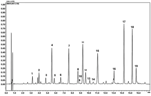
Figure 1 shows a representative chromatogram of BJe used in all studies presented in this PhD thesis. It was obtained by a UHPLC coupled online to an LCMSIT-TOF mass spectrometer. The peaks identify each compounds present in the extract. Their quantity is expressed as mg/g and listed in table 1, and their molecular structures are presented in figure 2.
Fig. 1. UHPLC chromatogram of BJe.
The flavanones neohesperidin, naringin, hesperetin and neoeriocitrin are the molecules present in the highest amount. The flavones rhoifolin and neodiosmin are also abundant, while C-glucosidic compounds are in a minor quantity.
33
peak compounds mg/g
1 Vicenin-2 11.61
2 Lucenin-2 4’-methyl ether 10.29
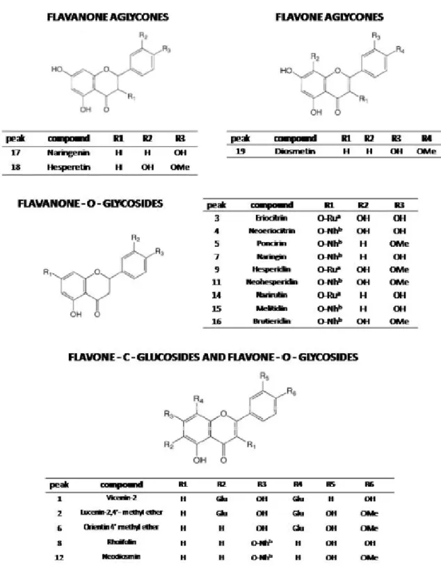
3 Eriocitrin 8.89 4 Neoeriocitrin 51.73 5 Poncirin 18.41 6 Orientin 4’ methylether 14.85 7 Naringin 91.90 8 Rhoifolin 19.96 9 Hesperidin 7.49 10 Isoquercitrin 2.5 11 Neohesperidin 98.5 12 Neodiosmin 12.39 13 Rhoifolin 4’-glucoside 2.55 14 Narirutin 4.97 15 Melitidin 79.47 16 Brutieridin 36 17 Naringenin 41.48 18 Hesperetin 53.84 19 Diosmetin 12.36
34
Fig. 2. Chemical structures of flavonoids found in the BJe. aRutinose (Ru); b
35
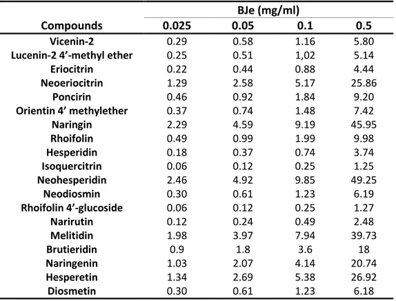
In table 2, there are the amounts of single flavonoids (expressed as µg) present in the BJe at different concentrations (0.025, 0.05, 0.1 and 0.5 mg/ml) used in this study.
BJe (mg/ml)
Compounds 0.025 0.05 0.1 0.5
Vicenin-2 0.29 0.58 1.16 5.80
Lucenin-2 4’-methyl ether 0.25 0.51 1,02 5.14
Eriocitrin 0.22 0.44 0.88 4.44 Neoeriocitrin 1.29 2.58 5.17 25.86 Poncirin 0.46 0.92 1.84 9.20 Orientin 4’ methylether 0.37 0.74 1.48 7.42 Naringin 2.29 4.59 9.19 45.95 Rhoifolin 0.49 0.99 1.99 9.98 Hesperidin 0.18 0.37 0.74 3.74 Isoquercitrin 0.06 0.12 0.25 1.25 Neohesperidin 2.46 4.92 9.85 49.25 Neodiosmin 0.30 0.61 1.23 6.19 Rhoifolin 4’-glucoside 0.06 0.12 0.25 1.27 Narirutin 0.12 0.24 0.49 2.48 Melitidin 1.98 3.97 7.94 39.73 Brutieridin 0.9 1.8 3.6 18 Naringenin 1.03 2.07 4.14 20.74 Hesperetin 1.34 2.69 5.38 26.92 Diosmetin 0.30 0.61 1.23 6.18
Tab. 2. Concentrations of flavonoids found in BJe at the indicated concentrations, expressed as µg.
36
4
.2
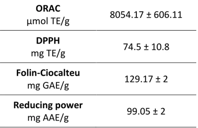
.Anti-oxidant capacity in cell-free models
The anti-oxidant and radical scavenging properties of BJe were determined using a series of chemical tests that results are shown in table 3.
ORAC µmol TE/g 8054.17 ± 606.11 DPPH mg TE/g 74.5 ± 10.8 Folin-Ciocalteu mg GAE/g 129.17 ± 2 Reducing power mg AAE/g 99.05 ± 2
Tab. 3. Anti-oxidant activity of BJe. Results are reported as mean ± S.E.M. of three
experiments performed in triplicate and expressed as standard equivalent/g of dried extract.
37
4
.3
.Protective effect of BJe from H
2O
2-induced oxidative stress in A549
cells
4.3.1. BJe prevents the H2O2-induced increase of ROS
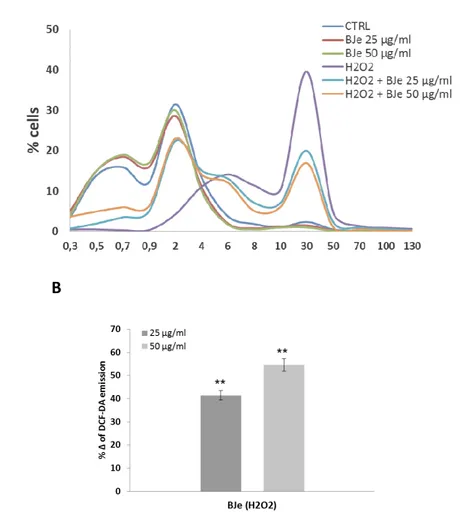
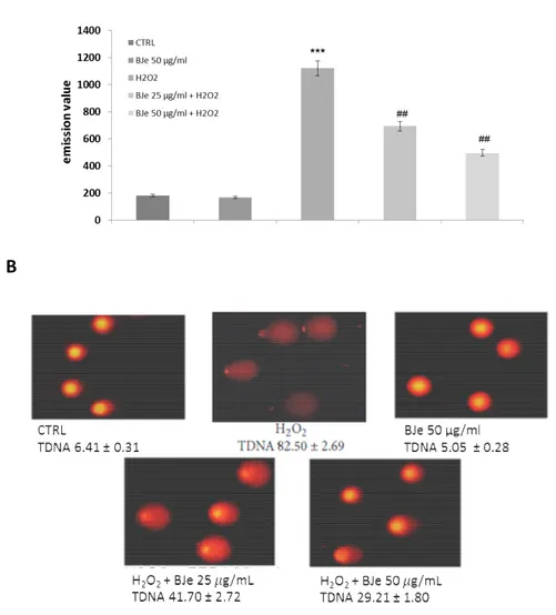
To study the potential protective effects of BJe in different compartments of oxidatively injured A549 lung epithelial cells, we first measured the intracellular content of ROS. As expected, DCF emission values in cells treated with BJe for 18 h did not differ significantly from the background values recorded in untreated cells (Fig. 3A). These results indicate that the extracts at both 25 and 50 µg/mL concentrations
did not trigger ROS generation. Instead, DCF emission values in H2O2-stressed cells
were up to 6.7-fold higher than those detected in untreated cultures, suggesting that there had been an increase in ROS generation (fig. 3A). Interestingly, as shown in fig.
3A, the presence of BJe reduced H2O2-induced oxidative stress, thus preventing the
increase in ROS. Indeed, the fluorescence emission curve for untreated culture shows a consistent peak on the left of the graph (proportional to the number of cells with low
emission values). In contrast, in A549 cells incubated with 200 µM H2O2 for 2 h a larger
number of cells with high emission values was detected (the purple peak on the right), indicating an increased ROS production. The curves obtained from the cells pre-treated
with BJe and then incubated with H2O2 show a smaller number of cells with high
emission values. Figure 3B summarizes data from the cytofluorimetric analyses presented in figure 3A, showing the percentage of ROS reduction in A549 cells
pretreated with BJe and then incubated with H2O2 in comparison to the cells exposed
to H2O2 alone (%Δ). The histograms in fig 3B illustrate that BJe significantly reduced
38
A
B
Fig. 3. Cytofluorimetric evaluation of intracellular ROS. A549 cells treated for 18 h
with BJe were oxidatively stressed by H2O2 200 M for additional 2 h. Results from BJe
treatments (A). The curves shifted rightward to higher emission values indicate the increase of ROS production. Data from A are expressed as percentage of reduction
(%Δ) of DCF-DA emission values in BJe-pretreated cells and then exposed to H2O2
compared to H2O2-stressed cells (B). The experiments were repeated at least three
times. ∗∗ <0.01 vs H2O2-treated cells.
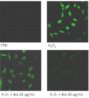
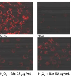
The data from FACS analyses were confirmed by CLSM observations. Figure 4 shows that, in comparison to oxidatively stressed A549 cells, the presence of BJe dampened the fluorescence emission proportionally to the concentration employed.
39
Fig. 4. Confocal laser scanning microscope images of DCF-DA-stained cells. A549 cells
grown on cell slides were pre-incubated with BJe and after 18 h were exposed to H2O2
200 µM. Green fluorescence represented the amounts of ROS. Images shown are representative of three independent experiments. 400x magnification.
4.3.2. BJe reduces the H2O2- caused cell death
As shown in figure 5, no differences between cell exposed to BJe and then treated or
untreated with H2O2 were recorded in PI emission values.
Fig. 5. Effect of BJe on cell death induced by H2O2. The cells were treated for 18 h with
BJe followed by incubation with 200 µM H2O2 for additional 2 h. PI-positive cells were
determined by flow cytometry collecting the emission signal in the FL-3 channel. Data
represent means ± S.E.M. of three separate experiments. ∗∗∗ <0.001 vs control cells
40
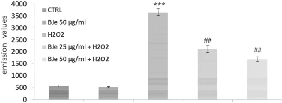
4.3.3. BJe counteracts lipid peroxidation induced by H2O2 in A549 cells
High lipid content of cell membranes makes them particularly susceptible to oxidative damage. Therefore we measured the lipid hydroperoxides to investigate the
consequences of the oxidative damage caused by H2O2 on the membrane lipids and
also to examine the effect of BJe. The level of lipid peroxidation in A549 cells increased
up to 6-fold after 2 h incubation with 200 µM of H2O2, an effect counteracted by the
pre-incubation with the extract (fig. 6). Interestingly, the DPPP emission value detected
in H2O2-stressed cells were lowered by up to 40 and 55% by pretreatment with BJe at
concentrations of 25 and 50 µg/mL, respectively (P<0.01).
Fig. 6. Cytofluorimetric evaluation of lipid hydroperoxides. The A549 cells incubated
for 18 h with BJe were oxidatively stressed with H2O2 200 µM for 2h and then loaded
by DPPP probe. The graph reports the mean of fluorescence expressed in arbitrary fluorescence units (AFU) of three independent experiments. Data represent the mean
± SEM of at least three separate experiments. ∗∗∗ <0.001 vs control cells and ## <0.01
vs H2O2-treated cells.
4.3.4. BJe attenuates the fall in Δ m induced by H2O2
Since mitochondrial membrane phospholipids are key targets for lipid peroxidation involving highly polyunsaturated side chains, we further evaluated the ability of BJe to restrain mitochondrial impairment. Figure 7 shows that incubation of A549 cells with