1
University of Calabria-Italy, Laboratory of Cardiovascular Pathophysiology, Dept. DiBEST University of Rouen Normandy-France, DC2N- French National Institute of Health and Medical
Research (INSERM) U1239 XXX Cycle of Doctorate in Life Sciences
Co-tutorship with the Normandy's Doctoral School of Integrative Biology, Health and Environment (EdN BISE 497)
Disciplines: Physiology and Biochemistry
Selenoprotein T as a novel cardiac modulator: from production to
expression measurement and pathophysiological implications
Director of Doctorate in Life Science Prof. Maria Carmela Cerra
Director of Doctoral School of Integrative Biology, Health and Environment Prof. François Dauphin
Ph.D. student
Dr. Carmine Rocca
Tutor
Co-tutor
Prof. Tommaso Angelone Dr. Youssef Anouar
2
This study was supported by “Galileo Project n. G16-31 37409RJ” Italian-French University
Index
Résumé français ... 6
1. Production du SelT recombinante (rSelT) ... 8
2. Enzyme-Linked Immunosorbent Assay (ELISA) pour la quantification de la SelT ... 10
3. Etudes cardiaques ex vivo ... 14
Summary ... 19
Introduction ... 23
1. Selenium and Selenocysteine in protein chemistry... 24
1.1 Selenium: biochemistry and physiology ... 24
1.2 Selenocysteine (Sec): reactivity and selectivity ... 26
1.3 Sec biosynthesis ... 28
1.3.1 Sec tRNA[Ser]Sec ... 28
1.3.2 Selenophosphate Synthetase ... 31
1.3.3 Sec Lyase ... 31
1.4 Sec incorporation into proteins ... 31
2. Structure and activity of mammalian selenoproteins... 34
2.1 Selenoproteins as ROS-detoxifying enzymes ... 37
2.1.1 Oxidants generation and oxidative stress ... 37
2.1.2 Antioxidant activity of selenoproteins ... 38
2.2 Pathophysiological implications of selenoproteins ... 39
2.2.1 Thioredoxin reductases (TrxRs) ... 40
2.2.2 Glutathione Peroxidases (GPxs) ... 41
2.2.3 Iodothyronine Deiodinases (DIOs) ... 42
2.2.4 Rdx family of selenoproteins... 43
3. The Rrx member Selenoprotein T (SelT or SELENOT) ... 44
3.1 Involvement of SelT in the regulation of Ca2+ homeostasis and neuroendocrine function ... 46
3.2 Involvement of SelT in the regulation of pancreatic β-cell function and glucose homeostasis ... 48
3.3 Role of SelT in neurodegeneration... 49
4. Oxidative stress in cardiovascular disease (CVD) ... 51
Aims of the doctoral project ... 53
Materials and methods ... 56
3
1.1 Cloning of rat SelT cDNA ... 57
1.2 Preparation of competent E. coli ... 57
1.3 Plasmid DNA purification and enzymatic digestion ... 57
1.4 Expression of rSelT in E. coli ... 58
1.5 Purification of 6His-rSelT protein under denaturing conditions and protein refolding ... 59
1.6 Post-refolding analysis: activity assessment of rSelT ... 60
1.7 Purity of rSelT ... 61
2. SelT Enzyme-Linked Immunosorbent Assay development for SelT detection ... 62
2.1 Production of sera against SelT ... 62
2.2 Western blot for determination of rabbit sera antibodies ... 62
2.3 Chequerboard titrations (CBTs) ... 63
2.4 I-ELISA for antibody measurement ... 63
2.5 IC-ELISA for antigen detection ... 64
2.5.1 Construction of a standard-inhibition curve ... 65
3. Ex vivo cardiac studies ... 65
3.1 Animals ... 65
3.2 Peptides and drugs ... 65
3.3 Isolated heart perfusion ... 66
3.4 Experimental protocols ... 67
3.4.1 PSELT-stimulated preparations ... 67
3.4.2 Ischemia/Reperfusion (I/R) studies ... 67
3.5 Assessment of myocardial injury ... 68
3.6 SDS-PAGE and Western blot ... 68
3.7 ELISA for ROS detection ... 69
3.8 Mitochondrial isolation ... 70
3.9 Immunofluorescence ... 70
4. Statistics ... 71
Results ... 72
1. Production of recombinant SelT ... 73
1.1 Enzymatic digestion of plasmid DNA ... 73
1.2 Expression of rSelT in E. coli ... 74
1.3 Determination of 6His-rSelT solubility ... 75
1.3.1 Solubilization of 6His-rSelT by the bacterial insoluble fraction ... 75
1.4 Purification of the expressed 6His-rSelT under denaturing conditions and protein refolding ... 77
1.5 Post-refolding analysis: activity assessment of rSelT ... 78
4
2.1 Assessment of sera ability to react with rSelT and C57Bl/6 mice tissue extracts ... 79
2.2 Determination of sera and rSelT working conditions by CBT ... 81
2.3 Measurement of SelT-specific antibodies by endpoint titer ELISA ... 84
2.3.1 Determination of the cutoff between negative and positive results for SelT-antibody ... 84
2.3.2 Determination of the antibody titers by I-ELISA ... 84
2.4 CBT for commercial SelT-antiserum ... 87
2.5 SelT detection by IC-ELISA ... 89
2.5.1 Standard antigen-inhibition curve ... 89
2.5.2 SelT detection in C57Bl/6 mice tissue extraxts ... 90
3. Ex vivo cardiac studies ... 93
3.1 SelT expression during heart ontogenesis ... 93
3.2 SelT expression during cardiac ischemia ... 95
3.3 Full length rSelT and PSELT effects on post-ischemic cardiac function ... 97
3.4 PSELT affects cardioprotective pathways ... 100
3.5 PSELT influence on apoptotic indexes ... 102
3.6 PSELT influence on cellular redox balance control ... 104
Discussion ... 107
1. Production of recombinant SelT ... 108
2. Development of Enzyme-Linked Immunosorbent Assay for SelT detection ... 109
3. Ex vivo cardiac studies ... 110
3.1 Cardiac expression of SelT during ontogenesis and under ischemic conditions ... 110
3.2 Cardioprotective effect of rSelT and PSELT ... 111
3.3 PSELT triggers various intracellular signaling mechanisms to provide cardioprotection ... 113
3.4 PSELT inhibits oxidative and nitrosative stress ... 114
Conclusions ... 115
References ... 116
Abbreviations ... 126
5
Doctorat en cotutelle de thèse
Université de Calabre, Italie
INSERM U1239, Université de Rouen-Normandie, France
Titre de la thèse
La sélénoprotéine T en tant que nouveau modulateur
cardiaque: de la production et la mesure de l'expression aux
implications physiopathologiques
Auteur
Dr. Carmine Rocca
Tuteur Co-tuteur
6
Résumé français
Les sélénoprotéines représentent une classe particulière d’enzymes contenant du sélénium et caractérisée par une réactivité chimique spécifique. Ces systèmes protéiques agissent comme des capteurs redox intercellulaires et sont largement reconnus comme médiateurs essentiels pour la prévention de maladies, y compris les pathologies cardiovasculaires. Parmi ces protéines, la sélénoprotéine T (SelT ou SELENOT) est une nouvelle enzyme localisée au niveau du réticulum endoplasmique (ER) appartenant à la famille des rédoxines, dont la suppression de gène chez la souris est associée à une létalité embryonnaire précoce. A travers son domaine catalytique redox (Cys-X-X-Sec), la SelT exerce une action antioxydante e cytoprotective essentielle au niveau cérébral durant l’embryogenèse et suite à des lésions tissulaires. De nombreuses études ont démontré l’implication de la SelT via son activité redox, dans les processus du différentiation et protection des cellules nerveuses et endocrines dans des conditions physiopathologiques spécifiques. En revanche, rien n’est connu sur l’expression et la fonction du cette protéine dans d’autres organes vitaux comme le cœur, un tissus physiologiquement sujet à un stress oxydatif élevé, surtout après un dommage d’ischémie- réperfusion (I/R).
Les études actuelles montrent un grand intérêt pour les molécules capables de réduire le stress oxydatif qui représente un des facteurs majeurs impliqués dans la pathogenèse des dommages myocardiques de l’I/R.
Plusieurs études ont démontré les effets bénéfiques au niveau cardiaque des antioxydants endogènes spécifiques tels que les enzymes catalase, superoxyde dimustase et glutathion peroxydase sur les cœurs isolés soumis à l’I/R. Dans ce contexte, les antioxydants exogènes incluant les substances contenant des groupements thiols, exercent une action protectrice significative contre le déséquilibre redox typique des lésions myocardiques induites par l’I/R (kalaycioglu et al., 1999; khaper et al., 1997).
A la lumière de ces observations, mon projet de thèse de doctorat a eu pour but de développer un immunodosage spécifique de la SelT et d’évaluer les implications physiopathologiques au niveau cardiovasculaire d’une forme de SelT recombinante (rSelT) et de son peptide dérivé PSELT (SelT 43-52) synthétisé chimiquement dans sa forme réduite, qui comprend le domaine catalytique redox Cys-Val-Ser-Sec.
7
• La première partie du travail a eu pour but de:
- optimiser la surexpression d’une forme recombinante de SelT de souris dans les cellules de
E. coli,
- calculer l’activité enzymatique exercée par la protéine pure.
• Le second but du cette thèse a été:
- le développement de deux essais spécifiques ELISA. En utilisant la protéine recombinante rSelT et un antisérum polyclonal de lapin contre rSelT, respectivement comme antigène et anticorps primaire, des dosages ELISA compétitif indirect (I-ELISA) pour évaluer les titres d’anticorps et indirect (C-ELISA) pour quantifier les niveaux des SelT solubles ont été mis au point.
• La troisième partie du ce projet a eu pour objectif de:
- déterminer l’expression de la SelT durant l’ontogenèse cardiaque et après un dommage ischémique ex vivo induit sur le cœur de souris adultes.
- évaluer l’effet cardioprotecteur exercé par la rSelT et PSELT en tant qu’agents pharmacologiques en post-conditionnement contre les dommages de l’I/R dans un modèle de cœur isolé de souris perfusé selon le méthode de Langendorff.
8
1. Production du SelT recombinante (rSelT)
Durant la première partie du travail, nous avons optimisé la production hétérologue d’une forme recombinante de SelT (rSelT) dans les cellules de E. coli. Dans ce but, le cDNA correspondant à la SelT a été amplifié à partir de cellules PC12 et ensuite cloné dans le vecteur pGEM-T; la protéine a été exprimée comme protéine de fusion His-tag dans E. coli. La rSelT a été purifiée par chromatographie d’affinité dans des conditions dénaturantes et immédiatement renaturée sur colonne en appliquant un protocole de renaturation en présence d’urée. La protéine purifiée a été identifiée comme une bande protéique correspondant au poids moléculaire de 23KDa (Fig. 1).
Fig. 1. Purification de 6His-rSelT dans les conditions dénaturantes et analysée sur gel SDS-PAGE à 12% suivi de coloration au bleu de Coomassie. Ligne 1. Marqueur, Ligne 2. Surnageant après lyse bactérienne avec
DNPI-10 (“cleared lysate”), Ligne 3. Lysat bactérien après la colonne (“flow-through”), Ligne 4Fraction après
tampon de lavage DNPI-20, Ligne 5. Fraction après tampon de lavage 7:1 (v/v) NPI-20/DNPI-20, Ligne 6. Dernières fractions (10 CV) après tampon de lavage NPI-20, Lignes 7-16. Fractions d'élution (0.5 ml) après tampon d’élution NPI-250.
Après le protocole de renaturation, rSelT a montré une activité enzymatique identique à celle de la tiorédoxine réductase (TrxR) de foie de souris utilisée comme contrôle. Nous avons utilisé un kit spécifique de TrxR puisque cette enzyme représente une des sélénoprotéines più importante avec ce type d’activité enzymatique (Dikiy et al, 2007).
1 2 3 4 5 6 7 8 9 10 11 12 13 14 15 16 15- 40- 25- 35- 55- 100-
70-9
Cette activité enzymatique est dépendante des résidus Cys/Sec du motif catalytique et est similaire à celle de la TRxR dont l’activité est inhibée par l’aurotiomalate (ATM), un inhibiteur sélectif (Fig. 2).
Fig. 2. Activité enzymatique exercée par rSelT (10 μg) avec un résidus Cys substituant un résidu de Sec dans le domaine catalitique redox (SelT CVSU/C) testé en utilisant un kit spécifique de la TrxR en comparaison avec une TrxR hépatique de souris (20 μl d’échantillon fourni de producteur), en présence et en absence d’aurotiomalate (ATM) 20 μM. L’absorption de substrat réduit a été mesurée a 405 nm. Le graphique montre un essai représentatif de 3 autres expériences qui ont montré des résultats similaires.
L’étude a permis d’obtenir, pour la première fois une forme de rSelT pure et active. time (min) O. D. 4 0 5 0 5 10 15 0.0 0.2 0.4 0.6 0.8 1.0 blank Trx TrxR + ATM rSelT rSelT + ATM TrxR: y=0.0353x + 0.3425 R2=0.999 rSelT: y=0.0332x + 0.3277 R2=0.995
10
2. Enzyme-Linked Immunosorbent Assay (ELISA) pour la quantification de
la SelT
Le développement de dosages immunologiques sélectifs pour certains antigènes et protéines pourrait être d’une grande aide pour des études aussi bien biologiques spécifiques que pour des applications cliniques. Actuellement un test immunologique spécifique pour évaluer les niveaux des SelT dans les échantillons biologiques n’est pas encore disponible.
La deuxième partie de ce travail a eu comme but celui de développer un ELISA indirect compétitif spécifique (IC-ELISA), utilisant la protéine recombinante et un antisérum polyclonal de lapin contre la rSelT, respectivement comme antigènes et anticorps primaires.
L’antiserum de lapin immunisé avec des fractions purifiées de SelT a réagi sélectivement avec l’immunogène homologue (rSelT); une réactivité élevée a été observée aussi avec les extraits protéiques de tissus murins (cœur, cerveau, testicule, foie, pancréas et rein) analysés par Western blotting, indiquant l’efficacité du protocole d’immunisation.
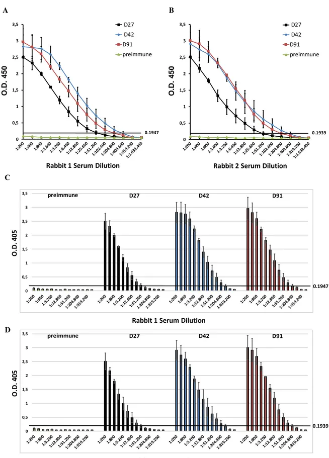
Afin de contrôler la qualité de la réaction des anticorps produits, les échantillons de sérum de lapin ont été prélevés dans divers intervalles du temps à partir de la première immunisation (au 27ème, 42ème et 91ème jour, indiqués respectivement comme D27, D42 et D91) et ont été analysés par ELISA indirect (I-ELISA). Ainsi, Il a été possible d’évaluer comment rSelT a généré durant toute la période d’immunisation une réponse immunogénique sélective chez les lapins, confirmant la génération d’un antisérum hyper-immun. L’absence de signal observée dans le sérum non-immun a été un indice d’une réactivité immunologique spécifique des anticorps produits. Ces données ont indiqué que le test L-ELISA développé était à même de discerner les antisera SelT-négatifs des anti-sera SelT-positifs (Fig. 3).
11
Fig. 3. A., B. Cinétique de réponse des anticorps (titres d’anticorps) de lapin n.1 et du lapin n.2. C., D. valeurs O.D. par rapport au cutoff O.D. de 0.1947 et O.D. de 0.1939 pour le lapin n.1 et lapin n.2, respectivement, mesurés par I-ELISA, aux divers intervalles après le protocole d’immunisation (D27, D42, D91). Chaque courbe
0 0,5 1 1,5 2 2,5 3 3,5 O .D. 4 5 0 D27 D42 D91 preimmune
Rabbit 1 Serum Dilution
0.1947 0 0,5 1 1,5 2 2,5 3 3,5 O .D. 4 5 0 D27 D42 D91 preimmune
Rabbit 2 Serum Dilution
0.1939 A B D 0 0,5 1 1,5 2 2,5 3 3,5 0.1947 O .D . 4 0 5
Rabbit 1 Serum Dilution
preimmune D27 D42 D91 0 0,5 1 1,5 2 2,5 3 3,5 0.1939 O .D . 4 05
Rabbit 2 Serum Dilution
12
représente la moyenne des valeurs O. D. ± erreur standard du lecteur effectuée en duplicats issus de deux expériences différentes.
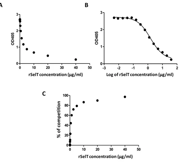
Sur la base du cette immunoréactivité d’antiserum élevée, un test IC-ELISA a été développé pour la détection de la SelT. Le test a été développé en utilisant la concentration du rSelT optimale en antigène recouvrant la microplaque, et la dilution optimale de l’antisérum comme anticorps primaire, déterminé par titrage en damier. L’objectif était la mesure de niveau des antigènes (SelT) utilisant le principe du ELISA compétitif. Pour la génération de la courbe standard d’inhibition d’antigène, nous avons utilisé des dilutions sériées d’une solution standard de rSelT (40-0.0048 µg/ml) (Fig. 4).
Fig. 4.A. Courbe standard d’inhibition de rSelT (0.0048-40 μg/ml) au moyen de IC-ELISA, avec l'axe des x sur une échelle linéaire B. Courbe standard d’inhibition de rSelT (0.0048-40 μg/ml) au moyen de IC-ELISA avec l’axe des x sur échelle logarithmique. C. Courbe standard d’inhibition de rSelT (0.0048-40 μg/ml) au moyen de
standard competitive curve
0 10 20 30 40 50
0 1 2 3
SelT concentration (ug/ml)
OD4
0
5
rSelT concentration (µg/ml)
standard competitive curve
-3 -2 -1 0 1 2
0 1 2 3
Log of SelT concentration (ug/ml)
OD4
0
5
Log of rSelT concentration (µg/ml)
A
B
C
0 10 20 30 40 50 0 20 40 60 80 100SelT concentration (ug/ml)
% o f c o m p e ti ti o n rSelT concentration (µg/ml) % o f co m p e ti ti o n
13
IC-ELISA exprimé comme régression non linéaire de pourcentage de compétition. Les trois graphiques ont été obtenus en utilisant le modèle sigmoïdal (logistique) à quatre paramètres de GraphPad.
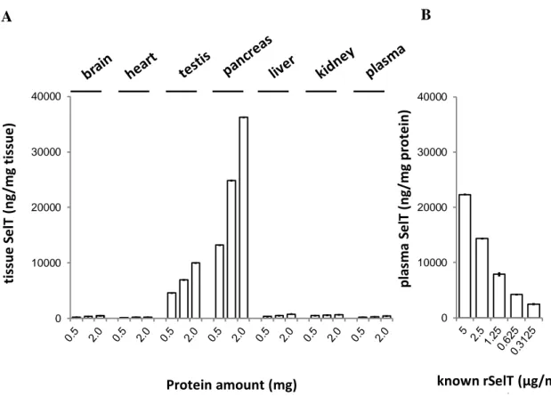
Dans le but d’évaluer si l’antisérum est capable de reconnaître la SelT dans les tissus biologiques, l'IC-ELISA a été appliqué aussi à des extraits protéiques de tissus de souris C57BI/6 (Fig. 5).
Fig. 5.A. Détection de la SelT par IC-ELISA dans les extraits protéiques de tissus de souris C57Bl/6 (cerveau, cœur, testicule, pancréas, foie, rein, plasma). B. Détection de SelT par IC-ELISA dans le plasma de souris C57Bl/6 pré-incubé avec différentes concentrations de rSelT (5-0.3125 μg/ml).
Les concentrations de SelT relevés au moyen d’IC-ELISA sont confirmées par le profil d’expression de la protéine dans les mêmes organes d’après les résultats rapportés précédemment par Tanguy et al., 2011 et Prevost et al., 2013. Ces résultats indiquent que l’essai ELISA développé pourrait représenter un outil à améliorer pour le développement d’un kit ELISA standardisé pour la quantification de la SelT dans le plasma et autres tissus.
0 10000 20000 30000 40000 tis su e S e lT ( n g/mg t is su e ) Protein amount (mg) 0 10000 20000 30000 40000 p la sma S e lT ( n g/mg p rot e in) known rSelT (µg/ml) µg) A B
14
D’autres études en cours visent à augmenter la sensibilité, la spécificité, et la précision du test dans le but d’obtenir une validation définitive du dosage.
3. Etudes cardiaques ex vivo
La troisième partie du travail a été réalisée pour étudier l’expression et la régulation de la SelT dans le cœur de souris durant l’ontogenèse et suite à un stress pathologique. En particulier, l’effet cardioprotecteur de la rSelT et par PSELT en tant qu’agent pharmacologique en post-conditionnement contre les dommages du myocarde induits par I/R, a été évalué dans un modèle de cœur de souris. Il est largement admis que chez les patients présentant un infarctus aigu du myocarde, l’extension de la zone infarcie pourrait être limitée par des interventions précoces de réperfusion du myocarde à travers l’intervention coronarienne percutanée (PCI). Toutefois, la restauration du flux sanguin après un épisode ischémique prolongé, peut aussi causer un dommage irréversible au myocarde dans un phénomène nommé ‘’dommage létal induit par réperfusion‘’ (Murphy
and Steennbergen, 2008; Hausenloy and Yellon, 2004). Les interventions réalisées au début
de la réperfusion pour préserver le tissu myocardique vital, sont décrits comme des traitements en post-conditionnement (Bice and Baxter, 2015). Pour les études cardiaques destinées à évaluer l’action pharmacologique post-conditionnement exercée par la rSelT et PSELT, nous avons utilisé le modèle du cœur de souris isolé et perfusé selon la méthode de Langendorff, et nous avons effectué des analyses d’immunohistochimie, western blot et ELISA.
Les premiers résultats intéressants montrent que la SelT est très abondante durant l’ontogenèse en phase embryonnaire dans le cœur de souris, alors qu’elle est réduite de manière significative chez les nouveau-nés et n’est pas détectée dans le cœur de l’adulte (Fig. 6). Par conséquent, la SelT pourrait avoir un rôle très important durant la croissance hyperplastique précoce des cardiomyocytes.
15
Fig. 6. Immunofluorescence de la SelT et de la calsequestrine-2 sur des tissus embryonnaires (E7), nouveau-né (P14) et adulte (3 mois de vie) de cœur de souris. La calsequestrine-2 a été utilisée comme marqueur du réticulum sarcoplasmique cardiaque. Les noyaux ont été marqués en bleu avec une solution DAPI.
Dans le cœur adulte de souris arrivé en phase terminale de maturation, l’expression de la SelT n’est pas essentielle sauf si le tissu est soumis à des conditions nocives comme un dommage par I/R, capable de déclencher une ré-expression de la protéine dans les cardiomyocytes (Fig. 7) Merge SelT Calsequestrin-2 Em b ry o N e w b o rn A d u lt Merge SelT Calsequestrin-2 Em b ry o N e w b o rn A d u lt Larger view A B C 0 0,5 1 1,5 2 R a ti o S e lT /β -t u b u lin d e n sito m e tr ic u n its * SelT 23 kDa 55 kDa β-tubulin R a ti o o f S e lT / -t u b u lin Emb ryo hear t Newb orn hear t Adul t hea rt 0.0 0.5 1.0 1.5 * ** *
16
Fig. 7. Immunofluorescence de la SelT et de la calsequestrine-2 sur des tissus cardiaques soumis àa un protocole Sham et I/R en ex vivo. La calsequestrine-2 a été utilisée comme marqueur de réticulum sarcoplasmique cardiaque. Les noyaux ont été marqués en bleu avec solution DAPI.
Ce résultat est en accord avec l’expression élevée des autres sélénoprotéines rencontrées au niveau du système nerveux (comme SelW, SelT et GPx) dans des conditions de stress particulières, comme celles associées à la protection contre le stress oxydatif (Petit et al.,
2003; Baek et al., 2005; Lee et al., 2008; Chung et al., 2009).
Les études de cardioprotection ont démontré que PSELT à une dose de 5nM, correspondant à la valeur de l’EC50 calculée en fonction des courbes concentration-réponse, et rSelT à la
même dose, induisent une cardioprotection comme agents pharmacologiques en post-conditionnement, comme révélé par une récupération post-ischémique de la contractilité (développement significatif de la pression ventriculaire gauche, dLVP) (Fig. 8A) et de la réduction de la zone d’infarctus (IS) (Fig. 8C), sans variation de l’indice de contracture cardiaque (pression endo-diastolique ventriculaire gauche, LVEDP) (Fig. 8B). Par contre, un peptide contrôle sans le site catalytique redox contenant la Sec qui a été remplacé par un résidu Ser, n’a conféré aucun effet cardioprotecteur (Fig. 8), mettant en évidence le rôle essentiel de la Sec dans la cardioprotection conférée par la SelT.
B A SelT GAPDH 23kDa 37kDa
SelT Calsequestrin-2 Merge
I/ R S h a m 10 μm Ra ti o S e lT /G A P DH Sham I/R 0.0 0.5 1.0 1.5 *
17
Fig. 8. A. Variation de dLVP e B. LVEDP. Les données sont exprimées comme la variation des valeurs dLVP et LVEDP (en mm Hg) à la fin de 120 min de réperfusion pour les cœurs soumis aux protocoles I/R et d’ I/R en présence de PSELT 5 nM, PSELT inerte et rSelT. C. Zone d’infarctus de cœurs soumis aux protocoles d’I/R e de I/R in presence di PSELT 5 nM, PSELT inerte et rSelT. La quantité de tissu nécrotique après 30 min d’ischemie globale et 120 min de réperfusion est expremée ene pourcentage par rapport à la masse de ventriculaire gauche (LV) (% IS/LV). p <0,05 (*), p <0,01 (**), p <0,001 (***), par One-Way ANOVA/Newman-Keuls Multiple Comparison Test.
Puisque PSELT et la rSelT à la même dose induisent une cardioprotection comparable, nous avons étudié le mécanisme d’action induit par le peptide. Les études de western blot ont
d L V P a t th e e n d o f re p e rf u s io n ( m m Hg ) I/R PSE LT 5 nM Full leng ht r SELT Iner t pep tide 0 20 40 60 80 100 * ** ** * L V E D P a t th e e n d o f re p e rf u s io n (m m H g ) I/R PS ELT 5 n M Full l engh t rS ELT Iner t pep tide 0 10 20 30 *** *** *** ** In fa rc t s iz e ( % I S /L V ) I/R PSEL T 5 nM Full leng ht r SEL T Iner t pep tide 0 20 40 60 80 100 *** * *** **
A
B
C
18
révélé que la cardioprotection s’accompagne d’une augmentation significative des niveaux de phosphorylation des kinases Akt, Erk1/2 et GsK3α-β et une diminution du niveau de phosphorylation de la protéine p38MAPK. PSELT exerce son effet cardioprotecteur en inhibant des facteurs pro-apoptotiques comme Bax, la caspase 3 et le cytochrome c, et en stimulant en même temps le facteur anti-apoptotique Bcl-2. En outre, PSELT réduit significativement divers marqueurs du stress oxydatif et nitrosatif induits par l’I/R, qui contribuent aux dommages cardiaques provoqués par les radicaux libres comme les enzymes xanthines oxydases et aldéhydes oxydases et le produit de nitration 3-nitrotyrosine. Ces résultats ont permis de déterminer le rôle de la SelT comme modulateur cardiaque, et d’identifier rSelT et PSELT comme des agents pharmacologiques efficaces capables de protéger en post-conditionnement le myocarde suite à un dommage ischémique.
19
Summary
Selenoproteins represent a particular class of selenium-containing enzymes with specific chemical reactivity, functioning as intracellular redox sensors; these proteins are increasingly recognized as essential mediators for disease prevention, including cardiovascular pathologies. Among these proteins, Selenoprotein T (SelT or SELENOT) is a novel thioredoxin-like endoplasmic-reticulum (ER) enzyme whose genetic ablation in mice results in early embryonic lethality. SelT exerts an essential antioxidant and cytoprotective action in the brain duringdevelopment and after injury, through its redox active catalytic site (Cys-X-X-Sec). Although a large body of evidence has now been accumulated showing that SelT affects, via its redox activity, the differentiation, function, the protection of nervous and endocrine cells in pathophysiological conditions, nothing is known about the expression and function of SelT in other vital organs such as the heart, which is normally severely exposed to oxidative stress, particularly as in the case of ischemia/reperfusion (I/R) damage. Therefore, the present doctoral project aimed to investigate, in a rat heart model, the pathophysiological cardiovascular implications of a recombinant form of SelT (rSelT) and its derived-peptide PSELT (SelT43-52), chemically synthetized in its reduced form,
which encompasses the key redox Cys-Val-Ser-Sec motif, and to evaluate the possible application of rSelT for the development of a specific immunoassay.
In the first part of the work, the production of a recombinant form of full-lenght rat SelT (rSelT) was optimized in E. coli cells; for this purpose, the cDNA corresponding to rat SelT was amplified from PC12 cells and cloned into the pGEM-T vector, and the protein was expressed as a His-tag fusion protein in E. coli. rSelT was purified under denaturing conditions and simultaneously renatured on-column by affinity chromatography.
Purified rSelT was detected as a protein band corresponding to the molecular weight of about 23 kDa. Re-folded rSelT exhibited a thioredoxin reducatase (TrxR)-like enzymatic activity comparable to that of a control rat liver TrxR, dependent on the Cys/Sec residues of the thioredoxin-like fold, as demonstrated by the ability of a selective inhibitor (aurothiomalate) to abrogate the enzymatic activity of both rSelT and TrxR.
20
Selective immunoassays for some antigens and proteins are instrumental for biological studies and clinical applications. So far, a specific immunoassay to evaluate SelT amounts in biological samples is not available. The second part of the work aimed to develop a specific indirect competitive enzyme-linked immunosorbent assay (IC-ELISA), using the recombinant protein and a rabbit polyclonal antiserum against rSelT, as antigen and primary antibody, respectively. Immunization of two rabbits against the refolded rSelT allowed the production of a polyclonal antiserum, whose immunoreactivity was tested by western blot and its titer was determined by an indirect ELISA (I-ELISA) and by using rSelT-coated plates. Optimal working conditions for rSelT concentration and primary antiserum dilution were determined by chequerboard titration, using the hyperimmune rabbit antiserum collected at different times after immunization. The developed I-ELISA and IC-ELISA may constitute a valuable tool to discern SelT-negative and positive sera and to quantify SelT in plasma or tissues, respectively.
The third part of the project aimed to determine the expression and the regulation of SelT in the rat heart during ontogenesis and after a pathological stress. In particular, the cardioprotective effect of rSelT and of PSELT against I/R injury in a rat heart model, as postconditioning agents, has been evaluated. It is widely known that the patients presenting acute myocardial infarction, the infarct size can be limited by early myocardial reperfusion via percutaneous coronary intervention (PCI), in order to preserve the left ventricular systolic function and to improve clinical outcome. However, restoring blood flow after a prolonged ischemic episode, may also paradoxically cause irreversible damage to the myocardium in a phenomenon known as “lethal reperfusion-induced injury” (Murphy and Steenbergen, 2008; Hausenloy and Yellon, 2004). Interventions applied at the onset of reperfusion to salvage the viable myocardium, are described as postconditioning treatments (Bice and Baxter, 2015). With the purpose to identify an optimal application for a myocardial reperfusion recovery strategy, several animal and human models were used to demonstrate that the administration of pharmacologically active agents at the onset of the reperfusion induces cardioprotection, named pharmacological postconditioning. This represents an approach able to recruit or mimic the well-established pathways associated with ischemic postconditioning, a maneuver, whose cardioprotective effects are largely described (Hausenloy, 2009). For the cardiac study aimed to describe the pharmacological postconditioning action of rSelT and PSELT, the isolated Langendorff rat heart model was
21
used and immunohistochemical, western blot, and ELISA analyses were carried out. First, it was found that SelT expression is very abundant in rat embryo, but is undetectable in the adult rat heart; however, protein expression tremendously increased after I/R stress. Both exogenous PSELT at 5 nM, corresponding to the EC50 concentration calculated on the basis
of concentration-response curves, and rSelT were able to induce pharmacological post-conditioning, as evidenced by a significant post-ischemic recovery of contractility (developed Left Ventricular Pressure, dLVP) and a reduction of infarct size (IS), without changes in cardiac contracture (Left Ventricular EndoDiastolic Pressure, LVEDP). In contrast, exogenous PSELT at a higher dose (100 nM) showed a reduced ability to protect the heart after an I/R insult compared to the 5-nM dose, while a control peptide lacking the redox site (Inert PSELT), in which the Sec residue was replaced with a Serine (Ser), did not confer cardioprotection. Since both PSELT 5 nM and rSelT induced comparable cardioprotection, the detailed mechanism of action for PSELT was investigated. Immunoblot analysis showed that cardioprotection is accompanied by a significant increase of phosphorylated Akt, Erk1/2 and Gsk3α-β, and a decrement of p38MAPK. PSELT inhibited the pro-apoptotic factors Bax, caspase 3 and cytochrome c, and stimulated the anti-apoptotic factor Bcl-2. Furthermore, PSELT significantly reduced several markers of I/R-induced oxidative and nitrosative stress, which contribute to free radical-mediated cardiac damage (i.e. xanthine oxidase, (XO), aldehyde oxidase-1, (AOX-1) and 3-nitrotyrosine, 3-NT). These results unraveled the role of SelT as a cardiac modulator and identified rSelT and PSELT as effective post-conditioning agents able to protect the heart after ischemic injury.
Overall:
1) The SelT immunoassays developed and related preliminary data indicate that this tool could be used for the immunodetection of SelT in physiological and pathological conditions. 2) The results obtained in the present doctoral project regarding the cardioprotective effects of rSelT and PSELT, provide new information on the implication of this protein and its derived peptide (redox site) in cardiac development, function and protection. These observations pave the way for future studies aimed to investigate the possible clinical relevance of rSelT and/or PSELT, which might represent a new class of drugs to be tested for reducing cardiac I/R injury. This is important in a medical context since it could allow
22
the development of new adjunctive therapies to be coupled with the reperfusion to reduce morbidity and mortality.
23
24
1. Selenium and Selenocysteine in protein chemistry
1.1 Selenium: biochemistry and physiology
Selenium (Se) is an essential micronutrient incorporated into selenoproteins. It has pleiotropic effects, ranging from antioxidant and anti-inflammatory effects to the synthesis of thyroid hormones (Rayman, 2012). The beneficial actions of selenium for the human health are clinically relevant and widely recognized with growing interest in understanding its biologic role for the prevention of diseases, and its potential use as a therapeutic agent (Rayman, 2012).
Compared with many other micronutrients, selenium intake varies in the world, ranging from deficit associated with selenium-deficiency diseases to harmful concentrations that may cause several disorders, such as alteration of the nervous system and paralysis (Johnson et al., 2010).
The effects of selenium are concentration-dependent, resulting in an essential antioxidant impact in the nanomolar-micromolar range to potentially pro-oxidant effects at concentrations above that required for maximal selenoprotein synthesis. At even higher levels, selenium compounds may accumulate and generate reactive oxygen species (ROS) in redox cycles with intracellular thiols, leading to oxidative stress and cellular damage, with consequent toxic effects (Vinceti et al., 2001). Recommendations for selenium intake average 60 μg per day for men and 53 μg per day for women (Rayman, 2004). Foods represent the major natural source of selenium and the reasons for the variable intake regard the soil selenium content and factors that determine its availability to the food chain including selenium speciation, soil pH and organic-matter content, and the presence of ions that can complex with selenium (Johnson et al., 2010). An additional very important aspect is that foods provide selenium in distinct combinations of chemical forms which in turn entail a different bioavailability (Rayman et al., 2008). The most important selenium species in vegetables are represented by selenomethionine (SeMet) and selenate/selenite (SeO42-,
SeO32-); minor species are selenocysteine (Sec), Se-methyl-selenocysteine (SeMCys) and
gamma-glutamyl-Se-methylselenocysteine (GGSeMCys) (Rayman et al., 2008). Selenate/ite, SeMet and SeCys are the most important species in animal products, with
25
widely variable proportions depending on the animal’s diet (Rayman et al., 2008). A simplified overview of selenium metabolism in mammals is represented in Fig.1.
Fig. 1. Selenium metabolism in mammalian. Dietary selenium metabolites are internalized into the cell, where, together with the existing intracellular pool, they become metabolized by different pathways, to yield selenide, which serves as the selenium source for Sec biosynthesis. (Se, selenium; GSSeSG, selenodiglutathione;
CH3SeH methylselenol; H2Se selenide; SeMet, selenomethionine; Sec, selenocysteine; GSH, glutathione; TrxR,
thioredoxin reductase; Trx, thioredoxin).
Until few years ago, most of the studies focusing on selenium status assessment evaluated only the total level of the element in tissues or body fluids. Plasma or serum selenium concentration was generally considered a biomarker of both selenium status and dietary intake in the short-term, while erythrocyte selenium better reflects the long-term status (Thomson, 2004). However, recently, it has been established that the total selenium concentration is not a complete representative index of the real functional activity of this micronutrient, because the element is incorporated in a large variety of proteins with different biological functions (Thomson, 2004); in addition, the distribution of selenium among selenoproteins is strongly dependent on a hierarchy in its incorporation, the average dietary intake, the speciation of selenium in food, health condition, age, other environmental factors and genetic polymorphism of selenoproteins (Kryukov et al., 2003).
26
In this regard, the two plasma/serum selenoproteins, namely selenoprotein P (SelP) and the isoform 3 of glutathione peroxidase (GPx3) are the most commonly used markers for the assessment of selenium status also because they can be determined with a not invasive procedure compared to tissue selenoproteins, which requires a biopsy (Roman et al.,
2014). Plasma SelP has been proposed as a biomarker (Hurst et al., 2010); however, the
concentration of SelP mostly reflects the short-term status of selenium in the organism because it has a plasma half-life of about 3-4 hours in the rat (Burk and Hill, 2005). The activity of GPx3, as well as the erythrocyte isoform 1 of glutathione peroxidase (GPx1), are well correlated with the total level of selenium in the blood (Thomson, 2004). Notably, the choice of a selenoprotein as biomarker must consider its specific biological function, which therefore provides partial information in terms of selenium bioactivity. In fact, the most effective biomarker should not be a single protein, but a set of combined parameters, applied to a specific problem associated with optimal selenium status, including an analysis of health parameters, endocrine and immunological status, selenoprotein polymorphism and other genetic and enviromental variables; so far, this is considered as the most important and promising approach for this purpose (Reszka et al., 2012).
1.2 Selenocysteine (Sec): reactivity and selectivity
Selenium represents a component of selenocysteine (Sec, U), the 21st amino acid in the
genetic code (Atkins and Gesteland, 2000, Böck et al., 1991) and 5-methylamino-2-selenouridine, a modified base found in transfer RNAs (tRNAs) of some prokaryote species (Wittwer et al., 1984). Among the trace elements used as cofactors for enzymes, selenium has unique properties. Unlike metal cofactors such as zinc and copper, it is actually part of the polypeptide chain as part of an amino acid. Sulfur and selenium are members of the chalcogen group of elements, in which selenium occupies the position immediately below sulfur on the periodic chart. Thus, the two elements have very similar chemical-physical properties, and can be involved in similar chemical reactions such as thiol/disulfide (and thiol-disulfide-like, that is selenol/disulfide or thiol/selenosulfide when selenium replaces sulfur) exchange reactions (Wessjohann et al., 2007). Unlike methionine, in which sulfur is in a relatively less reactive thioether form, the thiol group of cysteine is ionizable, with a negatively charged thiolate group generated after deprotonation, and this improves its
27
reactivity (Fig. 2A,B) (Poole, 2015). In addition, this thiol/thiolate group undergoes alkylation by electrophiles and oxidation by ROS and reactive nitrogen species (RNS), with consequent post-translational modifications that significantly alter its functions (Poole,
2015). The most important characteristic of Sec is its unique localization in catalytic sites of
enzymes. In selenoproteins, Sec function is partially preserved only when cysteine (Cys) replaces Sec; however, in most cases, the substitution of Sec with Cys leads to a marked reduction in catalytic efficiency (Hondal et al., 2013). Considering the functional predominance of Sec in proteins, many studies have been carried out to investigate its role and the exact nature of its chemical properties. A common view is that, since Sec and Cys differ for a single atom, the presence of selenium provides some superior chemical or physical properties, improving the functions of macromolecules (Hondal et al., 2013).
A
B
Fig. 2. A. Protonated (left) and deprotonated (right) forms of Cys and Sec B. Comparison of Cys and Sec structures, pKa values, and redox potentials.
28
The unique chemical-physical characteristics of Sec are related to its relatively low pKa value (5.2), with respect to the pKa of cysteine (Cys) (8.3) (Huber, 1967); this implies that most of the side chain selenols are deprotonated at physiological pH; another fact regards the lower redox potential of Sec, (E0 = -388 mV), (Nauser et al., 2006)] compared to that of
Cys (E0 = -220 mV) (Jocelyn., 1967) (Fig. 2). Consequently, Sec is readily oxidized under
physiological conditions to form the diselenide dimer, Sec. In addition, the largest atomic ray of selenium makes it highly polarizable, allowing it to act both as an electrophile and a nucleophile (Steinmann et al., 2010). These chemical properties confer to Sec its higher reactivity with respect to Cys, as also indicated by the higher catalytic activity exerted by the Sec-containing enzymes, which are typically 100- to 1000-fold more active than their cysteine (Cys) mutants (Kim and Gladyshev, 2005). This has been considered the reason for the presence of Sec in biological systems. However, some selenoproteins have orthologs in which Cys is used in place of Sec, and are catalytically competent as selenoproteins (Kim
and Gladyshev, 2005).
1.3 Sec biosynthesis
1.3.1 Sec tRNA[Ser]Sec
The majority of selenium biological effects are elicited by selenoproteins (Atkins and
Gesteland, 2000). Sec-containing proteins are present in all evolutionary lines: eukarya,
archaea, and eubacteria, as well as in certain types of viruses (Labunskyy et al., 2014). The pathway for Sec biosynthesis was revealed only in the last years by using different approaches based on a combination of comparative genomic, molecular, and structural analyses. The genetic code is redundant; thus, the translational termination is encoded by UGA, UAA, and UAG (Voet et al., 1999). However, in mitochondria, UGA encodes for tryptophan (Osawa et al., 1992), and in the nuclear genome, UGA also encodes for Sec. Thus, Sec is considered the 21st genetically encoded amino acid translated into proteins by
reading of the UGA codon (Lee et al., 1989). Sec is incorporated into protein by a tRNA molecule with an anti-codon complementary to UGA. Unlike the other 20 amino acids, an important property of Sec is that it is universally synthesized on its own tRNA designated as Sec tRNA[Ser]Sec (Leinfelder et al., 1989; Lee et al., 1989). Sec tRNA[Ser]Sec controls the
29
species. For Sec biosynthesis, tRNA[Ser]Sec is firstly aminoacylated with serine in a reaction
catalyzed by seryl-tRNA synthetase (SerRS) to form seryl-tRNA[Ser]Sec, and this provides the
backbone for Sec biosynthesis (Lee et al., 1989; Leinfelder et al., 1989).
The biological characteristics of tRNA[Ser]Sec make this tRNA different from all other tRNAs
(Hatfield et al., 1994). The structures of tRNA[Ser]Sec from E. coli, M. jannaschii, and H. sapiens are represented in clover leaf models in Fig. 3.
Fig. 3. Clover leaf models of eukaryotic, archaeal, and bacterial tRNAs. [Ser]Sec
The gene for tRNA[Ser]Sec is designated as Trsp; it is a single copy gene involved in
selenoprotein synthesis machinery. Human Trsp is localized on chromosome 19q13.2 q13.3 (Mitchell et al., 1992). Aminoacylation of tRNA[Ser]Sec with serine (Ser) represents the
first reaction which leads to Sec biosynthesis in eubacteria and eukaryotes (Labunskyy et
30
Fig. 4. Mechanism of Sec biosynthesis in eukaryotes and the Sec machinery-based pathway for synthesis of Cys. Phosphoseryl-tRNA kinase (PSTK) provides the phosphorylated intermediate PSer-tRNA[Ser]Sec used as a
substrate for Sec synthase (SecS). Selenophosphate (H2SePO3-) generated by Selenophosphate Synthetase-2
(SPS2) from selenite and ATP is used as a donor of active selenium for SecS. The de novo synthesis of Cys using the Sec biosynthetic machinery is shown on the bottom right.
The conversion of Ser on tRNA[Ser]Sec to selenocysteyl-tRNA[Ser]Sec is catalyzed by the enzyme
Sec synthase (SecS), which incorporates selenophosphate into the amino acid backbone forming Sec-tRNA. For details, see Fig. 4. The essential functional role of SecS in Sec biosynthesis on its tRNA is corroborated by a recent finding of two different homozygous mutations in the SecS gene in humans (Agamy et al., 2010), which were associated with progressive cerebello-cerebral atrophy in these patients. Despite the lack of tRNA[Ser]Sec as
a result of the whole-body Trsp gene knockout in mice results in embryonic lethality, patient mutations in SecS have been associated with a less dramatic phenotype (Wirth et
al., 2010). Based on the SecS structure, these mutations should be involved into the
31 1.3.2 Selenophosphate Synthetase
Selenophosphate synthetase (SPS) is involved in the de novo synthesis of selenophosphate and in Sec recycling through a selenium salvage system (Xu et al., 2007). In addition to its role in Sec biosynthesis, SPS has been implicated in Cys biosynthesis (Xu et al., 2010). In this context, it is important to note that Cys mutants of selenoproteins partially preserve their activities whereas other mutations disrupt their functions. Moreover, Cys was observed in place of Sec in several selenoproteins (i.e. TrxR1) in rodents subjected to selenium deprivation (Lu et al., 2009). During selenium deficiency, SPS is able to use sulfide instead of selenide and generate thiophosphate (H2SPO3-) as an active donor for the reaction
catalyzed by SecS (Fig.4). SecS then makes Cys tRNA[Ser]Sec from H
2SPO3- and
O-phosphoseryl-tRNA[Ser]Sec intermediates, and Cys is further inserted into selenoproteins at
the UGA-encoded sites instead of Sec. Since Cys is an essential amino acid in mammals, the synthesis of Cys on tRNA[Ser]Sec is a novel de novo pathway for the synthesis of this amino
acid (Xu et al., 2010).
1.3.3 Sec Lyase
Sec degradation is mediated by the enzyme Sec lyase (SCL), in a PLP-dependent reaction, where Sec is converted to L-alanine and elemental selenium (Mihara et al., 2000). SCL is highly conserved across species and is expressed in several tissues with the highest activity observed in liver and kidney; during its catalysis, SCL is able to discern Sec from Cys and Ser residues (Omi et al., 2010). Despite the role of SCL in selenium recycling from Sec, this enzyme seems to be involved in other processes, but its physiological function remains not completely understood.
1.4 Sec incorporation into proteins
Unlike the common process of tRNA aminoacylation, Sec is co-translationally incorporated into proteins due to the in-frame UGA codons present in selenoprotein mRNAs and this requires the formation of a quaternary complex (Leibundgut et al., 2005). In particular, Sec is introduced into selenoproteins by a complex mechanism that requires the Sec-tRNA[Ser]Sec
32
and several factors, including special trans-acting proteins and a cis-acting Sec insertion sequence (SECIS) element (Leibundgut et al., 2005) (Fig.5).
Fig. 5. Mechanism of Sec insertion in eukaryotes, showing the factors required for Sec incorporation into proteins in response to the UGA codon.
SECIS elements are cis-acting stem-loop RNA structures found in the 3'-untranslated regions of the mRNAs (Low et al., 1996); these elements are located immediately downstream of the UGA triplet nucleotide encoding Sec, within the coding region of selenoprotein genes (Böck et al., 2006). SECIS elements are factors that dictate recoding of UGA as Sec (Böck et al., 2000). In response to the SECIS element in selenoprotein mRNA, Sec-tRNA[Ser]Sec, which has an anticodon complementary to UGA, translates UGA as Sec. In
this complex process, at least two trans-acting factors are required for efficient recoding of UGA as Sec in eukaryotes: the SECIS binding protein-2 (SBP2) and the Sec-specific translation elongation factor (eEFSec). SBP2 is stongly associated with ribosomes and, via a specific RNA-binding domain in the central part of the protein, binds SECIS elements with high affinity and specificity (Copeland et al., 2000). SBP2 also interacts with eEFSec, which is involved in the recruitment of Sec-tRNA[Ser]Sec and allows the incorporation of Sec into
the nascent, growing polypeptide (Tujebajeva et al., 2000). Instead of SBP2 and eEFSec, bacteria use a Sec-specific translation elongation factor (named selenoprotein B, SelB in E.
coli) which directly recognizes SECIS and is required for the binding and the delivery of the
33
limiting factor for selenoprotein synthesis; in fact, it has been demonstrated that the
knockdown of SBP2 in mammalian cells, by using siRNA, is associated with a significantly
decreased expression of selenoproteins (Papp et al., 2006), while overexpression of SBP2 has been shown to enhance Sec incorporation (Low et al., 1996). These findings are supported by recent data regarding homozygous missence mutations in the SBP2 gene in patients with hypothyroidism caused by defects in the synthesis of type 2 deiodinase (DIO2), a selenoprotein involved in thyroid hormone activation (see later) (Dumitrescu et
al., 2005). This mutation impaired the SBP2-SECIS elements binding, which is necessary for
the Sec insertion into selenoproteins with a consequent defective synthesis of some selenoproteins, including DIO2, GPx1, and SelP (Bubenik et al., 2007). Other SECIS-binding proteins include ribosomal protein L30, eukaryotic initiation factor 4a3 (eIF4a3) and nucleolin (Labunskyy et al., 2014). It is important to emphasize that the requirement of several specialized factors that take part to the incorporation of Sec into proteins, suggests the evolutionary importance of selenoproteins, as well as the complexity of this biological process. An overview of selenoprotein synthesis is shown in Fig.6.
Fig. 6. Machinery involved in synthesis of selenoproteins. Selenium phosphorylated by SPS1 is used to
34
to the nucleus with many cofactors bound. The protein SBP2 binds to the SECIS element in the 3-UTR of selenoprotein mRNAs, and recruits the tRNASec complex along with bound cofactors. The assembled complex is transported from the nucleus for translation to protein (HSP90, heat-shock protein 90).
2. Structure and activity of mammalian selenoproteins
The early periods of selenoprotein research, were characterized by different experimental approaches aimed to analyze the presence of selenium by mass spectrometry and detection of radioactive 75Se, metabolically incorporated into proteins in the form of Sec
(Ballihaut et al., 2007). The first selenoprotein identified was the mammalian glutathione peroxidase 1 (GPx1) (Flohe et al., 1973), followed by the discovery of bacterial glycine reductase (Turner and Stadtman, 1973) and formate dehydrogenase (Andreesen et al.,
1973). Subsequently, thanks to a combination of bioinformatic and experimental
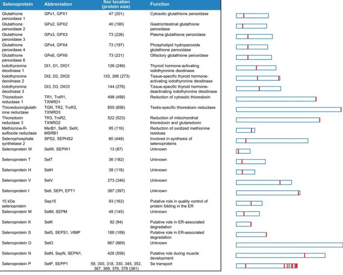
approaches, a number of selenoproteins have been identified in all life kingdoms and the full human selenoproteome encoded by 25 selenoprotein genes was characterized (Kryukov et al., 2003) (Fig. 7).
35
Fig. 7. Human selenoproteome. The relative length of selenoproteins and location of Sec within different proteins are shown on the right.
Since the selenoprotein genes contain UGA codons, these proteins are often misannotated in sequence databases (Kryukov et al., 2003). An experimental approach was based on the two characteristic genomic features typical of selenoproteins, the in-frame UGA codon encoding Sec residue and the SECIS element (Labunskyy et al., 2014). By searching candidate SECIS elements in completely sequenced genomes, and by analyzing the genomes of strongly related species for evolutionarily conserved SECIS elements belonging to orthologous selenoproteins, it was possible to identify selenoprotein genes through analyses of upstream coding sequences containing UGA (Labunskyy et al., 2014 and
references therein). In a second approach, selenoproteins were identified by searching the
in-frame UGA codons through analyses of sequences near to UGA in genomes which were completely sequenced (Labunskyy et al., 2014 and references therein). Considering that
36
almost all selenoproteins contain homologs in which Sec is replaced with Cys, this approach allowed the identification of selenoproteins independent from searching for SECIS elements (Labunskyy et al., 2014).
Selenoproteins are found in bacteria, archaea and eukaryota. However, some organisms do not use Sec, such as yeast and some plants, which lost the Sec insertion machinery during evolution (Lobanov et al., 2009). Based on the Sec location, mammalian selenoproteins can be classified into two groups (Fig. 8).
Fig. 8. Mammalian selenoproteins divided on the basis of Sec lacation. In one group Sec is located very close to the C-terminus, often in the C-terminal penultimate position. In the second group Sec is located in the N-terminal or middle regions of selenoproteins, often in a redox motif within a thioredoxin fold and have an α-helix downstream of Sec. Selenoprotein P, which has both an N-terminal redox Sec and multiple Sec residues in the C-terminal region (see fig.7), is not shown in this figure. Sec positions and selenoprotein lengths refer to human proteins. Close homologs of GPx1 (four selenoproteins), DI1 and TR1 (two each) are not shown.
One group of selenoproteins such as TrxRs and selenoproteins S, R, O, I, and K contains Sec in a site very close to the C-terminus of the protein. The other group [(including Glutathione Peroxidases (GPxs); Iodothyronine Deiodinases (DIOs); selenoproteins H, M, N, T, V, and W, SPS2, and selenoprotein Sep15)] has Sec in the N-terminus, often it has a thioredoxin fold structure, and some selenoproteins contain a C-X-X-U motif, corresponding to the thioredoxin catalytic active-site (Lu and Holmgren, 2009 and references therein).
37
Considering their structure, a key characteristic of all selenoproteins is the presence of Sec residues; except some cases, this amino acid is located in the enzyme active sites, whose function is to perform catalytic redox reactions (Arnér, 2010).
2.1 Selenoproteins as ROS-detoxifying enzymes
2.1.1 Oxidants generation and oxidative stress
On the basis of its electrochemical properties, molecular oxygen (O2) is a potent oxidizing
agent. When, at physiological temperature conditions an electron is added to form the superoxide radical (O2•–), oxygen becomes much more reactive (Fallab, 1967). Under
aerobic condition, cells continuously react with ROS, such as O2•–, H2O2, lipid and hydroxyl
radical (OH•), which derived from several metabolic reactions and counteract their effects through a wide range of antioxidant enzymes (Fallab, 1967). ROS can damage all cellular macromolecules; thus, harmful effectof these species occurs thanks to their ability to react with the Cys and Met residues present in proteins structure, thus inactivating their function, and to react with DNA and chromatin to cause mutations or double-stranded breaks in a phenomena overall known as “oxidative damage” (Hoshi and Heinemann,
2001). The current concept of “oxidative damage or oxidative stress” include pathways
related to the “nitrosative stress” and, considering their implication in cellular and extracellular metabolic processes, to the “metabolic stress” (Rahman et al., 2012). So far, the definition of oxidative stress confined to ROS has been also extended to RNS, such as nitric oxide (NO), peroxynitrite (ONOO−) and S-nitrosothiols (RSNO) (Rahman et al., 2012).
Cells have potent antioxidative systems for ROS detoxification and for repair of deleterious oxidative modifications on cellular structures (Sies, 1986). High and persistent ROS levels and/or inadequate antioxidant protection results in oxidative stress, cytotoxicity and apoptosis. However, little changes of oxidation–reduction (redox) cellular homeostasis are normally sufficient to activate a redox response-stress related pathway based on the upregulation and activation of protective enzymes (Winterbourn and Hampton, 2008). The redox signaling is an important part of the physiological cellular metabolism and it is an essential step in many signaling processes in not-stressed cells (Stone and Yang, 2006); in fact, several hormones, growth factors and cytokines induce ROS production in their
38
receptor-dependent mechanism of action. Based on the involvement of low ROS levels as part of intracellular signaling, the definition of oxidative damage has been recently refined as a “disruption of redox signaling and control” (Sies and Jones, 2007).
Therefore, it is widely accepted that an increase of the ROS and RNS-mediated oxidative damage to DNA and other biomolecules may impair the physiological functions of cells and tissues, leading to metabolic damage as the base for human aging and disease (Beckman
and Ames, 1999). Accordingly, oxidative stress has been associated with diverse
pathophysiological events, including cancer, renal disease, immunological pathologies, neurodegeneration and cardiovascular disease (Griendling and FitzGerald, 2003). Short-term oxidative stress may occur in tissues after exposition to specific stimuli, such as acute injury, infection and toxins. These injured tissues increase their expression of radical generating enzymes (i.e. xanthine oxidase and cyclooxygenase) activation of phagocytes, release of free iron, alteration in the electron transport chains of oxidative phosphorylation, leading to an excess of ROS production (Nathan and Cunningham-Bussel,
2013). The promotion and progression of complex and multifactorial pathologies, as well
as the side-effects of specific therapeutic regimes, are linked with a dramatic imbalance between ROS and the antioxidant defence system (Nathan and Cunningham-Bussel, 2013).
2.1.2 Antioxidant activity of selenoproteins
The precise functions of many selenoproteins are still unknown. In general, all selenoproteins with a known enzymatic activity, catalyse redox reactions by involving the oxidation of sulfhydryl groups and/or reduction of disulfides (Fig. 9); as ROS-detoxifying enzymes, selenoproteins belong to an array of antioxidants with different specific subcellular localization and chemical reactivities (Steinbrenner and Sies, 2009). In this context, all these antioxidant systems might exist in some hierarchical network or redox circuit, whose function is to maintain the overall cysteine proteome and the intracellular redox homeostasis (Jones and Go, 2011). In the mechanism of action mediated by selenoproteins in the H2O2 neutralization, these enzyme act through their unique ability to
trap and reduce H2O2 in a reaction mediated by selenols Se-H) to give selenenic acid
39
GSH; selenols are re-reduced by thiol-containing enzymes (Winterbourn and Hampton,
2008). Therefore, selenoproteins are well suited for controlling the formation of disulfide
bonds with specific Cys residues. In this process, due to the rapid oxidation kinetics of selenols, H2O2 in the cytosol completely reacts with Sec-containing proteins to produce Sec
selenenic acid before it can react with other potential targets (Winterbourn and Hampton,
2008). Consequently, the ability of selenoproteins to rapidly consume and neutralize H2O2,
result in a significant decrease of H2O2 levels and in a limitation of the space for its signal
transmission by diffusion in the cell (Winterbourn and Hampton, 2008).
2.2 Pathophysiological implications of selenoproteins
Selenoproteins are involved in many biological processes that are essential for life; therefore, this class of enzymes is considered critical for disease prevention, as in the case of cardiovascular disease, neurodegeneration, liver disease, cancer, immune and endocrine disorders (Papp et al., 2007). However, considering that the function of many mammalian selenoproteins is not completely known, their therapeutic potential so far has only limited applications (Papp et al., 2007). Many developed organoselenium compounds were tested as antioxidants, immunomodulators, anticancer and antihypertension agents. In this context, the organoselenium drug ebselen received particular attention, since it mimics the action of GPx (Muller et al., 1984). It is considered a peroxiredoxin-like agent, whose target is the thioredoxin system (Zhao and Holmgren, 2002); infact ebselen is reduced to selenol by TrxR or Trx and reoxidized by peroxides, including H2O2. Ebselen revealed a significant
action against H2O2 and smaller organic hydroperoxides with neuroprotective, antioxidant
and anti-inflammatory actions in a rat model of transient cerebral artery occlusion (Dawson
et al., 1995). These properties allowed an application of ebselen in the treatment of
patients with acute ischemic stroke (Papp et al., 2007 and references therein). Another important selenoenzyme of clinical interest is TrxR, another suitable target for anticancer therapies. It is required for cancer cell proliferation, it is highly expressed in cancer cells, and shows an active site that is accessible for reaction with reactive compounds (Papp et
al., 2007). Moreover, it has been demonstrated that many of the clinical anticancer drugs,
40
TrxR. This may pave the way for TrxR potential applications in cancer treatment (Mau and
Powis, 1992).
Overall, TrxRs, GPxs and DIOs are the three-best characterized selenoprotein families. They have different enzymatic activities, but all require reductants to provide the electrons for their catalytic redox cycle (Fig. 9).
Fig. 9. Putative catalytic mechanism of the three best characterized mammalian selenoproteins, TrxRs, GPxs and DIOs. GR, glutathione reductase.
2.2.1 Thioredoxin reductases (TrxRs)
TrxRs are members of the pyridine nucleotide-disulfide oxidoreductases that, together with thioredoxin (Trx), represent the major disulphide reduction system of the cell (Labunskyy
et al., 2014). Three TrxRs have been identified in mammals: TrxR1 in the cytosol/nucleus
(Gladyshev et al., 1996), TrxR2 in mitochondria (Lee et al., 1999), and thioredoxin glutathione reductase in the testes with the latter also possessing glutathione and


![Fig. 3. Clover leaf models of eukaryotic, archaeal, and bacterial tRNAs. [Ser]Sec](https://thumb-eu.123doks.com/thumbv2/123dokorg/2870289.9362/29.892.158.800.370.720/fig-clover-leaf-models-eukaryotic-archaeal-bacterial-trnas.webp)