EXTENDED REPORT
A deletion at
ADAMTS9-MAGI1 locus is associated
with psoriatic arthritis risk
Antonio Julià,
1José Antonio Pinto,
2Jordi Gratacós,
3Rubén Queiró,
4Carlos Ferrándiz,
5Eduardo Fonseca,
6Carlos Montilla,
7Juan Carlos Torre-Alonso,
8Lluís Puig,
9José Javier Pérez Venegas,
10Antonio Fernández Nebro,
11Emilia Fernández,
12Santiago Muñoz-Fernández,
13Esteban Daudén,
14Carlos González,
15Daniel Roig,
16José Luís Sánchez Carazo,
17Pedro Zarco,
18Alba Erra,
19José Luís López Estebaranz,
20Jesús Rodríguez,
21David Moreno Ramírez,
22Pablo de la Cueva,
23Francisco Vanaclocha,
24Enrique Herrera,
25Santos Castañeda,
26Esteban Rubio,
27Georgina Salvador,
28César Díaz-Torné,
29Ricardo Blanco,
30Alfredo Willisch Domínguez,
31José Antonio Mosquera,
32Paloma Vela,
33Jesús Tornero,
34Simón Sánchez-Fernández,
35Héctor Corominas,
16Julio Ramírez,
36María López-Lasanta,
1Raül Tortosa,
1Nuria Palau,
1Arnald Alonso,
1Andrés C García-Montero,
37Josep Lluís Gelpí,
38Laia Codó,
39Kenneth Day,
39Devin Absher,
39Richard M Myers,
39Juan D Cañete,
36Sara Marsal
1Handling editor Tore K Kvien ▸ Additional material is published online only. To view please visit the journal online (http://dx.doi.org/10.1136/ annrheumdis-2014-207190). For numbered affiliations see end of article.
Correspondence to Professor Sara Marsal, Rheumatology Research Group, Vall d’Hebron University Hospital, Pg Vall d’Hebron, 119-129, Barcelona 08035, Spain; [email protected] and Dr Juan D Cañete, Rheumatology Department, Hospital Clínic i Provincial and IDIBAPS, c/Villarroel 170, 08036, Barcelona, Spain; [email protected] SM and JDC are co-corresponding authors. Received 19 December 2014 Revised 23 April 2015 Accepted 23 April 2015 Published Online First 19 May 2015
▸ http://dx.doi.org/10.1136/ annrheumdis-2014-207187
To cite: Julià A, Pinto JA, Gratacós J, et al. Ann Rheum Dis 2015;74: 1875–1881.
ABSTRACT
Objective Copy number variants (CNVs) have been associated with the risk to develop multiple autoimmune diseases. Our objective was to identify CNVs associated with the risk to develop psoriatic arthritis (PsA) using a genome-wide analysis approach.
Methods A total of 835 patients with PsA and 1498 healthy controls were genotyped for CNVs using the Illumina HumanHap610 BeadChip genotyping platform. Genomic CNVs were characterised using CNstream analysis software and analysed for association using the χ2
test. The most significant genomic CNV associations with PsA risk were independently tested in a validation sample of 1133 patients with PsA and 1831 healthy controls. In order to test for the specificity of the variants with PsA aetiology, we also analysed the association to a cohort of 822 patients with purely cutaneous psoriasis (PsC).
Results A total of 165 common CNVs were identified in the genome-wide analysis. We found a highly significant association of an intergenic deletion between ADAMTS9 and MAGI1 genes on chromosome 3p14.1 ( p=0.00014). Using the independent patient and control cohort, we validated the association between ADAMTS9-MAGI1 deletion and PsA risk ( p=0.032). Using next-generation sequencing, we characterised the 26 kb associated deletion. Finally, analysing the PsC cohort we found a lower frequency of the deletion compared with the PsA cohort ( p=0.0088) and a similar frequency to that of healthy controls ( p>0.3).
Conclusions The present genome-wide scan for CNVs associated with PsA risk has identified a new deletion associated with disease risk and which is also differential from PsC risk.
INTRODUCTION
Psoriatic arthritis (PsA) is a chronic inflammatory
arthritis that affects 10–30% of patients with
psoriasis.1 2 To date, genome-wide association
studies (GWAS) as well as candidate gene studies have shown that both diseases share a substantial genetic component. However, sibling recurrence rates (λs) are much higher for PsA than psoriasis (PsA λs∼37 vs psoriasis λs∼7),3–5 indicating that additional, perhaps disease-specific, risk factors need to be identified.
GWAS based on single-nucleotide polymorph-isms (SNPs) have been highly successful in identify-ing >30 loci associated with psoriasis and PsA susceptibility.6–8 The cumulative risk exerted by these loci, however, is <50%,9 and additional genetic factors still need to be identified in order to explain the missing heritability. Strategies to com-plete the characterisation of the genetic architecture of psoriasis and PsA include the use of large sample sizes or the combination of different studies through metaanalysis,10 the deep sequence charac-terisation to identify rare variants with large effect sizes11 and, also, the analysis of other types of genetic variation that cannot be completely cap-tured by SNP-based genotyping platforms such as copy number variants (CNVs).
CNVs are fragments of DNA with sizes that range from hundreds of bases to several megabases, and that can either be absent (ie, deletions), repeated a certain number of times (ie, amplifications) or even rearranged.12Psoriasis was one of thefirst chronic
inflammatory diseases where CNVs were found to
be associated with the disease risk. The ampli fica-tions of the β-defensin genes on 8p23.1 region13 and the deletion affecting LCE3B and LCE3C genes14 have been clearly associated with psoriasis aetiology. The association of these CNVs with PsA aetiology, however, is still not clear,2 15 suggesting that they could participate in the chronic in flamma-tory processes in the skin rather than in the patho-logical process occurring in the joint.
In the present study, we have performed the first genome-wide analysis of CNVs in PsA. We havefirst analysed a discovery panel of 835 patients with PsA and 1498 healthy controls from the Spanish population using a microarray platform. The CNVs showing a more significant association to PsA risk were subse-quently selected and validated in an independent cohort of 1133 patients with PsA and 1831 healthy controls. In order to test for the specificity of the CNV association with PsA aeti-ology, we have also analysed a set of 822 psoriasis patients without arthritis. Using this approach, we have identified a new deletion associated with PsA risk that is not associated with purely cutaneous psoriasis (PsC).
PATIENTS AND METHODS
Study subjects
To identify new loci associated with psoriasis risk using the GWAS approach, we recruited 835 patients with PsA and 1498 healthy controls from the Spanish population. Patient and control individuals were obtained by the Immune-Mediated
Inflammatory Disease Consortium (IMIDC).16 The IMIDC is a
Spanish biomedical research collaboration project that includes biomedical and clinical researchers on rheumatology, dermatol-ogy and gastroenteroldermatol-ogy, and that is devoted to the study of prevalent autoimmune diseases. In the present study, a total of
26 rheumatology departments—15 in the GWAS stage and 11
additional in the replication stage—and 11 dermatology depart-ments from different university hospitals in Spain participated in the patient recruitment and clinical data collection. All patients with PsA included in this study had a clinical diagnosis made by a consultant rheumatologist. All patients with PsA were diagnosed according to the Classification Criteria for Psoriatic Arthritis criteria,17 were >18 years old—although the disease could have started earlier in life—and had at least 1 year of evo-lution of the disease. Exclusion criteria for the present study were (i) presence of any other inflammatory joint disease, (ii) presence of any inflammatory bowel disease and (iii) positivity of rheumatoid factor.
Control individuals for the GWAS stage were recruited from blood bank donors attending at 13 hospitals from different regions in Spain in collaboration with the Spanish National DNA Bank (http://www.bancoadn.org). Eligible individuals were screened for the presence of PsA or any other autoimmune dis-order, as well as for history of autoimmune disorders in first-degree relatives, and positive individuals were discarded from this study. Additionally, in order to increase the
‘hypernormal-ity’ of the control cohort,18 only individuals who were
>30 years old were included. In total, 1498 controls, 40% of whom were women, were analysed in the GWAS. Of note, >96% of the control individuals were >40 years old at the time of recruitment.
All patients and controls in the GWAS and replication cohorts were Caucasian European. In those cases where any of the four grandparents was not born in Spain, the individual was dis-carded from the study. The DNA samples from patients and controls in both stages of the study were obtained from whole blood samples.
A total of 1131 patients with PsA and 1831 controls were used to validate the most significant loci identified in the GWAS phase. Both cohorts were collected using the same clinical and epidemiological selection criteria as for the GWAS. Additionally, a sample of 822 patients diagnosed with psoriasis and without PsA (ie, PsC) was also analysed in the validation phase. All patients with PsC were diagnosed and recruited by a consultant dermatologist participating in the IMID Consortium.2Psoriasis
patients with plaque psoriasis affecting torso and/or extremities and with at least one year of duration were included. Patients with a single clinical localisation of plaque psoriasis (ie, scalp, face, palmoplantar), with exclusively inverse plaque psoriasis or
with an inflammatory bowel disease, were excluded from the
study. Finally, psoriasis patients diagnosed with PsA by a rheumatologist were excluded from this group.
Genome-wide CNV analysis
We performed a CNV genome-wide scan by using Illumina 610Quad Beadchips (Illumina, San Diego, California, USA), which contains a total of 620 901 probes. Sample genotyping was performed at the HudsonAlpha Institute for Biotechnology (Alabama, USA). After excluding mitochondrial as well as X and Y chromosome SNPs, a total of 600 470 probes were considered for GWAS CNV analysis.
Before proceeding to perform PsA risk analysis, we performed several quality control analysis steps. First, only those samples that had a >95% genotype completion rate were considered for analysis (99% of samples). Second, we used the SNPs genotype information to estimate the main axes of variation using the principal component analysis implemented in the Eigenstrat software.19With this approach, individuals showing a high devi-ation in any of the 10 top principal axes of varidevi-ation were con-sidered outliers and were consequently removed (>6 SDs from the centre of each component, n=42 outliers). Online supple-mentaryfigure S1 shows the patient with PsA and control distri-butions according to the first and second principal components after excluding the outliers.
Since CNV genotyping is subject to more technical biases than SNP genotyping, several additional quality control filters must be applied to the GWAS data. Following previous GWAS CNV studies,20 samples with a substantial deviation from the mean log Ratio (|m log R ratio| > 0.1) or with an excessive variability (σ log R ratio > 0.2), were excluded (n=556 indivi-duals, 23.8%). After applying all the quality control filters, a total of 658 patients with PsA and 1063 controls were finally
available for the CNV GWAS. Online supplementary figure S2
shows a schematic representation of the global CNV analysis
workflow.
CNV identification and genotyping was performed using
CNstream software (figure 1A).21CNstreamfirst applies a nor-malisation procedure to control for the presence of potential intensity biases from samples processed at different time points (ie, batch normalisation). It then applies a second normalisation step to minimise the difference of sensitivity between the two colour channels used to analyse each probe (ie, intensity normal-isation).22Once the data are normalised, CNstream jointly ana-lyses sets of consecutive probes (n=5 in this study) to identify the presence of a CNV in a particular region of the genome and generate a genotype call for each individual.
After genome-wide CNV identification and calling, the associ-ation with disease risk was tested using the genotypicχ2test. In the case of low-frequency CNVs (minor allele frequency <5%), the genotype counts for CNV homozygous (0N) as well as indi-viduals with 1 deletion (1N) were merged in a single CNV-positive group and compared with non-CNV carriers (2N). Statistical association analyses were performed using R software V.3.0.1.23
Targeted sequencing ofADAMTS9-MAGI1 locus
In order to characterise the chromosome 3q14.2 sequence har-bouring the deletion associated with PsA risk in the CNV GWAS, we performed a targeted resequencing analysis in
selected samples from the discovery phase. Next-generation sequencing was performed at the HudsonAlpha Institute for Biotechnology (Alabama, USA). A total of 100 patients with PsA and 100 control individuals were selected for sequencing of ADAMTS9-MAGI1 locus. The individuals were selected so that
deletion carriers and non-carriers—as determined by GWAS
genotyping—were equally present in both groups.
Consequently, 100 sequenced individuals carried one or two deletions (ie, 1N or 0N) and 100 individuals had no deletion (ie, 2N individuals).
The Illumina sequencing platform (Illumina, San Diego, USA) was used to characterise the deletion sequence. In order to iden-tify yet undiscovered variants in the two flanking genes that could be responsible for the observed association with PsA risk,
we also sequenced ADAMTS9 and MAGI1 genes and their 50
and 30 flanking sequences. First, the DNA quality of these
samples was assessed by running 1–3 mL on a 1% agarose gel
that contained 1× Sybr Green I dye (Life Technologies, USA).
Next we followed the CATCH-Seq procedure we reported
recently.24 In brief, we purified CTD-2216H2, CTD-2255G2,
CTD-2517E23, RP11-841H13, RP11-1080G20, RP11-411F5 and RP11-257J13 BAC DNAs that are commercially available (Life Technologies). BAC DNAs were pooled by percentage of the total target size (1.1 Mb) according to a 4mg total input mass, and the pool was sheared by E220 Covaris sonication. Linkers containing T7 promoter sequences were ligated to sheared BAC fragments, and biotinylated RNA probes were synthesised by in vitro transcription using a MEGAscript kit (Ambion) and biotin-11-UTP (Life Technologies) with T7-BAC fragments as template. Illumina libraries were prepared accord-ing to standard protocol usaccord-ing 24 inline barcoded adapters.
For capture of library within the ADAMTS9/MAGI1 chromo-some 3 region, hybridisation reactions were assembled with 4 barcoded libraries (125 ng of each), 20mg of Cot-1 DNA (Life
Technologies), 236 ng probe, 20 U SUPERase-In (Life
Technologies) and 2× hybridisation buffer in afinal volume of
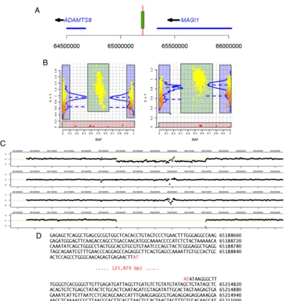
Figure 1 ADAMTS9-MAGI1 deletion characterisation. (A) Map of chromosome 3p14.1 region showing ADAMTS9 and MAGI1 genes (blue
segments), and the intergenic deletion (green rectangle) associated with psoriatic arthritis (PsA) risk. The horizontal red lines indicate the location of the two Taqman assays (Hs03225015_cn and Hs03225295_cn, red vertical lines) used to genotype the deletion. (B) CNStream deletion calling using multiple probes within the deletion sequence. This software method combines the copy number estimation of consecutive probes (left: probe at chr3:65 191 847 pb; right: probe at chr3: 65 196 123 pb). (C) Log2 intensity reads of six individuals for the chromosome 3 region harbouring the deletion. Starting from the top, thefirst two individuals show the characteristic drop in intensity corresponding to hemizygous (ie, 1N) individuals. The third, fourth and sixth individuals show the expected log2 reads of a 2N individual, with most of the intensities centred around 0. Finally, the fifth individual clearly shows the presence of an individual homozygous for the deletion, with no sequence reads mapping to this region of the chromosome. (D) Sequence of the reference and deletion alleles. Physical coordinates are on the reference human genome (build 37).
26mL incubated at 65°C for 70 h. Hybridisation reactions were
incubated with 25μL MyOne Streptavidin C1Dynabeads (Life
Technologies) for 30 min with frequent pulse vortexing. Bead captures were washed twice for 15 min each at room tempera-ture in 0.5 mL wash buffer 1 (1×SSC, 0.1% SDS), followed by four stringency wash steps at 65°C in 0.5 mL preheated wash buffer 2 (0.1×SSC, 0.1% SDS) for 10 min each. Captured libraries were eluted in 50μL 0.1 M NaOH and neutralised in 70μL 1 M Tris pH 7.5. Final libraries were cleaned with 1.8× solid-phase reversible immobilisation beads and eluted in 33mL water for assembly of library amplification PCR containing 1 mL Platinum Taq (Life Technologies) and 5mL 5 M betaine (Sigma)
in 50mL reactions (98°C 1 min, 95°C 30 s and 62°C 3.5 min
for 20 cycles). Final 4-plex library concentrations were deter-mined by KAPA QPCR (KAPA Biosystems) and adjusted to 15 nM each. Stock 4-plex libraries were pooled appropriately for final 24-plex libraries each for a single lane on HiSeq2000 sequencer (Illumina) using 50 bp paired end conditions.
Sequencing reads from individual samples were demuxed based on inline barcode sequences and aligned to the human reference genome (hg19) with BWA.25Relative read depth was calculated as the number of bases mapped to 100 bp windows per total bases mapped for a given sample. Then a log2 ratio between each sample at each 100 bp window to the mean of all samples at that window was used to plot a normalised read depth, representing the read depth relative to a theoretical diploid reference. The plots of these normalised read depths across the locus were used to confirm the presence of the dele-tion (figure 1B).
CNV replication analysis
Replication genotyping was performed using the TaqMan Genotyping System (Applied Biosystems, Foster City, California, USA). Two pre-designed Taqman CNV assays Hs03225015_cn and Hs03225295_cn were found to be located within the esti-mated deletion boundaries. In order to validate the two assays, we genotyped the group of 200 individuals that were previously used to sequence the deletion. The correspondence between the calls of the two CNVs between the Taqman and sequencing ana-lysis was 100%. Consequently, we used the two Taqman assays to genotype the ADAMTS9-MAGI1 deletion in an independent group of 1133 patients with PsA, 1831 healthy controls and 822 patients with PsC. Quality control measures similar to the GWAS were applied, including genotyping call rate >95%, sample completion rate >90% and Hardy–Weinberg disequilib-rium p value of control group p>0.001. The CNV genotype concordance between the two Taqman assays was >99%. Meta-analysis of the GWAS and replication association statistics was performed using METAL software.26
RESULTS
CNV identification and genotyping
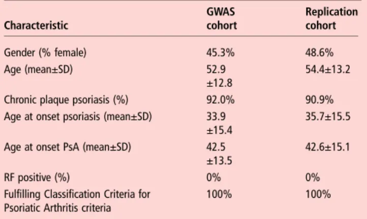
Table 1summarises the main features of the GWAS and replica-tion PsA patient cohorts.
Using a total of 658 patients with PsA and 1063 healthy
con-trols, we identified a total of 2674 CNV segments. After
merging segments belonging to the same genomic region (dis-tance <10 kb and/or r2>0.9), we performed the genotype calling in a total of 1953 different CNV regions. Among them, 165 CNVs appeared in >5% of the samples and were subse-quently used to test for association with PsA risk. Online supple-mentary table S1 describes the characteristics of these CNVs.
CNV GWAS for PsA risk
Using the group of common CNVs, we performed a GWAS for PsA risk. We found a very strong association signal at an inter-genic deletion located between the HLA-C and HLA-B genes ( p=5.37e-11, OR (95% CI) 2.08 (1.47 to 2.95), MAF=0.18,
figure 2). Since HLA-C locus is an established risk locus for PsA we sought to estimate the association of the deletion after cor-recting for the HLA haplotypes associated with psoriasis risk as
recently described.27 We found that, after correcting for
HLA-C*0602 and HLA-B*3801 alleles, the deletion was no longer associated with PsA risk ( p=0.81, see online supplemen-tary table S2). Consequently, this CNV was considered a proxy for the HLA allele association and was not included for replication.
We also found another highly significant association for a deletion located in the chromosome 3q14.2 intergenic region, between ADAMTS9 and MAGI1 genes ( p=0.00014, OR (95%
CI) 1.94 (1.37 to 2.75), MAF=0.04, figure 2). This genomic
region had not been previously associated to any SNP-based
Figure 2 Plot of the copy number variant (CNV) genome-wide association studies results. The−log10 p values (y-axis) are plotted for each of the CNVs identified by CNSstream. Each chromosome is coded in a different colour. The probes mapping the intergenic deletion in HLA-C/-B locus in chromosome 6 (light blue dots) were found to have a high significance; however, after correcting for HLA-C and HLA-B alleles this association disappeared. In chromosome 3, an intergenic deletion between genesADAMTS9 and MAGI1 (purple dot) shows a significant association that withstands multiple test correction.
Table 1 Phenotypic summary of GWAS and replication patient cohorts Characteristic GWAS cohort Replication cohort Gender (% female) 45.3% 48.6% Age (mean±SD) 52.9 ±12.8 54.4±13.2
Chronic plaque psoriasis (%) 92.0% 90.9%
Age at onset psoriasis (mean±SD) 33.9 ±15.4
35.7±15.5 Age at onset PsA (mean±SD) 42.5
±13.5
42.6±15.1
RF positive (%) 0% 0%
Fulfilling Classification Criteria for Psoriatic Arthritis criteria
100% 100%
GWAS, genome-wide association studies; PsA, psoriatic arthritis; RF, rheumatoid factor.
GWAS in PsA or any other related disease. After correcting for the number of CNVs analysed, the deletion association was still found to be significantly associated with PsA risk (p=0.023, Bonferroni multiple test correction). Consequently, we selected this region for replication in the independent data set of patients and controls. In the remaining group of CNVs, we found 12 additional CNV regions nominally associated with PsA risk ( p<0.05, see online supplementary table S3). However, after multiple test correction none of these variants was statistically significant and therefore they were not selected for replication. Additionally, evaluating the concordance between these CNVs
and neighbouring SNPs, we did not find a strong linkage
dis-equilibrium (LD) (r2>0.8) with markers previously associated with PsA, Ps or other autoimmune diseases.
CNV replication in the validation cohorts
The deletion genotypes determined using the quantitative RT-PCR assays showed a 100% concordance with the number of copies (0, 1 or 2) estimated using sequencing. We subse-quently used these two RT-PCR assays to genotype an independent cohort of 1133 patients with PsA and 1831 con-trols. We replicated the association of the ADAMTS9-MAGI1 intergenic deletion with PsA risk ( p=0.032, OR (95% CI) 1.3 (1.0 to 1.7), meta-analysis p=5.97e-5, OR (95% CI) 1.48 (1.21 to 1.82)).
Finally, comparing the frequencies of ADAMTS9-MAGI1 dele-tion of patients with PsA with patients with PsC, we also found a statistically significant increase of the deletion in the group of patients with PsA similar to that observed when comparing to healthy controls (freq PsA=11.0%, freq PsC=7.7%, freq con-trols=8.8%, p=0.0088). Accordingly, when comparing patients with PsC to healthy controls, we did notfind a statistically sig-nificant difference between the deletion frequencies of groups ( p=0.33).
ADAMTS9-MAGI1 deletion sequence characterisation
Using a next-generation sequencing approach, we characterised the deletion region in chromosome 3q14.2 associated with PsA risk. Using a sample of 100 patients with PsA and 100 controls selected to have a higher frequency of the deletion (in total, 100 deletion carriers vs 100 2N individuals), we determined the
physical extent and molecular nature of ADAMTS9-MAGI1 tion polymorphism. PCR capture and sequencing of the dele-tion breakpoints revealed that the deledele-tion removes 25 879 nucleotides (figure 1D).
In order to explore the LD pattern associated with the dele-tion and the reladele-tion with the twoflanking genes, we sequenced
ADAMTS9 and MAGI1 genes and their proximal 50 and 30
untranslated regions. LD analysis showed clearly that only very few close polymorphisms have moderate to high correlation with the deletion (r2>0.7, n=20 SNPs, figure 3). From the
>18 000 DNA variants identified after sequencing both
ADAMTS9 and MAGI1 genes, none showed a significant LD
with the deletion (figure 3). DISCUSSION
GWAS based on SNPs have been highly successful in identifying genetic variants associated with the risk to develop psoriasis and PsA. However, there is still a large fraction of the genetic basis of PsA that has not been identified. In the present study, we have performed a GWAS of CNVs to identify new genomic loci associated with PsA risk. Using a discovery cohort of Spanish patients with PsA and controls, we have found a significant asso-ciation of a deletion located between ADAMTS9 and MAGI1 genes with PsA risk. We have subsequently validated this associ-ation in an independent cohort of patients and controls. Furthermore, using a cohort of patients with psoriasis without concomitant arthritis we provide evidence that the deletion is specific for PsA.
ADAM metallopeptidase with thrombospondin type 1 motif 9 (ADAMTS9) gene belongs to a family of enzymes specialised in the degradation of the extracellular matrix. In particular, ADAMTS9 belongs to the family of the aggrecanases (which
also includes ADAMTS-1, ADAMTS-4, ADAMTS-6,
ADAMTS-8 and ADAMTS-15) that are specialised in the deg-radation of aggrecan, one of the main proteoglycan constituents of the cartilage extracellular matrix.28 Aggrecanase activity has been found to be associated with cartilage degradation in inflammatory joint diseases including PsA.29 30 Importantly, in vitro studies with chondrocytes have found that after
stimula-tion with tumour necrosis factor (TNF)-α and interleukin
(IL)-1, two of the most abundant cytokines in PsA synovium,31
Figure 3 Pairwise linkage disequilibrium (LD) between the deletion andflanking sequence variants. The LD (r2) between the deletion and the >18 000 variants identified after sequencing the regions flanking the copy number variants as well as ADAMTS9 and MAGI1 loci. From these results, it is clear that only a small proportion of close variants show a high correlation with the deletion (ie, r2>0.9, centre region of the plot) and that there is no variant within the transcribed or theflanking regions of ADAMTS9 (left region) or MAGI1 (right region) that could explain the observed association. The3p14.1 chromosome regions between these three loci and that were not sequenced are left blank.
ADAMTS9 was clearly the most highly induced among all the different aggrecanases.32 33 Consequently, genetic variants that influence the rates of matrix turnover in the cartilage and bone of the inflamed synovial joint could be crucial in determining the level of tissue degradation in PsA.
Membrane-associated guanylate kinase, WW and PDZ domain containing 1 (MAG1) is a member of the membrane-associated guanylate kinase family of genes. MAGI1 is known to be expressed in cell-to-cell contacts, acting as a scaffold protein to stabilise cadherin-mediated adhesions and has been found to be expressed in epithelial and endothelial tight junctions.34MAGI1 activity has been related to several pathological junction-associated processes, including oncogenic35 as well as virus-associated invasiveness.36 To date, there is no evidence of a direct implication of MAGI1 in PsA pathology or, even, in auto-immune diseases. There is evidence, however, that MAGI1 interacts with phosphatase and tensin homologue protein, a sig-nalling protein that participates in the negative regulation of regulatory T cells (Tregs),37which are master inhibitors of auto-immunity. While the implication of Treg has been clearly defined in rheumatoid arthritis or psoriasis aetiology,38there are yet no studies directly analysing the implication of this key immune regulator in PsA,39although there is recent evidence of their activity in the disease pathology.40 Clearly, future studies aimed at characterising the implication of MAGI1 activity in autoimmunity are necessary.
The deep sequence analysis found that there is very little cor-relation between the∼26 kb intergenic deletion associated with PsA risk and the polymorphisms located in the transcribed sequences and proximal regions of ADAMTS9 and MAGI1 genes. Also, our sequencing analysis clearly shows that very few neighbouring markers are in moderate or high LD with this CNV, suggesting that the deletion itself is the genetic variant implicated in the aetiology of PsA. Exploration of the chromo-some 3q14.2 deleted region in multiple biomedical databases
including ENCODE41 did not show regulatory evidence
asso-ciated with this variation. Also analysing available cis and trans-eQTL regulatory data sets42we did notfind an association between this deletion and the expression of other genes. However, it is increasingly becoming evident that a large frac-tion of regulatory variants in the genome will be only detected under the specific target tissue where they are expressed and, perhaps, only under the adequate stimulation.43 For example, ADAMTS9 expression in chondrocytes was found to be expressed only after stimulation by proinflammatory cytokines TNF and IL-1.32Additional studies aimed at characterising the functional implications of this deletion and their role in PsA aetiology are therefore warranted.
To date, there is evidence that the frequency and penetrance of multiple risk loci in PsA and psoriasis risk is different popu-lations with different ancestries.44It will be therefore necessary to evaluate the frequency and effect size of this deletion at 3p14.2 in other non-Caucasian populations. Also, the associ-ation of the ADAMTS9-MAGI1 CNV with different PsA phenotypes could be of high relevance. In our discovery cohort, we analysed the association of the deletion with axial disease, arthritis mutilans, gender, age of start of the disease and PsA familial aggregation, but we did notfind a significant association (data not shown). These results support that the deletion at chromosome 3q14.2 is specifically associated to the risk to develop PsA. It is possible, however, that once the
specific biological mechanisms influenced by this genetic
variation are identified, more targeted analysis will reveal association to other PsA phenotypes.
In the present study, we have performed thefirst CNV GWAS
in PsA. We have identified a new deletion in ADAMTS9-MAGI1
locus associated with PsA risk and we have validated this
associ-ation in an independent patient and control cohort.
Additionally, using a cohort of patients with PsC we have demonstrated that the variation is specifically associated with the development of PsA. The present study represents an important step in the characterisation of the common genetic variation associated with PsA.
Author affiliations
1Rheumatology Research Group, Vall d’Hebron Research Institute, Barcelona, Spain 2
Rheumatology Department, Complejo Hospitalario Juan Canalejo, A Coruña, Spain
3Rheumatology Department, Hospital Parc Taulí, Sabadell, Barcelona, Spain 4
Rheumatology Department, Hospital Universitario Central de Asturias, Oviedo, Spain
5
Dermatology Department, Hospital Universitari Germans Trias i Pujol, Badalona, Barcelona, Spain
6
Dermatology Department, Complejo Hospitalario Universitario de A Coruña, A Coruña, Spain
7
Rheumatology Department, Hospital Virgen de la Vega, Salamanca, Spain
8Rheumatology Department, Hospital Monte Naranco, Oviedo, Spain 9
Dermatology Department, Hospital de la Santa Creu i Sant Pau, Barcelona, Spain
10Rheumatology Department, Hospital de Jerez de la Frontera, Cádiz, Spain 11
UGC Reumatología, Instituto de Investigación Biomédica de Málaga (IBIMA), Hospital Regional Universitario de Málaga, Málaga, Spain
12
Department of Dermatology, Hospital Universitario de Salamanca, Salamanca, Spain
13
Rheumatology Department, Hospital Universitario Infanta Sofía, Madrid, Spain
14Dermatology Department, Hospital Universitario La Princesa, Madrid, Spain 15
Rheumatology Department, Hospital Universitario Gregorio Marañón, Madrid, Spain
16
Rheumatology Service, Hospital Moisès Broggi, Barcelona, Spain
17Dermatology Department, Hospital General Universitario de Valencia, Valencia,
Spain
18Rheumatology Department, Hospital Universitario Fundación Alcorcón, Madrid,
Spain
19Rheumatology Department, Hospital Sant Rafael, Barcelona, Spain 20
Dermatology Department, Hospital Universitario Fundación Alcorcón, Madrid, Spain
21
Rheumatology Department, Hospital Universitari de Bellvitge, Barcelona, Spain
22Dermatology Department, Hospital Universitario Virgen Macarena, Sevilla, Spain 23
Department of Dermatology, Hospital Universitario Infanta Leonor, Madrid, Spain
24Dermatology Department, Hospital Universitario 12 de Octubre, Madrid, Spain 25
Dermatology Department, Hospital Universitario Virgen de la Victoria, Málaga, Spain
26
Rheumatology Department, Hospital Universitario de La Princesa, IIS-Princesa, Madrid, Spain
27
Rheumatology Department, Centro de Salud Virgen de los Reyes, Sevilla, Spain
28Rheumatology Department, Hospital Mútua de Terrassa, Terrassa, Spain 29
Rheumatology Unit, Hospital de la Santa Creu i Sant Pau, Barcelona, Spain
30Rheumatology Department, Hospital Universitario Marqués de Valdecilla,
Santander, Spain
31Rheumatology Department, Complexo Hospitalario de Ourense, Ourense, Spain 32
Rheumatology Department, Complejo Hospitalario Hospital Provincial de Pontevedra, Pontevedra, Spain
33
Rheumatology Department, Hospital General Universitario de Alicante, Alicante, Spain
34
Rheumatology Department, Hospital Universitario Guadalajara, Guadalajara, Spain
35Rheumatology Department, Hospital La Mancha Centro, Alcázar de San Juan,
Spain
36Rheumatology Department, Hospital Clínic de Barcelona and IDIBAPS, Barcelona,
Spain
37Banco Nacional de ADN Carlos III, University of Salamanca, Salamanca, Spain 38
Life Sciences, Barcelona Supercomputing Centre, National Institute of Bioinformatics, Barcelona, Spain
39
HudsonAlpha Institute for Biotechnology, Alabama, USA
Correction notice This article has been corrected since it was published Online First. Dr Juan D Cañete has been included as a co-corresponding author.
Acknowledgements We thank the patients and clinical specialists collaborating in the IMID Consortium for participation.
Contributors All authors were involved in the design, analysis and interpretation of data. All authors revised the manuscript and gavefinal approval for its submission. SM and JDC shared senior authorship.
Funding This study was funded by the Spanish Ministry of Economy and Competitiveness, grant numbers: PSE-010000-2006-6 and IPT-010000-2010-36. Competing interests None declared.
Patient consent Obtained.
Ethics approval Local Institutional Review boards from all participating centres. This study was conducted in accordance with the Declaration of Helsinki principles Provenance and peer review Not commissioned; externally peer reviewed. REFERENCES
1 Gladman DD, Antoni C, Mease P, et al. Psoriatic arthritis: epidemiology, clinical features, course, and outcome. Ann Rheum Dis 2005;64(Suppl 2):ii14–17. 2 Julia A, Tortosa R, Hernanz JM, et al. Risk variants for psoriasis vulgaris in a large
case-control collection and association with clinical subphenotypes.Hum Mol Genet 2012;21:4549–57.
3 Gladman DD, Farewell VT, Pellett F, et al. HLA is a candidate region for psoriatic arthritis. Evidence for excessive HLA sharing in sibling pairs.Hum Immunol 2003;64:887–9.
4 Myers A, Kay LJ, Lynch SA, et al. Recurrence risk for psoriasis and psoriatic arthritis within sibships.Rheumatology (Oxford)2005;44:773–6.
5 Bhalerao J, Bowcock AM. The genetics of psoriasis: a complex disorder of the skin and immune system.Hum Mol Genet1998;7:1537–45.
6 Liu Y, Helms C, Liao W, et al. A genome-wide association study of psoriasis and psoriatic arthritis identifies new disease Loci.PLoS Genet2008;4:e1000041. 7 Ellinghaus E, Ellinghaus D, Stuart PE, et al. Genome-wide association study
identifies a psoriasis susceptibility locus at TRAF3IP2.Nat Genet2010;42:991–5. 8 Strange A, Capon F, Spencer CC, et al. A genome-wide association study identifies
new psoriasis susceptibility loci and an interaction between HLA-C and ERAP1. Nat Genet2010;42:985–90.
9 Chen H, Poon A, Yeung C, et al. A genetic risk score combining ten psoriasis risk loci improves disease prediction.PLoS One2011;6:e19454.
10 Ellinghaus E, Stuart PE, Ellinghaus D, et al. Genome-wide meta-analysis of psoriatic arthritis identifies susceptibility locus at REL.J Invest Dermatol2012;132:1133–40. 11 Sheng Y, Jin X, Xu J, et al. Sequencing-based approach identified three new
susceptibility loci for psoriasis.Nat Commun2014;5:4331.
12 Cook EH Jr, Scherer SW. Copy-number variations associated with neuropsychiatric conditions.Nature2008;455:919–23.
13 Hollox EJ, Huffmeier U, Zeeuwen PL, et al. Psoriasis is associated with increased beta-defensin genomic copy number.Nat Genet2008;40:23–5.
14 de Cid R, Riveira-Munoz E, Zeeuwen PL, et al. Deletion of the late cornified envelope LCE3B and LCE3C genes as a susceptibility factor for psoriasis.Nat Genet 2009;41:211–15.
15 Huffmeier U, Estivill X, Riveira-Munoz E, et al. Deletion of LCE3C and LCE3B genes at PSORS4 does not contribute to susceptibility to psoriatic arthritis in German patients.Ann Rheum Dis2010;69:876–8.
16 Julia A, Domenech E, Ricart E, et al. A genome-wide association study on a southern European population identifies a new Crohn’s disease susceptibility locus at RBX1-EP300.Gut2012;62:1440–5.
17 Taylor W, Gladman D, Helliwell P, et al. Classification criteria for psoriatic arthritis: development of new criteria from a large international study.Arthritis Rheum 2006;54:2665–73.
18 Morton NE, Collins A. Tests and estimates of allelic association in complex inheritance.Proc Natl Acad Sci U S A1998;95:11389–93.
19 Price AL, Patterson NJ, Plenge RM, et al. Principal components analysis corrects for stratification in genome-wide association studies.Nat Genet2006;38:904–9. 20 Peiffer DA, Le JM, Steemers FJ, et al. High-resolution genomic profiling of
chromosomal aberrations using Infinium whole-genome genotyping.Genome Res 2006;16:1136–48.
21 Alonso A, Julia A, Tortosa R, et al. CNstream: a method for the identification and genotyping of copy number polymorphisms using Illumina microarrays. BMC Bioinformatics2010;11:264.
22 Steemers FJ, Chang W, Lee G, et al. Whole-genome genotyping with the single-base extension assay.Nat Methods2006;3:31–3.
23 R Core Team. R: A language and environment for statistical computing. R Foundation for Statistical Computing: Vienna, 2013.
24 Day K, Song J, Absher D. Targeted Sequencing of Large Genomic Regions with CATCH-Seq.PLoS One2014;9:e111756.
25 Li H, Durbin R. Fast and accurate short read alignment with Burrows-Wheeler transform.Bioinformatics2009;25:1754–60.
26 Willer CJ, Li Y, Abecasis GR. METAL: fast and efficient meta-analysis of genomewide association scans.Bioinformatics2010;26:2190–1.
27 Okada Y, Han B, Tsoi LC, et al. Fine mapping major histocompatibility complex associations in psoriasis and its clinical subtypes.Am J Hum Genet2014;95: 162–72.
28 Kevorkian L, Young DA, Darrah C, et al. Expression profiling of metalloproteinases and their inhibitors in cartilage.Arthritis Rheum2004;50:131–41.
29 Lohmander LS, Neame PJ, Sandy JD. The structure of aggrecan fragments in human synovialfluid. Evidence that aggrecanase mediates cartilage degradation in inflammatory joint disease, joint injury, and osteoarthritis.Arthritis Rheum 1993;36:1214–22.
30 Arner EC, Pratta MA, Decicco CP, et al. Aggrecanase. A target for the design of inhibitors of cartilage degradation.Ann N Y Acad Sci1999;878:92–107. 31 Partsch G, Steiner G, Leeb BF, et al. Highly increased levels of tumor necrosis
factor-alpha and other proinflammatory cytokines in psoriatic arthritis synovial fluid. J Rheumatol 1997;24:518–23.
32 Demircan K, Hirohata S, Nishida K, et al. ADAMTS-9 is synergistically induced by interleukin-1beta and tumor necrosis factor alpha in OUMS-27 chondrosarcoma cells and in human chondrocytes.Arthritis Rheum2005;52:1451–60.
33 Uysal S, Unal ZN, Erdogan S, et al. Augmentation of ADAMTS9 gene expression by IL-1beta is reversed by NFkappaB and MAPK inhibitors, but not PI3 kinase inhibitors.Cell Biochem Funct2013;31:539–44.
34 Laura RP, Ross S, Koeppen H, et al. MAGI-1: a widely expressed, alternatively spliced tight junction protein.Exp Cell Res2002;275:155–70.
35 Zaric J, Joseph JM, Tercier S, et al. Identification of MAGI1 as a tumor-suppressor protein induced by cyclooxygenase-2 inhibitors in colorectal cancer cells.Oncogene 2012;31:48–59.
36 Kolawole AO, Sharma P, Yan R, et al. The PDZ1 and PDZ3 domains of MAGI-1 regulate the eight-exon isoform of the coxsackievirus and adenovirus receptor. J Virol2012;86:9244–54.
37 Walsh PT, Buckler JL, Zhang J, et al. PTEN inhibits IL-2 receptor-mediated expansion of CD4+ CD25+ Tregs. J Clin Invest 2006;116:2521–31.
38 Buckner JH. Mechanisms of impaired regulation by CD4(+)CD25(+)FOXP3(+) regulatory T cells in human autoimmune diseases.Nat Rev Immunol 2010;10:849–59.
39 Nograles KE, Brasington RD, Bowcock AM. New insights into the pathogenesis and genetics of psoriatic arthritis.Nat Clin Pract Rheumatol2009;5:83–91. 40 Ryder LR, Bartels EM, Woetmann A, et al. FoxP3 mRNA splice forms in synovial
CD4+ T cells in rheumatoid arthritis and psoriatic arthritis.APMIS 2012;120:387–96.
41 ENCODE Project Consortium. An integrated encyclopedia of DNA elements in the human genome.Nature2012;489:57–74.
42 Lappalainen T, Sammeth M, Friedlander MR, et al. Transcriptome and genome sequencing uncovers functional variation in humans.Nature2013;501:506–11. 43 Fairfax BP, Humburg P, Makino S, et al. Innate immune activity conditions the effect
of regulatory variants upon monocyte gene expression.Science2014;343:1246949. 44 Roberson ED, Bowcock AM. Psoriasis genetics: breaking the barrier.Trends Genet