Title: A phase I trial of oncolytic adenovirus ICOVIR-5 administered intravenously to cutaneous
and uveal melanoma patients.
Authors: M. García1*, R. Moreno2*, M. Gil-Martin3, M Cascallò2, 4, M. Ochoa de Olza3, C. Cuadra1, JM Piulats3, V. Navarro1, M Domenech3, R. Alemany2, and R. Salazar3
1Clinical Research Unit, Institut Català d'Oncologia-IDIBELL, L’Hospitalet, Barcelona, Spain; 2ProCure and Oncobell Programs, Institut Català d'Oncologia-IDIBELL, L’Hospitalet, Barcelona, Spain. 3Department of Medical Oncology, Oncobell Program, Institut Català d'Oncologia-IDIBELL, L’Hospitalet, Barcelona, Spain: 4VCN Biosciences. Sant Cugat del Valles. Barcelona. Spain
*Authorship note: M.García and R. Moreno contributed equally to this work.
Corresponding author:
Ramon Alemany. ProCure Program, Institut Català d’Oncologia, Avinguda Gran Via de l’Hospitalet, 199, 08907 L’Hospitalet de Llobregat, Barcelona, Spain. Tel +34932607462.
ABSTRACT
Oncolytic viruses represent a unique type of agents that combine self-amplification, lytic and immunostimulatory properties against tumors. A local and locoregional clinical benefit has been demonstrated upon intratumoral injections of an oncolytic herpes virus in melanoma patients, leading to its approval in USA and Europe for patients without visceral disease (up to stage IVM1a). However, in order to debulk and change the local immunosuppressive environment of tumors that cannot be injected directly, oncolyitc viruses need to be administered systemically. Among different viruses, adenovirus has been extensively used in clinical trials but with few evidences of activity upon systemic administration. Preclinical efficacy of a single intravenous administration of our oncolytic adenovirus ICOVIR5, an adenovirus type 5 responsive to the pRB pathway commonly deregulated in tumors, led us to use this virus in a dose-escalation phase I trial in metastatic melanoma patients. The results in 12 patients, treated with a single infusion of a dose up to 1E13 viral particles, show that ICOVIR5 can reach melanoma metastases upon a single intravenous administration but fails to induce tumor regressions. These results support the systemic administration of armed oncolytic viruses to treat disseminated cancer.
INTRODUCTION
Malignant Melanoma represents the ninth most common tumor in Europe with more than 100,000 new cases registered in 2012. 1 In advanced stages, the only approved chemotherapy is Dacarbazine whose response rate is around 10 %. 2 Recently, several treatments have been approved which are specifically targeting melanoma tumors with BRAF V600E or V600K mutations or blocking some forms of MEK. 3-5 Immune checkpoint blockers, ipilimumab, pembrolizumab and nivolumab, are also treatments recently approved due to their ability to improve overall survival. 6-8 In addition, Talimogene laherparepvec (T-VEC), a genetically modified oncolytic herpes simplex virus type 1, which secrets granulocyte-macrophage colony stimulating factor, is approved for local intralesional injection based on data that demonstrated shrinkage of lesions, showing the plausibility of a strategy based on oncolysis and immune system stimulation by viruses. 9
Oncolytic adenovirus-based therapy remains a promising approach to cancer treatment, as virus can infect tumor cells and selectively replicate in them, leading to oncolysis, and release of new viruses, which conduct to local and bloodstream spread and finally, induction of an immune reaction against the tumor. 10, 11 The existing experience with intravenous administration of oncolytic virus shows that high doses of adenovirus (≥ 1013 viral particles) in more than 300 cancer patients resulted in limited efficacy with mild toxicity characterized by a transient febrile peak, elevation of transaminases, and occasionally moderate transient nausea and flu-like symptoms.
ICOVIR-5 derives from the oncolytic adenovirus Ad- 24RGD 12, which contains two modifications compared to wild type Ad5: first, the E1A region responsible for pRB protein binding is deleted to abrogate the capability of E1A to release E2F from the pRB-E2F complex, characteristic of quiescent cells, contributing to the preferential replication of the virus in tumor and dividing cells. Second, an RGD sequence inserted at the HI loop of the fiber knob to target integrins at the surface of cancer cells as a primary receptor for virus entry. Two additional modifications were present in ICOVIR-5 13: replacement of the E1A promoter by the human E2F-1 promoter (insulated
from the E1A enhancer using the myotonic dystrophy insulator (DM-1)) and a consensus Kozak sequence inserted before the first codon of the E1A gene to boost E1A mRNA translation.
The main objective of this first-in-human study was to determine the maximum tolerated dose (MTD) of the infusion of conditionally replicating oncolytic adenovirus ICOVIR-5 in patients with advanced malignant melanoma. Secondary objectives were recommended dose, safety, toxicity profile, and preliminary efficacy data if any.
MATERIALS AND METHODS
This phase I trial of ICOVIR-5 was conducted at the Catalan Institute of Oncology (Barcelona, Spain) in accordance with the Declaration of Helsinki and the principles of Good Clinical Practice, and it was approved by The Clinical Research Ethics Committee of the Bellvitge University Hospital and the Spanish Medicines and Sanitary Products Agency (AEMPS). All participants provided written informed consent. This study was registered at clinicaltrials.gov with the identifier NCT01864759.
Patients
Eligible patients had uveal or cutaneous metastasic malignant melanoma. Patients could have received any prior treatment for metastatic disease. Inclusion criteria were histological proof of cancer, age ≥ 18 years, performance status 0 to 2 (Eastern Cooperative Oncology Group [ECOG] scale), absolute neutrophil count (ANC) greater than or equal to 1,500/L, platelet count greater than or equal to 100,000/L, normal function of the liver (bilirubin level < the upper limits of normal [ULN], AST/ALT level 2.5 times the ULN, alkaline phosphatase level < 2.5 times the ULN) and kidney (creatinine clearance ≥ 50 ml/min), and negative HIV serology. Patients with previous participation in studies with adenovirus, viral syndrome diagnosed two weeks before inclusion, concomitant chronic immunosuppressant medication, concomitant malignant hematology disease, Li Fraumeni Syndrome or germ known deficit pRb pathway were excluded.
Study Design
The study was conducted using an accelerated escalating phase I design with cohorts of 1 patient if no grade >2 toxicity was found, and then with 3-6 patient cohorts after the first grade 3 toxicity finding. A Dose-Limiting Toxicity (DLT) observation period of 4 weeks was established before the entry of the first patient at the next dose level. If one out of three patients experienced DLT during the treatment cycle, at least one more additional patient was to be treated at this dose
level and up to 6 patients could be sequentially treated at this dose level. The Maximum Tolerated Dose (MTD) was defined as the dose level at which two of the three to six treated patients experienced DLT and the recommended dose (RD) was the highest dose level at which less than 2 of 6 patients experienced DLT. Toxicity was classified according to the Common Toxicity Criteria of the National Cancer Institute (NCI CTC, version 3.0). Dose Limiting Toxicity was defined as: ANC <0.5 x 10⁹ / L for more than 5 days, febrile neutropenia as defined by the NCI CTC 3.0, platelet count <25 x 109 / L, any other non-hematologic grade 3-4 toxicity except for alopecia, nausea/vomiting unresponsive to specific treatment, or any toxicity grade 2 unrecovered at 6 weeks of treatment.
Treatment
ICOVIR-5 was administered as a single infusion in an inpatient regimen. Before administration, ICOVIR-5 was thawed and diluted in 50ml 0.9% NaCl for intravenous administration. Patients were monitored in a specific isolation room at the hospital until acute events were solved.
Dose levels: 1a, 1x10¹¹ viral particles (vp); 2a, of 3.3x10¹¹vp; 3a, of 1x10¹²vp; 4a, of 3.3x10¹²vp and 5a, of 1x10¹³vp. A dose level of 1x10¹⁰ (-1a) was planned if dose 1a became limiting.
Study assessments
All patients were evaluated by physical examination, ECG and chest X-ray. Complete blood cell (CBC) count, liver, and kidney function tests and urine analysis were obtained before treatment, on days 1, 2, 3, 5, 12, 19, and at the end of the study (day 26). Study of extent of disease was performed by computed tomography (CT) or magnetic resonance imaging (MRI) of the chest, liver and abdomen, and bone scan with X-ray if hot spots were observed. Assessments of tumor lesions were made at 4 weeks of treatment applying the criteria recommended for evaluation of response in solid tumors (RECIST 1.1). Tumor biopsies were obtained if feasible at the beginning of the study and at day 5 post-treatment.
ICOVIR-5
ICOVIR-5 has been previously described. 13 It derives from the oncolytic adenovirus Ad- 24RGD 12, currently in clinical trials against glioblastoma under the name DNX-2401 (by DNAtrix Therapeutics, Houston Tx). ICOVIR-5 was constructed by homologous recombination in bacteria and transfection in HEK293 cells. Then, it was plaque-purified and amplified in A549 cells, and purified using a CsCl gradient. The viral genomic structure was verified by restriction analysis. The DM-1 insulator, E2F-1 promoter, Kozak sequence, E1A- 24 deletion, and RGD fiber were sequenced. The clinical batch of ICOVIR-5 was manufactured at the Center for Cell & Gene Therapy at Baylor College of Medicine (Houston, TX, USA) under GMP conditions using A549 cells. Virus titer was adjusted to 1x10¹² vp/ml in 20 mM Tris pH 8.0, 25 mM NaCl, 2,5% glycerol, aliquoted in sterile vials, and stored at -80 oC with continuous monitoring of temperature.
Functional virus titer was 5.33x10¹⁰ plaque-forming units (PFU) / ml, which corresponds to 18.7 virus particle / functional particle bioactivity ratio. No wild type Ad5 or other adventitious viruses were detected by PCR.
Virus pharmacokinetics
Immediately after the end of virus administration (time 0), and at 30min, 1h, 2h, 4h, 6h, and 24h after virus administration, blood samples were taken using 4ml Lithium Heparin Vacuette tubes (Greiner Bio-One, Monroe, NC, USA) and total DNA was isolated by the QIAamp DNA Mini and QIAamp DNA Blood Mini Kits (QIAGEN, Valencia, CA) in triplicate according to the manufacturer protocol. Four microliters of DNA were used to quantify genome copy number by Quantitative Real Time-PCR using ICOVIR-5 specific primers (ICO5F2-5’-GAT TTG GCG CGT AAA AGT G -3' which overlaps E2F1 promoter and the reverse ICO5R2-5'-CGG CCA TTT CTT CGG TAA TA -3' which overlaps with the E1A gene, generating a 126 bp amplicon). Real Time PCR consisting of 10 min at 95 oC and 40 cycles (95 oC, 15 s; 60 oC s; 72 oC, 10 s) was performed on the LightCycler® 480 II
(Roche, Basel, Switzerland), using LightCycler 480 SYBR Green I Master (Roche) and analyzed with LightCycler® 480 Software release 1.5.0 SP4 (Roche). A control PCR without DNA was included to monitor reagent contamination.
For genome copy quantification, blood from a donor was spiked with a 10-fold serial dilution of ICOVIR-5 to generate a standard curve from 1x10¹⁰ vp/ml to 1x10² vp/ml. This curve and negative controls of blood without virus were processed with the same protocol as patient samples and analyzed in the same Real Time PCR to quantify genome copies. Precision, accuracy, and limits of detection (LOD) and quantification (LOQ) were established using the standard curve in a previous experiment. LOD, the lowest concentration of genomes in the standard curve that can be detected, was 1x10³ genomes/ml. LOQ, the lowest concentration of genomes in the standard curve which maintains linearity with the standard curve (slope 0.8-1.2), was 1x10⁴ genomes/ml.
Analysis of virus shedding
Blood, urine, sputum, and stool samples were collected before administration and on days 1, 2, 5, 12, 19, and 26 post-treatment. Samples were analyzed when available due to compliance of protocols. Among them, sputum and stool presented a higher frequency of lack of compliance. DNA was extracted with the following kits: QIAamp DNA Mini and QIAamp DNA Blood Mini Kits for blood; QIAamp Viral RNA Mini kit for urine after concentration with Millipore Amicon Ultra Centrifugal Filters Ultracel 50k (Millipore; Darmstadt, Germany); QIAampMinElute Virus Spin for sputum; and QIAamp DNA Stool for stool (all kits from QIAGEN). All samples were processed in triplicate.
A 10-fold serial dilution standard curve for each type of sample was generated diluting ICOVIR-5 in the corresponding sample obtained from non-treated donors. Standard curves and negative controls (samples without virus) were processed as patient samples.
Isolated DNA (4 microliters) were used to quantify the genome copy number using the same Quantitative Real Time-PCR program described above for pharmacokinetics.
The LOQ and LOD were previously determined for the different samples. For blood, LOQ and LOD were the same described above for pharmacokinetics. For urine, LOQ and LOD were established as 1.25x10⁴ genomes/ml. For sputum, LOD 1x10³ genomes/ml and LOQ was 1x10⁴ genomes/ml. For stool LOD was 1x10³ genomes/g and LOQ was 1x10⁴ genomes/g.
ICOVIR-5 genomes in tumor biopsies
Patient tumor biopsies were obtained when possible by core needle biopsy (CNB) from accessible skin lesions at day 5 post-treatment to detect ICOVIR-5. Samples were digested with proteinase K and DNA extracted using QIAamp DNA Mini and QIAamp DNA Blood Mini Kits according to the manufacturer instructions. A 10-fold serial dilution standard curve was generated diluting ICOVIR-5 with sterile phosphate buffered saline (PBS, Life Technologies, Carlsbad, CA, USA) and processed as tumor samples.
Isolated DNA (4 microliters) were used to quantify the genome copy number using the same Quantitative Real Time-PCR program as above for pharmacokinetics. The LOD was 103genomes/sample and the LOQ 104genomes/sample.
Analysis of cytokines in blood.
Cytokines IL-6 and IL-10 were chosen for the analysis as possible predictors of immunologic response and toxicity. Basal and post-treatment levels (6h, 24h, 48h, day 5, day 12, day 19, and day 26) of cytokines IL-6 and IL-10 in patient serum were quantified using ELISA assays (R&D; Minneapolis, MN, USA). Patient peripheral blood was collected in Vacuette Z Serum Sep Clot Activator tubes (Greiner Bio-One, Monroe, NC, USA), allowed to coagulate for 30 minutes and centrifuged at 1500 g for 10 minutes. Serum samples were harvested and stored at –80 oC until
analysis. ELISA assays were carried out according to manufacturer’s protocols. Each sample was analyzed in triplicate.
Anti-ICOVIR-5 neutralizing antibodies
Neutralizing antibodies in serum against adenovirus type 5 were analyzed before treatment and at day 19 after ICOVIR-5 administration. Samples of serum were heated at 56 °C for 30 minutes in order to inactivate complement. A 2-fold serial dilution of inactivated samples from 1/10 to 1/5120 was performed in Dulbecco’s modified Eagle’s medium in 96-well plates (final volume 50
l) in quadruplicates. Serum dilutions were incubated for 1h at room temperature with 2.5x10⁴ transducing units (TU) of AdTL, an adenovirus type 5 E1-deleted vector expressing luciferase and enhanced green fluorescent protein (EGFP). 14 Afterwards, 1x10⁵ HEK-293 cells per well were added (to obtain a proportion or multiplicity of infection of 0.25 TU/cell) and cultured at 37 °C and 5% CO2. After 24h of infection, media was removed and cells washed once with PBS. Finally, the level of luciferase in each sample was quantified using a Victor X5 Multilabel Plate Reader (PerkinElmer, Waltham, MA, USA). The percentage of neutralization was calculated with the formula (% neutralization = [1-((LUsample-LUnegative control)/(LUpositive control-LUnegative control))] x 100.
As a negative control we used the luciferase units (LU) of a non-infected well and as a positive control we used the LU of an infected well without serum. The neutralizing antibody titers were expressed as the reciprocal of the serum dilution closest to 50% neutralization of the infection. Every serum sample was quantified in quadruplicate.
Statistical analysis
Descriptive statistics were used for safety analyses for all patients who received one dose of ICOVIR-5. Categorical and continuous data were summarized with frequencies and percentages. The efficacy population included all patients with a baseline assessment and a post-baseline tumor assessment. Fisher´s exact test was used to correlate neutralizing antibodies with clinical responses and linear regression analysis to correlate viral doses with cytokine levels.
RESULTS
Patient characteristics, tolerability, and efficacy
Thirteen patients were enrolled in the study and twelve were treated at five dose levels (Table 1). Patient #05 was screened but not treated due to early progression. All 12 treated patients were evaluable for toxicity. Patients’ characteristics were as follows: 7 males and 5 females; the median age was 51 (range 40-80) and the ECOG performance status was 0 (5 patients) or 1 (7 patients). All patients had evaluable or measurable disease. Six patients (50%) had received at least one prior chemotherapy regimen for metastatic melanoma, and 4 (33%) patients had received previous immunotherapy (supplementary table 1). Six patients had uveal melanoma and six cutaneous melanoma. All patients received one treatment injection and were observed for 4 weeks, except for one (#02) who did not complete one cycle of observation due to early progression. This patient #02, following the protocol, was replaced, although he was considered evaluable for toxicity. The patient, in the context of a rapid progression, presented a disseminated intravascular coagulation (DIC) not related to the treatment but to the progression of the underlying disease.
Table 2 lists the most significant all-cycle non-hematological toxicities observed in the study. At dose levels 1a to 3a no relevant toxicity was observed. The grade 2 toxicity observed at level 4a allowed to proceed to dose escalation. Acute toxicity was mainly a flu-like syndrome with fever, chills, arthromyalgia, headache, nausea and vomiting, and diarrhea, which began in the first 4-6 hours after infusion and during 2-4 days. The first patient at the level 5a (patient #08) experienced toxicity as transaminitis grade 3 at day +1. In addition, this patient presented grade 3 thrombocytopenia at day +4. On day +2, transaminitis was recovered to grade 1 and thrombocytopenia lasted to day +12. This dose level was then expanded to 6 patients. Patient #13 presented the second DLT as hepatic grade 3 toxicity (grade 3 AST elevation which lasted two days and recovered to grade 0 at day +12). Both patients had normal transaminases level before treatment, and only patient #08 had metastatic liver involvement at the beginning of the study.
According to protocol, this level (5a: 1x10¹³) was declared MTD, and the recommended phase 2 dose was defined at the inferior dose level 4a (3.3x10¹²). Other significant toxicities at this dose level included: grade 2 neutropenia in patient #08 and grade 2 AST elevation in both patients. With regard to efficacy, the eleven patients who had received treatment and had undergone at least one disease evaluation (performed day 26) were evaluated. No objective responses, partial or complete, were observed. At the lower dose levels two patients had stable disease, patient #03 until day 100 post-treatment and patient #06 until day 433 after treatment. At the highest dose level (1x10¹³ vp) stable disease was observed in 5 out of 6 patients, patient #09 until day 56, patient #010 until day 94, patient # 11 until day 131, patient # 12 until day 84, and patient #013 until day 112 post-treatment (Table 3). Comparing the survival of cutaneous melanoma patients that never received targeted therapy or anti-PD1 antibodies after participating in the trial versus uveal melanoma patients, the survival probability was 3.7 times longer for the uveal melanoma patients (Supplementary figure 1. Median 271 days for uveal vs 73 days for cutaneous, HR 0.15; 95% CI 0.026-0.85).
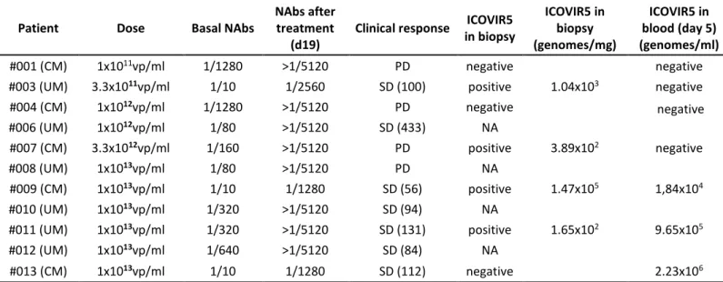
Neutralizing antibodies
The levels of Anti-Ad5 neutralizing antibodies were analyzed before treatment and day 19 post-treatment in sera (Table 3). All patients developed an antiviral immune response as shown by the increase of neutralizing antibodies after virus administration regardless of the basal level and the dose of ICOVIR-5 administered. The increase of titer did not correlate with the dose of ICOVIR-5 administered. Only the three patients with lower basal levels of NAbs did not reach the upper limit of quantification (>5120). Finally, for the highest dose cohort, where stable diseases were seen, no correlation was observed between the pre-existence of basal neutralizing antibodies and the stabilization of the disease using Fisher’s exact test (p=1).
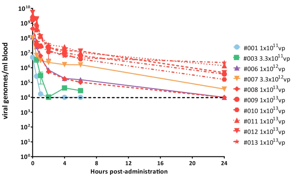
Pharmacokinetics of ICOVIR-5 in patients’ blood was monitored for the first 24h post-administration by Quantitative Real Time-PCR using the specific set of primers for the ICOVIR-5 genome. The level of ICOVIR-5 in blood correlated with the dose administered (Fig 1). Patient #01 treated with 1x10¹¹vp and patient #03 treated with 3.3x10¹¹vp had circulating viral genomes up to 1h (1.69x10⁴genomes/ml) and 6h (2.85x10⁴genomes/ml) post-administration, respectively (patient #01 had detectable but not quantifiable at 6h), with no evidence of viral genomes in blood after 24h post-administration. Patient #06 treated with 1x10¹²vp had quantifiable viral genomes at 6h post-administration (1.56x10⁵genomes/ml) and detectable levels at 24h post-administration. Patient #07 administered with 3.3x10¹²vp showed quantifiable viral genomes at 24h post-treatment (3.61x10⁴genomes/ml). Finally, 5 out of the 6 patients of the last cohort (treated at 1x10¹³vp) had quantifiable virus genomes at 24h post-treatment (9.18x10⁵genomes/ml ± 8.67x10⁵ genomes/ml), with only one patient (patient #08) with detectable but not quantifiable levels at this time point.
Virus shedding
In all patients from the lower dose levels (one patient per each dose of 1x10¹¹, 3.3x10¹¹, and 1x10¹²vp) ICOVIR-5 genomes could not be detected in any of the samples analyzed (blood, urine, sputum, and stool).
In patient #07 treated with 3.3x10¹²vp virus DNA was detected in blood (although not quantifiable) and in sputum (3.96x10³ genomes/ml) at 48h post-treatment. Both types of samples became negative for the presence of virus in the next time point analyzed, at 5 days post-treatment, and remained negative.
In patients treated at the highest dose level (1x10¹³vp) an increase in the virus shedding was detected (Fig 2). In blood, 24h post-treatment 5 out of the 6 patients presented quantifiable viral genomes (9.18x10⁵genomes/ml ± 8.67x10⁵genomes/ml) and one patient (patient #08) presented detectable but not quantifiable levels. At 48h post-treatment, 4 out of 5 patients had quantifiable
virus genomes (2.90x10⁵genomes/ml ± 3.70x10⁵genomes/ml) and one patient (patient #08) had detectable but not quantifiable levels. At 5 days post-treatment, 4 out of 6 patients had quantifiable viral genomes (8.64x10⁵genomes/ml ± 9.94x10⁵genomes/ml) and the remaining 2 patients (patients #08 and #10) had detectable but not quantifiable levels. Virus genomes could be detected up to day 6 (patients #08 and #11) or day 8 (patients #09 and #10) after virus administration. Surprisingly, in the remaining 2 patients (patients #012 and #013), virus genomes were detected until the end of the treatment (day 26 after virus administration). In urine, virus DNA was detected only in patient #09. This patient had 1.16x10⁶genomes/ml at 24h, 1.51x10⁵genomes/ml at 48h, and 2.16x10⁵genomes/ml at day 5 after treatment. In stool, 24h post-administration virus genomes were detected in 4 out of 4 patients analyzed (patients #09, #010, #011, and #012, 5.89x10⁴genomes/g ± 4.35x10⁴genomes/g); 48h post-administration virus was detected in 2 (#09 and #012) out of 3 (patients #09, #010 and #012) analyzed (6.98x10⁴genomes/g ± 3.90x10⁴genomes/g); and at day 5 post-administration virus was detected in 1 (patient #013) out of 3 (patients #09, #012 and #013), but did not reach a quantifiable level. Finally, in sputum at 24h post-administration viral genomes were detected in 2 (patients #09 and #010) out of 3 (patients #08, #09 and #010) samples analyzed (6.09x10⁴genomes/ml ± 5.87x10⁴genomes/ml); at 48h post-administration in 2 (patients #010 and #012) out of 4 (patients #08, #09, #010 and #012) samples analyzed (3.43x10⁴genomes/ml and one not quantifiable); and at day 5 post-administration in 1 (patient #012) out of 5 (patients #08, #09, #010, #012, and #013) samples analyzed (3.91x10⁴genomes/ml). All samples from days 12, 19, and 26 were negative.
Cytokine analysis
Analysis of serum IL6 and IL10 levels was performed before and after treatment (6h, 24h, 48h, day 5, day 12, day 19, day 26 post-ICOVIR-5 administration) (Fig 3). An increase of IL6 levels was detected for all patients in the first 24h post-administration, likely reflecting an early innate immune response against the virus. At 6h post-administration, there was a correlation between
the viral dose used and the IL6 level detected (22.6 pg/ml for 1x10¹¹vp, 28.8 pg/ml for 3.3x10¹¹vp, 31.5 pg/ml for 1x10¹²vp, 49.9 pg/ml for 3.3x10¹²vp, and a mean of 303 pg/ml for 1x10¹³vp; lineal regression IL6 concentration versus amount of ICOVIR-5 administered, r=0.97 p<0.005). The peak of IL6 level observed for most patients was lower than 100 pg/ml. Only 3 patients showed a peak higher than 200 pg/ml (patients #01, #08, #010). Among them, only in patient #08 (1x10¹³vp) this higher IL6 elevation was accompanied with a dose-limiting toxicity, as a transaminitis grade 3 at 24h post-treatment. Serum IL10 was also measured as it is another biomarker of inflammation commonly detected upon systemic adenovirus administrations, although with anti-inflammatory properties contrary to the pro-inflammatory IL6. A peak of IL10 was detected in all patients, reaching higher levels with higher doses. As previously reported, the peak of IL10 (24h) was delayed compared to IL6 (6h). 15 At 24h post-administration, there was a correlation between the viral dose and the IL-10 level detected (lineal regression IL10 concentration versus amount of ICOVIR-5 administered, r=0.81; p<0.05). Patient #013 (1x10¹³vp) had the highest IL10 peak (530 pg/ml) and showed a dose-limiting toxicity as a transaminitis grade 3 at 24h post-treatment. There was a correlation of IL6 levels at 6h and IL10 at 24h (r=0.93; p<0.05).
Detection of ICOVIR-5 in tumor biopsies
One core needle biopsy (CNB) was obtained from skin lesions of cutaneous melanoma patients and liver lesions of uveal melanoma patients. Biopsies were obtained at day 5 post-treatment in patients treated with 1x10¹¹vp, 3.3x10¹¹vp, 3.3x10¹²vp, and 3 (patients #09, #011 and #013) out of the 6 patients from the highest dose of 1x10¹³vp. DNA was extracted from biopsies and the presence of viral genomes analyzed by qPCR using specific primers for ICOVIR-5. Virus DNA in metastasis was not detected in the patient treated with the lower 1x10¹¹vp dose. In contrast, virus DNA was detected in patients treated with 3.3x10¹¹vp and 3.3x10¹²vp, and in 2 (patients #09 and #011) out of 3 patients from the highest 1x10¹³vp dose (Table 3). In these 2 patients the presence of virus DNA in blood at the time of biopsy (day 5 post-administration) precludes a clear
conclusion on tumor transduction. However, in the 2 patients with lower virus doses (3.3x10¹¹vp and 3.3x10¹²vp) no viral DNA was detected in peripheral blood at day 5 post-administration suggesting that the detected virus derives from tumor cells and not from blood.
DISCUSSION
The use of oncolytic viruses to treat cancer, known as virotherapy, is an old idea born after the observation of cancer remissions upon viral infections. 16 After up and downs, the concept has been revisited in the last 20 years using viruses, such as Vaccinia Virus, Herpes Simplex Virus (HSV), Measles, and Adenoviruses (Ad), designed to target and replicate more selectively in cancer cells, and wild type viruses with a natural selectivity for tumor cells such as Reovirus, Parvovirus, or Coxsackievirus A21, among others. Intratumoral injection of these viruses in patients has proven the concept that when a virus reaches a tumor mass it can cause tumor lysis. 17 However, tumor targeting upon systemic administration is still a challenge.
This first-in-humans phase 1 study shows that the intravenous administration of the conditionally replicative adenovirus ICOVIR-5 is well tolerated. A dose of 1x10¹³vp of ICOVIR-5 induced transaminitis in 2 out of 6 patients, and thrombocytopenia in one of them, while a dose of 3.3x10¹²vp can be safely administered without
DLT.
The primary objective of the trial was to recommend a phase II dose. According to the protocol, the dose level in which no DLT was observed would be taken as a safe dose level and therefore the recommended phase II dose. Our results indicate that for ICOVIR5 this dose is 3.3x10¹² vp.Mild liver toxicity has been previously described with similar intravenous administered viruses. 15, 18, 19 ICOVIR-7, an oncolytic adenovirus similar to ICOVIR-5, produced up to grade 2 liver transaminases elevation in 13 of 21 patients, although most of the patients received treatment by intratumoral injection and only 3 intravenously. 20 For ICOVIR-5 transaminitis was the main toxicity limiting the dose escalation beyond the 1x10¹³vp dose. This transient transaminitis observed in 2 patients, started at days 1 and 5 post-treatment, and persisted for 1 and 7 days, respectively.Systemic administration has been attempted in clinical trials using Newcastle Disease Virus HSV, reovirus, picornavirus, measles, Vaccinia Virus 21 and adenovirus. 19 Out of eight evaluated
patients in a recent trial of an oncolytic Vaccinia Virus administered systemically, two proved positive for virus in biopsy. 21 Still, the inadequate viral delivery was identified as a main factor for insufficient antitumor efficacy. Adenovirus has been extensively used in gene therapy and virotherapy clinical trials. Among several clinical trials with systemically administered Onyx015 only occasional and transient responses were observed in some colorectal carcinoma patients. 22, 23 With CG7870, an oncolytic adenovirus designed for prostate carcinoma with E1A under the probasin promoter and E1B under the PSA promoter, the serum PSA level dropped 25% to 49% transiently in 5 out of the 23 intravenously treated patients, 4 of them at the highest doses (6x10¹¹ to 6x10¹² vp). 19 Focusing on tumor targeting, occasional evidence for systemic tumor targeting has been reported. Reid et al. injected ONYX-015 intra-arterially in 11 patients with liver metastases of colorectal cancer and biopsies at day 4 post-infusion resulted negative for virus. 22 Similar results were obtained in the phase II trial after infusing 2x10¹²vp as a maximum feasible dose on multiple days (1, 8, 22, 50, and 78). 23 It was concluded that improving delivery and replication efficiency would be needed to obtain evidences of intratumoral replication. Hamid et al. infused intravenously ONYX-015 to 18 patients with liver metastases of colorectal carcinoma. 18 The autopsy of one patient that died 56 h after virus administration showed very few cells positive for virus in the tumors. Nemunaitis et al. infused ONYX-015 intravenously in 10 patients with lung metastases reaching much higher doses (up to 2x10¹³vp). One analyzed biopsy in a patient treated with 2x10¹²vp resulted positive for virus presence 5 days post-infusion. 15 A more potent virus, CG7870, where E1A and E1b were regulated by the probasin and the PSA promoters respectively, was infused intravenously in prostate carcinoma patients up to a dose of 6x10¹²vp, identified as the MTD, but no biopsies were analyzed for tumor targeting. 19 Lack of antitumor efficacy in these clinical trials led to abandon the systemic approach with oncolytic adenoviruses based on adenovirus serotype 5 with an unmodified capsid. After an interval of several years, a survey of clinical trials ongoing in 2014 identified ICOVIR-5, ColoAd1, and VCN01 (all of them with capsids different from Ad5-wild type) as oncolytic Ads used intravenously, and no additional
adenoviruses were added to this list in 2015. 24, 25 However, the Ad-D24RGD oncolytic adenovirus, with the same RGD-modified capsid as ICOVIR-5, has been used intratumorally 26, intraperitoneally 27, and in a combination of intratumoral and intravenous routes 20, 28. Evidence of systemic tumor targeting in non-injected tumors distant from injected tumors was observed in an autopsy of one patient (C200) that died 19 days after injection in multiple tumor sites. 28 With regard to oncolytic adenoviruses with other capsid modifications, metastatic tumor targeting has been demonstrated in a patient treated intravenously with a serotype 5/3 chimeric fiber. 28 The biodistribution analysis in patient autopsy samples indicated a fair capability of tumor targeting of oncolytic adenoviruses even when injected intratumorally. 28 This consistent detection of virus in occasional patient autopsies suggests that the small samples that can be analyzed in a biopsy may underestimate the tumor targeting potential of the virus. In this context it is noteworthy our detection of viral genomes in four out of seven analyzed biopsies, in two cases with a negative viremia. The same can be concluded from the more frequently reported observation of secondary peaks of viremia compared to positive biopsies, which suggest virus replication in tumors. We did not observe secondary peaks of viremia even in the four patients where the virus had been detected in the tumor at day 5. In two patients (#012 and #013) viremia levels were positive until the end of the study (day 26). Among them, one (patient #013) had a tumor biopsy analyzed at day 5 and it resulted negative. Although preliminary, our results indicate that the presence of virus in the tumor does not necessarily leads to secondary peaks of viremia.
In our study, pharmacokinetics analysis indicates that there is a correlation between exposure and the administered dose, since larger doses lead to longer time to virus elimination. In addition, the study shows that the virus shedding is also dose-dependent: the presence of the virus is higher in the last dose level, but at the recommended dose level it is found in blood and sputum at 48 hours, indicating effective spread of the virus. The analysis shows that neutralizing antibody titers were not dose-dependent and pretreatment levels did not correlate with tumor response.
All patients had an early onset of innate immune response, with a correlation between dose and levels of IL6. As reported, the peak of IL10 was delayed compared to IL6. IL6 and IL10 levels correlated with the dose and between them. Patients with grade 3 transaminitis (patients #08 and #013) showed highest peaks of IL6 and IL10. This toxicity was very short in time and no systemic inflammatory response syndrome was observed.
As a most relevant finding, viral genomes were detected in the biopsy of metastatic lesions of four patients, indicating that oncolytic adenovirus can achieve tumor targeting of metastases upon intravenous administration. With regards to clinical activity, no tumor responses were observed, but 7 stabilizations of disease were seen out of 11 patients, 5 of them among the 6 patients treated at the highest dose. Despite this low antitumor activity, the longer survival of uveal melanoma patients compared to cutaneous melanoma patients was intriguing as the expected survival before the emergence of targeted therapy (BRAF/MEK inhibitors) and anti-PD1 antibodies for those patients should be the same. 29 Although the number of patients is very small and definitive conclusions cannot be taken, this trend suggests a superior activity of systemic ICOVIR5 against metastatic uveal melanoma compared to metastatic cutaneous melanoma. It could be speculated that this could be related to the shared hepatotropism of adenovirus and uveal melanoma metastases or/and that uveal melanoma liver metastases are more prone to be controlled by the immune system once infected by the virus. Low antitumor efficacy has been previously reported with oncolytic adenoviruses. By intratumoral administration head and neck patients ONYX-015 produced 3 partial and 2 minor responses and 8 disease stabilizations. 30 In 23 pancreatic cancer patients produced six minor responses and 10 stabilizations. 31 In the phase 1 study of intratumoral Telomelysin in various solid tumors, one partial and seven stable disease responses were reported. 32 By intraperitoneal injection of Ad5-∆24-RGD, 15 disease stabilizations were observed among 21 malignant gynecologic patients. 27Finally, among 35 non-invasive bladder cancer patients receiving CG0070 intravesically, 17 showed complete responses. 33 By
intravenous administration, ONYX-015 induced one mixed response and 9 disease stabilizations in 10 patients. 15 In a subsequent phase II trial, seven out to 18 patients experienced stable disease. 18 Systemic administration of CG7870 in 23 prostate cancer patients induced some decrease in serum PSA in 5 patients, with no complete or partial PSA responses. 19 With ICOVIR7 20, out of two evaluated patients that were treated only by the intravenous route, one head and neck tumor patient had a 10% decrease of tumor size.
There is little experience with oncolytic adenoviruses applied to melanoma, and none after systemic administration alone. When oncolytic adenovirus Ad5/3-D24-GMCSF was administered to 9 patients with melanoma (50% of dose intratumoral and 50% intravenous), one out of four evaluable patients showed a minor response (15% size decrease) in an injected lesion. 34
Our study adds interesting information to the development of virotherapy with oncolytic adenoviruses. One point of growing interest is the potential combination of oncolytic viruses with immunotherapy, as the virus-induced immune response may favor the infiltration of lymphocytes in tumors. In this setting, it is imperative for the virus to reach all tumor sites systemically to promote the infiltration on “cold” tumors characterized by the lack of tumor-infiltrating lymphocytes. Our results indicate that systemic tumor targeting of metastases with oncolytic adenoviruses is feasible using the intravenous administration route. Although armed oncolytic adenoviruses may be needed to obtain sufficient tumor debulking by oncolysis before antiviral immunity neutralizes the virus, our results encourage further studies by systemic administration. We are currently exploring the use of a hyaluronidase-armed oncolytic adenovirus (VCN-01) with a slightly different capsid than ICOVIR-5 (RGD in the fiber shaft instead of the HI-loop) by systemic administration in cancer patients.
ACKNOWLEDGEMENTS
Thanks to Mei Zhuyong, Deborah L. Lyon, and Suzanne M. Poole from the Center for Cell and Gene Therapy at Baylor College of Medicine for GMP production of ICOVIR-5; ICO nurses Olimpia
Garcia and Susana Lorente for caring of patients; Maria Perayre from the ICO Pharmacy for her support in ICOVIR-5 storage and processing; Enric Espunya, Nuria Arilla and Josep Ferrés from HIPRA for importing ICOVIR-5; Isabel Gomez at IDIBELL and Nuria Benitez at ICO for project management; Mercè Monfar and Miguel Chillon from the Vector Unit Production of the Center of Animal Biotechnology and Gene Therapy of Bellaterra for ICOVIR-5 production and characterization; Manuel Ramirez, Luis Madero, and Javier García Castro for sharing FIS grants for copayment for GMP ICOVIR-5production; and Sol Ruiz from the Spanish Drug Agency for regulatory guidance.
REFERENCES
1.
International Agency for Research on Cancer. GLOBOCAN 2012. Estimated
Cancer Incidence, Mortality, and Prevalence Worlwide in 2012 2012;Lyon, France:
International Agency for Research on Cancer:Available from: http://globocan.iarc.fr.
Accessed July 1, 2016.
2.
Chapman PB, Einhorn LH, Meyers ML et al. Phase III multicenter randomized trial
of the Dartmouth regimen versus dacarbazine in patients with metastatic melanoma.
Journal of clinical oncology : official journal of the American Society of Clinical
Oncology 1999;17:2745-2751.
3.
Hauschild A, Grob JJ, Demidov LV et al. Dabrafenib in BRAF-mutated metastatic
melanoma: a multicentre, open-label, phase 3 randomised controlled trial. Lancet
2012;380:358-365.
4.
Flaherty KT, Robert C, Hersey P et al. Improved survival with MEK inhibition in
BRAF-mutated melanoma. The New England journal of medicine 2012;367:107-114.
5.
Long GV, Stroyakovskiy D, Gogas H et al. Combined BRAF and MEK inhibition
versus BRAF inhibition alone in melanoma. The New England journal of medicine
2014;371:1877-1888.
6.
Hodi FS, O'Day SJ, McDermott DF et al. Improved survival with ipilimumab in
patients with metastatic melanoma. N Engl J Med 2010;363:711-723.
7.
Robert C, Long GV, Brady B et al. Nivolumab in previously untreated melanoma
without BRAF mutation. N Engl J Med 2015;372:320-330.
8.
Robert C, Ribas A, Wolchok JD et al. Anti-programmed-death-receptor-1 treatment
with pembrolizumab in ipilimumab-refractory advanced melanoma: a randomised
dose-comparison cohort of a phase 1 trial. Lancet 2014;384:1109-1117.
9.
Andtbacka RH, Kaufman HL, Collichio F et al. Talimogene Laherparepvec
Improves Durable Response Rate in Patients With Advanced Melanoma. J Clin Oncol
2015;33:2780-2788.
10.
Uusi-Kerttula H, Hulin-Curtis S, Davies J et al. Oncolytic Adenovirus: Strategies
and Insights for Vector Design and Immuno-Oncolytic Applications. Viruses
2015;7:6009-6042.
11.
Alemany R, Cascallo M. Oncolytic viruses from the perspective of the immune
system. Future microbiology 2009;4:527-536.
12.
Suzuki K, Fueyo J, Krasnykh V et al. A conditionally replicative adenovirus with
enhanced infectivity shows improved oncolytic potency. Clin Cancer Res 2001;7:120-126.
13.
Cascallo M, Alonso MM, Rojas JJ et al. Systemic toxicity-efficacy profile of
ICOVIR-5, a potent and selective oncolytic adenovirus based on the pRB pathway.
Molecular therapy : the journal of the American Society of Gene Therapy
2007;15:1607-1615.
14.
Alemany R, Curiel DT. CAR-binding ablation does not change biodistribution and
toxicity of adenoviral vectors. Gene Ther 2001;8:1347-1353.
15.
Nemunaitis J, Cunningham C, Buchanan A et al. Intravenous infusion of a
replication-selective adenovirus (ONYX-015) in cancer patients: safety, feasibility and
biological activity. Gene Ther 2001;8:746-759.
16.
Russell SJ, Peng KW, Bell JC. Oncolytic virotherapy. Nature biotechnology
2012;30:658-670.
17.
Kaufman HL, Kohlhapp FJ, Zloza A. Oncolytic viruses: a new class of
immunotherapy drugs. Nature reviews Drug discovery 2015;14:642-662.
18.
Hamid O, Varterasian ML, Wadler S et al. Phase II trial of intravenous CI-1042 in
patients with metastatic colorectal cancer. Journal of clinical oncology : official journal of
the American Society of Clinical Oncology 2003;21:1498-1504.
19.
Small EJ, Carducci MA, Burke JM et al. A phase I trial of intravenous CG7870, a
replication-selective, prostate-specific antigen-targeted oncolytic adenovirus, for the
treatment of hormone-refractory, metastatic prostate cancer. Molecular therapy : the
journal of the American Society of Gene Therapy 2006;14:107-117.
20.
Nokisalmi P, Pesonen S, Escutenaire S et al. Oncolytic adenovirus ICOVIR-7 in
patients with advanced and refractory solid tumors. Clin Cancer Res 2010;16:3035-3043.
21.
Downs-Canner S, Guo ZS, Ravindranathan R et al. Phase 1 Study of Intravenous
Oncolytic Poxvirus (vvDD) in Patients With Advanced Solid Cancers. Molecular therapy :
the journal of the American Society of Gene Therapy 2016;24:1492-1501.
22.
Reid T, Galanis E, Abbruzzese J et al. Intra-arterial administration of a
replication-selective adenovirus (dl1520) in patients with colorectal carcinoma metastatic to the liver:
a phase I trial. Gene Ther 2001;8:1618-1626.
23.
Reid T, Galanis E, Abbruzzese J et al. Hepatic arterial infusion of a
replication-selective oncolytic adenovirus (dl1520): phase II viral, immunologic, and clinical
endpoints. Cancer research 2002;62:6070-6079.
24.
Pol J, Bloy N, Obrist F et al. Trial Watch:: Oncolytic viruses for cancer therapy.
Oncoimmunology 2014;3:e28694.
25.
Pol J, Buque A, Aranda F et al. Trial Watch-Oncolytic viruses and cancer therapy.
Oncoimmunology 2016;5:e1117740.
26.
Lang FF, Conrad C, Gomez-Manzano C et al. Phase I Clinical Trialof Oncolytic
Virus Delta-24-RGD (DNX-2401) with Biological Endpoints: Implications for
Viro-Immunotherapy. In: Neuro Oncol. 2014; pp. v159-v167.
27.
Kimball KJ, Preuss MA, Barnes MN et al. A phase I study of a tropism-modified
conditionally replicative adenovirus for recurrent malignant gynecologic diseases. Clin
Cancer Res 2010;16:5277-5287.
28.
Koski A, Bramante S, Kipar A et al. Biodistribution Analysis of Oncolytic
Adenoviruses in Patient Autopsy Samples Reveals Vascular Transduction of Noninjected
Tumors and Tissues. Molecular therapy : the journal of the American Society of Gene
Therapy 2015;23:1641-1652.
29.
Kuk D, Shoushtari AN, Barker CA et al. Prognosis of Mucosal, Uveal, Acral,
Nonacral Cutaneous, and Unknown Primary Melanoma From the Time of First Metastasis.
The oncologist 2016;21:848-854.
30.
Ganly I, Kirn D, Eckhardt G et al. A phase I study of Onyx-015, an E1B attenuated
adenovirus, administered intratumorally to patients with recurrent head and neck cancer.
Clin Cancer Res 2000;6:798-806.
31.
Mulvihill S, Warren R, Venook A et al. Safety and feasibility of injection with an
E1B-55 kDa gene-deleted, replication-selective adenovirus (ONYX-015) into primary
carcinomas of the pancreas: a phase I trial. Gene Ther 2001;8:308-315.
32.
Nemunaitis J, Tong AW, Nemunaitis M et al. A phase I study of
telomerase-specific replication competent oncolytic adenovirus (telomelysin) for various solid tumors.
Molecular therapy : the journal of the American Society of Gene Therapy
2010;18:429-434.
33.
Burke JM, Lamm DL, Meng MV et al. A first in human phase 1 study of CG0070,
a GM-CSF expressing oncolytic adenovirus, for the treatment of nonmuscle invasive
bladder cancer. J Urol 2012;188:2391-2397.
34.
Bramante S, Kaufmann JK, Veckman V et al. Treatment of melanoma with a
serotype 5/3 chimeric oncolytic adenovirus coding for GM-CSF: Results in vitro, in
rodents and in humans. International journal of cancer 2015;137:1775-1783.
Figure legends
Figure 1. Quantification of ICOVIR-5 pharmacokinetics in blood by Real-Time PCR. In dose levels
1a, 2a, 3a and 4a only viral dose are indicated. For dose level 5a, patient number identification is also indicated. The dashed lines represent the lower limit of quantification of ICOVIR-5 genomes in blood. Samples were analyzed in triplicates.
Figure 2. Quantification of ICOVIR-5 genomes in high-dose patients (1x10¹³vp) by Real-Time PCR in: A) Blood, B) Urine, C) Stool and D) Sputum. The dashed lines represent the lower limit of
quantification (LOQ) and detection (LOD) of ICOVIR-5 genomes. Samples were analyzed in triplicates. # : Sample detectable but not quantifiable.
Figure 3. Analysis of IL6 and IL10 in serum samples by ELISA. Analysis of IL6 (A) and IL10 (B) in
serum samples on days 0, 1, 2, 5, 12, 19 and 26 post-ICOVIR-5 administration. Samples were analyzed in triplicates.
Figure 1. García et al
0
4
8
12
16
20
24
10
010
110
210
310
410
510
610
710
810
910
10#001 1x10
11vp
#003 3.3x10
11vp
#006 1x10
12vp
#007 3.3x10
12vp
#008 1x10
13vp
#009 1x10
13vp
#010 1x10
13vp
#011 1x10
13vp
#012 1x10
13vp
#013 1x10
13vp
Hours post-administration
vi
ra
l g
en
om
es
/m
l b
lo
od
Blood
24h 48h Day 5 Day 1 2 Day 1 9 Day 2 6 100 101 102 103 104 105 106 107Tim e after ICOVIR -5 adm inistration
viral genome / ml
A)
Urine
24h 48h Day 5 Day 1 2 Day 1 9 Day 2 6 100 101 102 103 104 105 106 107Tim e after ICOVIR -5 adm inistration
B)
LOQ
LOQ/LOD
LOD
Stool
h h y 5 2 9 6 100 101 102 103 104 105 106 107LOQ
LOD
C)
Sputum
h h y 5 2 9 6 100 101 102 103 104 105 106 107LOQ
LOD
D)
Figure 2. García et al
#
#
viral genome / g
viral genome / ml
viral genome / ml
sa l 6h 24h 48 h y 5 y 1 2 y 1 9 y 2 6 sa l 6h 24h 48 h y 5 y 1 2 y 1 9 y 2 6 sa l 6h 24h 48 h y 5 y 1 2 y 1 9 y 2 6 sal 6h 24 h 48 h y 5 y 1 2 y 1 9 y 2 6 sal 6h 24 h 48 h y 5 y 1 2 y 1 9 y 2 6 sal 6h 24 h 48 h y 5 y 1 2 y 1 9 y 2 6 sal 6h 24 h 48 h y 5 y 1 2 y 1 9 y 2 6 sal 6h 24 h 48 h y 5 y 1 2 y 1 9 y 2 6 sal 6h 24 h 48 h y 5 y 1 2 y 1 9 y 2 6 sal 6h 24 h 48 h y 5 y 1 2 y 1 9 y 2 6 0 25 50 75 100 300 350 400 450 500 550
IL 1 0
pg /m lA)
B)
Figure 3 García et al
ba sa l 6h 24h 48h day 5 da y 1 2 da y 1 9 da y 2 6 ba sa l 6h 24h 48h day 5 da y 1 2 da y 1 9 da y 2 6 ba sa l 6h 24h 48h day 5 da y 1 2 da y 1 9 da y 2 6 ba sa l 6h 24h 48h day 5 da y 1 2 da y 1 9 da y 2 6 ba sa l 6h 24h 48h day 5 da y 1 2 da y 1 9 da y 2 6 ba sa l 6h 24h 48h day 5 da y 1 2 da y 1 9 da y 2 6 ba sa l 6h 24h 48h day 5 da y 1 2 da y 1 9 da y 2 6 ba sa l 6h 24h 48h day 5 da y 1 2 da y 1 9 da y 2 6 ba sa l 6h 24h 48h day 5 da y 1 2 da y 1 9 da y 2 6 ba sa l 6h 24h 48h day 5 da y 1 2 da y 1 9 da y 2 6 0 20 40 60 80 100 200 300 400 500 600 700IL 6
pg /m l #3 3.3X10¹¹vp #1 1X10¹¹vp #6 1X10¹²vp #7 3.3X10¹²vp #8 1X10¹³vp #9 1X10¹³vp #10 1X10¹³vp #11 1X10¹³vp #12 1X10¹³vp #13 1X10¹³vpSupplementary figure 1. Kaplan-Meier survival analysis of cutaneous (n=4) vs uveal (n=6) melanoma patients.
Table 1. Dose levels and DLTs
Patient Dose (vp) Level Toxicity DLT Decision taken by investigators’ committee
#01 1x10¹¹ 1a Grade 3 lymphopenia
Grade 2 neutropenia No
Grade 3 toxicity not associated to treatment Decision: escalation to the next level
#02 3.3x10¹¹ 2a Grade 3 asthenia No The patient did not complete the cycle.
Decision: replacement of patient #03 3.3x10¹¹ 2a Grade 1 toxicity No Decision: escalation to the next level
#04 1x10¹² 3a Grade 2 thrombocytopenia No
Grade 3 thrombocytopenia out of the cycle. By relevance, the decision is replacement of patient.
#06 1x10¹² 3a Grade 3 neutropenia No Transient (<24 hours) grade 3 toxicity.
Decision: escalation to the next level #07 3.3x10¹² 4a < Grade 2 toxicity No Grade 2 unrelated asthenia
Decision: escalation to the next level #08 1x10¹³ 5a Grade 3 transaminitis
Grade 3 thrombocytopenia Yes Expansion of cohort by DLT
#09 1x10¹³ 5a No
#10 1x10¹³ 5a No
#11 1x10¹³ 5a No
#12 1x10¹³ 5a No
#13 1x10¹³ 5a Grade 3 transaminitis Yes End of study due to two DLT.
MTD declared level Vp: viral particles, DLT: dose limiting toxicity; MTD: maximum tolerated dose
Table 2. Number of patients with nonhematologic toxicity, considering the worst grade toxicity for each patient (n=12).
Toxicity (grade) Symptoms 1 2 3 4 Fever 8 1 0 0 Stomatitis 1 1 0 0 Myalgia/arthralgia 2 1 0 0 Headache 1 0 0 0 Asthenia 4 2 1 0 Rash 2 0 0 0 Diarrhea 1 0 0 0 Constipation 1 0 0 0 Hypertension 1 1 0 0 Edema 1 0 1 0 Nausea/ Vomiting 2 0 0 0 Renal failure 0 1 0 0 Chills 2 0 0 0 Transaminitis 0 1 2 0 Hypocalcemia 0 1 0 0 Hypophosphatemia 0 1 0 0 Hypomagnesemia 1 0 0 0 Hypokalemia 1 0 0 0
Table 3. Anti-Ad5 neutralizing antibodies, clinical responses, and viral genomes in biopsies.
Patient Dose Basal NAbs
NAbs after treatment
(d19)
Clinical response ICOVIR5 in biopsy ICOVIR5 in biopsy (genomes/mg) ICOVIR5 in blood (day 5) (genomes/ml) #001 (CM) 1x10¹¹vp/ml 1/1280 >1/5120 PD negative negative
#003 (UM) 3.3x10¹¹vp/ml 1/10 1/2560 SD (100) positive 1.04x10³ negative
#004 (CM) 1x10¹²vp/ml 1/1280 >1/5120 PD negative negative #006 (UM) 1x10¹²vp/ml 1/80 >1/5120 SD (433) NA #007 (CM) 3.3x10¹²vp/ml 1/160 >1/5120 PD positive 3.89x10² negative #008 (UM) 1x10¹³vp/ml 1/80 >1/5120 PD NA #009 (CM) 1x10¹³vp/ml 1/10 1/1280 SD (56) positive 1.47x10⁵ 1,84x10⁴ #010 (UM) 1x10¹³vp/ml 1/320 >1/5120 SD (94) NA #011 (UM) 1x10¹³vp/ml 1/320 >1/5120 SD (131) positive 1.65x10² 9.65x10⁵ #012 (UM) 1x10¹³vp/ml 1/640 >1/5120 SD (84) NA #013 (CM) 1x10¹³vp/ml 1/10 1/1280 SD (112) negative 2.23x10⁶
Patient: CM, cutaneous melanoma; UM, uveal melanoma. Nabs: anti-adenovirus neutralizing antibodies. >1/5120 upper limit of detection, Vp: viral particles, PD: progressive disease, SD: stable disease (until day indicated); NA: not available.