Francesca Piras
Techniques of Immunoflourescence and Confocal Microscopy applied to the study of Syngnathids gonadal structure And development and to the Dopaminergic control of the reproduction in Teleosts
PhD Thesis in Environmental Biology – University of Sassari, 2014 – XXVII cycle
Contents
Abstract
Riassunto
Introduction
Reproductive Biology
Dopaminergic control of reproduction
List of Papers
I.
Biagi , F., Piras, F., Farina, V., Zedda, M., Mura, E., Floris, A., Franzoi, P., Fausto A. M., Taddei, A. R. and Carcupino, M. 2014. Testis structure, spermatogenesis and sperm morphology in pipefishes of the genus Syngnathus. – Acta Zoologica doi: 10.1111/azo.1210825
II.
Piras, F., Biagi, F., Floris, A., Farina,V., Zedda, M., Franzoi, P. and Carcupino M.Intra- and inter-males variability of mature sperm traits analysed in two brackish water populations of the pipefish Syngnathus abaster (Syngnathidae).
(Submitted and Revised)
38
III.
Piras, F., Biagi, F., Taddei, A.R., Fausto, A.M., Farina,V., Zedda, M., Floris, A., Franzoi, P. and Carcupino M. Male gonads morphology, spermatogenesis and sperm ultrastructure of the seahorse Hippocampus guttulatus (Syngnathidae).(Manuscript)
65
4
5
6
6
14
3
Francesca Piras
Techniques of Immunoflourescence and Confocal Microscopy applied to the study of Syngnathids gonadal structure And development and to the Dopaminergic control of the reproduction in Teleosts
PhD Thesis in Environmental Biology – University of Sassari, 2014 – XXVII cycle
IV.
Biagi, F., Piras, F., Franzoi, P., Taddei, A.R., Fausto, A.M., Farina,V., Zedda, M., Floris, A., and Carcupino, M. Morphology and ultrastructure of the male gonad in the straight-nosed pipefish Nerophis ophidion (Syngnathidae).(Manuscript)
92
V.
Piras, F., Carcupino, M., Caboni, F., Biagi, F. and Spiga S.Characterization of the Dopaminergic system in adult and young males of pipefish Syngnathus. (Manuscript)122
Francesca Piras
Techniques of Immunoflourescence and Confocal Microscopy applied to the study of Syngnathids gonadal structure And development and to the Dopaminergic control of the reproduction in Teleosts
PhD Thesis in Environmental Biology – University of Sassari, 2014 – XXVII cycle
Abstract
Reproduction is an indispensable function for the perpetuation of the species and is perfomed with extraordinarily diverse reproductive strategies (adaptations that improve the chances of fertilization and/or increase the survaival rate of offsprings), each of wich is under control of a sophisticated network of regulatory signals. The array of resporductive strategies among teleosts are extraordinarily diverse and, among them, those of syngnathids are peculiar. The Syngnathidae are a small family of brackish and freshwater species. They have attracted attention for decades due to their unique morphology, remarkable camouflage ability and the distinctive phenomenon of male pregnancy. Females deposit eggs, on or into a male incubation area, on the tail (subfamily Urophori) or on the trunk (subfamily Gastrophori). Syngnathids Mediterranean species only belong to Syngnathus, Hippocampus and Nerophis genera. Very little is known about the male reproductive biology and on the nervous system control on the reproductibe cycle of these fish. The aims of this work are therefore to clarify: 1) relevant aspects of male reproduction in both internal and external brooder species, such as those belongin to Syngnathus and Hippocanous (Urophori) and Nerophis genera (Gastrophori); 2) characterize the Dopaminergic system in adult and young males of the pipefish Syngnathus abaster. The aims have been achieved using different techniques of light, confocal and electron microscopy.
5
Francesca Piras
Techniques of Immunoflourescence and Confocal Microscopy applied to the study of Syngnathids gonadal structure And development and to the Dopaminergic control of the reproduction in Teleosts
PhD Thesis in Environmental Biology – University of Sassari, 2014 – XXVII cycle
Riassunto
La riproduzione è una funzione indispensabile per la conservazione della specie ed è attuata attraverso differenti strategie riproduttive controllate da una sofisticata rete segnali di regolatori. Nei teleostei troviamo strategie riproduttive straordinariamente differenti, tra queste, quelle dei Singnatidi sono particolarmente peculiari. I Syngnathidi sono una piccola famiglia d'acqua dolce, marina e salmastra. Sono particolarmente studiati per la loro affascinante morfologia, per la straordinaria capacità di mimetizzarsi e per il fenomeno della gravidanza maschile. Le femminile depositano le uova all’interno di un area incubatrice, sulla coda (sottofamiglia Urophori) o sulla superficie ventrale del tronco (sottofamiglia Gastrophori) dei maschi. Nel Mediterraneo troviamo le specie appartenenti ai generi
Syngnathus, Hippocampus e Nerophis. Le informazioni a carico della biologia riproduttiva
maschile e del controllo del sistema nervoso sul ciclo riproduttivo di questi pesci sono molto scarse. Gli obiettivi di questo lavoro sono quindi chiarire: 1) gli aspetti rilevanti della riproduzione maschile in entrambe le specie a fecondazione interna ed esterna, in particolare nei generi Syngnathus, Hippocampus (Urophori) e Nerophis (Gastrophori); 2) caratterizzare il sistema dopaminergico in maschi adulti e giovani del pesce ago Singnathus abaster. Gli obiettivi di questo lavoro sono stati raggiunti utilizzando tecniche di microscopia ottica classica, confocale ed elettronica.
Francesca Piras
Techniques of Immunoflourescence and Confocal Microscopy applied to the study of Syngnathids gonadal structure And development and to the Dopaminergic control of the reproduction in Teleosts
PhD Thesis in Environmental Biology – University of Sassari, 2014 – XXVII cycle
Introduction
The general aim of my PhD thesis is to contribute to the knowledge of the male reproductive biology of some teleostean species belonging to the family Syngnathidae, studying the maturation of both male gonads and gametes and how the nervous system, and particularly the dopamine, acts in the various phases of the reproductive cycle.
Reproductive Biology
The family Syngnathidae (pipefishes, seahorses and seadragons) are well known for their highly specialized morphology, and the diversity of morphological forms. This exceptional morphological variation is reflected in the current taxonomy of the group: 14 of the 54 currently recognized syngnathid genera are monotypic (Froese and Pauly 2010), and the majority of genera are composed of fewer than three species.
The small teleostean family is also known for the remarkable adaptations for paternal care. The female deposits eggs directly onto a specialized brooding area or into a pouch of the male body (Breder and Rosen 1966). This evolutionary innovation ensures males complete confidence of paternity (Jones and Avise 1997; Jones et al., 1998, 1999), but at a level of paternal investment that exceeds that of most other vertebrates (Breder and Rosen 1966). The brooding structures vary in complexity in five steps, from: (1) a simple unprotected ventral area for gluing, (2) individual membranous egg compartments, (3) protection of eggs in a pouch with pouch plates, (4) bilateral pouch folds that grow together into a closed pouch, to (5) the most complex and completely enclosed brooding pouch of seahorses (Dawson 1985). There is a further significant difference among species in that brooding may occur on the tail (Urophori: A-type) or on the abdomen (Gastrophori: B-type)
7
Francesca Piras
Techniques of Immunoflourescence and Confocal Microscopy applied to the study of Syngnathids gonadal structure And development and to the Dopaminergic control of the reproduction in Teleosts
PhD Thesis in Environmental Biology – University of Sassari, 2014 – XXVII cycle
(Herald 1959). Among the Gastrophori, brooding structures only vary in complexity from step 1 to step 3 (Dawson 1985).
Syngnathidae has traditionally been included as a member of the order Gasterosteiformes, which includes 11 families in two suborders: the Gasterosteoidei, with the Hypoptychidae, Gasterosteidae and Aulorhynchidae; and the Syngnathoidei, with the Indostomidae, Aulostomidae, Fistulariidae, Macroramphosidae, Centriscidae, Pegasidae, Solenostomidae and Syngnathidae ( c.f. Wilson and Orr 2011).
Studies based on morphological characters have proposed monophyly of the order Gasterosteiformes and suggested sister groups based on weak evidence, and while the close relationships of super families is well supported, the relationships among these family pairs remains unclear (Pietsch, 1978; Johnson and Patterson 1993; Orr, 1995; Keivany and Nelson 2006).
In contrast, studies based on molecular data clearly refute the monophyly of the Gasterosteiformes, placing gasterosteoids close to the cottoid–zoarcoid lineage (Imamura and Yabe 2002), excluding the Indostomidae, and placing both groups distant from syngnathoids. In addition, while the sister group of syngnathoids remains unknown and the relationships among syngnathoid lineages are poorly resolved, the family Solenostomidae (ghost pipefishes) has been faithfully recovered as the sister group of the Syngnathidae in both morphological and molecular analyses (c.f. Wilson and Orr 2011).
The evolutionary relationships among members of the family Syngnathidae have been resolved with greater confidence. According to Herald (1959), the Syngnathidae diverged early in its evolution into tail (Urophori) and trunk-brooding (Gastrophori) species. Following the development of a rudimentary form of male brooding in both these lineages, brood-pouch complexity evolved in parallel in the Gastrophori and Urophori, resulting in the fully enclosed pouch of the seahorse and the highly developed brooding structures found in
Francesca Piras
Techniques of Immunoflourescence and Confocal Microscopy applied to the study of Syngnathids gonadal structure And development and to the Dopaminergic control of the reproduction in Teleosts
PhD Thesis in Environmental Biology – University of Sassari, 2014 – XXVII cycle
some gastrophorine species. Herald (1959) suggested that the brooding structures of urophorine pipefish with partially enclosed brood pouches could be further subdivided into monophyletic lineages according to their method of closure (inverted, semi-inverted, overlapping and everted) and proposed a multistage model by which the fully enclosed pouch of the seahorse was derived from pipefish ancestors with an everted brooding structure. Molecular data obtained performing mtDNA analyses (Wilson et al., 2001, 2003; Wilson and Orr 2011) supported the evolutionary model of the family characterized an early divergence of the two groups, trunk- and tail-brooding lineages, (Urophori and Gastrophori). Those data, however, suggested that several major pouch types within each of these lineages had independent evolutionary origins, challenging the phylogenetic model proposed by Herald (1959). mtDNA sequence data also supported a close evolutionary relationship between
Syngnathusand Hippocampus, contradicting Herald’s (1959) theory on the origin of the
seahorse brood pouch.The most up-to-date phylogenetic tree of the family Syngnathidae,
based on mtDNA sequence data, is provided in the paper of Wilson and Orr (2011).
The members of the family Syngnathidae inhabit coastal tropical and temperate warm waters of seas and oceans and also occur in river estuaries. They have an elongated body entirely covered by bone plates connected to each other to form belts (rings). Only 294 of the 558 nominal species of syngnathids are presently considered to be valid (Froese and Pauly 2010).
Syngnathids are present in the Mediterranean sea with only three genera: Nerophis,
Syngnathus and Hippocampus. The genus Syngnathus comprises six species, i.e. Syngnathus
acus, S. abaster, S. tenuirostris, S. phlegon, S. typhle, S.tenionotus, whereas the other two
genera are represented with only two species each, i.e. Nerophis ophidion, N. maculatus,
9
Francesca Piras
Techniques of Immunoflourescence and Confocal Microscopy applied to the study of Syngnathids gonadal structure And development and to the Dopaminergic control of the reproduction in Teleosts
PhD Thesis in Environmental Biology – University of Sassari, 2014 – XXVII cycle
immigrant from the Red Sea, and Syngnathus rostellatus, immigrant from the Atlantic Ocean have been newly recorded (Gokoglu et al., 2004).
Seahorses are exclusively marine species, whereas pipefishes, especially those belonging to the genus Syngnathus, inhabit sea and brackish waters, and in some case, even freshwaters (i.e. S. abaster).
The high level of adaptability to different habitats of the Syngnathus species is closely related to the high and particular plasticity of their body structure, which determines different local morphotypes (D’Ancona 1934; Tortonese 1970). To this, morphological plasticity may be attributed the numerous controversies on the systematic of this genus. A variable number of Mediterranean species (from sixteen to nine) was reported during the 19th century (Kaup 1856; Duméril 1870). A more accurate revision of the genus based on the analysis of 11 different morphological characters recognized in the Mediterranean and Black sea, 10 species only (D’Ancona 1934).
In the syngnathids studied to date, males of species with less complex brooding structures (e.g., Nerophis ophidion) is suggested spend less energy on their young than do those brooding embryos in enclosed pouches with placenta-like structures (Berglund et al., 1986; Masonjones 2001; Carcupino et al., 1997, 2002). According to Wilson et al. (2003), it is important to recognize that parental expenditures are not necessarily equivalent to parental investment, because expenditures such as parental guarding may not necessarily carry a fitness cost. However, in many cases, time and energy expenditures may be positively correlated and, in general, a large expenditure will often carry larger costs and therefore represent a higher parental investment. If increasing pouch complexity results in a general increase in male parental investment relative to females, male pregnancy predisposes males to limit female reproductive success; sexual selection may then operate more strongly on females and female sexual signals may evolve (sex-role reversal). Sex-role reversal, which
Francesca Piras
Techniques of Immunoflourescence and Confocal Microscopy applied to the study of Syngnathids gonadal structure And development and to the Dopaminergic control of the reproduction in Teleosts
PhD Thesis in Environmental Biology – University of Sassari, 2014 – XXVII cycle
is found in fishes, insects, amphibians and birds (Oring and Lank 1986; Simmons 1995; Berglund and Rosenqvist 2003), is characterized by intense mating competition among females, which should also favor the evolution of sexual dimorphism, where females are larger and more colorful than males. On the contrary, conventional sex role, which represents the most common pattern in nature, is characterized by male – male competition and female choice, and is traditionally viewed as being due to females limiting the reproductive potential of males.
Although several pipefishes are sex-role reversed (e.g., Nerophis ophidion,
Stigmatopora nigra, Syngnathus typhle), with females that are more vividly colored and
striped than males, some other species retain conventional sex roles (e.g., Hippichthys
penicillus) (see
R
osenqvist and Berglund 2011). One notable exception to this pattern is the genus Hippocampus, in which, although these species have the highest degree of pouch development, sex-role reversal has not yet been documented (Vincent and Sadler 1995; Kvarnemo et al., 2000; Masonjones and Lewis 2000).All species however show elaborate courtship behaviour involving only one partner (monogamy) or multiple partners (polygamy). Genetic analyses have discovered a wide variety of genetic mating patterns in pipefishes and seahorses (Jones and Avise 1997a, b, 2001; Jones et al., 1999; Wilson 2006). The broad-nosed pipefish Syngnathus typhle, for example, exhibits multiple mating by males as well as by females. Hence, S. typhle is characterized by a polygynandrous mating system, where both sexes mate multiply over the course of a single pregnancy. In a related American species, the gulf pipefish Syngnathus
scovelli, males received eggs from only one female per pregnancy, whereas females mated
with several males, a classic case of polyandry (Jones and Avise 1997a). Classical polyandry may also be the most common pattern found in Gastrophori pipefishes, as for instance,
11
Francesca Piras
Techniques of Immunoflourescence and Confocal Microscopy applied to the study of Syngnathids gonadal structure And development and to the Dopaminergic control of the reproduction in Teleosts
PhD Thesis in Environmental Biology – University of Sassari, 2014 – XXVII cycle
monogamy such as the seahorse Hippocampus angustus (Jones et al., 1998). A mating system strictly monogamous, which may occur in most seahorses, at least within each brooding period (Foster and Vincent 2004), may also be present in some pipefish species, as reported in Hippichthys penicillus, Corythoichthys haematopterus (Watanabe et al., 1997;
Matsumoto and Yanagisawa 2001; Sogabe and Yanagisawa 2007).
Although in synagnthids, male parental investment seems bigger relative to females,
H. abdominalis and H. erectus oocytes are relatively large compared to the eggs of the
majority of other marine teleost species (Pankhurst and Conroy 1987; Selman at al., 1999; Poortenaar et al., 2004). Large egg size is generally reflected in low fecundity and a more significant investment in parental care.
Syngnathids are also unique among teleosts in their ovarian structure. The ovary consists of a rolled follicular sheet, which has stem cell compartments, called the germinal ridge, running along the entire length of the edge of the follicular sheet (Wallace and Selman 1981; Begovac and Wallace 1987; Selman et al., 1991). Oocyte development starts at the germinal ridge, and developing follicles are arranged in sequence according to their development (Begovac and Wallace 1988). Distinct differences in ovarian structure have been reported in syngnathids. Some species have a single germinal ridge at one edge of the follicular sheet, with the most advanced follicles at the opposite edge, referred to as the mature edge. This type of ovary has been reported only in three species of congeneric pipefish, Syngnathus typhle and S. scovelli. and S. schlegeli (Begovac and Wallace 1987; Sogabe and Ahnesjo 2011; Sogabe et al., 2013), and one species of the sister genus
Hippichthys (Hippichthys spicifer; see Ishihara and Tachihara 2009). Other species, such
as Hippocampus erectus, Corythoichthys haematopterus, Nerophis ophidion, and
Urocampus nanus) have two germinal ridges, one at each edge of the follicular sheet,
Francesca Piras
Techniques of Immunoflourescence and Confocal Microscopy applied to the study of Syngnathids gonadal structure And development and to the Dopaminergic control of the reproduction in Teleosts
PhD Thesis in Environmental Biology – University of Sassari, 2014 – XXVII cycle
edges (Selman et al., 1991; Sogabe et al., 2008, 2012; Sogabe and Ahnesjo 2011). Furthermore, it has been reported that the mode of egg production varies among syngnathids in relation to differences in ovarian structure. In species with a single germinal ridge, oocyte maturation occurs asynchronously, and the number of mature eggs increases continuously over time (i.e., the asynchronous type; Begovac and Wallace 1988; Sogabe and Ahnesjö 2011). On the other hand, in species with two germinal ridges, oocyte maturation occurs in group(s), and mature eggs are produced in batches or at one time before spawning (i.e., the group-synchronous type; Sogabe et al., 2008, 2012; Sogabe and Ahnesjo 2011; Sogabe et
al., 2013). The mode of egg production has important implications for how eggs are spawned,
and thus a close link between ovarian structure, mode of egg production, and mating pattern (i.e., spawning frequency of females) (Sogabe et al., 2008; Sogabe and Ahnesjö 2011).
Scheme of the structure of the ovary in the pipefish Syngnathus scovelli (from Begovac and Wallace, 1987). (a) Arteriole: (v) vein; (vs) ventral side; (gc) germinal ridge; (ds) dorsal side; (lv) lynphatic vessel; (sm) smooth muscle fibers; (o) oocyte; (ce) coelomic epithelium; (ow) ovarian wall; (t) theca; (f) follicles; (e) egg.
13
Francesca Piras
Techniques of Immunoflourescence and Confocal Microscopy applied to the study of Syngnathids gonadal structure And development and to the Dopaminergic control of the reproduction in Teleosts
PhD Thesis in Environmental Biology – University of Sassari, 2014 – XXVII cycle
The male reproductive apparatus of syngnathids still largely remains a mystery to investigators, and, at present, conflicting data are reported for both testis and sperm morphology.
Testes of restricted lobular and unrestricted tubular type have been reported in
Microphis brachyurus lineatus (Kaup) (Miranda-Marure et al., 2004), Syngnathus abaster
Risso and Syngnathus acus L. (Carcupino et al., 1999) and Phyllopteris taeniolatus (Forsgren and Young 2008) respectively. In addition in the last two species, the germinal epithelium has been reported to be organized in the typical spermatocysts (Carcupino et al., 1999), whereas in Syngnathus schlegeli Kaup these structures seem to be absent, or at least, difficult to be recognized (Watanabe et al., 2000).
Large droplets-containing cells characterize the testis of several syngnathid species. These cells, however, are reported to have different localization. In S. schlegeli, they appear to be localized in the germinal epithelium only (Watanabe et al., 2000), whereas in S. abaster and S. acus they have been also observed inside the lumen, where they have been interpreted as germinal cells in different developmental stages, associated to a semicystic spermatogenesis (Carcupino et al., 1999).
The functional sperm of most syngnathids examined up to now have been categorized as introsperm type (Watanabe et al., 2000; Van Look et al., 2007; Biagi et al., 2008; Dzyuba et
al., 2008), which has elongated head and long flagellum, and is typical of internal fertilizing
fish (Jamieson 1991). This type of sperm is the unique type found in a brackish water population of S. Abaster, S. acus and N. ophidion (Carcupino et al., 1999; Ah-King et al., 2006). Only one type of sperm has also been reported in M. brachyurus lineatus. In this species however, sperm seem to be of the aflagellate type (Miranda-Marure et al., 2004).
Syngnathus schlegeli and Hippocampus kuda, seem to have two sperm types: functional
Francesca Piras
Techniques of Immunoflourescence and Confocal Microscopy applied to the study of Syngnathids gonadal structure And development and to the Dopaminergic control of the reproduction in Teleosts
PhD Thesis in Environmental Biology – University of Sassari, 2014 – XXVII cycle
Van Look et al., 2007). The latter, which is typical of external fertilizing fish (Jamieson 1991), is characterized by a large spherical head. Three different morphotypes of sperm, differing in flagellum length, head length and head wide, have been reported in a freshwater population of S. abaster (Dzyuba et al., 2008). Among them however, only the longest seem to be functional.
To shed light on these topics, I have analysed the male gonad and mature sperm morphology of six syngnathid species; five internal brooder species, i.e. Syngnathus abaster,
S. acus, S. tenionotus, S. typhle, and Hippocampus guttulatus, and one external brooder
species, i.e. Nerophis ophidion, using different light and electron microscopic techniques. I particularly intended to ascertain in all above mentioned species:
(i) the testis structure, and spermatogonia localization,
(ii) the germinal epithelium organization,
(iii) the morphology and localization of Leydig cells
(iv) the spermatogenetic process and sperm structure (v) the presence of polymorphic sperm.
Dopaminergic control of reproduction
Dopamine is one of the major neurotransmitters of the nervous system. Only some of its functions are conserved among different vertebrate groups, and this is reflected in the anatomical aspects of DA systems in the brain of different mammal taxa (Yamamoto and Vernier 2011). In mammals, DA-containing neurons are located in two midbrain nuclei, the ventral-tegmental-area (VTA) and the substantia nigra (SNc). In these areas, they are
15
Francesca Piras
Techniques of Immunoflourescence and Confocal Microscopy applied to the study of Syngnathids gonadal structure And development and to the Dopaminergic control of the reproduction in Teleosts
PhD Thesis in Environmental Biology – University of Sassari, 2014 – XXVII cycle
involved in the control of several behavioral processes, such as learning and memory (Wise
2004; Hyman et al., 2006), social behavior (Young et al., 2011; O’Connell and Hofmann
2011), and the selection of motor programs (Joshua et al., 2009; Vidal-Gadea et al., 2011). VTA plays a pivotal role in the reward pathway while SNc is involved in the extrapyramidal control of movements.
In mammals, DA neurons are located in the mesencephalon and into the basal diencephalon, have a common developmental origin. In contrast, dopaminergic cell groups are not located in the midbrain of teleost, which makes establishing functionally similar cell groups between the mammalian mesencephalic dopaminergic neurons and dopaminergic cell groups in teleostes exceedingly difficult (Wullimann and Mueller 2004; O’Connell and
Hofmann 2011; Yamamoto and Vernier 2011).
The neuroendocrine control of reproduction is regulated in fishes, as in mammals, by the hypotalamus-pitituary-gonadal axis. Neuroendocrine cells, localized in the hypothalamus, synthetize the Gonadotropin-Release-Hormone (GnRH) (Yu et al., 1991), which stimulates the release of gonadotropins (GtH) from the pituitary gland (Kobayashi et al., 1997). In most teleostes species, Dopamine (DA) modulates pituitary activity (Chaang et al., 1983; Peter and Fryer 1983) by inhibiting GtH release (Dufour et al., 2010). In teleostes DA-releasing neurones are located mainly in three areas: the olfactory bulb, the diencephalon and the telencephalon. DAergic neurons, which are involved in the regulation of pituitary function, are localized in the preoptic area (POA) (Fremberg et al., 1977), as shown in Acipenser sturio
and in some condroittis (Adrio et al., 2002;Meek et al., 1989).
The POA is a region between telencephalon and diencephalon and in this area we can recognize one magnocellular and one parvocellular nuclei. Most of the studies on the role of DA in fish brain are focused on the control on gonadal development
(Hernandez-Francesca Piras
Techniques of Immunoflourescence and Confocal Microscopy applied to the study of Syngnathids gonadal structure And development and to the Dopaminergic control of the reproduction in Teleosts
PhD Thesis in Environmental Biology – University of Sassari, 2014 – XXVII cycle
Rauda et al., 1999), and on sexual differentiation (Gagnè and Blaise 2003). DA activity changes with the development and the reproductive cycle and it is probably controlled by environmental cues as well as by endogenous signals (Dufour et al., 2010).
A phenomenon particularly well studied in many fish (Astatotilapia burtoni) is the remarkable plasticity of GnRH secreting neurons in the POA, respect with the social environment. Males exhibit two distinctive phenotypes based on social status, correspond with the soma size of GnRH neurons in the POA (Francis et al.,1993). This mechanism of socially induced cell size change provides the potential for relatively quick adaptive changes in the neuron-endocrine system.
Fig. Brain organization in fish
The present thesis aimed at investigating, in the POA of S. abaster: 1) the existence
of a DA inhibitory tone on the endocrine reproductive axis, and 2) if there are modifications in morphometric parameters of DAergic neurones (as shown for GnRH ones in the same area) in correlation with sexual maturity. To better understand where dopamine acts for regulating the reproductive process, in this species I have determined the distribution of the
17
Francesca Piras
Techniques of Immunoflourescence and Confocal Microscopy applied to the study of Syngnathids gonadal structure And development and to the Dopaminergic control of the reproduction in Teleosts
PhD Thesis in Environmental Biology – University of Sassari, 2014 – XXVII cycle
dopaminergic cells (by tyrosine hydroxylase immunohistochemistry), and I have verifty if these cells suffer alterations analysing morphometric parameters (Area, Perimeter, Circularity and cell perfield).
References
Adrio, F., Anadón, R. and Rodríguez-Moldes, I. 2002. Distribution of tyrosine hydroxylase (TH) and dopamine beta-hydroxylase (DBH) immunoreactivity in the central nervous system of two chondrostean fishes (Acipenser baeri and Huso huso). – Journal of
Comparative Neurology 448: 280–97.
Ah-King, M., Elofsson, H., Kvarnemo, C., Rosenqvist, G., and Berglund, A. 2006. Why is there no sperm competition in a pipefish with externally brooding males? Insights from
sperm activation and morphology. – Journal of Fish Biology 68: 1–5.
Begovac, P. C. and Wallace, R.A. 1988. Stages of oocyte development in the pipefish,
Syngnathus scovelli. – Journal of Morphology 197: 353–369.
Begovac, P.C. and Wallace, R.A. 1987. Ovary of the pipefish, Syngnathus scovelli. –Journal
of Morphology 193: 117–133.
Berglund, A. and Rosenqvist, G. 2003. Sex role reversal in pipefish. –Advances in the Study
of Behavior 32: 131–167.
Berglund, A., Rosenqvist, G. and Svensson, I. 1986. Mate choice, fecundity and sexual dimorphism in two pipefish species (Syngnathidae). – Behavioral Ecology and
Sociobiology 19: 301–307.
Biagi, F., Addis, A., Floris, A., Corso, G., Franzoi, P., Torricelli, P. and Carcupino, M. 2008. Sperm number and structure of the black-striped pipefish Syngnathus abaster (Teleostei, Syngnathidae). In Collodel, G. and Moretti, E. (Eds): Sperm Morphology and Pathology,
pp. 1–11. Research Signpost, Kerala India.
Breder, C.M. and Rosen, D.E. 1966. Modes of Reproduction in Fishes. T.F.H. Publications, p. 941. Neptune City, New Jersey.
Carcupino M., Baldacci A., Mazzini M. and Franzoi, P. 2002. Functional significance of the male brood pouch in the reproductive strategies of pipefishes and seahorses: a
Francesca Piras
Techniques of Immunoflourescence and Confocal Microscopy applied to the study of Syngnathids gonadal structure And development and to the Dopaminergic control of the reproduction in Teleosts
PhD Thesis in Environmental Biology – University of Sassari, 2014 – XXVII cycle
morphological and ultrastructural comparative study on three anatomically different pouches. – Journal of Fish Biology. 61: 1465- 1480.
Carcupino M., Baldacci, A.L., Mazzini M. and Franzoi P. 1997. Morphological characterization of the male brood pouch of Syngnathus abaster Risso (Teleostea, Syngnatidae), before, during and after egg incubation. Tissue Cell. 29 : 21-30.
Carcupino, M., Baldacci, A., Corso, G., Franzoi, P., Pala, M. and Mazzini, M. 1999. Testis
structure and symplastic spermatid formation during spermatogenesis of pipefish. – Journal
of Fish Biology 55: 334–353.
Chaang, J.P., Cook, A.F. and Peter, R.E. 1983. Influence of catecholamines on gonadotropin secretion in goldfish, Carassius auratus. – General and Comparative Endocrinology 1:22– 31.
Dawson, C.E. 1986. Syngnathidae. In Fishes of the North-eastern Atlantic and the Mediterranean (Whitehead, P. J. P., Bauchot, M. L., Hereau, J. C., Nielsen, J. and Tortonese, E., eds), pp. 628–639. Paris: UNESCO.
Dawson, C.E. 1985. Indo-pacific Pipefishes (Red Sea to the Americas). Ocean Springs, MS:
Gulf Coast Research Laboratory.
D’Ancona, U. 1934. Le specie mediterranee del genere Syngnathus (Mediterranean species of the genus Syngnathus). Memoria Comitato Talassografico Italiano, pp.1-79
Dum´eril, A. 1870. Histoire Naturelle des Poissons ou Ichthyologie Générale. Paris: Libraire Encyclopedique de Boret.
Dufour, S., Sebert, M.E., Weltzien, F.A., Rousseau, K. and Pasqualini, C. 2010. Neuroendocrine control by dopamine of teleost reproduction. – Journal of Fish Biology
76:129–60.
Dzyuba, B.B., Van Look , K.J.W., Kholodnyy, V.S., Satake, N., Cheung, S. and Holt, W.V. 2008.Variable sperm size and motility activation in the pipefish, Syngnathus abaster; adaptations to paternal care or environmental plasticity? – Reproduction, Fertilility and
Development 20: 474–482.
Forsgren, K.L. and Young, K.A. 2008. Gonadal morphology of the weedy seadragon,
Phyllopteryx taeniolatus (Lacépède): characterisation of ovarian and testicular maturation.
19
Francesca Piras
Techniques of Immunoflourescence and Confocal Microscopy applied to the study of Syngnathids gonadal structure And development and to the Dopaminergic control of the reproduction in Teleosts
PhD Thesis in Environmental Biology – University of Sassari, 2014 – XXVII cycle
Foster, S.J. and Vincent, A.C.J. 2004. Life history and ecology of seahorses: implications for conservation and management. – Journal of Fish Biology 65: 1–61.
Francis, R.C., Soma, K. and Fernald, R.D. 1993. Social regulation of the brain-pituitary-gonadal axis. – Proceedings of the National Academy of Sciences of the United States of
America 16: 7794–8.
Fremberg, M., Meurling, P., Van Veen, T. and Hartwig, H.G. 1977. The adrenergic innervation of the pituitary of the eel, Anguilla anguilla, with special reference to the control of the pars intermedia. –Acta physiologica Scandinavica Supplementum 452:109–11. Froese, R. and Pauly, D. 2010. (eds) Fish base. World Wide Web electronic
Pubblication.www.fishbase.org.
Gagné, F. and Blaise, C. 2003. Effects of municipal effluents on serotonin and dopamine levels in the freshwater mussel Elliptio complanata. – Comparative Biochemistry and
Physiology Part C: Pharmacology, Toxicology 136:117–25.
Gokoglu, M., Bodur, T. and Kaya, Y. 2004. First record of Hippocampus fuscus and
Syngnathus rostellatus (Osteichthyes: Syngnathidae) from the Anatolian coast
(Mediterranean Sea).– Journal Of The Marine Biological Association Of The United
Kingdom 84: 1093–1094.
Herald, E. S. 1959. From pipefish to seahorse – a study of phylogenetic relationships. –
Proceedings of the California Academy of Sciences 29: 465–473.
Hernandez-Rauda, R., Rozas, G., Rey, P., Otero, J. and Aldegunde, M. 1999. Changes in the pituitary metabolism of monoamines (dopamine, norepinephrine, and serotonin) in female and male rainbow trout (Oncorhynchus mykiss) during gonadal recrudescence. –
Physiological and Biochemical Zoology 72:352–9.
Hyman, S.E., Malenka, R.C. and Nestler, E.J. 2006. Neural mechanisms of addiction: the role of reward related learning and memory. – The Annual Review of Neuroscience 29: 565–98.
Imamura, H. and Yabe, M. 2002. Demise of the Scorpaeniformes (Actinopterygii: Percomorpha): an alternative phylogenetic hypothesis. –Bulletin of Fisheries Sciences,
Francesca Piras
Techniques of Immunoflourescence and Confocal Microscopy applied to the study of Syngnathids gonadal structure And development and to the Dopaminergic control of the reproduction in Teleosts
PhD Thesis in Environmental Biology – University of Sassari, 2014 – XXVII cycle
Ishihara, T. and Tachihara, K. 2009. The maturity and breeding season of the bellybarred pipefish, Hippichthys spicifer, in Okinawa-jima Island rivers. –Ichthyology Research
56:388–393.
Jamieson, B.G.M. 1991. Fish Evolution and Systematics: Evidence from Spermatozoa, p. 319. Cambridge University Press, Cambridge.
Jones, A.G. and Avise, J.C. 1997a. Microsatellite analysis of maternity and the mating system in the gulf pipefish Syngnathus scovelli, a species with male pregnancy and sex-role reversal.
– Molecular Ecology 6: 203–213.
Jones, A.G. and Avise, J.C. 1997b. Polygynandry in the dusky pipefish Syngnathus floridae
revealed by microsatellite DNA markers. – Evolution 51: 1611–1622.
Jones, A.G., Walker, D. and Avise, J.C. 2001. Genetic evidence for extreme polyandry and
extraordinary sex-role reversal in a pipefish. – Proceedings of the Royal Society B 268:
2531–2535.
Jones, A. G., Rosenqvist, G., Berglund, A. and Avise, J. C. 1999. The genetic mating system
of a sex-role-reversed pipefish (Syngnathus typhle): a molecular inquiry. –Behavioral
Ecology and Sociobiology 46: 357–365.
Jones, A.G., Kvarnemo, C., Moore, G.I., Simmons, L.W. and Avise, J.C. 1998. Microsatellite evidence for monogamy and sex-biased recombination in the Western Australian seahorse
Hippocampus angustus. – Molecular Ecology 7: 1497–1505.
Johnson, D.G. and Patterson, C. 1993. Percomorph phylogeny: a survey of acanthomorphs
and a new proposal. –Bulletin of Marine Science 52: 554–626.
Joshua, M., Adler, A. and Bergman, H. 2009. The dynamics of dopamine in control of motor behavior. – Current Opinion in Neurobiology 19: 615–620.
Keivany, Y. and Nelson, J.S. 2006. Interrelationships of Gasterosteiformes (Actinopterygii,
Percomorpha). – Journal of Ichthyology 46: S84–S96.
Kaup, J.J. 1856. Catalogue of Lophobranchiate Fish in the Collections of the British Museum. London: Taylor and Francis.
21
Francesca Piras
Techniques of Immunoflourescence and Confocal Microscopy applied to the study of Syngnathids gonadal structure And development and to the Dopaminergic control of the reproduction in Teleosts
PhD Thesis in Environmental Biology – University of Sassari, 2014 – XXVII cycle
Kobayashi, M., Furukawa, K., Kim, M.H. and Aida, K. 1997. Induction of male-type gonadotropin secretion by implantation of 11-ketotestosterone in female goldfish. –
General and Comparative Endocrinology 108: 434–445.
Kvarnemo, C., Moore, G.I., Jones, A.G., Nelson, W.S. and Avise, J. C. 2000. Monogamous pair bonds and mate switching in the Western Australian seahorse Hippocampus
subelongatus. – Journal of Evolutionary Biology 13: 882–888.
Masonjones, H.D. 2001. The effect of social context and reproductive status on the metabolic rates of dwarf seahorses (Hippocampus zosterae). – Comparative Biochemistry and
Physiology 129: 541–555.
Masonjones, H.D. and Lewis, S.M. 2000. Differences in potential reproductive rates of male and female seahorses related to courtship roles. – Animal Behaviour 59: 11–20.
Matsumoto, K. and Yanagisawa, Y. 2001. Monogamy and sex role reversal in the pipefish –
Corythoichthys haematopterus. – Animal Behaviour 61: 163–170.
Mccoy, E.E., Jones, A.G. and Avise, J.C. 2001. The genetic mating system and tests for cuckoldry in a pipefish species in which males fertilize eggs and brood offspring externally. – Molecular Ecology 10: 1793-1800.
Meek, J., Joosten, H.W. and Steinbusch, H.W. 1989. Distribution of dopamine immunoreactivity in the brain of the mormyrid teleost Gnathonemus petersii. – Journal of
Comparative Neurology 15: 362–8
Miranda-Marure, M.E., Martínez-Peréz, J.A. and Brown-Peterson, N.J. 2004. Reproductive biology of the opossum pipefish, Microphis brachyurus lineatus, in Tecolutla estuary,
Veracruz, Mexico. – Gulf and Caribbean Research 16: 101–108.
O’Connell, L.A., Fontenot, M.R. and Hofmann, H.A. 2011. Characterization of the dopaminergic system in the brain of an African cichlid fish, Astatotilapia burtoni. – Journal
of Comparative Neurology 519: 75–92.
Oring, L.W. and Lank, D. B. 1982. Sexual selection, arrival times, philopatry, and site fidelity in the polyandrous Spotted Sandpiper. Behavioral. – Ecology and Sociobiology 10: 185-l 91.
Orr, J.W. 1995. Phylogenetic relationships of Gasterosteiform fishes (Teleostei: Acanthomorpha). PhD Thesis, University of Washington, Seattle, WA, USA.
Francesca Piras
Techniques of Immunoflourescence and Confocal Microscopy applied to the study of Syngnathids gonadal structure And development and to the Dopaminergic control of the reproduction in Teleosts
PhD Thesis in Environmental Biology – University of Sassari, 2014 – XXVII cycle
Pankhurst, N.W. and Conroy, A. 1987. Seasonal changes in reproductive condition and plasma levels of sex steroids in the blue cod, Parapercis colias (Bloch and Schneider) (Mugiloididae). – Fish Physiology and Biochemistry 4: 15–26.
Peter, R.E. and Fryer, J.N. 1983. Endocrine functions of the hypothalamus of actinopterygians. In Fish Neurobiology, Higher Brain Areas and Functions , vol 2, pp 165– 201. Eds Davis R.E. Northcutt. Ann Arbor: University of Michigan Press.
Pietsch, T. W. 1978. Evolutionary relationships of the sea moths (Teleostei: Pegasidae) with
a classification of Gasterosteiform families. –Copeia 1: 517–529.
Poortenaar, C.W., Woods, C.M.C., James, P.J., Giambartolomei, F.M. and Lokman, P.M. 2004. Reproductive biology of female big-bellied seahorses. –Journal of Fish Biology 64: 717–725.
Rosenqvist, G. and Berglund, A. 2011. Sexual signals and mating patterns in Syngnathidae. – Journal of Fish Biology 78: 1647– 61.
Selman, K., Wallace, R.A. and Player, D. 1991. Ovary of the seahorse, Hippocampus erectus. –Journal of Morphology 209: 285–304.
Simmons, L.W. 1995. Relative parental expenditure, potential reproductive rates, and the control of sexual selection in katydids. – American Naturalist 145: 797–808.
Sogabe, A. and Ahnesjo, I. 2011. The ovarian structure and mode of egg production in two polygamous pipefishes: a link to mating pattern. – Journal of Fish Biology 78: 1833–1846. Sogabe, A. and Yanagisawa, Y. 2007. Sex-role reversal of a monogamous pipefish without higher potential reproductive rate in females. – Proceedings of the Royal Society B 274:
2959–2963.
Sogabe, A., Takata, H. and Kobayashi, Y. 2013. Ovarian structure and mode of egg
production in the sea weed pipefish Syngnathus schlegeli (Syngnathidae). – Ichthyological
Research 1:85-88.
Sogabe, A., Kawanishi, R., Takata, H. and Kobayashi, Y. 2012. Basic reproductive biology of the barbed pipefish Urocampus nanus (Syngnathidae) under laboratory conditions. –
23
Francesca Piras
Techniques of Immunoflourescence and Confocal Microscopy applied to the study of Syngnathids gonadal structure And development and to the Dopaminergic control of the reproduction in Teleosts
PhD Thesis in Environmental Biology – University of Sassari, 2014 – XXVII cycle
Sogabe, A., Matsumoto, K., Ohashi, M., Watanabe, A., Takata, H., Murakami, Y., Omori, K. and Yanagisawa, Y. 2008. A monogamous pipefish has the same type of ovary as observed in monogamous seahorses. – Biology Letters 4: 362–365.
Tortonese, E. 1970. Osteichthyes (Pesci ossei) Parte prima (Osteichthyes (Bony fishes) First part). In: pp. 564. Fauna d’Italia.
Van Look, K.J., Dzyuba, B., Cliffe, A., Koldewey, H.J. and Holt, W.V. 2007. Dimorphic sperm and the unlikely route to fertilisation in the yellow seahorse. – Journal of
Experimental Biology 210: 432–437.
Vidal-Gadea, A., Topper, S., Young, L., Crisp, A., Kressin, L., Elbel, E., Maples, T., Brauner, M., Erbguth, K., Axelrod, A., Gottschalk, A., Siegel, D. and Pierce-Shimomura, J.T. 2011.
Caenorhabditis elegans selects distinct crawling and swimming gaits via dopamine and
serotonin. – Proceedings of the National Academy of Sciences of the United States of
America 108: 17504–17509.
Vincent, A.C.J. and Sadler, L.M. 1995. Faithful pair bonds in wild seahorses, Hippocampus
whitei. – Animal Behaviour 50: 1557–1569.
Wallace, R.A. and Selman, K. 1981. Cellular and Dynamic Aspects of Oocyte Growth in Teleosts. – American Zoologist 21: 325-343.
Watanabe, S., Hara, M. and Watanabe, Y. 2000. Male Internal Fertilization and Intro sperm-like Sperm of the Seaweed Pipefish (Syngnathus schlegeli). – Zoological Science 17: 759– 767.
Watanabe, S., Watanabe, Y. and Okiyama, M. 1997. Monogamous mating and conventional sex roles in Hippichthys penicillus (Syngnathidae) under laboratory conditions. –
Ichthyology Research 44: 306–310.
Wilson, A.B. 2006. Genetic signature of recent glaciation on populations of a near-shore
marine fish species (Syngnathus leptorhynchus). – Molecular Ecology 15: 1857–1871.
Wilson, A.B. and Orr, J.W. 2011. The evolutionary origins of Syngnathidae: pipefishes and
seahorses. – Journal of Fish Biology 78:1603–1623.
Wilson, A.B., Ahnesjö, I., Vincent, A. and Meyer, A. 2003. The dynamics of male brooding, mating patterns, and sex-role in pipefish and seahorses (Family Syngnathidae). –Evolution
Francesca Piras
Techniques of Immunoflourescence and Confocal Microscopy applied to the study of Syngnathids gonadal structure And development and to the Dopaminergic control of the reproduction in Teleosts
PhD Thesis in Environmental Biology – University of Sassari, 2014 – XXVII cycle
Wilson, A.B., Vincent, A., Ahnesjo, I. and Meyer, A. 2001. Male pregnancy in seahorses and pipefishes (family Syngnathidae): rapid diversification of paternal brood pouch morphology inferred from a molecular phylogeny. – Journal of Heredity 92:159–166. Wise, R.A. 2004. Dopamine, learning and motivation. – Nature Reviews Neuroscience 5:
483–94.
Wullimann, M.F. and Mueller, T. 2004. Teleostean and mammalian forebrains contrasted: evidence from genes to behavior. –Journal of Comparative Neurology 475: 143–162. Yamamoto, K. and Vernier, P. 2011. The evolution of dopamine systems in Chordates. –
Frontiers in Neuroanatomy 5: 21.
Young, J.J. and Shapiro, M.L. 2011. The orbitofrontal cortex and response selection. –
Annals of the New York Academy of Sciences 1239: 25–32.
Yu, K.L., Rosenblum P.M. and Peter R.E. 1991. In vitro release of gonadotropin-releasing hormone from the brain preoptic anterior hypothalamic region and pituitary of female goldfish. – General and Comparative Endocrinology 8: 256.
25
Francesca Piras
Techniques of Immunoflourescence and Confocal Microscopy applied to the study of Syngnathids gonadal structure And development and to the Dopaminergic control of the reproduction in Teleosts
PhD Thesis in Environmental Biology – University of Sassari, 2014 – XXVII cycle
Francesca Piras
Techniques of Immunoflourescence and Confocal Microscopy applied to the study of Syngnathids gonadal structure And development and to the Dopaminergic control of the reproduction in Teleosts
27
Francesca Piras
Techniques of Immunoflourescence and Confocal Microscopy applied to the study of Syngnathids gonadal structure And development and to the Dopaminergic control of the reproduction in Teleosts
Francesca Piras
Techniques of Immunoflourescence and Confocal Microscopy applied to the study of Syngnathids gonadal structure And development and to the Dopaminergic control of the reproduction in Teleosts
29
Francesca Piras
Techniques of Immunoflourescence and Confocal Microscopy applied to the study of Syngnathids gonadal structure And development and to the Dopaminergic control of the reproduction in Teleosts
Francesca Piras
Techniques of Immunoflourescence and Confocal Microscopy applied to the study of Syngnathids gonadal structure And development and to the Dopaminergic control of the reproduction in Teleosts
31
Francesca Piras
Techniques of Immunoflourescence and Confocal Microscopy applied to the study of Syngnathids gonadal structure And development and to the Dopaminergic control of the reproduction in Teleosts
Francesca Piras
Techniques of Immunoflourescence and Confocal Microscopy applied to the study of Syngnathids gonadal structure And development and to the Dopaminergic control of the reproduction in Teleosts
33
Francesca Piras
Techniques of Immunoflourescence and Confocal Microscopy applied to the study of Syngnathids gonadal structure And development and to the Dopaminergic control of the reproduction in Teleosts
Francesca Piras
Techniques of Immunoflourescence and Confocal Microscopy applied to the study of Syngnathids gonadal structure And development and to the Dopaminergic control of the reproduction in Teleosts
35
Francesca Piras
Techniques of Immunoflourescence and Confocal Microscopy applied to the study of Syngnathids gonadal structure And development and to the Dopaminergic control of the reproduction in Teleosts
Francesca Piras
Techniques of Immunoflourescence and Confocal Microscopy applied to the study of Syngnathids gonadal structure And development and to the Dopaminergic control of the reproduction in Teleosts
37
Francesca Piras
Techniques of Immunoflourescence and Confocal Microscopy applied to the study of Syngnathids gonadal structure And development and to the Dopaminergic control of the reproduction in Teleosts
Francesca Piras
Techniques of Immunoflourescence and Confocal Microscopy applied to the study of Syngnathids gonadal structure And development and to the Dopaminergic control of the reproduction in Teleosts
PhD Thesis in Environmental Biology – University of Sassari, 2014 – XXVII cycle
39
Francesca Piras
Techniques of Immunoflourescence and Confocal Microscopy applied to the study of Syngnathids gonadal structure And development and to the Dopaminergic control of the reproduction in Teleosts
Francesca Piras
Techniques of Immunoflourescence and Confocal Microscopy applied to the study of Syngnathids gonadal structure And development and to the Dopaminergic control of the reproduction in Teleosts
41
Francesca Piras
Techniques of Immunoflourescence and Confocal Microscopy applied to the study of Syngnathids gonadal structure And development and to the Dopaminergic control of the reproduction in Teleosts
Francesca Piras
Techniques of Immunoflourescence and Confocal Microscopy applied to the study of Syngnathids gonadal structure And development and to the Dopaminergic control of the reproduction in Teleosts
43
Francesca Piras
Techniques of Immunoflourescence and Confocal Microscopy applied to the study of Syngnathids gonadal structure And development and to the Dopaminergic control of the reproduction in Teleosts
Francesca Piras
Techniques of Immunoflourescence and Confocal Microscopy applied to the study of Syngnathids gonadal structure And development and to the Dopaminergic control of the reproduction in Teleosts
45
Francesca Piras
Techniques of Immunoflourescence and Confocal Microscopy applied to the study of Syngnathids gonadal structure And development and to the Dopaminergic control of the reproduction in Teleosts
Francesca Piras
Techniques of Immunoflourescence and Confocal Microscopy applied to the study of Syngnathids gonadal structure And development and to the Dopaminergic control of the reproduction in Teleosts
47
Francesca Piras
Techniques of Immunoflourescence and Confocal Microscopy applied to the study of Syngnathids gonadal structure And development and to the Dopaminergic control of the reproduction in Teleosts
Francesca Piras
Techniques of Immunoflourescence and Confocal Microscopy applied to the study of Syngnathids gonadal structure And development and to the Dopaminergic control of the reproduction in Teleosts
49
Francesca Piras
Techniques of Immunoflourescence and Confocal Microscopy applied to the study of Syngnathids gonadal structure And development and to the Dopaminergic control of the reproduction in Teleosts
Francesca Piras
Techniques of Immunoflourescence and Confocal Microscopy applied to the study of Syngnathids gonadal structure And development and to the Dopaminergic control of the reproduction in Teleosts
51
Francesca Piras
Techniques of Immunoflourescence and Confocal Microscopy applied to the study of Syngnathids gonadal structure And development and to the Dopaminergic control of the reproduction in Teleosts
Francesca Piras
Techniques of Immunoflourescence and Confocal Microscopy applied to the study of Syngnathids gonadal structure And development and to the Dopaminergic control of the reproduction in Teleosts
53
Francesca Piras
Techniques of Immunoflourescence and Confocal Microscopy applied to the study of Syngnathids gonadal structure And development and to the Dopaminergic control of the reproduction in Teleosts
Francesca Piras
Techniques of Immunoflourescence and Confocal Microscopy applied to the study of Syngnathids gonadal structure And development and to the Dopaminergic control of the reproduction in Teleosts
55
Francesca Piras
Techniques of Immunoflourescence and Confocal Microscopy applied to the study of Syngnathids gonadal structure And development and to the Dopaminergic control of the reproduction in Teleosts
Francesca Piras
Techniques of Immunoflourescence and Confocal Microscopy applied to the study of Syngnathids gonadal structure And development and to the Dopaminergic control of the reproduction in Teleosts
57
Francesca Piras
Techniques of Immunoflourescence and Confocal Microscopy applied to the study of Syngnathids gonadal structure And development and to the Dopaminergic control of the reproduction in Teleosts
Francesca Piras
Techniques of Immunoflourescence and Confocal Microscopy applied to the study of Syngnathids gonadal structure And development and to the Dopaminergic control of the reproduction in Teleosts
59
Francesca Piras
Techniques of Immunoflourescence and Confocal Microscopy applied to the study of Syngnathids gonadal structure And development and to the Dopaminergic control of the reproduction in Teleosts
Francesca Piras
Techniques of Immunoflourescence and Confocal Microscopy applied to the study of Syngnathids gonadal structure And development and to the Dopaminergic control of the reproduction in Teleosts
61
Francesca Piras
Techniques of Immunoflourescence and Confocal Microscopy applied to the study of Syngnathids gonadal structure And development and to the Dopaminergic control of the reproduction in Teleosts
Francesca Piras
Techniques of Immunoflourescence and Confocal Microscopy applied to the study of Syngnathids gonadal structure And development and to the Dopaminergic control of the reproduction in Teleosts
63
Francesca Piras
Techniques of Immunoflourescence and Confocal Microscopy applied to the study of Syngnathids gonadal structure And development and to the Dopaminergic control of the reproduction in Teleosts
Francesca Piras
Techniques of Immunoflourescence and Confocal Microscopy applied to the study of Syngnathids gonadal structure And development and to the Dopaminergic control of the reproduction in Teleosts
65
Francesca Piras
Techniques of Immunoflourescence and Confocal Microscopy applied to the study of Syngnathids gonadal structure And development and to the Dopaminergic control of the reproduction in Teleosts
PhD Thesis in Environmental Biology – University of Sassari, 2014 – XXVII cycle
Francesca Piras
Techniques of Immunoflourescence and Confocal Microscopy applied to the study of Syngnathids gonadal structure And development and to the Dopaminergic control of the reproduction in Teleosts
PhD Thesis in Environmental Biology – University of Sassari, 2014 – XXVII cycle
Male gonads morphology, spermatogenesis and sperm ultrastructure of the seahorse
Hippocampus guttulatus (Syngnathidae).
Francesca Piras1,Francesca Biagi1,Anna Rita Taddei2, Anna Maria Fausto3, Vittorio farina3,
Marco Zedda4, Antonello Floris3, Piero Franzoi5, Marcella Carcupino1
1. Dipartimento di Scienze dalla natura e del Territorio, Università di Sassari.
2. Centro Interdipartimentale di Microscopia Elettronica (CIME). Università della Tuscia, Viterbo.
3. Dipartimento per l’Innovazione nei sistemi Biologici, Agro-alimentari e Forestali,
Università della Tuscia, Viterbo
4. Dipartimento di Medicina Veterinaria, Università di Sassari.
5. Dipartimento di Scienze Ambientali, Informatica e Statistica, Università Ca’ Foscari di Venezia.
Corresponding Author
Marcella Carcupino, Dipartimento di Scienze della Natura e del Territorio,
Via Muroni 25, 07100 Sassari, Italy. Tel ++39 079 228664; Fax ++39 079 228665 E-mail: [email protected]
67
Francesca Piras
Techniques of Immunoflourescence and Confocal Microscopy applied to the study of Syngnathids gonadal structure And development and to the Dopaminergic control of the reproduction in Teleosts
PhD Thesis in Environmental Biology – University of Sassari, 2014 – XXVII cycle Abstract
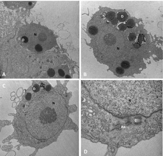
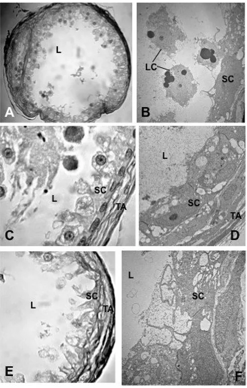
Testes morphology, spermatogenetic process and mature sperm ultrastructure were analysed in adult males of Hippocampus guttulatus using both light and transmission electron microscopy. The H. guttulatus testis is organized in a single large germinal compartment, with a central lumen and an external tunica albuginea. In germinal epithelium, spermatcysts only contain spermatogonia and primary spermatocytes. Inside the testis lumen, together with mature sperm, two types of large mono-nucleate cells were recognizable. The first type was constituted by aflagellate cells with cytoplasm rich in droplets; the second type was represented by mono-flagellate cells with cytoplasm containing a less amount of droplets. Both types of cells were interpreted as developing germ cells precociously released inside the testis lumen, where their maturation was completed. Functional sperm consisted of three distinct portions; the cylindrical head, the midpiece and the flagellum. These and previous data about the same topic reported on other syngnathids species were compared and discussed.
Francesca Piras
Techniques of Immunoflourescence and Confocal Microscopy applied to the study of Syngnathids gonadal structure And development and to the Dopaminergic control of the reproduction in Teleosts
PhD Thesis in Environmental Biology – University of Sassari, 2014 – XXVII cycle Introduction
Seahorses and their close relatives pipefishes and seadragon (Family Syngnathidae) occupy a very interesting position in the field of reproductive biology of bony fishes. Several interesting features characterize Syngnathids. They show peculiar parental care, with male pregnancy (Breder and Rosen 1966); have heterogeneous mating system varying from monogamy to different types of polygamy, associated with conventional or inverted sex roles) ( c.f. Jones et al. 1999) and display an atypical organization of both female and male gonads (Begovac and Wallace 1987, 1988; Selman et al. 1991; Carcupino et al. 1999; Sogabe et al. 2008; ; Sogabeet al. 201, Biagi et al. 2014).
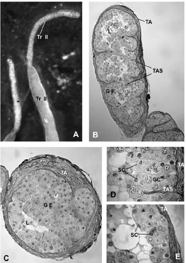
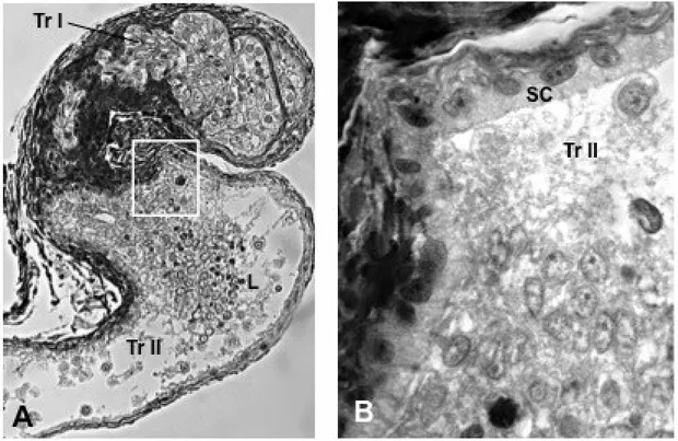
Numerous seminiferous lobules or tubules, which are connected to the main sperm duct via an efferent duct system generally characterize the teleost testes (Parenti and Greir 2004; Schulz et al. 2010). The efferent duct system collects and, sometimes stores, the spermatozoa. The syngnathids testes lack an of efferent duct system. Testes, at least several species belonging to the Syngnathus genus, i.e. Syngnathus abaster, S. typhle, S. tenuirostris, S. acus, are constituted by a single seminiferous compartment of unrestricted lobular types, which continue into a sperm duct. The two sperm ducts converge posteriorly to form a single main duct, which run parallel to the urethra and open independently in the apex of a urogenital papilla. The latter is located caudal to the anus, hidden by numerous skin folds arranged radially to the anal opening (personal observation). Two sperm ducts, originating from the last portion of the testis and converging posteriorly to form a single main duct which open in an urogenital papilla, also characterized the reproductive systems of other syngnathids species such as Nerophis ophidion and Hippocampus guttuatus, although the male gonad organization in these species has not be analysed in details. However, at least in Hippocampus kuda, testes have a morphology similar to that reported for the Sygngnathus species (Laksanawimol 2004).
69
Francesca Piras
Techniques of Immunoflourescence and Confocal Microscopy applied to the study of Syngnathids gonadal structure And development and to the Dopaminergic control of the reproduction in Teleosts
PhD Thesis in Environmental Biology – University of Sassari, 2014 – XXVII cycle
In the testis of both non–brooding and brooding males of H. kuda, examined during the reproductive season, spermatogonia and primary spermatocytes werefound along the entire length of the testis, while secondary spermatocytes and spermatids were only found inside the lumen. This seem to confirm that in syngnathids of Hippocampus genus, as well as those of
Syngnathus, the testis organization is of unrestricted lobular type and the spermatogenetic
process is of the semicystic type (Carcupino et al. 1999; Biagi et al. 2014).
This type of spermatogenesis is currently known in few species, belonging to several teleostean groups (Selman and Wallace 1986; Bazzoli and Godinho 1991; Mattei et al. 1993, Manni and Rasotto 1997; Yoneda et al. 1998, Carcupino et al. 1999, Giacomello et al. 2008; Srivastava and Singh 1994; Andrade et al. 2001; Mazzoldi 2001, Muñoz et al. 2002; Sàbat 2002; Laksanawimol 2004; García-López et al. 2005; Hernández et al. 2005; Shahin 2006; Sabat et al. 2009; Magalhaes et al. 2011). It consists of a precocious open of the germinal cysts, causing an asynchronous maturation of spermatids and the simultaneous presence of germ cells at different developmental stages inside the testis lumen. Moreover, in Syngnathus species, developing germ cells inside the lumen are mono- and, more frequently, poly-nucleate and poly-flagellate cells. Individualization of mature sperm seems to occur at the end of spermiogenesis, so the cytokinesis seems to be abolished or at least delayed (Carcupino et al. 1999; Biagi et al. 2014). Poly-nucleate developing germ cells has not been reported in H.
kuda (Laksanawimol 2004).
Syngnathids are also known to produce a very low number of sperm. The functional sperm : egg ratio has been estimated to be about 191 : 1 in S. abaster (Dzyuba et al. 2008) and even much lower 5 : 1 in Hippocampus kuda (Van Look et al. 2007). These values are numerous orders of magnitude lower than estimated in the zebrafish (Danio rerio) (48000 : 1), which was considered to have one of the lower sperm concentration in fish (Stokley et al.
Francesca Piras
Techniques of Immunoflourescence and Confocal Microscopy applied to the study of Syngnathids gonadal structure And development and to the Dopaminergic control of the reproduction in Teleosts
PhD Thesis in Environmental Biology – University of Sassari, 2014 – XXVII cycle
1996). Probably due to this low sperm concentration, no sperm were observed in the testis
lumen of both non-brooding and brooding male of H. kuda by Laksanawimol (2004), whereas dimorphic sperm have been reported in the same species by Van Look et al. (2007). In the latter study, however no data on sperm ultrastructure were shown. Sperm polymorphism was also reported in a freshwater population of Syngnathus abaster (Dzyuba et al. 2008), which belong to a monophyletic lineage within the urphorine subfamily including Syngnathus and to Hippocampus speies (Wilson and Orr 2011). Recently we have demonstrated that in two population the brackish water form of the same species S. abaster mature sperm are great variable in their morphometric traits, but they can not be distinguished in different morphotypes (Piras et al. sumitted).
The aim of this paper was to analyse the male gonad morphology, the spermatogenetic process and both sperm traits and ultrastructure in the seahorse Hippocampus guttulatus. Results obtained could be also contribute to shed light to the presence of sperm polymorphism in Syngnathids.
Materials and Methods
Sampling
Four adult male of Hippocampus guttulatus were sampled from Venice lagoon (Veneto), during the reproductive period (May-September 2013). Alive specimens, delivered to the laboratory within 3 h, were sacrificed by exposure to the anaesthetic 3-aminobenzoic acid ethyl ether (MS-222, Sigma-Aldrich) for 10 min, and then processed for microscopic analysis.