Accepted Manuscript
Title: Models of Heart Failure Based on the Cardiotoxicity of Anticancer Drugs
Author: Valentina Mercurio, Flora Pirozzi, Edoardo Lazzarini, Giancarlo Marone, Paola Rizzo, Giulio Agnetti, Carlo G Tocchetti, Alessandra Ghigo, Pietro Ameri
PII: S1071-9164(16)30023-9
DOI: http://dx.doi.org/doi: 10.1016/j.cardfail.2016.04.008 Reference: YJCAF 3753
To appear in: Journal of Cardiac Failure Received date: 5-2-2016
Revised date: 12-4-2016 Accepted date: 13-4-2016
Please cite this article as: Valentina Mercurio, Flora Pirozzi, Edoardo Lazzarini, Giancarlo Marone, Paola Rizzo, Giulio Agnetti, Carlo G Tocchetti, Alessandra Ghigo, Pietro Ameri, Models of Heart Failure Based on the Cardiotoxicity of Anticancer Drugs, Journal of Cardiac Failure (2016), http://dx.doi.org/doi: 10.1016/j.cardfail.2016.04.008.
This is a PDF file of an unedited manuscript that has been accepted for publication. As a service to our customers we are providing this early version of the manuscript. The manuscript will undergo copyediting, typesetting, and review of the resulting proof before it is published in its final form. Please note that during the production process errors may be discovered which could affect the content, and all legal disclaimers that apply to the journal pertain.
Models of heart failure based on the cardiotoxicity of anticancer drugs
Short title: Anticancer drugs and models of heart failure
Valentina Mercurio1, Flora Pirozzi1, Edoardo Lazzarini2, Giancarlo Marone3, Paola Rizzo4, Giulio Agnetti5,6, Carlo G Tocchetti1, Alessandra Ghigo7, Pietro Ameri2
1
Division of Internal Medicine, Department of Translational Medical Sciences, Federico II University, Naples, Italy; 2Laboratory of Cardiovascular Biology, Department of Internal Medicine, University of Genova, Genova, Italy; 3Department of Clinical Medicine and Surgery, Federico II University, Naples, Italy; 4Department of Morphology, Surgery and Experimental Medicine and Laboratory for Technologies of Advanced Therapies, University of Ferrara, Ferrara, Italy; 5Johns Hopkins University, Cardiology, Baltimore, MD, USA; 6Department of Biomedical and Neuromotor Sciences, University of Bologna, Bologna, Italy; 7Department of Molecular Biotechnology and Health Sciences, University of Torino, Torino, Italy
Address for correspondence:
Prof. Carlo Gabriele Tocchetti, MD, PhD Department of Translational Medical Sciences Federico II University
Via Pansini 5, Edificio 2 80131 Napoli, NA, Italy Phone: +39 081 746 2270 Fax: +39 081 746 2282
HIGHLIGHTS
Heart failure (HF) is a common complication of oncological treatments
Models of HF can be classified based on the mechanisms of cardiotoxicity of antineoplastic drugs
Myocyte-intrinsic forms include ROS generation, mitochondrial, DNA and metabolic damages
Paracrine forms include blockade of ErbB2, VEGFR and PDGFR
Anthracyclines and trastuzumab can also induce HF by affecting the CPC population
ABSTRACT
Heart failure (HF) is a complication of oncological treatments that may have dramatic clinical impact. It may acutely worsen a patient’s condition, or it may present with delayed onset, even years after treatment, when cancer has been cured or is in stable remission. Several studies have addressed the mechanisms of cancer therapy-related HF (CTHF) and some have led to the definition of disease models that hold valid for other and more common types of HF. Here, we review these models of HF based on the cardiotoxicity of antineoplastic drugs, and classify them in cardiomyocyte-intrinsic, paracrine, or potentially secondary to effects on cardiac progenitor cells (CPC). The first group includes HF due to the combination of oxidative stress, mitochondrial dysfunction and activation of the DNA damage response, which is typically caused by anthracyclines, and HF resulting from deranged myocardial energetics, such as that triggered by anthracyclines and sunitinib. Blockade of the neuregulin-1/ErbB4/ErbB2, vascular endothelial growth factor (VEGF)/VEGF receptor and platelet-derived growth factor (PDGF)/PDGF receptor pathways by trastuzumab, sorafenib and sunitinib is proposed as paradigm of CTHF associated with alterations of myocardial paracrine pathways. Finally, anthracyclines and trastuzumab are also presented as examples of antitumor agents that induce HF by affecting the CPC population.
Keywords: heart failure; antineoplastic drugs-induced cardiotoxicity; anthracyclines; receptor
tyrosine kinase
INTRODUCTION
Recent advances in antineoplastic treatments have rendered cancer curable in a sizable percentage of subjects, which is likely to further increase in the next years. In many other cases prolonged remission is achieved, leaving patients free of disease for a considerable amount of time. Unfortunately, these improvements take their toll in terms of emerging chronic side effects of antineoplastic agents, which can predominate once a tumor is eliminated or durably controlled [1]. Asymptomatic reduction in left ventricular (LV) function and heart failure (HF) are prototypical complications of cancer therapies that may have long-lasting impact [2]. Much effort has been expended in trying to pinpoint the mechanisms of cancer therapy-related HF (CTHF). This substantial body of work has allowed the identification of potential approaches to tackle the cardiotoxicity of antitumor agents, and additional ones are expected to be investigated in the next future. On the other hand, such an approach has also contributed to uncover important insights into the pathogenesis of HF in general. Among the many mechanisms that have been reported for CTHF, here we focus on those which have proven to be also relevant for other, more common forms of HF. We classify them as cardiomyocyte-intrinsic and paracrine, depending on whether they primarily affect cardiomyocytes or paracrine signals regulating cardiomyocytes, respectively. Furthermore, we propose disturbance of the cardiac progenitor cell (CPC) compartment as a third, stand-alone potential event in CTHF that may apply to other types of HF (Table 1). Each model is discussed referring to the anticancer agents that typically cause it, although it may also explain the cardiotoxicity of other drugs.
CARDIOMYOCYTE-INTRINSIC MECHANISMS Oxidative stress and DNA damage
Anthracyclines currently constitute key components of chemotherapeutic regimens for the treatment of different adult and pediatric cancers, such as leukemia and lymphomas as well as many solid tumors, including breast cancer [3]. These agents are amongst the most cardiotoxic anti-cancer drugs. Anthracycline cardiotoxicity can manifest acutely, early after infusion, thus requiring either modification or withdrawal of anti-cancer regimens [4]. Luckily, this is a relatively rare complication of chemotherapy, occurring in less than 9% of all patients suffering treated with anthracyclines, and recent evidence clearly demonstrates that anthracycline side effects are usually dose-dependent and more frequently detected within the first year after completing the treatment [5,6]. Pre-existent heart diseases and advanced age represent major risk factors for CTHF and recent studies also highlight the existence of a gender-related predisposition. Interestingly, a significantly higher risk for subclinical cardiotoxicity has been found in females than in males [7-9] and this could be ascribed, at least in cellular models, to testosterone-mediated protection from anthracycline-induced senescence of cardiomyocytes [10] (see Table 2 for a comprehensive list of risk factors).
Cardiotoxicity is a well-known adverse effect of anthracyclines and the research performed in the last 50 years has started to shed light on the molecular pathways involved. From a pathophysiological point-of-view, anthracyclines induce cardiomyocyte death, primarily apoptosis and necrosis, through several molecular mechanisms, including but not limited to induction of oxidative stress, activation of DNA damage responses and impairment of mitochondrial biogenesis and metabolism (Figure 1) [11]. The maladaptive changes occurring in surviving myocytes, as well as those occurring in the extracellular matrix, eventually lead to pathological LV remodeling, with dilatation and impairment of contractility, up to decline in systolic function and development of clinical HF.
Anthracycline-mediated myocyte damage has traditionally been attributed to the production of reactive oxygen species (ROS) with a subsequent increase in oxidative stress, which in turn causes membrane lipid peroxidation, vacuolization, irreversible damage, and myocyte replacement by fibrous tissue [2,3,12-18]. This primarily stems from the susceptibility of anthracyclines to be rapidly reduced to unstable metabolites (such as doxorubicin-semiquinone) which in turn react with oxygen to generate hydrogen peroxide and superoxide. Alternatively, the redox properties of anthracyclines have been ascribed to their ability to chelate free intracellular iron and form iron-doxorubicin complexes which, in turn, react with oxygen and trigger ROS production. Finally, anthracyclines can directly interfere with the activity of major iron-transporting and -binding proteins [19]. On these grounds, doxorubicin has been recently shown to impair the expression of the mitochondrial iron exporter, ABCB8, thus promoting mitochondrial iron accumulation and ROS production [20]. Accordingly, overexpression of ABCB8 in vitro, in isolated cardiomyocytes, and
in vivo, in the hearts of transgenic mice, decreases mitochondrial iron and cellular ROS and protects
against induced cardiomyopathy. Intriguingly, hearts from patients with doxorubicin-related heart dysfunction have significantly higher mitochondrial iron levels than hearts from patients with other types of cardiomyopathies or normal cardiac function [20]. These results thus suggest that mitochondrial iron accumulation and oxidative stress constitute a major hallmark of doxorubicin-induced cardiomyopathy.
A recent study indicates that anthracycline-induced ROS formation may additionally result from the interaction of the drug with the beta isozyme of topoisomerase II (Top2), an enzyme responsible for managing DNA tangles and supercoils, in cardiomyocytes [21] (Figure 1). Of the two Top2 isoforms, Top2-alpha and Top2-beta, Top2-alpha is overexpressed in proliferating cancerous cells, but not in quiescent tissues, and it is thought to be the molecular target of doxorubicin antitumor activity. By binding both Top2-alpha and DNA, doxorubicin forms a ternary Top2-doxorubicin-DNA cleavage complex, which in turn triggers the death of tumor cells. Unfortunately, doxorubicin also interacts with cardiac Top2-beta, the only isoform expressed by
adult mammalian cardiomyocytes, and the Top2-beta-doxorubicin-DNA complex induces DNA double strand breaks, ultimately promoting cardiomyocyte death [14]. The hypothesis that doxorubicin-mediated cardiomyopathy is mediated by cardiomyocyte Top2-beta is supported by studies showing that cardiomyocyte-specific Top2-beta deletion protects cardiomyocytes from doxorubicin-induced DNA double strand breaks and eventually results in almost complete protection against the development of doxorubicin-induced progressive heart failure in mice [14].
Mechanistically, it is likely that Top2-beta binding by doxorubicin, and the ensuing DNA break, lead to the activation of the DNA damage response and in turn, of the tumor suppressor protein p53. This enzyme, which is responsible for the activation of DNA repair proteins, can also repress genes involved in mitochondrial biogenesis, such as Ppargc1, and oxidative phosphorylation, ultimately leading to defective organelle biogenesis and metabolic failure [14]. Intriguingly, doxorubicin-activated p53 further contributes to this metabolic derangement by inhibiting the proper recycling of dysfunctional mitochondria via autophagy, a catabolic mechanism facilitating degradation of cytoplasmic proteins as well as old/damaged organelles. As a consequence, doxorubicin-injured mitochondria accumulate in the myocardium, leading to increased production of harmful reactive oxygen species (ROS) and ultimately cardiomyocyte death. In support of this view, p53-deficient mice show less decline of cardiac functional reserve after treatment with doxorubicin [22]. Notably, p53-null mice display preserved mitochondrial integrity and cardiac function also with increasing age. These data thus suggest that p53-mediated inhibition of autophagy may represent a major mechanism underlying not only anthracycline-related cardiomyopathies, but heart failure progression in general [22].
Besides p53, anthracyclines can also trigger the mitogen activated protein kinase (MAPK) cascade through reactive oxygen species and Ca2+ [23,24]. Notably, within MAPK family, ERK is known to protect cardiomyocytes from apoptosis, whereas p38 MAPK is involved in the induction of cardiomyocyte death [23]. Further studies are awaited to clarify the role of these enzymes and of other uncharacterized signaling pathways in anthracycline-mediated cardiomyocyte damage.
Nevertheless, these data indicate that the molecular mechanisms underlying anthracycline-related cardiomyopathy may be also relevant for other and more common forms of HF and suggest that timely innovative pharmacological interference with the primary mechanisms of cardiac injury, such as p53 activation, may represent a common strategy to fight heart disease.
Deranged myocardial energetics
Among other mechanisms involved in anthracycline-induced cardiotoxicity, recent studies have identified the role of deranged myocardial energetics, expressed by a reduced phosphocreatine/ATP ratio, which precedes LV dysfunction [25]. Indeed, anthracyclines are able to oxidize sulphydryl groups of creatine kinase (CK), diminishing its function, thus lowering myocardial energetics [25] and cauing cardiac dysfunction. More studies on such an interesting mechanism could provide a target for novel preventive therapies. Indeed, overexpression of myofibrillar CK in mice with HF caused by aortic banding significantly improved cardiac function [26], suggesting a key role for CK in HF prevention and treatment. Accordingly, the same group showed that CK overexpression also improved myocardial energetics, contractile function and survival in a mouse model of doxorubicin-induced cardiotoxicity [27]. These results provide novel strategies for limitation of anthracycline-related cardiotoxicity.
Anthracyclines can also alter cardiac energy metabolism by reducing the level of 5’ AMP-activated protein kinase (AMPK, involved in the response to energy stress) and phosphorylation of anti-acetyl-CoA carboxylase (ACC), resulting in impairment of fatty acid oxidation [28]. The mechanisms underlying inhibition of AMPK require further investigation.
Interestingly, sunitinib, a tyrosine kinase inhibitor (TKI), has been shown to inhibit AMPK as an off target effect which could lead to ATP depletion and affect contractility [29,30]. In pressure-overloaded mice treated with sunitinib, Chu and coworkers [31] showed opening of the mitochondrial permeability transition pore and marked mitochondrial swelling with derangements of the normal mitochondrial structure in cardiomyocytes. Also, direct sunitinib administration on
different myocardial preparations produces a dose-dependent negative inotropic effect, paralleled by a decline in intracellular Ca2+ and enhancement of ROS generation [32,33]. Interestingly, our preliminary data show that CK might play a role in the modulation of sunitinib myocardial effects [34].
PARACRINE MECHANISMS
Disruption of the NRG-1/ErbB4/ErbB2 system
Cardiotoxicity of anticancer agents targeting ErbB2 is one of the best examples of how cardiac side effects of an oncological treatment have prompted major advances in cardiac biology and pathology, with specific reference to HF.
ErbB2 (also known as HER2/Neu) is a membrane receptor belonging to the epidermal growth factor receptor family, which also includes ErbB1, ErbB3, and ErbB4. It is normally activated by the interaction (dimerization) with another ErbB receptor, which occurs after the latter one is stimulated by a ligand. [35]. In up to 30% of breast cancers, ErbB2 is overexpressed and transmits tumor growth-promoting intracellular signals regardless of the presence of an ErbB ligand (Figure 2) [36]. This has led to the design of ErbB2-targeting drugs, the first-in-class being trastuzumab, a humanized monoclonal antibody against ErbB2 [37]. Unexpectedly, trastuzumab was associated with LV dysfunction and HF in a substantial proportion of treated women in the pivotal clinical trials, with the highest incidence of cardiac complications in patients who concurrently received anthracyclines [38]. This outcome fueled a considerable body of research on the role of ErbB2 in the heart: in a decade, it was discovered that ErbB2 is part of a signaling system fundamental to maintain post-natal cardiac homeostasis.
It is now known that microvascular endothelial cells of the adult myocardium release neuregulin-1 (NRG-1, particularly the NRG-1β isoform) in response to stimuli such as mechanical strain [39,40]. NRG-1 acts on contiguous cardiomyocytes in a paracrine way and triggers the dimerization of ErbB2 with ErbB4, which is followed by a number of cellular events contributing
to cardiac homeostasis and adaptation to stress [41-43] (Fig 2). After the observation that trastuzumab was cardiotoxic in many recipients, mice with cardiac-specific disruption of the NRG-1/ErbB4/ErbB2 system were created via conditional deletion of the ErbB2 gene [44,45]. These animals proved to be viable and displayed normal cardiac morphogenesis at birth, but developed a dilated cardiomyopathy with systolic dysfunction in early adulthood [44,45]. Therefore, it was postulated that trastuzumab causes cardiomyocyte damage and, eventually HF, by inhibiting the NRG-1/ErbB4/ErbB2 axis in the heart, and that this is more likely to happen if cardiomyocytes are simultaneously exposed to another source of stress, like anthracyclines [46-48]. In principle, the same may be true when other types of cardiac stress are present, e.g. hypertension.
Several criticisms have subsequently been raised to this paradigm, which probably does not fully explain the cardiac side effects of trastuzumab and other ErbB2-blocking agents [49]. Nevertheless, it may still hold valid and – most important - has paved the way for further studies of NRG-1/ErbB4/ErbB2 that have moved the focus from cancer therapy-related HF to HF of any etiology, and new therapeutic perspectives have been opened. For instance, in a mouse model of LV pressure overload, ErbB4 and ErbB2 mRNA and protein were shown to decrease significantly during the transition from compensatory LV hypertrophy to HF [50]. Consistently, ErbB2 and ErbB4 expression and phosphorylation (a marker of receptor activation) was reported to be reduced in LV myocardium from HF patients, compared to that from organ donors [51]. Interestingly, it was also found that LV unloading after LV assist device implantation restored ErbB4 and ErbB2 levels [51,52]. In apparent contrast with these investigations, enhanced phosphorylation of ErbB4 and ErbB2 was observed in pacing-induced HF in dogs [53]. However, the intracellular mediators downstream of ErbB4 and ErbB2, Akt and ERK1/2, were inactivated, pointing anyway to disabled NRG-1/ErbB4/ErbB2 signaling. NRG-1 expression was higher in HF than in control conditions in most studies [51,53].
Overall, these findings suggest that impaired NRG-1/ErbB4/ErbB2 activity is implicated in the pathogenesis of HF at least in two ways: i) HF may ensue from the use of medications blocking
the NRG-1/ErbB4/ErbB2 pathway, like trastuzumab; ii) ErbB4/ErbB2 downregulation and/or uncoupling from intracellular signaling cascades occurs during the natural history of any type of HF in spite of normal or increased NRG-1, possibly contributing to disease progression. Moreover, it has been recently shown that catecholamines, that typically rise with the onset of cardiac dysfunction and following anthracycline treatment [54,55], can enhance ErbB2 expression in cardiomyocytes, thus rendering these cells more “targetable” by trastuzumab, thus vulnerable to the drug toxic effects [56]. These data may support the use of beta-blockers to prevent trastuzumab cardiotoxicity [57]. Indeed, a retrospective study by Seicean et al.indicated that coincidental use of β-blockers was able to lower the incidence of symptomatic HF in breast cancer patients treated with trastuzumab, anthracyclines, or both [58]. Pharmacological prevention with β-blockers is currently being tested in clinical trials [57; 58; 59] with bisoprolol (MANTICORE 101-Breast) [60], carvedilol (NCT01009918), and metoprolol (NCT01434134, NCT00806390) to prevent or treat trastuzumab-induced cardiomyopathy [57]. Interestingly, preliminary data from the PRADA (Prevention of cardiac dysfunction during adjuvant breast cancer therapy) trial presented at the 2015 American Heart Association Scientific Sessions showed that blocking just β1 with metoprolol may not provide sufficient protection [61], thus supporting the use of an unbiased β1- and β2-blocker [56].
Based on the evidence that NRG-1 exerts cardioprotective actions via ErbB4/ErbB2 and that the activity of these receptors is defective in HF, it has been postulated that NRG-1 and NRG-1 analogs may be employed for HF treatment. Intravenous administration of recombinant human NRG-1 and of the glial growth factor 2 (GGF2) isoform of NRG-1β improved cardiac performance and reduced LV dimensions in various animal models of HF [62-64]. Since NRG-1 therapy was beneficial in experimental ischemic HF even when it was started after myocardial infarction, it is conceivable that it induces reverse remodeling of the failing heart and does not merely oppose LV dilatation [62,64]. In addition, NRG-1 appears to directly inhibit cardiac fibroblasts and prevent fibrosis [64].
Early-phase clinical studies showed that infusion of recombinant human NRG-1 is safe, well tolerated, and favorably affects LV volumes and EF until up to 3 months after treatment [65,66]. Nonetheless, it should be noted that systemically administered NRG-1 might serve as growth factor for cancer cells or microfoci. Additional ongoing clinical trials will hopefully address this safety concern and will generate new evidence about the efficacy of NRG-1 in HF [40].
VEGF/VEGF-R and PDGF/PDGF-R blockade
Sunitinib and sorafenib are TKIs that target more than 30 kinases, and this explains their high rate of toxicities. Sunitinib targets VEGF receptors (VEGFR) 1–3, platelet-derived growth factor receptor (PDGFR), c-Kit, FMS-like tyrosine kinase-3, colony-stimulating factor-1 receptor (CSF-1R), and the product of the human RET gene (RET, mutated in medullary thyroid carcinomas/ multiple endocrine neoplasia). It is indicated in metastatic renal cancer and in imatinib-resistant gastrointestinal stromal tumor (GIST) and pancreatic neuroendocrine tumors that cannot be treated with surgery [2,29,32,67]. The main kinases inhibited by clinically relevant concentrations of sorafenib are VEGFR, PDGFR, Raf – 1/B-Raf, c-Kit, and FLT3 in in vitro kinase assays. Sorafenib is used to treat patients with advanced renal cell carcinoma after failure of prior therapy with interferon alfa or interleukin-2, or patients that are considered unsuitable for such therapy; it is also indicated for the treatment of liver cancer [2,29,32,67].
Beside the aforementioned AMPK, the fundamental target of these drugs is vascular endothelial growth factor (VEGF) signaling. Since VEGF is both an important regulator of cardiomyocyte function and growth, and of the integrity and expansion of the coronary and systemic circulation (Fig 2) [13,17,29,67-72], not surprisingly inhibiting VEGF signaling can induce cardiotoxic effects, mainly hypertension, thrombo-embolism, LV dysfunction, and HF [73,74]. Indeed, the heart depends on adequate perfusion for its normal function, and, similarly to cancer, relies on the integrity of HIF-1 (a transcriptional activator that is sensitive to cellular hypoxia and mediates many cellular and systemic homeostatic responses to hypoxia [75]) and
VEGF pathways [13,17,29,67-72]. In particular, inhibiting HIF-1 with p53 produced LV dysfunction after chronic pressure overload [24], while conditional expression of a VEGF scavenger led to microvessel rarefaction and myocardial hibernation that could be fully reversed even months after turning off the expression of the scavenger [76,77]. These findings point out that the heart is sensitive to anti-angiogenic drugs, particularly with pressure overload.
Inhibition of VEGF would also be responsible for the most common cardiovascular side effect of antiangiogenic drugs, namely hypertension. Indeed, VEGF signaling is important for the regulation of endothelial function and nitric oxide generation; its inhibition therefore abolishes normal vasodilation [78]. Other effects of VEGF inhibition may include induction of endothelial cell death and rarefaction of resistance vessels [2; 79]. Hypertension is due to mechanisms similar to those involved in the anticancer effects of antiangiogenic drugs, and therefore may also be considered as a marker for these drugs’efficacy [80].
Sunitinib has a high incidence of cardiotoxic effects. Besides its programmed action on VEGF, these important side effects can be also explained by the drug’s inhibition of off-target kinases, such as ribosomal S6 kinase (RSK), which brings to subsequent activation of the intrinsic apoptotic pathway [29].
Inhibition of PDGFR could be another mechanism related to sunitinib and sorafenib induced toxicity, since PDGFR also plays a critical role in cell survival and cardioprotection during pathologic stress (Fig 2). Exposure of mice lacking PDGFR (PDGFR‐β KO) to afterload stress results in LV dilation, decreased cardiac function, and pulmonary congestion compared with controls. This is also accompanied by increased apoptosis and decreased expression of pro‐angiogenic genes [81]. PDGFR inhibition is also known to impair angiogenesis, and leads to microvascular dysfunction through loss of pericytes [82].
Sunitinib and sorafenib share many mechanisms of cardiotoxicity, although sorafenib may have additional effects through inhibition of the Rapidly Accelerated Fibrosarcoma (RAF) kinases that normally promote cell survival [83]. It has been demonstrated in animal models that
cardiac-specific deletion of RAF-1 results in left ventricular (LV) dilatation and reduced contractile function [84].
Interestingly, a recent study addressed additional mechanisms of sorafenib-induced cardiotoxicity. The authors found that mice treated with sorafenib showed a decreased 2-week survival after myocardial infarction (MI) compared to vehicle-treated controls. In particular, sorafenib cardiotoxicity resulted from myocyte necrosis rather than from any direct effect on myocyte function, with surviving myocytes undergoing pathological hypertrophy. Inhibition of c-kit+ stem cell proliferation was also advocated as a potential, exacerbating factor that decreased endogenous cardiac repair [85].
All things considered, it is clear that cardiotoxicity from antiangionenic drugs is a complex side effect achieved by a combination of inhibitions of the activities of different Tyrosine Kinases that have important roles both in tumor cell proliferation and cardiovascular homeostasis (Table 3).
EFFECTS OF CARDIAC PROGENITOR CELLS
Emerging evidence seems to indicate that the cardiotoxicity of anti-cancer drugs is not solely restricted to cardiomyocytes but may also affect resident cardiac progenitor cells [86,87]. Although the heart has long been considered a definitively differentiated organ, without regenerative capacity, some studies have reported the presence of stem cells in the myocardium, known as cardiac progenitor cells (CPCs; see also [88] for extensive review of CPC function in health and disease). These cells seem to be self-renewing, clonogenic, and multipotent, and have been suggested to be implicated in the constant and physiological renewal of myocytes, endothelial cells (ECs), smooth muscle cells (SMCs), and fibroblasts. Furthermore, CPCs could potentially contribute to myocardial regeneration in pathological states. For instance, they regenerate cardiomyocytes and coronary vessels partly restoring the structure of the infarcted myocardium [86]. Nevertheless, although their identification has triggered a first wave of enthusiasm, the actual existence of CPCs and their regenerative capacity have been questioned and are still controversial [89]. In particular, there is
considerable controversy regarding whether c-kit+ cells are even cardiomyocyte progenitors [90,91,92]. Further studies are thus required to conclusively prove the therapeutic potential of these regenerative approaches, also considering other stem cells populations, mainly Sca-1+ cardiac progenitor cells, but also committed mesenchymal precardiomyocytes positive for Nkx2.5 or Isl1 [93].
Despite these controversial views, recent studies have proposed CPCs as an additional cellular target responsible of doxorubicin-induced cardiomyopathy [86] (Figure 3). Doxorubicin has been reported to interfere with the endothelial differentiation capacity of CPCs mediated by the CCL2/CCR2 pathway, as this drug reduces CCL2 expression and promotes the depletion of cardiac erythropoietin (EPO), which binds to CPCs and seems to be responsible for maintaining an active CCL2/CCR2 system [93]. Accordingly, mice exposed to doxorubicin display attenuated CCL2/CCR2 activation as well as reduced EPO levels in the cardiac microenvironment. Of note, mice prone to developing heart failure as a result of reduced cardiac STAT3 expression (cardiomyocyte-restricted deficiency of STAT3) show similar defects, accompanied by increased activity of MMP-12 which is responsible for the proteolytic cleavage of CCL2 [93]. These data thus support the view that deregulation of EPO/CCL2/CCR2 signaling might represent a general mechanism underlying CPC dysfunction in heart failure. In agreement, EPO expression is downregulated by 4-fold in hearts from patients with non-ischemic HF [94]. More importantly, this work envisages the possibility of exploiting EPO supplementation as a therapeutic strategy to reduce the risk of HF not only during anticancer therapies, but also in other disease conditions. EPO treatment in a low dose that does not increase the hematocrit (hematocrit-inactive) is indeed sufficient to upregulate CCL2, restore endothelial differentiation of CPCs, andpreserve the cardiac microvasculature and cardiac function in both doxorubicin-treated and STAT3-KO mice [93].
Impaired CPC differentiation may be also responsible of the late-onset doxorubicin-induced cardiotoxicity which represents a major concern in pediatric oncology. Juvenile mice exposed to doxorubicin, using a cumulative dose that does not induce acute cardiotoxicity, display impaired
vascular development, resulting in abnormal vascular architecture in the hearts with less branching and decreased capillary density. This in turn results in an adult heart that is more susceptible to stress as both physiological (exercise) and pathological (myocardial infarction) stress induce late-onset cardiomyopathy in the adult doxorubicin-treated mice. Notably, when subjected to myocardial infarction, doxorubicin-treated mice display fewer progenitor cells in the infarct border zone and fail to increase capillary density in the injured area, thus supporting a model where doxorubicin treatment reduces proliferation and differentiation of the progenitor cells into cells of cardiac lineages [95].
Mechanistically in CPCs, anthracycline exposure has been shown to increase ROS production and induce DNA damage, expression of p53, telomere attrition and apoptosis (Figure 3). In addition, doxorubicin promotes cell cycle arrest at the G2/M transition, by decreasing cyclin D1, cdk4 and phosphorylated Rb levels, in a dose- and time-dependent manner [96]. In agreement, p16INK4a, also known as cyclin-dependent kinase inhibitor 2A, is upregulated in patients who died of doxorubicin-induced heart failure and indicates that the majority of CPCs are senescent [86,96]. Doxorubicin also induces profound changes in global gene expression of CPCs, including genes that are implicated in drug efflux and cell protection from toxic agents, such as the ATP-binding cassette ABC transporter Abcg2/Mdr1, and genes involved in self-renewal and progenitor cell expansion [86,96]. Inhibition of CPC division in combination with accumulation of oxidative DNA damage, growth arrest, cellular senescence, and apoptosis ultimately leads to an almost complete depletion of the CPC pool within the myocardium over a period of 6 weeks after doxorubicin exposure. Intriguingly, supplementation of syngeneic progenitor cells has been proven effective in opposing the progression of doxorubicin cardiotoxicity in a rat model and suggests that CPC repopulation of the depleted myocardium after aggressive chemotherapy may represent a valuable option to rescue the cardiomyopathic heart [96].
Finally, the other cardiotoxic antineoplastic agent, trastuzumab, has also been reported to impair the cardiomyogenic and angiogenic capacities of CPCs. Although clinically relevant
concentrations of trastuzumab have only minor effects on proliferation, apoptosis, or size of the c-kit-positive CPC subpopulation, in vitro assays indicate diminished potential for cardiogenic differentiation and impaired ability to form microvascular networks in trastuzumab-treated cells. Consistently, trastuzumab treatment impairs the functional benefits of CPCs injected into the border zone of acutely infarcted mouse hearts [87].
Overall, these findings propose cardiac progenitor cells as one of the cellular targets responsible for chemotherapy-induced cardiomyopathy and envisage a CPC repopulation strategy for therapeutic intervention. Further studies, however, are awaited to clarify the debatable nature and therapeutic potential of this cardiac cell subpopulation.
CONCLUSIONS
Many of anti-cancer drugs induced toxicities share paradigmatic mechanisms of LV dysfunction. New, key cardiovascular signaling pathways involved in myocardial homeostasis have been more deeply understood through the study of the side effects caused by these drugs. Indeed, understanding the relevance of such pathways might prove important in order to study and develop novel therapeutic approaches for heart failure patients and improve longevity and quality of life in cancer patients. It is likely that along with the growing development of targeted cancer therapies, novel toxicities will be discovered [67], but in the same time these unfortunate findings may unveil unexplored myocardial signalings, thus allowing for parallel development in novel potential HF treatment strategies, including, for instance, genetic manipulation, miRNAs, gene transfer [13,16,27,97-106].
ACKNOWLEDGEMENTS
Grants 12SDG9210000 and 16IRG27240002 from the American Heart Association, Magic that Matters from Johns Hopkins University and RFO, University of Bologna to GA; “Excellent Young PI” grant from Compagnia di San Paolo to AG.
DISCLOSURES
All authors have materially participated in the article preparation, All authors have approved the final article.
REFERENCES
1. Bloom MW, Hamo CE, Cardinale D, Ky B, Nohria A, Baer L, Skopicki H, Lenihan DJ, Gheorghiade M, Lyon AR, Butler J. Cancer Therapy-Related Cardiac Dysfunction and Heart Failure: Part 1: Definitions, Pathophysiology, Risk Factors, and Imaging. Circ Heart Fail. 2016; 9:e002661.
2. Suter TM, Ewer MS. Cancer drugs and the heart: importance and management. Eur Heart J 2013; 34:1102-11
3. Menna P, Pax OG, Chello M, Covino E, Salvatorelli E, Minotti G. Anthracycline cardiotoxicity. Expert Opin Drug Saf 2012; 1:S21-36.
4. Rochette L, Guenancia C, Gudjoncik A, Hachet O, Zeller M, Cottin Y, Vergely C. Anthracyclines/trastuzumab: new aspects of cardiotoxicity and molecular mechanisms. Trends Pharmacol Sci. 2015; 36:326-48.
5. Ewer MS, Ewer SM. Cardiotoxicity of anticancer treatments. Nat Rev Cardiol 2015; 12:620.
6. Cardinale D, Colombo A, Bacchiani G, Tedeschi I, Meroni CA, Veglia F, Civelli M, Lamantia G, Colombo N, Curigliano G, Fiorentini C, Cipolla CM. Early detection of anthracycline cardiotoxicity and improvement with heart failure therapy. Circulation 2015; 131:1981-8.
7. Lanzarini L, Bossi G, Laudisa ML, Klersy C, Aricò M. Lack of clinically significant cardiac dysfunction during intermediate dobutamine dises in long term childhood cancer survivors exposed to anthracyclines. Am Heart J 2000. 140:315-23.
8. Sorensen K, Levitt G, Sebag-Montefiore D, Bull C, Sullivan I. Cardiac function in Wilms’tumor survivors. J Clin Oncol 1995; 13:1546-56.
9. Silber JH, Jakacki RI, Larsen RL, Goldwein JW, Barber G. Increased risk of cardiac dysfunction after anthracyclines in girls. Med Pediatr Oncol 1993; 21:477-9.
10. Altieri P, Barisione C, Lazzarini E, Garuti A, Bezante GP, Canepa M, Spallarossa P, Tocchetti CG, Bollini S, Brunelli C, Ameri P. Testosterone Antagonizes Doxorubicin-Induced Senescence of Cardiomyocytes. J Am Heart Assoc 2016; 5:pii:e002383.
11. Ghigo A, Li M, Hirsch E. New signal transduction paradigms in anthracycline-induced cardiotoxicity. Biochim Biophys Acta. 2016: S0167-4889: 30011-8.
12. Li T, Singal PK. Adriamycin-induced early changes in myocardial antioxidant enzymes and their modulation by probucol. Circulation 2000; 102:2105-10.
13. Molinaro M, Ameri P, Marone G, Petretta M, Abete P, Di Lisa F, De Placido S, Bonaduce D, Tocchetti CG. Recent Advances on Pathophysiology, Diagnostic and Therapeutic Insights in Cardiac Dysfunction Induced by Antineoplastic Drugs. Biomed Res Int 2015; 2015:138148.
14. Zhang S, Liu X, Bawa-Khalfe T, Lu LS, Lyu YL, Liu LF, Yeh ET. Identification of the molecular basis of doxorubicin-induced cardiotoxicity. Nat Med 2012; 18:1639-42.
15. Sawyer DB. Anthracyclines and heart failure. N Engl J Med 2013; 368:1154-6.
16. Salvatorelli E, Menna P, Minotti G. Managing anthracycline-induced cardiotoxicity: beginning with the end in mind. Future Cardiol.2015; 11:363-6
17. Ky B, Vejpongsa P, Yeth ET, Force T, Moslehi JJ. Emerging paradigms in cardiomyopathies associated with cancer therapies. Emerging paradigms in cardiomyopathies associated with cancer therapies. Circ Res 2013; 113:754-64.
18. Shalkey-Hahn V, Lenihan DJ, Ky B. Cancer therapy-induced cardiotoxicity: basic mechanisms and potential cardioprotective therapies. J Am Heart Assoc 2014;3:e000665.
19. Gammella E, Maccarinelli F, Buratti P, Recalcati S, Cairo G. The role of iron in anthracycline cardiotoxicity. Front Pharmacol 2014; 5:25.
20. Ichikawa Y. Ghanefar M, Bayeva M, Wu R, Khechaduri A, Naga Prasad SV, Mutharasan RK, Naik TJ, Ardehali H. Cardiotoxicity of doxorubicin is mediated through mitochondrial iron accumulation. J Clin Invest 2014; 124:617-30.
21. Lyu YL, Kerrigan JE, Lin CP, Azarova AM, Tsai YC, Ban Y, Liu LF. Topoisomerase IIbeta mediated DNA double-strand breaks: implications in doxorubicin cardiotoxicity and prevention by dexrazoxane. Cancer Res 2007; 67:8839-46.
22. Hoshino A, Mita Y, Okawa Y, Ariyoshi M, Iwai-Kanai E, Ueyama T, Ikeda K, Ogata T, Matoba S. Cytosolic p53 inhibits Parkin-mediated mitophagy and promotes mitochondrial dysfunction in the mouse heart. Nat Commun 2013; 4:2308.
23. Zhu W, Zou Y, Aikawa R, Harada K, Kudoh S, Uozumi H, Hayashi D, Gu Y, Yamazaki T, Nagai R, Yazaki Y, Komuro I. MAPK superfamily plays an important role in daunomycin-induced apoptosis of cardiac myocytes. Circulation 1999; 100:2100-2107.
24. Sano M. Minamino T, Toko H, Miyauchi H, Orimo M, Qin Y, Akazawa H, Tateno K, Kayama Y, Harada M, Shimizu I, Asahara T, Hamada H, Tomita S, Molkentin JD, Zou Y, Komuro I. p53-induced inhibition of HIF-1 causes cardiac dysfunction during pressure overload. Nature 2007;446: 444-448
25. Maslov MY, Chacko VP, Hirsch GA, Akki A, Leppo MK, Steenbergen C, Weiss RG. Reduced in vivo high-energy phosphates precede adriamycin-induced cardiac dysfunction. Am J Physiol Heart Circ Physiol 2010; 299: H332-7.
26. Gupta A, Akki A, Wang Y, Leppo MK, Chacko VP, Foster DB, Caceres V, Shi S, Kirk JA, Su J, Lai S, Paolocci N, Steenbergen C, Gerstenblith G, Weiss RG. Creatine kinase-mediated
improvement of function in failing mouse hearts provides causal evidence the failing heart is energy starved. J Clin Invest 2012;122: 291-302.
27. Gupta A, Rohlfsen C, Leppo MK, Chacko VP, Wang Y, Steenbergen C, Weiss RG. Creatine kinase-overexpression improves myocardial energetics, contractile dysfunction and survival in murine doxorubicin cardiotoxicity. PLoS One 2013;8: e74675.
28. Tokarska-Shalattner M, Zaugg M, da Silva R, Lucchinetti E, Schaub MC, Wallimann T, Schlattner U. Acute toxicity of doxorubicin on isolated perfused heart: response of kinases regulating energy supply. Am J Physiol Heart Circ Physiol 2005; 289:H37-H47.
29. Force T, Krause DS, Van Etten RA. Molecular mechanisms of cardiotoxicity of tyrosine kinase inhibition. Nat Rev Cancer 2007; 7: 332-344.
30. Kerkela R, Woulfe KC, Durand JB, Vagnozzi R, Kramer D, Chu TF, Beahm C, Chen MH, Force T. Sunitinib-induced cardiotoxicity is mediated by off-target inhibition of AMP-activated protein kinase. Clin Transl Sci 2009; 2:15-25.
31. Chu TF, Rupnick MA, Kerkela R, Dallabrida SM, Zurakowski D, Nguyen L, Woulfe K, Pravda E, Cassiola F, Desai J, George S, Morgan JA, Harris DM, Ismail NS, Chen JH, Schoen FJ, Van den Abbeele AD, Demetri GD, Force T, Chen MH. Cardiotoxicity associated with tyrosine kinase inhibitor sunitinib. Lancet 2007 ;370:2011-9.
32. Tocchetti CG, Gallucci G, Coppola C, Piscopo G, Cipresso C, Maurea C, Giudice A, Iaffaioli RV, Arra C, Maurea N. The emerging issue of cardiac dysfunction induced by antineoplastic angiogenesis inhibitors. Eur J Heart Fail 2013; 15: 482-9
33. Rainer PP Doleschal B, Kirk JA, Sivakumaran V, Saad Z, Groschner K, Maechler H, Hoefler G, Bauernhofer T, Samonigg H, Hutterer G, Kass DA, Pieske B, von Lewinski D, Pichler M. Sunitinib causes dose-dependent negative functional effects on myocardium and cardiomyocytes. BJU Int 2012; 110:1455-62.
34. Tocchetti CG, Leppo M, Bedja D, Wang Y, Weiss RG, and Paolocci N. Cardiac Overexpression of Creatine Kinase Improves Cardiomycytes Function in Heart Failure and During Increased Redox Stress. Circulation Research. 2015;117:A338.
35. Odiete O, Hill MF, Sawyer DB. Neuregulin in cardiovascular development and disease. Circ Res 2012; 111:1376-85.
36. Slamon DJ, Clark GM, Wong SG, Levin WJ, Ullrich A, McGuire WL. Human breast cancer: correlation of relapse and survival with amplification of the HER-2/neu oncogene. Science 1987; 235:177-82.
37. Denegri A, Moccetti T, Moccetti M, Spallarossa P, Brunelli C, Ameri P. Cardiac toxicity of trastuzumab in elderly patients with breast cancer. J Geriatr Cardiol in press
38. Slamon DJ, Leyland-Jones B, Shak S, Fuchs H, Paton V, Bajamonde A, Fleming T, Eiermann W, Wolter J, Pegram M, Baselga J, Norton L. Use of chemotherapy plus a monoclonal antibody against HER2 for metastatic breast cancer that overexpresses HER2. N Engl J Med 2001 344:783-92.
39. Lemmens K, Segers VF, Demolder M, De Keulenaer GW. Role of neuregulin-1/ErbB2 signaling in endothelium-cardiomyocyte cross-talk. J Biol Chem 2006; 281:19469-77. 40. Lim SL, Lam CS, Segers VF, Brutsaert DL, De Keulenaer GW. Cardiac
endothelium-myocyte interaction: clinical opportunities for new heart failure therapies regardless of ejection fraction. Eur Heart J 2015; 36:2050-2060.
41. Fukazawa R, Miller TA, Kuramochi Y, Frantz S, Kim YD, Marchionni MA, Kelly RA, Sawyer DB. Neuregulin-1 protects ventricular myocytes from anthracycline-induced apoptosis via erbB4-dependent activation of PI3-kinase/Akt. J Mol Cell Cardiol 2003; 35:1473-9.
42. Adam L, Vadlamudi R, Kondapaka SB, Chernoff J, Mendelsohn J, Kumar R. Heregulin regulates cytoskeletal reorganization and cell migration through the p21-activated kinase-1 via phosphatidylinositol-3 kinase. J Biol Chem 1998; 273:28238-46.
43. Kuramochi Y, Guo X, Sawyer DB. Neuregulin activates erbB2-dependent src/FAK signaling and cytoskeletal remodeling in isolated adult rat cardiac myocytes. J Mol Cell Cardiol 2006; 41:228-35.
44. Crone SA, Zhao YY, Fan L, Gu Y, Minamisawa S, Liu Y, Peterson KL, Chen J, Kahn R, Condorelli G, Ross J Jr, Chien KR, Lee KF. ErbB2 is essential in the prevention of dilated cardiomyopathy. Nat Med 2002; 8:459-65.
45. Ozcelik C, Erdmann B, Pilz B, Wettschureck N, Britsch S, Hübner N, Chien KR, Birchmeier C, Garratt AN. Conditional mutation of the ErbB2 (HER2) receptor in cardiomyocytes leads to dilated cardiomyopathy. Proc Natl Acad Sci U S A 2002 Jun 25;99(13):8880-5.
46. Sawyer DB, Zuppinger C, Miller TA, Eppenberger HM, Suter TM. Modulation of anthracycline-induced myofibrillar disarray in rat ventricular myocytes by neuregulin-1beta and anti-erbB2: potential mechanism for trastuzumab-induced cardiotoxicity. Circulation 2002; 105:1551-4.
47. de Korte MA, de Vries EG, Lub-de Hooge MN, Jager PL, Gietema JA, van der Graaf WT, Sluiter WJ, van Veldhuisen DJ, Suter TM, Sleijfer DT, Perik PJ. 111Indium-trastuzumab visualises myocardial human epidermal growth factor receptor 2 expression shortly after anthracycline treatment but not during heart failure: a clue to uncover the mechanisms of trastuzumab-related cardiotoxicity. Eur J Cancer. 2007 Sep;43(14):2046-51
48. Ewer MS, Ewer SM. Troponin I provides insight into cardiotoxicity and the anthracycline-trastuzumab interaction. J Clin Oncol 2010; 28:3901-4.
49. De Keulenaer GW, Doggen K, Lemmens K. The vulnerability of the heart as a pluricellular paracrine organ: lessons from unexpected triggers of heart failure in targeted ErbB2 anticancer therapy. Circ Res 2010; 106: 35-46.
50. Rohrbach S, Niemann B, Silber RE, Holtz J. Neuregulin receptors erbB2 and erbB4 in failing human myocardium -- depressed expression and attenuated activation. Basic Res Cardiol 2005;100: 240-9.
51. Rohrbach S, Yan X, Weinberg EO, Hasan F, Bartunek J, Marchionni MA, Lorell BH. Neuregulin in cardiac hypertrophy in rats with aortic stenosis. Differential expression of erbB2 and erbB4 receptors. Circulation 1999; 100:407-12.
52. Uray IP, Connelly JH, Thomázy V, Shipley GL, Vaughn WK, Frazier OH, Taegtmeyer H, Davies PJ. Left ventricular unloading alters receptor tyrosine kinase expression in the failing human heart. J Heart Lung Transplant 2002; 21:771-82.
53. Doggen K, Ray L, Mathieu M, Mc Entee K, Lemmens K, De Keulenaer GW. Ventricular ErbB2/ErbB4 activation and downstream signaling in pacing-induced heart failure. J Mol Cell Cardiol 2009; 46:33-8.
54. Nousiainen T, Vanninen E, Jantunen E, Remes J, Ritanen E, Vuolteenaho O, Hartikainen J. Neuroendocrine changes during the evolution of doxorubicin-induced left ventricular dysfunction in adult lymphoma patients. Clin Sci (Lond) 2001; 101:601-7.
55. Jeon TJ, Lee JD, Ha JW, Yang WI, Cho SH. Evaluation of cardiac adrenergic neuronal damage in rats with doxorubicin-induced cardiomyopathy using iodine-131 MIBG autoradiography and PGP 9.5 immunohistochemistry. Eur J Nucl Med 2000; 27:686-93.
56. Sysa-Shah P, Tocchetti CG, Gupta M, Rainer PP, Shen X, Kang BH, Belmonte F, Li J, Xu Y, Guo X, Bedja D, Gao WD, Paolocci N, Rath R, Sawyer DB, Naga Prasad SV, Gabrielson K.
Bidirectional cross-regulation between ErbB2 and β-adrenergic signalling pathways. Cardiovasc Res 2016;109:358-73.
57. Nohria A. β-Adrenergic blockade for anthracycline- and trastuzumab-induced cardiotoxicity: is prevention better than cure? Circ Heart Fail 2013; 6:358-61.
58. Seicean S, Seicean A, Alan N, Plana JC, Budd GT, Marwick TH. Cardioprotective effect of beta-adrenoceptor blockade in patients with breast cancer undergoing chemotherapy: follow-up study of heart failure. Circ Heart Fail 2013;6:420 – 426.
59. Kalam K, Marwick TH. Role of cardioprotective therapy for prevention of cardiotoxicity with chemotherapy: a systematic review and meta-analysis. Eur J Cancer 2013; 49:2900-9
60. Pituskin E, Haykowsky M, Mackey JR, Thompson RB, Ezekowitz J, Koshman S, Oudit G, Chow K, Pagano JJ, Paterson I. Rationale and design of the Multidisciplinary Approach to Novel Therapies in Cardiology Oncology Research Trial (MANTICORE 101--Breast): a randomized, placebo-controlled trial to determine if conventional heart failure pharmacotherapy can prevent trastuzumab-mediated left ventricular remodeling among patients with HER2+ early breast cancer using cardiac MRI. BMC Cancer 2011;11:318
61. Gulati G, Heck SL, Ree AH, Hoffmann P, Schulz-Menger J, Fagerland MW, Gravdehaug B, von Knobelsdorff-Brenkenhoff F, Bratland Å, Storås TH, Hagve TA, Røsjø H, Steine K, Geisler J, Omland T.Prevention of cardiac dysfunction during adjuvant breast cancer therapy (PRADA): a 2 × 2 factorial, randomized, placebo-controlled, double-blind clinical trial of candesartan and metoprolol. Eur Heart J 2016. pii: ehw022. [Epub ahead of print]
62. Liu X, Gu X, Li Z, Li X, Li H, Chang J, Chen P, Jin J, Xi B, Chen D, Lai D, Graham RM, Zhou M. Neuregulin-1/erbB-activation improves cardiac function and survival in models of ischemic, dilated, and viral cardiomyopathy. J Am Coll Cardiol 2006; 48:1438-47.
63. Li B, Zheng Z, Wei Y, Wang M, Peng J, Kang T, Huang X, Xiao J, Li Y, Li Z. Therapeutic effects of neuregulin-1 in diabetic cardiomyopathy rats. Cardiovasc Diabetol 2011;10:69. 64. Galindo CL, Kasasbeh E, Murphy A, Ryzhov S, Lenihan S, Ahmad FA, Williams P,
Nunnally A, Adcock J, Song Y, Harrell FE, Tran TL, Parry TJ, Iaci J, Ganguly A, Feoktistov I, Stephenson MK, Caggiano AO, Sawyer DB, Cleator JH. Anti-remodeling and anti-fibrotic effects of the neuregulin-1β glial growth factor 2 in a large animal model of heart failure. J Am Heart Assoc 2014; 3:e000773.
65. Gao R, Zhang J, Cheng L, Wu X, Dong W, Yang X, Li T, Liu X, Xu Y, Li X, Zhou M. A Phase II, randomized, double-blind, multicenter, based on standard therapy, placebo-controlled study of the efficacy and safety of recombinant human neuregulin-1 in patients with chronic heart failure. J Am Coll Cardiol 2010; 55:1907-14.
66. Jabbour A, Hayward CS, Keogh AM, Kotlyar E, McCrohon JA, England JF, Amor R, Liu X, Li XY, Zhou MD, Graham RM, Macdonald PS. Parenteral administration of recombinant human neuregulin-1 to patients with stable chronic heart failure produces favourable acute and chronic haemodynamic responses. Eur J Heart Fail 2011;13:83-92.
67. Cheng H, Force T. Molecular mechanisms of cardiovascular toxicity of targeted cancer therapeutics. Circ Res 2010; 106:21-34.
68. Eschenhagen T, Force T, Ewer MS, de Keulenaer GW, Suter TM, Anker SD, Avkiran M, de Azambuja E, Balligand JL, Brutsaert DL, Condorelli G, Hansen A, Heymans S, Hill JA, Hirsch E, Hilfiker-Kleiner D, Janssens S, de Jong S, Neubauer G, Pieske B, Ponikowski P, Pirmohamed M, Rauchhaus M, Sawyer D, Sugden PH, Wojta J, Zannad F, Shah AM. Cardiovascular side effects of cancer therapies: a position statement from the heart failure association of the European Society of Cardiology. Eur J Heart Fail 2011; 13:1-10.
69. Curigliano G, Cardinale D, Suter T, Plataniotis G, de Azambuja E, Sandri MT, Criscitiello C, Goldhirsch A, Cipolla C, Roila F; ESMO Guidelines Working Group. Cardiovascular
toxicity induced by chemotherapy, targeted agents and radiotherapy: ESMO Clinical Practice Guidelines. Ann Oncol 2012;23 Suppl 7:vii155-66.
70. Folkman J. Tumor angiogenesis: therapeutic implications. N Engl J Med 1971; 285:1182-1186.
71. Folkman J. Angiogenesis: an organizing principle for drug discovery? Nat Rev Drug Discov 2007; 6:273-286.
72. Marone G, Granata F. Angiogenesis, lymphangiogenesis and clinical implications. Preface. Chem Immunol Allergy 2014; 99:XI-XII.
73. Schmidinger M, Zielinski CC, Vogl UM, Bojic A, Bojic M, Schukro C, Ruhsam M, Hejna M, Schmidinger H. Cardiac toxicity of sunitinib and sorafenib in patients with metastatic renal cell carcinoma. J Clin Oncol 2008; 26:5204-5212.
74. Welti J, Loges S, Dimmeler S, Carmeliet P. Recent molecular discoveries in angiogenesis and antiangiogenic therapies in cancer. J Clin Invest 2013; 123:3190-200.
75. Semenza GL, Agani F, Feldser D, Iyer N, Kotch L, Laughner E, Yu A. Hypoxia, HIF-1, and the pathophysiology of common human diseases. Adv Exp Med Biol 2000;475:123-30. 76. Walsh K, Shiojima I. Cardiac growth and angiogenesis coordinated by intertissue
interactions. J Clin Invest 2007; 117:3176-3179.
77. May D, Gilon D, Djonov V, Itin A, Lazarus A, Gordon O, Rosenberger C, Keshet E. Transgenic system for conditional induction and rescue of chronic myocardial hibernation provides insights into genomic programs of hibernation. Proc Natl Acad Sci USA 2008; 105:282-287.
78. Ku DD, Zaleski JK, Liu S, Brock TA. Vascular endothelial growth factor induces EDRF-dependent relaxation in coronary arteries. Am J Physiol 1993;265(2 Pt 2): H586–H592 79. Steingart RM, Bakris GL, Chen HX, Chen MH, Force T, Ivy SP, Leier CV, Liu G, Lenihan
patients receiving vascular endothelial growth factor signaling pathway inhibitors. Am Heart J 2012;163:156 – 163.
80. Mir O, Ropert S, Alexandre J, Goldwasser F. Hypertension as a surrogate marker for the activity of anti-VEGF agents. Ann Oncol 2009;20:967–970.
81. Chintalgattu V, Ai D, Langley RR, Zhang J, Bankson JA, Shih TL, Reddy AK, Coombes KR, Daher IN, Pati S, Patel SS, Pocius JS, Taffet GE, Buja LM, Entman ML, Khakoo AY. Cardiomyocyte PDGFR‐beta signaling is an essential component of the mouse cardiac response to load‐induced stress. J Clin Invest 2010; 120:472-484.
82. Chintalgattu V, Rees ML, Culver JC, Goel A, Jiffar T, Zhang J, Dunner K Jr, Pati S, Bankson JA, Pasqualini R, Arap W, Bryan NS, Taegtmeyer H, Langley RR, Yao H, Kupferman ME, Entman ML, Dickinson ME, Khakoo AY. Coronary microvascular pericytes are the cellular target of sunitinib malate-induced cardiotoxicity. Sci Transl Med 2013; 5:187ra69.
83. Ratain MJ, Eisen T, Stadler WM, Flaherty KT, Kaye SB, Rosner GL, Gore M, Desai AA, Patnaik A, Xiong HQ, Rowinsky E, Abbruzzese JL, Xia C, Simantov R, Schwartz B, O'Dwyer PJ. Phase II placebo‐controlled randomized discontinuation trial of sorafenib in patients with metastatic renal cell carcinoma. J Clin Oncol 2006; 24:2505-2512.
84. Yamaguchi O, Watanabe T, Nishida K, Kashiwase K, Higuchi Y, Takeda T, Hikoso S, Hirotani S, Asahi M, Taniike M, Nakai A, Tsujimoto I, Matsumura Y, Miyazaki J, Chien KR, Matsuzawa A, Sadamitsu C, Ichijo H, Baccarini M, Hori M, Otsu K. Cardiac-specific disruption of the c-raf-1 gene induces cardiac dysfunction and apoptosis. J Clin Invest 2004;114:937– 43.
85. Duran JM, Makarewich CA, Trappanese D, Gross P, Husain S, Dunn J, Lal H, Sharp TE, Starosta T, Vagnozzi RJ, Berretta RM, Barbe M, Yu D, Gao E, Kubo H, Force T, Houser SR. Sorafenib cardiotoxicity increases mortality after myocardial infarction. Circ Res 2014;114:1700-12.
86. Piegari E, De Angelis A, Cappetta D, Russo R, Esposito G, Costantino S, Graiani G, Frati C, Prezioso L, Berrino L, Urbanek K, Quaini F, Rossi F.Doxorubicin induces senescence and impairs function of human cardiac progenitor cells. Basic Res Cardiol 2013; 108:334.
87. Barth AS, Zhang Y, Li T, Smith RR, Chimenti I, Terrovitis I, Davis DR, Kizana E, Ho AS, O'Rourke B, Wolff AC, Gerstenblith G, Marbán E.. Functional impairment of human resident cardiac stem cells by the cardiotoxic antineoplastic agent trastuzumab. Stem Cells Transl Med 2012; 1:289-97.
88. Tang XL, Li Q, Rokosh G, Sanganalmath S, Chen N, Ou Q, Stowers H, Hunt G, Bolli R. Long-Term Outcome of Administration of c-kitPOS Cardiac Progenitor Cells After Acute Myocardial Infarction: Transplanted Cells Do Not Become Cardiomyocytes, but Structural and Functional Improvement and Proliferation of Endogenous Cells Persist for At Least One Year. Circ Res 2016. pii: CIRCRESAHA.115.307647. [Epub ahead of print]
89. van Berlo JH, Molkentin JD. An emerging consensus on cardiac regeneration. Nat Med 2014;20:1386-93.
90. van Berlo JH, Kanisicak O, Maillet M, Vagnozzi RJ, Karch J, Lin SC, Middleton RC, Marbán E, Molkentin JD. c-kit+ cells minimally contribute cardiomyocytes to the heart. Nature. 2014;509:337-41.
91. van Berlo JH, Molkentin JD. Most of the Dust Has Settled: cKit+ Progenitor Cells Are an Irrelevant Source of Cardiac Myocytes In Vivo. Circ Res. 2016;118:17-9.
92. Sultana N, Zhang L, Yan J, Chen J, Cai W, Razzaque S, Jeong D, Sheng W, Bu L, Xu M, Huang GY, Hajjar RJ, Zhou B, Moon A, Cai CL. Resident c-kit(+) cells in the heart are not cardiac stem cells. Nat Commun. 2015;6:8701.
93. Hoch M, Fischer P, Stapel B, Missol-Kolka E, Sekkali B, Scherr M, Favret F, Braun T, Eder M, Schuster-Gossler K, Gossler A, Hilfiker A, Balligand JL, Drexler H, Hilfiker-Kleiner D.
Erythropoietin preserves the endothelial differentiation capacity of cardiac progenitor cells and reduces heart failure during anticancer therapies. Cell Stem Cell 2011;9:131-43.
94. Kittleson MM, Minhas KM, Irizarry RA, Ye SQ, Edness G, Breton E, Conte JV, Tomaselli G, Garcia JG, Hare JM. Gene expression analysis of ischemic and nonischemic cardiomyopathy: shared and distinct genes in the development of heart failure. Physiol Genomics 2005; 21:299-307.
95. Huang C, Zhang X, Ramil JM, Rikka S, Kim L, Lee Y, Gude NA, Thistlethwaite PA, Sussman MA, Gottlieb RA, Gustafsson AB. Juvenile exposure to anthracyclines impairs cardiac progenitor cell function and vascularization resulting in greater susceptibility to stress-induced myocardial injury in adult mice.Circulation 2010; 121:675-83.
96. De Angelis A, Piegari E, Cappetta D, Marino L, Filippelli A, Berrino L, Ferreira-Martins J, Zheng H, Hosoda T, Rota M, Urbanek K, Kajstura J, Leri A, Rossi F, Anversa P. Anthracycline cardiomyopathy is mediated by depletion of the cardiac stem cell pool and is rescued by restoration of progenitor cell function. Circulation 2010; 121:276-92.
97. Bollini S, Smart N, Riley PR. Resident cardiac progenitor cells: at the heart of regeneration. J Mol Cell Cardiol 2011;50:296-303.
98. Braunwald E. The war against heart failure: the Lancet lecture. Lancet 2015; 385:812-24.
99. Tarone G, Balligand JL, Bauersachs J, Clerk A, De Windt L, Heymans S, Hilfiker-Kleiner D, Hirsch E, Iaccarino G, Knöll R, Leite-Moreira AF, Lourenço AP, Mayr M, Thum T, Tocchetti CG. Targeting myocardial remodelling to develop novel therapies for heart failure: a position paper from the Working Group on Myocardial Function of the European Society of Cardiology. Eur J Heart Fail 2014;16:494-508
100. Kumarswamy R, Thum T. Non-coding RNAs in cardiac remodeling and heart failure. Circ Res 2013;113:676-89.
101. Pleger ST, Brinks H, Ritterhoff J, Raake P, Koch WJ, Katus HA, Most P. Heart failure gene therapy: the path to clinical practice. Circ Res 2013;113:792-809.
102. Tocchetti CG, Carpi A, Coppola C, Quintavalle C, Rea D, Campesan M, Arcari A, Piscopo G, Cipresso C, Monti MG, De Lorenzo C, Arra C, Condorelli G, Di Lisa F, Maurea N. Ranolazine protects from doxorubicin-induced oxidative stress and cardiac dysfunction. Eur J Heart Fail 2014;16:358-66.
103. Lemmens K, De Keulenaer GW. Paving new paths for neuregulin-1-assisted cardiac regenerative medicine. Focus on "Improving murine embryonic stem cell differentiation into cardiomyocytes with neuregulin-1: differential expression of microRNA". Am J Physiol Cell Physiol 2011;301(1):C16-7.
104. Wang JX, Zhang XJ, Feng C, Sun T, Wang K, Wang Y, Zhou LY, Li PF. MicroRNA-532-3p regulates mitochondrial fission through targeting apoptosis repressor with caspase recruitment domain in doxorubicin cardiotoxicity. Cell Death Dis 2015;6:e1677.
105. Das A, Durrant D, Salloum FN, Xi L, Kukreja RC. PDE5 inhibitors as therapeutics for heart disease, diabetes and cancer. Pharmacol Ther 2015;147:12-21.
106. Freedman NJ, Ginsburg GS. Novel—and “neu”—therapeutic possibilities for heart failure”. J Am Coll Cardiol 2006;48:1448-50.
FIGURE LEGENDS
Figure 1: Cardiomyocyte-intrinsic molecular mechanisms underlying anthracycline cardiotoxicity.
Anthracyclines, such as doxorubicin (DOX), lead to cardiotoxicity by promoting the production of reactive oxygen species (ROS), via direct (unstable DOX metabolites, such as doxorubicin-semiquinone, react with oxygen and generate hydrogen peroxide and superoxide) and indirect (DOX chelates free iron and modulates the activity/expression of major iron-transporting/binding proteins) mechanisms. Alternatively, DOX can interact with cardiomyocyte topoisomerase 2β (Top2β), an enzyme responsible for managing DNA tangles and supercoils, thus inducing DNA double-strand breaks. DNA damage, in turns, activates the tumor suppressor protein p53, which is responsible for the activation of DNA repair proteins, but also of repression of genes involved in mitochondrial biogenesis/recycling and oxidative phosphorylation pathways. Finally, DOX can accumulate within mitochondria of cardiomyocytes and exacerbates metabolic failure of these organelles. Altogether, these molecular events contribute to cardiomyocyte death and ultimately to cardiac dysfunction.
Abbreviations: ROS, reactive oxygen species; TOP2β, topoisomerase 2β.
Figure 2: Paracrine molecular mechanism underlying main TKI cardiotoxicity.
Adapted from [13], [17] and [106].
Therapies targeting paracrine signaling network at several levels with antibodies such as trastuzumab or with TKIs are able to perturb the potentially protective action of VEGF, PDGF and NRG in the myocardium. In particular, neuregulin-1 (NRG1) binding brings to dimerization between the neuregulin receptor ErbB4 and its co-receptor ErbB2. ErbB2 and ErbB4 phosphorylate each other on tyrosyl residues activating several signaling pathways, including those involving Akt that inhibits apoptotic signaling and promotes cellular hypertrophy, while ERK is able to phosphorylate transcription factors thus promoting gene transcription.
Figure 3. Effects of anthracyclines on cardiac progenitor cell function.
Anthracyclines, such as doxorubicin (DOX), interfere with different functions of CPCs. DOX impairs the endothelial differentiation capacity of CPCs by downregulating CCL2/CCR2 signaling, either directly (DOX reduces CCL2 expression) or indirectly (DOX promotes the depletion of a key regulator of CCL2/CCR2, cardiac erythropoietin). Furthermore, DOX dampens CPC proliferation by downregulating key controllers of cell cycle progression, such as cyclin D1, cdk4 and phosphorylated Rb. Finally, DOX-dependent ROS production and DNA damage contribute to the activation of the tumor suppressor protein p53 ultimately driving to telomere attrition and apoptosis. Abbreviations: ROS, reactive oxygen species; CPCs, cardiac progenitor cells; CCL2, chemokine ligand 2; CCR2, C-C chemokine receptor type 2; Rb, retinoblastoma protein; cdk4, cyclin-dependent kinase 4.
Table 1. Chemotherapeutic agents commonly associated with cardiotoxicity
Proposed mechanisms Chemotherapeutic drug
Cardiomyocyte intrinsic Anthracyclines, sunitinib
Paracrine Trastuzumab, sunitinib, sorafenib
Effects on cardiac progenitor cells Anthracyclines, trastuzumab
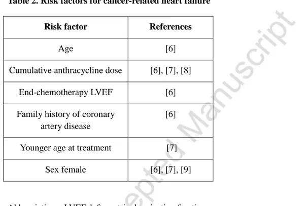
Table 2. Risk factors for cancer-related heart failure
Risk factor References
Age [6]
Cumulative anthracycline dose [6], [7], [8]
End-chemotherapy LVEF [6]
Family history of coronary artery disease
[6]
Younger age at treatment [7]
Sex female [6], [7], [9]
Table 3 (modified from [67])
Cardiovascular role of the main kinases inhibited by antiangiogenic drugs
Kinases Role in Cardiovascular system Inhibitors
VEGFR Contribution to cardiomyocyte function and growth and to the integrity and expansion of the coronary and systemic circulation; stimulation of endothelial growth, migration, and survival
Sunitinib and sorafenib
PDGFR Contribution to cell survival and
cardioprotection during stress conditions, regulation of angiogenesis
Sunitinib and sorafenib
AMPK Energy production Sunitinib
RAF Promoting cell survival Sorafenib
c-kit Contribution to homing of CSC to sites of
post-MI injury, CSC differentiation, and
cardiomyocyte terminal differentiation.
Sunitinib and sorafenib
ribosomal S6 kinase (RSK)
signals survival through inhibitory
phosphorylation of the pro-apoptotic factor
BAD