CELLULAR Na
+,K
+-ATPase MEDIATES
UNCONVENTIONAL EXPORT OF HIV-1 TAT PROTEIN
Silvia Agostini
Ph.D. Thesis
In
Molecular Biology
Supervisor:
Prof. Mauro Giacca
Scuola Normale Superiore
A Me,
Alla voglia di conoscere e
al piacere di capire
che mi accompagnano
TABLE OF CONTENTS
SYNOPSIS...11
INTRODUCTION ...17
HIV life cycle... 17
Role of intercellular protein trafficking in the biology of complex
retroviruses ... 25
HIV-1 Tat...26
Tat- mediated transcriptional activation ...27
Pleiotropic activities of extracellular Tat ...33
Neurodegenerative disorders ...36
Kaposi’s sarcoma ...39
The immune response ...41
HIV-1 Vpr ...42
Intracellular activities of Vpr...42
Cytotoxic effect of extracellular Vpr ...45
Vpr and AIDS dementia complex ...46
HIV-1–associated nephropathy...47
Role of Vpr in AIDS-related insulin resistance/lipodystrophy syndrome...47
Effects on immune system ...48
HIV-1 Nef ...50
Intracellular activities of Nef...51
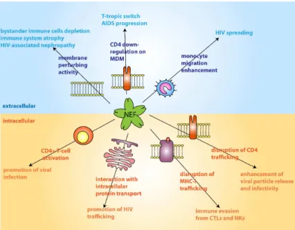
Pathological effects of extracellular Nef...52
Perturbation of membrane structure ...53
M-tropic to T-tropic switch ...54
Extracellular Nef increases the migration of monocytes ...55
HIV-associated nephropathy: role of Nef...56
HTLV-1 Tax...56
Tax intracellular activities: oncogenesis ...57
Biological effects of extracellular Tax ...59
Neurotoxic effects. ...60
Pro-inflammatory activity...62
HFV Bet ...62
Properties of extracellular Bet ...64
Internalization of extracellular Tat and other proteins and peptides 65
Cell surface receptors for extracellular Tat internalization...68Molecular mechanism of Tat internalization...70
Tat as a vehicle for transcellular transduction of proteins, nucleic acids and other cargos in vitro and in vivo...73
Protein secretion ... 74
TABLE OF CONTENTS
ER-Golgi trafficking...77
The Golgi apparatus ...78
Unconventional pathways of protein secretion...80
Signal peptide containing proteins: bypassing COPII vesicles or Golgi apparatus ...81
Secretion of proteins lacking the signal peptide for ER targeting: the “unconventional pathway” ...82
Cytokines: interleukin-1β, thioredoxin and macrophage migration inhibitory factor ...85
Pro-angiogenic growth factors: FGF-1 and FGF-2 ...87
Galectins: components of the extracellular matrix ...91
Other secretory proteins exported by non conventional means: HIV-Tat, Herpes simplex VP22 and foamy virus Bet...93
Leishmania HASPB ...94
Homeodomain-containing transcription factors and HMG (high mobility group) chromatin-binding proteins ...95
Cytoplasmic clearance of unfolded proteins by non classical secretion...97
Non classical secretion in yeast. ...98
Targeting motifs and regulation of non classical protein export ... 101
Caspase-1: a common regulator of unconventional protein secretion? ... 105
Emerging roles of GRASP proteins ... 106
Unconventional secretion and evolution: integrating complex functions ... 109
Na
+,K
+-ATPase structure, functions and regulation... 110
Na+,K+-ATPase structure... 111
Na+,K+-ATPase functions and regulation ... 114
Na+,K+-ATPase interaction with other proteins ... 116
Na+,K+-ATPase and cardiac glycosides ... 118
Na+,K+-ATPase signaling functions ... 119
MATERIALS AND METHODS ...123
Plasmid constructs... 123
Recombinant fusion proteins ... 125
Synthetic peptides ... 126
In vitro
GST-pull down assays ... 127
TABLE OF CONTENTS
Antibodies and reagents... 128
Fluorescence microscopy... 129
FRET analysis... 129
Cell cultures and transfections ... 130
Drug treatment ... 131
Secretion assay ... 131
Flow cytometry ... 132
Immunoprecipitation ... 132
CAT ELISA ... 133
Virus preparation... 133
Infections ... 134
Luciferase assay ... 135
Statistical analysis ... 135
RESULTS ...139
Heterologous proteins fused to HIV-1 Tat are released from the
expressing cells... 139
Kinetics of Tat-fusion protein release ... 141
Tat secretion is independent from HSPG binding ... 142
Tat secreted from HEK293Tcells is able to transactivate LTR-CAT
genes in HL3T1 cells... 144
Tat is excluded from the Golgi apparatus... 146
TABLE OF CONTENTS
The cellular Na
+,K
+-ATPase inhibitor ouabain blocks extracellular
release of Tat86 and Tat11 fusion proteins ... 148
Ouabain-sensitive interaction of Tat with the Alpha1 subunit of the
cellular Na
+,K
+-ATPase... 151
Ouabain treatment does not affect Alpha1 or Tat intracellular
localization... 152
Ouabain does not interfere with Tat internalization... 154
Visualization of the interaction of Tat with the Na
+,K
+-ATPase Alpha1
subunit by fluorescence resonance energy transfer (FRET) ... 155
Alpha1 catalytic function is not necessary for Tat release... 157
Tat-Alpha1 interaction is crucial for Tat secretion ... 158
The Tat basic region binds the Alpha1 C-terminal domain ... 160
Interaction between Tat and the cytoplasmic portion of Alpha1
C-terminal domain ... 162
3D modeling of C-terminal peptides in Alpha1 subunit tertiary
structure... 164
Tat fails to co-immunoprecipitate an Alpha1 mutant lacking the
C-terminus... 165
Alpha1 deletion mutant fails to rescue Tat86 secretion in ouabain
treated cells ... 167
A fusion protein corresponding to the Na
+,K
+-ATPase Alpha1
C-terminal loops binds Tat and impairs transactivation... 169
Internalization of fluorescent peptides... 172
Synthetic peptides corresponding to the Na
+, K
+-ATPase Alpha1
C-terminal loops block Tat export... 174
Synthetic peptides corresponding to the Na
+, K
+-ATPase Alpha1
C-terminal loops inhibit HIV-1 replication ... 177
TABLE OF CONTENTS
DISCUSSION ...183
Characterization of the unconventional pathway of Tat secretion.. 184
Ouabain blocks extracellular Tat release ... 187
The role of the Na
+,K
+-ATPase in extracellular Tat release ... 188
Intercellular trafficking and lentiviral biology: Why is Tat secreted?
... 195
SYNOPSIS
11
SYNOPSIS
The Tat (Trans activator) protein of the human immunodeficiency virus type 1 (HIV-1) is a 101 amino acid polypeptide acting as a powerful transcriptional activator of viral geneexpression. The protein binds a highly structured region of nascent RNA transcripts and, from here, directs the assembly of processive transcription complexes and promotes chromatin modification at the HIV-1 LTR (Long Terminal Repeat) promoter. Besides this fundamental role in the control of HIV-1 gene expression, Tat also possesses the unusual property of trafficking in and out of the cells. In particular, the capacity of being internalized by the cells when present in the extracellular compartment, which depends on the integrity of the basic domain of the protein (aa 49-57), has been extensively characterized and constitutes the basis for its biotechnological utilization for the delivery of heterologous proteins, drugs, viral vectors and nanoparticles (Becker-Hapak
et al. 2001; Tasciotti et al. 2003; Fittipaldi and Giacca 2005; Tasciotti and Giacca 2005). Some of the studies that elucidated the molecular mechanisms of extracellular Tat internalization also noticed that cells constitutively expressing Tat release this protein into the cell culture supernatant (Becker-Hapak et al. 2001; Tyagi et al. 2001; Tasciotti et al.
2003; Tasciotti and Giacca 2005). However, the mechanisms underlying extracellular Tat release have so far remained elusive.
The aim of this project was to explore the molecular mechanisms responsible for Tat export from the expressing cells, with the ultimate purpose to understand the significance of such mechanism in the biology of HIV-1 infection and exploit the ensuing information for the improvement of the trafficking properties of Tat-fused protein cargos.
SYNOPSIS
12
1) When cells were transfected with expression vectors encoding fusion constructs corresponding to a reporter protein fused to full-length HIV-1 Tat or to an 11 amino acid peptide encompassing its basic domain, a significant fraction of these proteins was found in the cell culture supernatants. Secretion had a rapid kinetics, different from that of proteins secreted through the endoplasmic reticulum (ER)-Golgi route, and was dependent on the integrity of the Tat basic domain.
2) Neither drugs blocking canonical protein secretion nor drugs interfering with intracellular vesicle trafficking blocked extracellular Tat release, indicating that this process occurred through a Golgi-independent, unconventional secretion route.
3) Tat and Tat-fusion protein release was impaired by ouabain. This drug is a specific inhibitor of the cellular Na+,K+-ATPase, an enzyme built into the plasma membrane which catalyzes ATP hydrolysis coupled with Na+ and K+ transfer through the membrane against the electrochemical gradient. Ouabain is known to also impair the unconventional secretion of FGF-2 (Fibroblast Growth Factor 2) (Florkiewicz et al. 1998; Dahl et al. 2000). 4) The Tat basic domain was found to specifically bind the catalytic (alpha) subunit of the Na+,K+-ATPase. This interaction appeared essential for extracellular Tat release and was disrupted by ouabain treatment.
5) Interaction between Tat and the Na+,K+-ATPase required integrity of the Carboxy-terminal domain of the latter protein. In particular, this region contains three short intracytoplasmic peptide stretches (of 19, 12, and 16 amino acids) that are juxtaposed in the protein three-dimensional structure, which were all required to bind Tat.
6) Intracellular expression of a novel fusion protein corresponding to a linear combination of the three Tat-binding, Na+,K+-ATPase peptide regions blocked Tat transactivation.
SYNOPSIS
13 7) A mixture of soluble synthetic peptides corresponding to the three Tat-binding, Na+,K+-ATPase peptide regions blocked Tat release.
8) The same mixture of peptides, when added to CD4+ T-cells infected with a wild type strain (HIV-1BRU) impaired HIV-1 infection.
Taken together, these results shed light on the molecular mechanism responsible for extracellular Tat release, disclose the importance of inhibiting this process in the viral life cycle and identify a novel tool for pharmacological and/or gene-based therapy of HIV-1 infection.
Chapter 1
INTRODUCTION
17
INTRODUCTION
HIV life cycle
The Human Immunodeficiency Virus type 1 and 2 (HIV-1 and HIV-2) belongs to the Lentiviral genus of Retroviridae, and is the etiological agents of the Acquired Immunodeficiency Syndrome (AIDS). HIV establishes a persistent infection by means of provirus integration in the host genome and rapid mutation of viral genes encoding for proteins eliciting host immune response. Even in presence of effective antiviral treatment and strong immune response, however, the ability of the virus to establish a latent infection during the early stages of the disease grants the persistence of the virus in the host organism.
The HIV genome is 10 kilobases long and encodes a number of structural proteins as well as several regulatory and accessory proteins which are not found in gamma- and alpha-retroviruses (Fig. 1.1); consequently, HIV replication cycle is far more complex, showing many regulatory mechanisms and strategies aimed at viral persistence.
Viral replication requires the transcription of the proviral genome and the synthesis of several proteins necessary for the assembly and the budding of the viral progeny. HIV transcription depends upon the interaction between a number of cellular transcription factors and co-activators with regulatory sequences contained in the viral Long Terminal Repeat (LTR) promoter; such interactions lead to the assembly of a stable transcription complex stimulating several transcription rounds by RNA polymerase II.
The structural genes, gag, pol and env, common to all other retroviruses, encode the proteins necessary for virion assembly, as well as the enzymes required for genome replication, proviral integration and polyprotein cleavage. The gag, pol and env genes are transcribed into polyproteins that are subsequently processed by viral (Gag and Pol) or cellular (Env)
INTRODUCTION
18
proteases. The gag gene encodes the core proteins (Capsid CA, Nucleocapsid NC and p6) and the Matrix protein (MA), while the env gene encodes the glycoproteins gp120 and gp41, and the pol gene encodes three essential enzymes for viral replication: Reverse Transcriptase (RT), Integrase (IN) and Protease (PR).
In addition to these structural genes, the HIV genome expresses several accessory and regulatory proteins: Vif (Viral infectivity Factor), Vpr (Viral Protein R), Vpu (Viral Protein U) (HIV-1), Vpx (Viral Protein X) (HIV-2), Tat, Rev and Nef (Negative Factor).
Fig. 1.1: HIV-1 genome
HIV has been reported to infect a broad range of cellular types in vitro; however, in vivo, infection is restricted to CD4+ T-lymphocytes and cells of the monocyte-macrophage lineage.
Infection starts with the fusion between HIV envelope and the cell plasma membrane; the surface gp120 protein on the viral envelope binds the CD4 receptor on the host cell surface, inducing conformational changes and promoting the binding to chemokine receptors that serve as co-receptors for HIV-1 infection (Kwong et al. 1998). These co-receptors are both critical for virus entry, and crucial for determining the tropism among CD4+ cell types. HIV-1 uses for its infection two chemokine receptors: CCR5, which binds macrophage-tropic (R5) viruses, and CXCR4, which binds T-cell tropic (X4) viruses. While R5 viruses are responsible of mucosal and intravenous transmission of HIV-1, X4 isolates appear only in the late stages of infection, with the appearance of the immunodeficiency (Scarlatti et al. 1997) .
INTRODUCTION
19 Viral gp120 is bound to gp41, a transmembrane protein which assembles as a trimer on the virion envelope. The interaction between gp120, CD4 and the co-receptor induces a conformational change in gp41, leading to the exposition of three peptide fusion domains. These domains are able to spear the plasma membrane and promote the fusion of the viral envelope, leading to the release of the HIV core in the host cytoplasm (Chan and Kim 1998). Upon entry, the virus undergoes the uncoating process, which generates the viral reverse transcription complex, composed by the viral genome, RT, IN, MA, NC, Vpr and other host proteins (Karageorgos et al. 1993); MA protein is then phosphorylated and interacts with actin microfilaments (Bukrinskaya
et al. 1998), while Vpr serves to stabilize the reverse transcription complex (Ohagen and Gabuzda 2000).
The completion of the reverse transcription leads to the formation of the Pre-integration complex (PIC), which is composed of viral cDNA, IN, MA, Vpr and RT (Miller et al. 1997). As HIV, unlike classical retroviruses, is able to infect non-dividing cells, such as differentiated macrophages, the PIC needs to be translocated in the nucleus through an intact nuclear envelope. Viral proteins Vpr, IN and MA possess in their sequence a nuclear localization signal, and are all involved in mediating nuclear import of the PIC (Bukrinsky
et al. 1993; Heinzinger et al. 1994; Gallay et al. 1997); however, whether these proteins act in a cooperative manner, or have individual roles in different target cells is still unclear.
Inside the nucleus, IN mediates the integration of the viral DNA in the cellular genome. IN, as well as other proteins involved in PIC formation, binds specific sequences at the end of the viral cDNA, named “att sites”, and removes two nucleotides left by the terminal transferase activity of RT. No primary sequence of the cellular DNA has been shown as a preferential target for IN and integration seems to occur at random in the host genome.
INTRODUCTION
20
Fig. 1.2: scheme of the different fates of the viral DNA after retrotranscription (Butler et al. 2001)
Other than integration, the viral DNA may follow three different fates, neither leading to the formation of a functional provirus (Fig. 1.2). The viral ends may join to form a 2-LTR ring, or the viral genome may undergo homologous recombination, producing a single LTR ring, or the viral DNA may integrate into itself, forming a rearranged circular structure. All these circular structures are non infectious, even if some of them are transcribed and produce Tat and Nef proteins (Wu and Marsh 2001).
Eukaryotic DNA inside cells is assembled into nucleosomes to form chromatin, which can be found in at least two different functional forms: a condensed form, named heterochromatin, generally lacking transcriptional activity, and a decondensed form (euchromatin) that provides the necessary environment for DNA regulatory processes such as transcription. As HIV integrates randomly in the host cell genome, the provirus can be found in chromatin domains with different condensation states; integration in heterochromatic regions may lead to latent infections, while viral integration in euchromatin may lead to transcriptionally active form of infection (Adams
INTRODUCTION
21 The 5’ LTR of the integrated provirus acts as the viral promoter, as it contains the binding sites for several positive transcription factors; in the absence of the viral Tat protein, however, the binding of such factors is not sufficient to promote the active transcription of viral genes. The binding of transcription factors to the promoter elements results in the correct positioning of RNA polymerase II complex at the initiation site and in the assembly of a pre-initiation complex. At this point transcription starts in a non-processive way, as the polymerase produces mostly short, non polyadenylated RNAs containing at the 5’ end a hairpin structure, named trans-activation-responsive region (TAR). Tat is a powerful trans-activator of provirus transcription, acting by binding the TAR region and promoting the production of poly-adenylated, full-length RNA viral genome.
Tat-activated transcription gives birth to different transcripts derived from the splicing of the viral genome; the first transcripts to appear after infection are completely spliced and are exported into the cytoplasm following the fate of cellular mRNAs (Cullen 1998). These are the shortest viral mRNAs, and encode for Tat, Rev and Nef proteins. Incompletely spliced RNAs cannot be exported from the nucleus as they are blocked by the cellular machinery controlling the integrity of splicing processes; unspliced and single spliced transcripts persist in the nucleus due to the presence of defective splice sites, and to the inhibitory effected exerted by Rev on the splicing process (Luo et al. 1994; Powell et al. 1997). Longer transcripts encode for Gag, Pol and Env proteins, as well as they constitute the viral genome; thus, they are necessary for the production of a viral progeny. The export of these transcripts into the cytoplasm depends on the expression of the Rev protein (Pomerantz et al. 1992), which is able to shuttle between the nucleus and the cytoplasm. Rev binds the viral transcripts through the interaction with an RNA stem-loop structure named Rev responsive element (RRE), located in the env gene (Malim et al. 1990). Upon binding to the RRE, Env protein assembles in a multimer which associates with the cellular CRM-1 and Ran
INTRODUCTION
22
proteins. Ran is a small GTP/GDP binding protein, whose GDP binding form is mainly located in the cytoplasm; Ran-GTP, instead, is mainly found in the nucleus. The Rev/CRM-1/Ran-GTP complex associated with the viral RNA interacts with the pore complex allowing nuclear export, coupled to the hydrolysis of Ran-bound GTP (Cullen 1998). By this mechanism, Rev promotes the cytoplasmic translocation of unspliced or single spliced viral RNAs, allowing the synthesis of all required viral proteins.
Fig. 1.3: overview of HIV-1 life cycle (reproduced from Quade Paul, Echo Medical Media)
Upon translation, all viral proteins necessary for virion assembly together with RNA genomes are transported to the plasma membrane, close to lipid rafts domain where the building of the viral particles takes place. The gp120/gp41 complex is translocated through the ER-Golgi pathway, while
INTRODUCTION
23 the Gag-Pol polyproteins are targeted to the membrane by means of myristylation of Gag, resulting in the attachment of the secretory vesicles to the plasma membrane (Gottlinger et al. 1989). The resulting virions budding from the membrane are still incomplete, and their maturation is exploited by the viral Protease, which first cleaves Gag-Pol and then, from the Gag and Pol precursors, originates several products: the single core proteins, the MA protein and the viral enzymes. The proteolytic activity ends when the virion is detached from the host cell and results in the formation of mature infectious virions.
Fig. 1.4: Assembly of the new virions at the cell surface (Ono and Freed 2005)
During the replication cycle, the auxiliary proteins of HIV-1 play a crucial role in regulating the different steps of the intracellular viral pathway.
Nef is encoded by completely spliced transcripts and is targeted to the plasma membrane by myristylation of its N-terminus. The first demonstrated activity of Nef is the down-regulation of CD4 receptor. Even if the real advantage of removing the CD4 receptor is still unclear, this mechanism is
INTRODUCTION
24
supposed to protect the infected cells from the immune system and to facilitate the release of the virions. In addition to CD4, Nef has been demonstrated to promote degradation of MHC-I that presents viral epitopes to cytotoxic T lymphocytes (CTLs), thus impairing the CTL-mediated immune response leading to the lysis of HIV infected cells (Collins et al. 1998). Vif protein is a small protein of 23 Kda affecting infectivity but not viral particle production. As the protein is encoded by single-spliced viral transcript, its expression is Rev-dependent. Upon expression, the protein accumulates in the cytoplasm and interacts with the plasma membrane through its C-terminal region. While mutant viruses lacking Vif show regular cell-to-cell transmission, they show diminished infectivity. It has been recently demonstrated that Vif promotes the degradation of APOBEC3G, a cellular factor involved in the RNA editing process (Sheehy et al. 2002); in the absence of Vif, this factor is packaged into virions and introduces point mutations in the viral RNA, thus impairing virion infectivity.
Vpr is expressed at later stages of the life cycle and is packaged in all HIV-1 virions by the p6 core protein. Vpr is incorporated in the PIC complex and contributes to its nuclear import, thus promoting HIV-1 replication in non-dividing cells (Popov et al. 1998). Moreover, Vpr induces cell cycle arrest in the G2 phase, by inhibiting the p34-cyclin B kinase activity (Re et al. 1995). As the LTR is more active in the G2 phase, viral transcription is enhanced in cells blocked by the action of Vpr.
Vpu is a transmembrane protein found only in HIV-1. like Nef, Vpu exerts CD4 downregulating activity, promoting its ubiquitination and subsequent proteasomal degradation (Margottin et al. 1998). In addition, Vpu enhances virus release inducing the budding and the detachment of virions from the plasma membrane.
For more detailed insights into other properties of the HIV-1 accessory proteins, see below.
INTRODUCTION
25 Role of intercellular protein trafficking in the biology of complex retroviruses
The relatively simple life cycle delineated above is not a fully adequate description of the replication cycle of all the members of the Retroviridae family of viruses. Retroviruses belonging to the complex retroviruses category, which includes all lentiviruses and spumaviruses, as well as HTLV-I (Human T-cell Leukaemia Virus) and related viruses, are distinguished from MLV (Murine Leukaemia Virus) and other simple retroviruses by a number of characteristics. These include the peculiar genomic characterization with the presence of accessory genes, the complex pattern of splicing of the mRNA of genomic length and the special regulation of both transcription and splicing. In addition, the accessory proteins of these viruses exert a number of effects on the infected cells, ranging from modulation of the immune response to inhibition of apoptosis (Cullen 1991a; Cullen 1991c).
While the biological activity of regulatory proteins in complex retroviruses in terms of transcriptional regulation and/or modulation of viral functions has been widely dissected and understood, only in recent years the focus has shifted to the notion that several of these proteins (namely, HIV-1 Tat, Vpr and Nef, HTLV-1 Tax and HFV -Human Foamy Virus- Bet) can be detected in the extracellular environment, and could be responsible of a number of pleiotropic effects, often observed during the time course of the infection, and not directly related to the cytopathic effects of the viruses.
The notion that a protein could have distinct functions inside and outside the cell membrane is not new, and has been recently proposed that this duality of roles could be part of a general regulation of complex functions, such as organ development or tissue homeostasis (Radisky et al. 2009).
The biological significance of intercellular trafficking of these proteins is still unclear. However, this characteristic elicits interest, since it might be variably involved in the mechanisms of viral pathogenesis.
INTRODUCTION
26
The main features of the retroviral proteins showing the capacity of intercellular trafficking are summarized in the following Sections.
HIV-1 Tat
Similar to most animal DNA viruses, upon infection of the host cell, HIV-1 needs to carry out transcription of viral genes using the cellular transcriptional apparatus as well as cellular transcription factors and co-activators.
The Tat proteins of HIV-1 and HIV-2 serve as powerful transcriptional activators of viral transcription; Tat is a small protein of 86-101 aminoacids (the widely used HXB2 strain contains a point mutations thereby stopping at aminoacid 86) (Neuveut and Jeang 1996; Jeang et al. 1999); the coding sequence is encoded by two exons. The expression of Tat is essential for the transcription of viral genes and thereby for viral replication.
So far no crystal structure of the protein has been produced; NMR (Nuclear Magnetic Resonance) spectroscopy (Bayer et al. 1995) and structural prediction point out a highly flexible structure and does not indicate any evident secondary structure. Five different domains have been identified, based on the amino acid distribution in the protein sequence, and on conservation studies comparing homologous proteins from other lentiviruses (Fig. 1.5). In the first exon (aa 1-72) there are an N-terminal acidic domain (aa 1-21), a cystein-rich domain (aa 22-37), a core region (aa 38-48) and a basic domain, highly conserved and rich in Arginine and Lysine residues (aa 49-57). The second exon starts at position 73 and has a variable sequence among different strains. The minimal LTR transactivating domain is fully contained in the first exon.
INTRODUCTION
27 Fig. 1.5: HIV-1 tat protein domains (the sequence of the basic domain is highlighted)
Tat- mediated transcriptional activation
Following reverse-transcription and integration into the host genome, the HIV-1 proviral sequence is organized in a chromatin structure which is repressive of viral transcription (Marzio and Giacca 1999; He et al. 2002); such repression is relieved by a number of extracellular stimuli leading to cellular activation. Tat transactivation of viral gene expression is a critical step in the life cycle of the virus.
In spite of continuous HIV-1 replication in all phases of disease, the activity of the LTR promoter at the single cell level is strictly regulated and significantly correlated with the level of host cell activation. The expression of a sufficient amount of the Tat protein leads to a very strong activation of the LTR, making this sequence a highly efficient promoter of viral expression. At the 5’ end of all viral mRNAs, Tat interacts with TAR, a region comprising nucleotides 1-60 (considering 1 the first nucleotide of the transcribed RNA) (Berkhout et al. 1989; Cullen 1990). The Tat-TAR interaction occurs between the basic domain of the protein and three nucleotides that form a bulge near the apex of the TAR stem (Rana and Jeang 1999). The control of transcription is obtained through the concerted action of several cellular transcription factors, bound to regulatory sequences in the LTR. The main function of Tat is to induce a chromatin modification at the LTR promoter, and to mediate the recruitment of an elongation-competent RNA polymerase II.
INTRODUCTION
28
A number of cellular proteins have been reported to interact with Tat and to modulate its transcriptional activity. These proteins include general transcription factors (TATA binding protein -TBP-, TAFII250, TFIIB, TFIIH) (Kashanchi et al. 1994; Veschambre et al. 1995; Parada and Roeder 1996; Veschambre et al. 1997; Weissman et al. 1998), RNA polymerase II (Wu-Baer et al. 1995), Sp1 (Jeang et al. 1993), the cyclin subunit of positive transcription elongation factor (PTEFB) cyclinT1 (Wei et al. 1998), and several different co-activators possessing histone acetyl-transferase (HAT) activity.
In particular, Tat activates HIV transcription from the LTR through at least two different mechanisms (Marcello et al. 2001).
The first one involves cellular HATs and their recruitment at the level of the LTR promoter; here they can relieve the repression induced by chromatin conformation on the HIV promoter (Verdin 1991; Verdin et al. 1993). Such repressive activity and the consequent viral latency have been ascribed to the positioning of nucleosomes in the promoter region. Whichever the site of integration is, nucleosomes in the 5’ LTR are positioned with respect of the
cis-acting regulatory elements, and define two nucleosome-free regions, encompassing nucleotides -256 to -3 and +141 to +256 (Van Lint et al.
1996); these regions are separated by a nucleosome named nuc-1 blocking efficient transcription in the silent provirus. Nuc-1 is rapidly and efficiently disrupted during transcriptional activation, leaving a large open chromatin region (Fig. 1.6).
INTRODUCTION
29 Fig. 1.6: Tat recruits HATs thus determining the acetylation of the N-terminal tails of histone 3 and 4
(A) Nuc-1 blocking efficient transcription in the silent provirus
(B) Nuc-1 is rapidly and efficiently disrupted during transcriptional activation
The positioning of nuc-1 on the transcription start site, and its disruption during transcriptional activation show the critical role played by chromatin modification in viral latency and the subsequent switch to active viral replication.
Complexes containing HATs mitigate nucleosomal repression at the level of specific promoters by means of acetylation of the N-terminal tails of histones, thus inducing destabilization of histone-DNA interactions. The HAT proteins involved in the TAR-dependent Tat transactivation include p300 and the cAMP-response element binding protein (CREB)-binding protein (CBP) (Marzio et al. 1998), the p300/CBP associated factor P/CAF (Benkirane et al.
1998), the general control non-derepresssible-5 (GCN-5) (Col et al. 2001), the TIP60 protein (Kamine et al. 1996), and the general transcription factor TAFII250 (Weissman et al. 1998). As a consequence of Tat-mediated recruitment of HATs at the level of the viral promoter, LTR-proximal
INTRODUCTION
30
nucleosomes are acetylated and viral expression is activated (Lusic et al.
2003).
The second mechanism mediating Tat-induced activation is the interaction with human cyclin T1 (Wei et al. 1998), the cyclin subunit of CDK9 (Cyclin Dependent Kinase 9) in PTEFB. A homologous of this complex was originally identified in D. melanogaster as a kinase necessary for the transcription of several genes (Marshall and Price 1992).
HIV-1 LTR contains several binding sites for a number of transcription factors (such as AP-1 (activator protein 1), COUP-TF (Chicken ovalbumin upstream promoter transcription factor), Ets, LEF-1 (lymphoid enhancer binding factor 1) NFAT (Nuclear factor of activated T-cell 1), Rel/NFkB, Sp1, USF (upstream stimulatory factor) and TFIID). However, in the absence of Tat expression, there is no viral expression and the LTR promoter produces only short, non polyadenylated RNA comprising the TAR structure (Cullen 1991b). DNA footprinting experiment have demonstrated that such DNA sites are bound to cellular transcription factors in both silent and activated HIV-1 infected cells (Demarchi et al. 1993), suggesting a mechanism of transcriptional regulation going beyond chromatin accessibility to transcription factors. The inability to produce full length mRNA is due to the lack of processivity of the hypo-phosphorilated RNA polymerase II. When Tat is expressed, it interacts with both the TAR element and the cyclin T1, thus promoting CDK9-mediated phosphorylation of the RNA polymerase II CTD (Carboxy Terminal Domain) and overcoming the low processivity of the enzyme. The interaction between Tat and cyclin T1 increases the specificity and the affinity of binding of Tat to the TAR element, thus forming a ternary complex which guarantees the presence on the HIV promoter of the kinase activity required for efficient viral transcription (Fig. 1.7). Moreover, Tat shifts the equilibrium towards CTD phosphorylation, by inhibiting a CTD phosphatase (Marshall et al. 1998).
INTRODUCTION
31 Fig. 1.7: Tat binding to cyclinT1/CDK9 complex promotes the hyperphosphorylation of the RNA polymerase II CTD
PTEFB kinase activity also results in the dissociation of NTEF, negative transcription elongation factor which associates with the CTD impairing the processivity of RNA polymerase II (Fujinaga et al. 2004). This complex is found associated with the viral LTR in the early phases of viral transcription (Ping and Rana 2001): one of its components, named DSIF (DRB sensitivity inducing factor) specifically binds the hypophosphorylated CTD, while NELF (negative elongation factor) associates with the TAR region through an RNA recognition domain. When Tat recruits PTEFB at the site of viral transcription, CDK9 phosphorylates DSIF, NELF and the CTD thus relieving NTEF mediated inhibition of RNA polymerase II processivity.
Tat also possesses a direct role in the activation of viral gene expression by modifying the site of CDK9-induced phosphorylation on the CTD of RNA polymerase II (Zhou et al. 2000), and stimulating the activity of the mRNA capping enzyme Mce1 (Chiu et al. 2002). In the absence of Tat, CDK9 only phosphorylates the CTD on Ser2; when Tat is present, however, CDK9 changes its substrate specificity and phosphorylates the CTD at Serine2 and Serine5, thus promoting polymerase hyperphosphorylation. As the mammalian capping enzyme (Mce1) only binds phosphorylated CTD, this
INTRODUCTION
32
hyperphosphorylation results in an added stability of Mce1 to the transcription machinery leading to a higher guanylyl transferase activity. Moreover, Tat binds directly to Mce1 with its C-terminal domain, stimulating its enzymatic activities and promoting its recruitment to the transcription initiation site. Finally, capping promotes efficient splicing and polyadenylation of viral RNAs protecting them from exonucleolytic decay. The activity of Tat on cellular HATs in not limited to the promotion of their association with the LTR promoter: Tat is also a substrate for acetylation by p300/CBP, PCAF and GCN5 (Kiernan et al. 1999; Ott et al. 1999; Col et al.
2001; Bres et al. 2002; Dorr et al. 2002; Mujtaba et al. 2002). p300/CBP and GCN5 acetylate lysines 50 and 51 within the basic, RNA binding domain, regulating the binding with the TAR structure; pCAF instead acetylates lysine 28 in the activation domain, thus modulating the interaction of Tat with the CDK9/cyclin T1 complex. Mutation in these lysines impairs virus replication. The effects of acetylation at different lysine residues are various: acetylation of lysine 28 impairs Tat interaction with PCAF, while strengthening the binding with PTEFB; while acetylation of Lys 50 promotes the dissociation from the TAR (Kiernan et al. 1999) and creates a new binding site for PCAF (Mujtaba et al. 2002).
The proposed model (Bres et al. 2002) (Figure 1.8) for the events triggered by Tat at the 5’ LTR is the following: non acetylated Tat binds PCAF and is acetylated at lysine 28; following acetylation, Tat dissociates from PCAF and binds PTEFB, and the latter complex interacts with the TAR RNA, leading to the hyperphosphorylation of the CTD of RNA polymerase II, which is associated with the site of initiation. Subsequently, p300 acetylates lysine 50, inducing the dissociation from the TAR and the formation of a TAT/PTEFB/PCAF complex which remains associated with the elongation machinery. GCN5 also acetylates Tat on Lysine 50 and 51, leading to increased activation of the LTR (Col et al. 2001).
INTRODUCTION
33 Fig. 1.8: proposed model for the modulation of Tat activity by cellular HATs PCAF and p300 (Bres et al. 2002)
Pleiotropic activities of extracellular Tat
Besides being a powerful regulator of viral transcription, Tat also possesses the peculiar property of entering cells when present in the extracellular medium (Frankel and Pabo 1988; Green and Loewenstein 1988). The early experiments were performed by assessing the ability of exogenous Tat to activate an LTR-driven reporter gene, thus implying that not only the protein is able to cross the cell membrane, but also that it is transported in the nucleus in a transcriptionally active form (Frankel and Pabo 1988).
In addition to entering cells when present in the extracellular compartment, that has been demonstrated to be secreted from expressing cells, following an ER-Golgi independent pathway as its sequence lacks an N-terminal signal
INTRODUCTION
34
peptide necessary for “classical” secretion (Chang et al. 1997; Tyagi et al.
2001).
Recent findings have suggested that Tat is displayed on the envelope of HIV-1 virions (Marchio et al. 2005). Thus, once it is released from HIV-1 infected cells, Tat interacts with heparan sulfate proteoglycans (HSPGs) on the cell surface and accumulates on cell membrane lipid rafts. An additional event that supports the presence of Tat on viral envelope is that the intracellular Gag polyprotein associates with lipid rafts (Ono and Freed 2005) and virion budding occurs through these membrane microdomains, producing viruses with cholesterol-rich membranes (Nguyen and Hildreth 2000; Campbell et al. 2001). The presence of Tat on the viral surface might have an effect on HIV infectivity by facilitating virus adsorption with its HSPG binding activity (Nappi et al. 2009). Moreover, recent evidence suggest that the presence of Tat bound to extracellular HSPGs of endothelial cells, may promote adhesion and subsequent extravasation of lymphoid cells (Urbinati et al. 2009).
The fact that a viral peptide possesses the ability to easily cross cell membranes thus allowing its intercellular trafficking raises several questions about whether this ability could have been evolved to enhance viral infectivity, or to somehow interfere with the host immune response. A reasonable possibility is that HIV-1 evolved this property in order to prime non infected cells for their primary infection, thus creating a tissue environment suitable for viral replication. Several evidences have been reported on the ability of Tat to induce the transcription of cellular genes. In this manner, secreted Tat present in the extracellular environment might exert a number of pleiotropic effects. Indeed, Tat induces the secretion of cytokines (Zauli et al. 1992; Lotz et al. 1994; Nabell et al. 1994; Scala et al.
1994; Westendorp et al. 1994; Opalenik et al. 1995) and their receptors (Pocsik et al. 1992; Puri and Aggarwal 1992; Purvis et al. 1992), modulates the survival, proliferation and migration of several cell types (Ensoli et al.
INTRODUCTION
35 1990; Ensoli et al. 1993; Zauli et al. 1993; Lafrenie et al. 1997), possesses angiogenic activity both in vitro and in vivo (Albini et al. 1995; Albini et al.
1996; Corallini et al. 1996), and inhibits antigen-specific lymphocyte proliferation (Viscidi et al. 1989; Subramanyam et al. 1993).
Fig. 1.9: Role of intra- and extracellular Tat in AIDS progression and in AIDS-associated pathologies
Probably some of the above mentioned effects have important implications for the pathogenesis of HIV disease in an autocrine or paracrine fashion, in particular in the neuropathogenic role of HIV in the infection of the central nervous system (Sabatier et al. 1991; Taylor et al. 1992; Weeks et al. 1995). Whether these effects are mediated by the interaction of Tat with cell surface receptors (Vascular endothelial growth factor receptor -VEGFR- 1 and 2, integrin and chemokine receptors above others) and the consequent activation of intracellular signal transduction, or are a consequence of the
INTRODUCTION
36
transcriptional activation following Tat internalization remains poorly understood.
In addition to the above effects, Tat release might be involved in the induction of immunosuppression during the course of HIV disease.
Neurodegenerative disorders
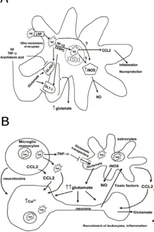
AIDS-related neurodegenerative disorders are found in one third of adults and half of the children affected with the disease. Productive infection of the central nervous system (CNS) occurs in macrophages and microglia, but is also found in astrocytes. The deterioration of brain tissue (leading do dysfunction of cognition, dementia and eventually paralysis) however is found even in absence of a productive infection in the neuronal tissue, or of malignancies and opportunistic infections (Price et al. 1988). The discrepancy between the lack of detectable HIV-1 and the severity of brain damage in infected individuals has led to the hypothesis that damage might due to the release of viral products from HIV-infected cells as macrophages or microglia; such molecules could trigger a pathological alteration of neuronal cells by disrupting the normal pattern of gene expression.
INTRODUCTION
37 Transforming growth factor-β1 (TGF-β1) gene expression is stimulated in HIV-1 positive individuals (Kekow et al. 1990); it has been suggested that this growth factor could act as a chemoactractant, recruiting infected monocytes in the brain, thus enhancing CNS dysfunction, and altering the expression of neurotoxic cytokines. Moreover, monocyte infiltration in the CNS, even in the absence of monocyte infection, is associated with AIDS- induced dementia, probably due to the toxic effect exerted by monocyte-derived molecules.
As the Tat protein, upon secretion from infected cells, can penetrate uninfected cells, it could alter the expression of several genes. For example, its upregulating effect on TGF-β1 (Rasty et al. 1996) and MCP-1 (monocyte chemoactractant protein-1) (Conant et al. 1998) were demonstrated both in vitro and in the mouse brain, thus suggesting a key role for secreted Tat in monocytic infiltration and in subsequent neurotoxin release.
Fig. 1.11: Tat interaction with uninfected monocytes (King et al.
2006)
Extracellular Tat also up-regulates CCR5 chemokine receptor on human peripheral blood monocytes, an event capable of enhancing the migratory response and the transmigration of these cells across a tissue model of blood-brain barrier in vitro (Weiss et al. 1999). Moreover, Tat is able to
INTRODUCTION
38
induce the expression and the extracellular release from activated macrophages and astrocytes of Tumor Necrosis Factor-α (TNF-α), the levels of which closely correlate with the severity of dementia (Chen et al. 1997; New et al. 1998).
Fig. 1.12: Tat neurotoxic activities (A) Tat interaction with astrocytes
(B) Tat neurotoxic effect is mediated by an interplay between different cell types triggered by Tat pleiotropic activities (King et al. 2006)
In order to better define the functional damage induced by Tat in the CNS, several groups have examined both neuronal injury and spatial learning. They found that the intracerebroventricular injection of Tat leads to the
INTRODUCTION
39 attenuation of spatial learning and correlates with the suppression of Long Term Potentiation (LTP) (Li et al. 2004). Unlike what had been previously described by other authors (Sabatier et al. 1991), in this study the intra-hippocampal injection of Tat did not lead to neuronal cell loss, thus suggesting that Tat induces neuronal dysfunction before causing neuronal cell death.
Kaposi’s sarcoma
Kaposi’s sarcoma (KS) is an angioproliferative disease very frequent and aggressive when found in HIV infected individuals. All forms of KS possess similar histological features, showing the presence of spindle-shaped cells, vascular smooth cell, endothelial cells, fibroblasts, inflammatory cells and high vascularization. KS lesions are associated with infection by HHV8 (human herpes virus 8) and are characterized by a complex interplay of cytokines (Interleukin (IL)-1, IL-6, Interferon (IFN) - α, TNF-α), angiogenic growth factors (among others, FGF-2, vascular endothelial growth factor (VEGF)-A and VEGF-C), extracellular matrix components and integrins (Fife and Bower 1996; Nickoloff and Foreman 1996).
The evidence that KS is much more frequent and aggressive in HIV infected individuals originally led to the hypothesis that somehow HIV infection itself might play an important role in favoring KS onset and progression.
Tat mimics the activity of fibronectin and vitronectin by binding their cell surface receptors through its RGD (Arg-Gly-Asp) motif; however, the heparin binding activity of its basic domain is also involved in the process of migration (Barillari et al. 1993).
Moreover, Tat is able to mimic extracellular proteins involved in adhesion, migration and growth of both KS and endothelial cells, thus potentially enhancing angiogenesis and KS progression. However Tat is not able to induce a response in primary endothelial cells unless they are activated by
INTRODUCTION
40
inflammatory cytokines (IFN-γ, TNF-α and IL-1β) (Fiorelli et al. 1999), which in turn activate the release of FGF-2.
In addition to cellular adhesion, integrins play a key role in angiogenesis, by modulating migration, apoptosis and response to growth factors of endothelial cells. Tat RGD domain is able to induce endothelial cells migration, mimicking the effect of vitronectin and fibronectin, stimulating the expression of matrix metalloproteinase (MMP)-2 (Toschi et al. 2001), and of focal adhesion kinases necessary for the invasion of the extracellular matrix. In addition to these activities, the binding of Tat to integrins enhance the cellular response to FGF-2 (Barillari et al. 1999).
The basic domain of Tat binds extracellular matrix heparan-sulfate proteoglycans (HSPGs) thus facilitating the interaction between the RGD domain and the integrins; on the other hand, binding of Tat to the HSPG retrieves soluble FGF-2 from the extracellular matrix, thus promoting cell growth (Folkman et al. 1988) (similar to Tat, FGF-2 binds HSPG upon secretion). Finally, the basic domain can bind the VEGFR-2, thus stimulating its phosphorylation and promoting endothelial cells proliferation (Mitola et al.
2000).
Fig. 1.13: Model for the role of Tat in the pathogenesis of acquired AIDS-associated Kaposi’s sarcoma (Foreman 2001)
INTRODUCTION
41
The immune response
Several studies on HIV-infected patients showed a marked increase of the inflammatory response in the brain tissue, due to the effect of Tat on the expression of several pro-inflammatory cytokines and growth factors, such as the aforementioned IFN-γ, TNF-α, IL-1β, as well as TGF-β, 2, 6, IL-8, IL-10, CXC-chemokine receptor 4, IL-2 and IL-4 and TNF-α receptors. Moreover, HIV infected individuals display a significant immune hyperactivation, leading to lymphocyte proliferation, expression of T cell activation antigens and increased cytokine expression.
During the early stages of HIV infection, Tat is selectively taken up by mature dendritic cells (Fanales-Belasio et al. 2002), where it induces a upregulation of the major MHC genes and of Th1 cytokines and β chemokines, therefore promoting the antigen presenting capacity of these cells, and enhances the recruitment of T cells by means of reprogramming gene expression in dendritic cells (Izmailova et al. 2003). Since the chemokines induced by Tat recruit activated T cells and macrophages, HIV can use the dendritic cells as vehicles to infect its ultimate targets of infections.
As HIV cannot infect resting T- cells, Tat could also facilitate the spread of HIV infection by upregulating the expression of IL-2, thus promoting the priming of naive T-cells (Ott et al. 1997).
A number of different and sometimes controversial studies suggest several immunosuppressive activities for extracellular Tat, which may account for the immunosuppression observed in AIDS disease.
These activities range from apoptosis of peripheral blood cells (Li et al.
1995), to impairment of natural killers (NK) cells (Poggi et al. 1998; Zocchi
et al. 1998), to the inhibition of antigen-specific T-cell activation response (Viscidi et al. 1989), to the upregulation of the production in peripheral blood monocytes of IL-10 (Bennasser and Bahraoui 2002), a highly
INTRODUCTION
42
immunosuppressive cytokine the levels of which correlate with disease progression in HIV infected individuals.
Despite this intense research on the properties of extracellular Tat, however, the issue whether Tat might be directly involved in the induction of immunosuppression in HIV-1 infected patients still remains unsolved. HIV-1 Vpr
Vpr is a small protein of 96 aminoacids (14 kDa) rich in basic residues, which is well conserved in HIV-1, HIV-2 and SIV (Simian Immunodeficiency Virus) (Tristem et al. 1992). Its real functions during the natural course of infection are still under debate; nonetheless, the role of Vpr in AIDS pathogenesis appears crucial, as vpr null mutants show decreased viral replication and delayed disease progression in rhesus monkeys experimentally infected with SIV (Tristem et al. 1992). As a result of extensive study in a number of in vitro, in vivo and ex-vivo systems, Vpr has been shown to play, despite its small size, a wide number of functions during viral replication, ranging from nuclear import of the pre-integration complex, to cell cycle progression, to transactivation of the viral LTR as well as of host genes.
Intracellular activities of Vpr
Along the viral life cycle, a first activity of Vpr is to influence the fidelity of reverse transcription: in addition to a potential role in the initiation step of the reverse transcription (Stark and Hay 1998), Vpr was shown to modulate the in vivo mutation rate of HIV-1 by interacting with the nuclear form of uracil DNA glycosylase (UNG2) (Mansky et al. 2000), an enzyme involved in the base excision repair pathway that specifically removes the RNA base uracil from DNA. The association of Vpr with UNG2 in virus-producing cells allows the incorporation of a catalytically active enzyme into HIV-1 particles, where UNG2 may directly influence the reverse transcription accuracy (Mansky et al. 2000), contributing to the ability of HIV-1 to replicate in
INTRODUCTION
43 primary macrophages and nondividing cells (which express low levels of UNG and contain relatively high levels of dUTP) (Sandgren et al. 2002). Despite the lack of any identifiable canonical nuclear localization signal (NLS), Vpr displays evident karyophilic properties and is rapidly targeted to the host cell nucleus after infection (Lochelt et al. 1993). One possibility is that Vpr primarily serves to dock the PIC at the nuclear envelope, while IN and MA act in cooperation with the central DNA flap to target the viral DNA to the nucleus.
In addition to its non conventional NLS for targeting into the nucleus, Vpr is a dynamic mobile protein able to shuttle between the nucleus and cytoplasmic compartments (Jenkins et al. 2001; Sherman et al. 2001; Sherman et al. 2003). The exact role of the nuclear export signal (NES) in the function of Vpr is not known but since Vpr is rapidly imported into the nucleus after biosynthesis, the NES could redirect it into the cytoplasm for subsequent incorporation into virions during the late budding step of the virus life cycle (Jenkins et al. 2001; Sherman et al. 2003).
A further important biological activity of SIV and HIV Vpr proteins is related to their ability to induce an arrest in the G2 phase of the cell cycle of infected proliferating human and simian T cells (Di Marzio et al. 1995; He et al. 1995; Jowett et al. 1995; Re et al. 1995; Bartz et al. 1996; Planelles et al.
1996). The biological significance of this arrest during the natural infection is not well understood, but as the HIV-1 LTR seems to be more active in the G2 phase, the G2 arrest may confer a favorable cellular environment for efficient transcription of HIV-1. Such increase transcriptional activity from the viral LTR in arrested cells expressing Vpr (Subbramanian et al. 1998; Gummuluru and Emerman 1999; Hrimech et al. 1999), could be mediated through cis-acting elements, found in the LTR promoter which are bound by Vpr.
Though no specific DNA sequence targeted by Vpr has been yet identified (Zhang et al. 1998; Kichler et al. 2000), Vpr displays high affinity for nucleic
INTRODUCTION
44
acids; Vpr may function as an adaptor molecule for an efficient recruitment of transcriptional co-activators (such as GRE, p300/CBP) to the HIV-1 LTR promoter and thus enhances viral replication. Additionally, it may be involved in the activation of host cell genes inducing cellular pathways in relation with the AIDS pathogenesis.
HIV infection causes a depletion of CD4+ T cells in AIDS patients, which results in a weakened immune system, impairing its ability to fight infections. The major mechanism for CD4+ T cell depletion is programmed cell death, or apoptosis; even though the exact contribution of Vpr as a pro-apoptotic factor responsible for the T cell depletion observed in the natural course of HIV infection is still unknown, it was repeatedly evidenced that Vpr has cytotoxic potential and is able to induce apoptosis in many in vitro systems. However, controversial results indicating that Vpr can also act as negative regulator of T cell apoptosis have been reported (Ayyavoo et al.
1997; Conti et al. 1998).
Other laboratories have then shown that synthetic Vpr, as well as truncated Vpr polypeptides, are able to induce apoptosis by directly acting on the mitochondria, leading to the permeabilization of the mitochondrial membrane and subsequent dissipation of the mitochondrial transmembrane potential (∆Ψm) (Jacotot et al. 2000). This direct effect of Vpr is related to its ability to interact physically with the adenine nucleotide translocator (ANT), a component of the permeability transition pore of mitochondria localized in the inner mitochondrial membrane. The interaction between Vpr and ANT triggers permeabilization of the inner membrane followed by permeabilization of the outer mitochondrial membrane with consequent release of soluble intermembrane proteins, such as cytochrome c and apoptosis inducing factors, in the cytosol.
INTRODUCTION
45 Fig. 1.14: Overview of Vpr biological activities
Cytotoxic effect of extracellular Vpr
Extracellular or free Vpr that exists in cell-free and virus-free state was detected in great amounts in sera and cerebrospinal fluid (CSF) of HIV-1 infected patients (Levy et al. 1994). However, it has not yet been clearly established whether extracellular Vpr results from breakdown of infected cells and virus particles or release from infected cells. Interestingly, some studies found that purified intracellular Vpr could enter the cells when added to cultured cells (Levy et al. 1995; Huang et al. 2000).
The carboxyl-terminal end of Vpr was shown to cause structural defects in cell membranes, indicated by osmotic sensitivity and gross cell enlargement (Macreadie et al. 1995); these effects were dependent on the sequence HFRIGCRHSRIG, containing two H(S/F)RIG motifs (Macreadie et al. 1995). When extracellular peptides containing the above sequence were added, they rapidly entered yeast cells and caused cell membrane permeabilization
INTRODUCTION
46
and death in a variety of yeast cells (Macreadie et al. 1996). Extracellular addition of synthetic Vpr peptides containing the H (F/S) RIG repeat motif showed similar consequence in mammalian cells. When this peptide was added externally to human CD4+ cells, it induced mitochondrial membrane permeabilization, dissipation of mitochondrial transmembrane potential, morphological changes, the formation of apoptotic bodies and breaks of DNA chain (Arunagiri et al. 1997).
According to these last findings, the existence of circulating Vpr in infected individuals, might also explain the toxic effects exerted by Vpr on bystander, uninfected cells.
Vpr and AIDS dementia complex
Relatively high levels of Vpr were detected in the CSF of HIV-infected patients with neurological defects (Levy et al. 1995); as macrophages and microglia of human brain are infiltrated during HIV-1 infection (Nottet and Gendelman 1995; Nottet 1999), these cells are thought to release extracellular Vpr to the CSF. Additionally, infected astrocytes may also release Vpr (for a review on the effect of HIV on the neural tissue, see (Jones and Power 2006)).
Based on several studies demonstrating the neurotoxicity of extracellular Vpr, the protein was then shown to exhibit channel-forming capacity in the membrane of intact cells when added extracellularly. Moreover, recombinant extracellular Vpr associated directly with the plasmalemma of hippocampal neurons caused a large inward cation current and depolarization of the plasmalemma, finally resulting in cell death (Piller et al. 1998). The first 40 amino acid residues of N-terminal domain of Vpr (N40) affect the ion channel activity thus showing cytotoxic effects, while the C-terminal domain is involved in rectification of Vpr currents. Moreover, the N40 has the cytotoxic ability similar to intact Vpr, including depolarization of the
INTRODUCTION
47 plasmalemma and cell death in cultured hippocampal neurons (Piller et al.
1999).
Extracellular Vpr also showed toxic effects on brain activity; when added to mixed embryonic rat brain cultures, Vpr-induced cell death was observed. Similarly, Vpr-induced cell death was also observed in enriched primary cortical rat astrocytes, and it was considered that death was mainly caused by necrotic mechanism (Huang et al. 2000). On the other hand, extracellular Vpr was shown to induce the death of human neuronal cells through apoptosis (Patel et al. 2000).
HIV-1–associated nephropathy
HIV-associated nephropathy (HIVAN) is unique in that the etiology of podocyte injury is genetically well defined, namely caused by HIV-1 itself. Patients with HIVAN present with heavy proteinuria and rapid progression to end-stage renal failure with collapsing focal segmental glomerulosclerosis and microcystic tubular dilation (Bourgoignie 1990; Humphreys 1995; Klotman 1999; Ross and Klotman 2002; Kimmel et al. 2003; Weiner et al.
2003; Ross and Klotman 2004).
Transgenic mice carrying the HIV-1 long terminal repeat, tat and vpr, but not mice that carrying an HIV-1 genome defective in vpr, develop glomerulosclerosis. Selective podocyte expression of either Vpr or Nef alone can induce podocyte injury that leads to glomerulosclerosis; in addition, a prominent synergistic effect of these two proteins on podocyte injury was reported (Zuo et al. 2006).
Role of Vpr in AIDS-related insulin resistance/lipodystrophy syndrome
Recent advances in the development of the nucleotide and non-nucleotide analogues acting as reverse transcriptase inhibitors (NRTIs) and the non-peptidic viral protease inhibitors (PI), and their introduction in the management of patients with AIDS, either alone or in combination, have
INTRODUCTION
48
dramatically improved the clinical course of the disease and prolonged life expectancy in patients with AIDS. The increase in life expectancy associated with the long-term use of the above antiviral agents, however, has generated novel morbidities and complications. Central among them is the quite common AIDS-related insulin resistance and lipodystrophy syndrome, which is characterized by a striking phenotype and marked metabolic disturbances.
A role for the Vpr protein in these disorders has been suggested by a recent study which investigated the capacity of Vpr to co-activate the glucocorticoid receptor, which potentiates the action of glucocorticoid hormones, thereby inducing tissue glucocorticoid hypersensitivity (Kino and Chrousos 2004). Vpr interacts with novel 14-3-3 proteins, promoting their interaction with Cdc (Cell division cycle) 25 and subsequently suppressing their transcriptional activity by segregating Cdc25 into the cytoplasm. The same study also showed that Vpr suppresses the association of 14-3-3 with other partner molecules, in particular the FoxO (Forkhead box O) transcription factors. Since the FoxO proteins function as negative transcription factors for insulin, Vpr may cause resistance of tissues to insulin. Through these two newly identified functions of Vpr, namely, coactivation of glucocorticoid receptor activity and inhibition of insulin effects on FoxO proteins, Vpr may participate in the development of AIDS-related insulin resistance/lipodystrophy syndrome.
Effects on immune system
Cellular immunity, specifically MHC-restricted CTL responses, is thought to play an intrinsic role in protection and clearance of many viral infections. Though HIV-1 infection is controlled by the immune response initially, the immune system fails to clear the virus and ultimately loses control of viremia through unclear mechanisms (Koup et al. 1994; Pantaleo et al. 1994; Fauci 1996; Barouch et al. 2000; Piguet and Trono 2001; Letvin et al. 2002; Letvin
INTRODUCTION
49 and Walker 2003; Nickle et al. 2003). Furthermore, there is evidence that the host immune response is compromised early in HIV infection.
Vpr could be a possible important player in the compromised immune control of HIV as it exerts significant effects on cellular proliferation, differentiation, regulation of apoptosis, modulation of cytokine production and transcription in vitro. Many of these Vpr-mediated cellular events have been observed in a wide variety of cell lineages, suggesting that Vpr targets basic eukaryotic cellular pathways.
As an extracellular delivered protein, Vpr down-modulates the expression of several immunologically important molecules including CD40, CD80, CD83 and CD86 costimulatory molecules on MDM and MDDC (monocyte-derived dendritic cells) (Muthumani et al. 2005), suggesting that Vpr could interfere with DC (dendritic cells) functions or maturation, or both.
The potential consequences of failed APC maturation during HIV infection could be significant: for instance, Vpr-mediated CD40 repression could contribute to the inability of the host immune system to continue to mount an effective response against HIV, as the engagement of CD40L on APC by CD40 on CD4+ or CD8+ cells were shown to ‘condition’ the APCs for antigen-specific CTL activation, and facilitate induction of a memory T cell response. The CD80 and CD86 molecules are surface glycoproteins and members of the immunoglobulin superfamily, which are only expressed on professional APCs (Sharpe and Freeman 2002). Blocking these costimulatory signals leads to T cell unresponsiveness; therefore, despite efficient migration of APCs into regional lymph nodes, the effects of Vpr may influence both presentation and activation of T cells, thus influencing viral clearance. This unique property of Vpr could ensure infection in a T cell rich environment without immune clearance and favor viral propagation.
Similarly, it has been observed that Vpr affects the expression of the CD33 antigen, a transmembrane surface protein specific for myeloid lineage cells, which can strongly influence the antigen presentation properties of DCs.