PhD Program in
Biotechnologies for Human Health
Thermo-responsive methylcellulose based hydrogel for
regenerative medicine applications
Candidate: Andrea Cochis
Tutor: Prof.ssa Lia Rimondini
Phd coordinator: Prof. Claudio Santoro
2013-2014
Cycle 26°
1
INDEX.
P
REFACE pag.3C
HAPTER1
. Thermo-reversible methylcellulose-based Hydrogel production, mechanicalcharacterization and biocompatibility evaluation pag.6
Introduction pag.7
Materials and Methods pag.9
Results and Discussion pag.13
Conclusions pag.19
Acknowledgements pag.20
References pag.20
C
HAPTER2
. MC hydrogel as 3D matrix for the bioreactor-guided in vitro production of artificialcartilage pag.21
Introduction pag.22
Materials and Methods pag.25
Results and Discussion pag.28
Conclusions pag.33
Acknowledgements pag.33
References pag.34
C
HAPTER3
. MC hydrogel as scaffold for the in vitro biofabrication of implantable proto-tissuescell sheets pag.36
Introduction pag.37
2
Results and Discussion pag.43
Conclusions pag.48
Acknowledgements pag.48
References pag.48
C
HAPTER4
. MC hydrogel-derived cell sheets (CS) for skin regeneration pag.50Introduction pag.51
Materials and Methods pag.54
Results and Discussion pag.56
Conclusions pag.63
Acknowledgements pag.64
3
PREFACE.
Background.
Tissue engineering is a very attractive branch of modern medicine. In fact, it represent not only a promising vehicle to face the “regenerative medicine” requests, but it is also a very sophisticate tool for the in vitro studies of complex biological systems such as tissues development and regeneration. A great step forward in tissue engineering advancement is represented by the improvement of biomimetic materials that are able to active interact with cells in order to provide microenvironments suitable for the clinical application and the basic theory progress. From this point of view, hydrogels are nowadays considered as a very interesting materials thanks to their biocompatibility and controllable mechanical features. Their utilization range from the 3D matrix for cells development to the drug delivery systems. Thus, the hydrogel development course is an excellent possibility for the making of a platform for biological and clinical investigations.
Aim and presentation of the Thesis.
In this PhD Thesis, it will be reported the three years activities of the candidate. The main purposes has been the development of a novel hydrogel with thermo-sensitive and thermo-reversible features. After that first crucial step, the selected hydrogel has been tested for biomedical purposes facing different applications. Therefore, the Thesis has been divided into 4 different chapter in order to better detail the succeeding steps that are all related to each others. In turn, each of the 4 chapters is characterized by a specific introduction, materials and methods and results and discussion sections with the purpose to minutely explain the biological hypothesis encountered and the strategies used to face them. As support of the selected techniques, a chapter specific references section was spent to each one.
• In the Chapter 1, it will be described the methylcellulose-based (MC) thermo-reversible hydrogel development from a mechanical point of view. The purpose was to select the best salts-MC w/v compositions in order to produce a hydrogel able to reverse the physical phase (from liquid to gelation and vice-versa) at 37°C that is the optimal temperature for cells cultivation. Furthermore, the selected hydrogel cytocompatibility was first verified in vitro and then the biocompatibility evaluation was extended to the in vivo immunological reaction study. Experimental procedures of this part were performed in the Biomedical Materials Laboratory of the Health Sciences Department in Novara for the biological part and in the Laboratory of Materials Sciences of the Polytechnics of Milan for the mechanical assays part.
4
• After the MC-hydrogel biocompatibility confirmation, in the Chapter 2, it will be showed a first biological application for the in vitro production of artificial cartilage. In this section, the MC hydrogel has been used in combination with a porous polyurethane scaffold (PU) as 3D matrix for mesenchymal stem cells (MSCs) chondrogenesis. The MSCs differentiation has been carried out using a specific bioreactor able to promote chondrogenesis by the appliance of compression and shear forces on the PU-hydrogel-MSCs composite. By this way, it was possible to produce artificial cartilage via mechanical stimulation, avoiding the use of biochemical stimulation. The successful results were an important proof of the MC-hydrogel suitability as 3D matrix for cells development. All the experiments were performed in the Musculoskeletal Regeneration Laboratory of the AO Research Institute of Davos Platz (Switzerland) within the COST-NAMABIO exchange program (European grant to the candidate).
• In the Chapter 3, it will be described the use of the hydrogel as scaffold for the in vitro biofabrication of implantable cell sheets (CS). Therefore, it will be detailed the protocol used to cultivate a monolayer of cells onto the MC-hydrogel surface and the subsequent spontaneous detachment allowed by the hydrogel temperature-guided inversion phase. CS were successfully produced, collected and their ability to adhere to a new substrate was in vitro confirmed. Afterwards, CS were in vivo implanted into recipient mice; thanks to the presence of cells natural extracellular matrix (ECM), the CS were able to adhere to the naïve tissue without the use o sutures. The successful implantation was confirmed by the neo-vascularization occurred after 2 weeks. Thus, the suitability of the MC-hydrogel for the biofabrication of implantable CS was confirmed. All the procedures were performed in the Biomedical Material Laboratory.
• Finally, in the Chapter 4, it will be detailed the MC-hydrogel derived CS technique application for skin regeneration. For this purpose, a complex system composed of a human recombinant elastin (HELP) layer and gingival derived primary human fibroblasts (HGF) was realized. The HELP-HGF CS was detached from MC-hydrogel surfaces and in vitro characterized. Afterwards, HELP-HGF CS were implanted into the dorsal skin of nude mice carry a 1cm diameter skin excision mimicking a 3rd degree burn. The CS were attached by fibrin glue avoiding the use of sutures and after 1and 3 weeks the regenerative potency of the CS was confirmed as a complete repair of the skin excision was noticed. These findings represented a strong proof of the CS technology suitability for soft tissues repair. The experiments were performed in the Biomedical Materials Laboratory, while the HELP were kindly produced and
5
provided by the Laboratory of Recombinant proteins of the Life Sciences Department of the University of Trieste.
Finally, the candidate declare that all the presented data were original and that all animals surgical procedures were performed after local ethical committee approval, following pre-approved surgical procedures.
6
Chapter 1.
Thermo-reversible methylcellulose-based Hydrogel
production,
mechanical
characterization
and
biocompatibility evaluation.
In collaboration with the Polytechnics of Milan.
7
1.1
INTRODUCTION
1.1.1. Hydrogels characteristics.
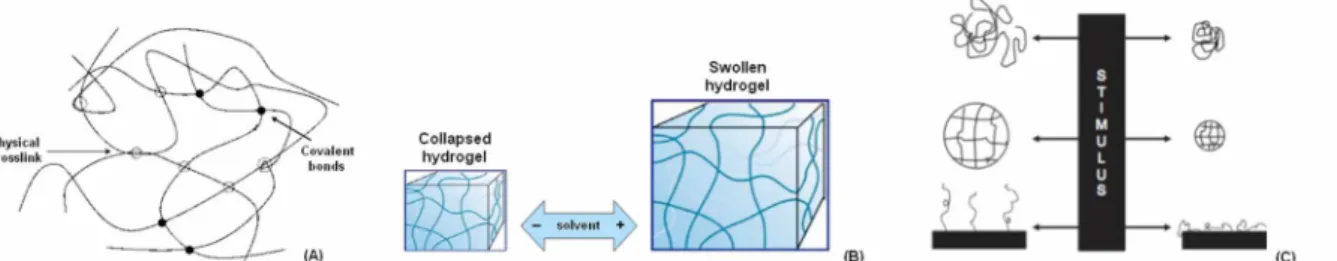
Between the polymeric materials class, a very interesting role is played by the hydrogels. They can be defined as 3D macromolecular structures able to retain a high amount of solvent thanks to their thermodynamic affinity with it. In fact, hydrogels are composed by complex reticular networks supported by physic, ionic and covalent interactions [1-3]. Therefore, hydrogels present both high-energy (covalent bonds) and low-high-energy (hydrophobic interactions) crosslinks (Figure 1A). However, it is very important the presence of high-energy covalent bonds between polymer chains since they confer to the hydrogel the ability to absorb the solvent without losing their macromolecular integrity (Figure 1B) [2].
Hydrogels can be composed by a multiple repetition of a single polymer (homopolymer) or by the presence of different polymers (copolymer); moreover, it is possible to distinguish between hydrogels presenting a random polymer chains network (amorphous), a partly ordered network (semi-crystalline) or a complete ordered network (crystalline) [4, 5].
Another important hydrogels peculiarity is related to the chemical groups that strongly condition the ionic charge. From this point of view, it is possible to divide hydrogels in neutral (no charge), positive/negative charged (presenting a prevailing (+) or (-) charge) or ampholytes when they have the ability to act both as positive or negative charged responding to an environmental stimulus. Neutral hydrogels develop a complex 3D structure thanks to the energy balance between the solvent osmotic force and the deformation energy of the polymer chains network. In the case of charged hydrogels, the ability to absorb solvent is mainly leaded by two driven forces: the electrostatic repulsion between the polymer chains charges and the osmotic force derived by the presence of charges into the solvent-polymer solution [4, 5].
1.1.2. Hydrogels as smart materials.
A novel interesting class of materials suitable for biomedical applications consists on the “smart” materials. These particular polymers are able to range their physical/chemical properties following an external stimulus such as pH or temperature changing [6-9] (Figure 1C). Some hydrogels belong to this peculiar order of materials. In fact, they present two distinct phases (Figure 1B): in the (I) collapsed phase, the hydration solvent is repulsed since the interactions energy between the polymer chains are predominant to the solvent-polymer ones. In the (II) swollen phase, the hydration solvent is absorbed because the polymer chains-hydration solvent become predominant to the chains-chains ones [7]. The passage from a collapsed to a swollen state correspond to a physical change of the
8
hydrogel from a liquid phase, in which the polymer is solved, to a gel phase. In the gel phase, the hydrogel finally tend to jellify because of the polymer chains network reticulation.
Figure 1 A-C. Schematic representation of the low/high energy network supporting hydrogel macromolecular structure (A). Hydrogel ability to absorb solvent without losing the polymer chains crosslink (B). Smart materials are able to vary their chemical/physical phase following an external stimuli (C).
(A=modified from [3], B=modified from [6], C=modified from [9])
1.1.3. Methylcellulose-derived thermo-reversible hydrogels.
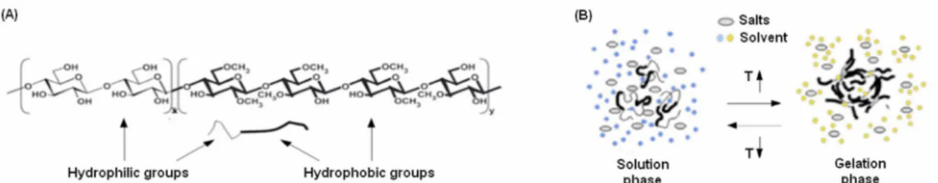
Methylcellulose (MC) is commonly derived from cellulose by the substitution of hydroxyl groups (-OH) with methoxy groups (-OCH3) in a two-steps process involving first sodium hydroxide and then methyl chloride [10]. At the end of the process, MC achieve a structure characterized by the presence of both hydrophilic (-OH) and hydrophobic (-OCH3) groups (Figure 2A). Accordingly, the MC phase in solution is directly determined by the system temperature (T) [10-11]. In fact, when T>55°C MC became not solvable due to the prevalence of the polymer chains hydrophobic interactions; on the opposite, when T<20°C MC results as solvable as the hydrophilic interactions between solvent and polymer chains are predominant on the hydrophobic ones. So, by mixing the MC powder in a defined weight/volume (w/v) percentage in a solvent, it is possible to obtain smart hydrogels that are able to undergo a solution-gelation (sol-gel) phase change guided by the system temperature [12-14].
The presence of salts in the hydrogels hydration solvent (that is normally water) is very useful to improve the stability of the hydrogel and it is also a tool to determine the decrease of the phase-changing temperature. In fact, when the T is raised, the system tend to absorb the heat and to convert it into the energy necessary to originate the MC chains-chains crosslink. The water molecules tend to accumulate close to the salts reducing the total number of hydrophilic interactions. Therefore, the energy that is necessary to broke these hydrophilic interactions and undergo MC chains-chains crosslink is lower and the system T for the phase-change decrease (Figure 2B) [12-14].
As in the presence of salts the system sol-gel phase transition is more easy controlled and require lower T, it is very important to underline that this phenomenon results as reversible. Is in fact
9
possible with MC-based hydrogels to carry out the sol-gel transition and return to the sol phase by lowering the T for a gel-sol phase transition [12].
Figure 2 A-B. Methylcellulose chemical structure characterized by the presence of both hydrophilic (left) and hydrophobic (right) groups (A). Representative scheme of the sol-gel phase transition of MC-based hydrogels in the presence of salts and solvent (B).
(Modified from [4])
In conclusion, it is possible to consider the MC-based hydrogel as a smart thermo-reversible material sensitive to the system temperature.
1.1.4. Aim of the work.
In this first part of the Thesis, thermo-responsive hydrogels composed of MC and different salt solutions were prepared and characterized. Rheological analysis were performed to determine variations of complex viscosity (η*), conservative shear modulus (G’) and viscous shear modulus (G”) of hydrogels after temperature increase. The thermo-reversible features of MC-based hydrogels was also investigated by the inversion test and rheological characterization. Selected prepared MC-based hydrogels were preliminary in vitro tested to investigated possible cytotoxicity towards mouse fibroblasts. Afterwards, gel-phase MC hydrogels biocompatibility were in vivo evaluated into wild type mice.
1.2. MATERIALS and METHODS.
1.2.1. Materials.
Methylcellulose (MC, Methocel A4M, η =4000 mPa×s for a 2% w/v aqueous solution at 20°C) was kindly supplied by The Dow Chemical Company. All basic chemicals were obtained from Sigma-Aldrich unless mentioned otherwise.
1.2.2. Thermo-reversible MC-based hydrogel preparation.
Aqueous MC solutions in different concentrations (Table 1) were prepared with the addition of selected salts (sodium sulphate, Na2SO4, sodium phosphate, Na3PO4, calcium chloride, CaCl2) or
10
Phosphate Buffered Saline (PBS), varying their concentration in the MC solution (Table 1).
In the following, the composition of the prepared and characterized MC hydrogels will be summarized by acronyms in which the first number is referred to the MC concentration (% w/v), the letters indicate the salt and the last number is referred to the salt or PBS concentration (moles/l for salts, g/l for PBS). The preparation of the MC hydrogels consisted in three main steps, as reported in Figure 3A: 1) preparation of the saline solution; 2) addition of the MC to the saline solution; 3) hydration of the MC powder (i.e. sol phase).
Step 1: preparation of saline solution: saline solutions were prepared mixing the appropriate quantity of the salt with distilled water or PBS (Table 1) at 50°C under magnetic stirring.
Step 2: mixing: the MC was added to the saline solution under stirring at 50°C using the dispersion technique [15], to allow a homogenous distribution of the MC powder in the solution. The MC suspension was poured in tissue culture polystyrene Petri dishes or in the wells of TCPS multi-well plate.
Step 3: MC suspension hydration: to allow the complete hydration of the MC powder, after the mixing step, the suspension was cooled down to 4°C. At 30-35°C, depending on the MC hydrogel composition, MC powder started to hydrate and the viscosity of the solution increased (gel phase). The prepared MC solutions (Table 2) were then stored at 4°C overnight to allow the complete hydration of the MC powder, thus obtaining the hydrogel in the sol phase.
1.2.3. Gelation test.
The physical gelation of MC hydrogels was observed visually using the inversion method already described in literature [13, 15]. Briefly, specimens (10 ml each, n=3) of MC hydrogels (Table 2) were put in different Falcon tubes (15 ml), and heated up to 40°C in a standard bath. Temperature was then decreased down to 20°C, at approximately 0.5°C/min. At 37 and 20°C, the Falcon tube
11
was inclined of 90° and the possible flow of the MC solution was observed. At each temperature, the solutions/gels were allowed to equilibrate for 1 h. The gelation criterion was defined as the temperature at which the clear solution did not flow upon inversion of the Falcon tube [b].
1.2.4. Rheological characterization.
Rheological characterization of MC hydrogels was performed with a rotational rheometer (AR-1500ex, TA Instruments, USA), using a flat plate geometry (diameter=2 cm, working gap=1 mm). A home-made isolation chamber in polymethyl methacrilate (Plasting srl, Segrate, MI, IT) was designed and assembled to the rheometer to prevent the possible dehydration of the MC hydrogels during the test. Tests were performed using five samples for each considered MC hydrogel composition. To investigate the rheological properties of the MC hydrogels, dynamic viscosity (η*), storage shear modulus (G’) and viscous shear modulus (G”) were registered over the temperature range 5-50°C, with a temperature ramp of 5°C/min. The oscillation frequency during the temperature ramp was held at 1 Hz. Thermo-reversibility characteristic of the MC hydrogels was studied with a first run increasing the temperature from 4 up to 40°C and a second run decreasing the temperature down to 4°C (temperature ramp=10°C/min, oscillation frequency=1 Hz).
1.2.5. In vitro cytotoxicity evaluation.
In vitro cytotoxicity of the MC hydrogel extracts was assessed using murine fibroblasts (L929,
ECACC No. 85011425). The hydrogels with the highest salt concentration (Table 3) were prepared under laminar hood using saline solutions previously sterilized with antibacterial filter (Minisart 20 nm, Sartorius Stedim Biotech). Then, the obtained hydrogels were sterilized by UV light exposure (t=30 min). L929 fibroblasts were cultivated in Dulbecco’s Modified Eagle’s Medium (DMEM, Sigma-Aldrich) supplemented with 10% foetal bovine serum (Sigma) and 1% penicillin/streptomycin at 37°C, 5% CO2; when cells reached 80-90% confluence, they were detached by trypsin-EDTA solution, harvested and used for experiments. Cylindrical (diameter=10 mm, thickness=3 mm, n=3) samples of different considered hydrogels compositions (Table 3) were put into the wells of a 24 TCPS multiwell plate (CellStar, VWR-PBI International) and submerged for 1 week with 2 ml of complete medium.
Afterwards, surnatants were collected from different samples and used to cultivate cells (cell density=1 x 10⁴ cells/well, cell suspension=500
12
µl/well) for 24, 48 and 72 h. At each time-point, cells viability was evaluated by the (3-(4,5-Dimethylthiazol-2-yl)-2,5-diphenyltetrazolium bromide) colorimetric metabolic assay (MTT, Sigma). Briefly, 50 µl of MTT solution (3 mg/ml in PBS) were added to each well and incubated for 4 h in the dark. Then, surnatants were removed, formazan crystals were dissolved with 100 µl of dimethyl sulphoxyhde (DMSO), and 50 µl were spotted into a 96 TCPS multiwell plate. Sample optical density (O.D.) was evaluated by spectrophotometer (SpectraCount, Packard Bell) at 570 nm. Cells cultivated with fresh DMEM were used as positive control and their O.D. was considered as 100% viability. Furthermore, cells morphology was observed 72 h after seeding by light microscope (Leica AF6500, Leica Instruments). Experiments were performed 3 times using triplicates.
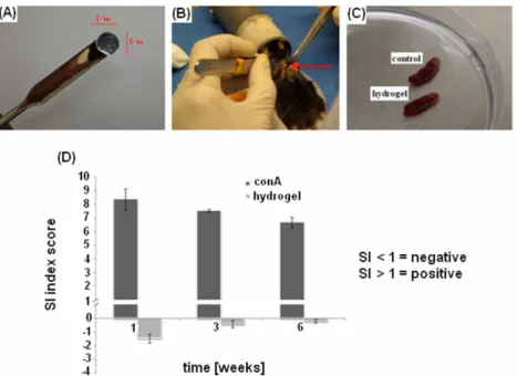
1.2.6. In vivo biocompatibility evaluation.
Considering the mechanical assays results and the promising cytocompatibility evaluation data, the 8% w/v Na2SO4 0.05M was selected as the best composition for further in vivo biocompatibility investigations. One centimetre diameter specimens of the selected hydrogel were subcutaneously implanted into a pocket realized in the dorsal skin of 6-8 weeks old mice (C57BL/6JOlaHsd, wild type, Harlan Laboratories) (Figure 8 A-B). After 1, 3 and 6 weeks cellular immune responses were determined by using a lymphocyte proliferation assay in tissue culture wells [16]. Spleens were harvested and single cell suspensions prepared by tissue disruption. Lymphocytes were washed, counted, and assessed for viability in trypan blue counting fluid, and adjusted to a suspension of 2.5 × 10⁶ cells/mL in Minimal Essential Medium Alpha modification (α-MEM, Sigma) supplemented with 10% FBS, 1% antibiotics. Triplicate aliquots were dispensed in wells previously coated with 100µl of hydrogel solution or buffer alone. Positive controls consisted of cells stimulated with the mitogen ConA (5 mg/mL) in additional wells to confirm that they were competent. Plates were incubated at 37°C, 5% CO2 for 48 hours. Cells viability was evaluated by the MTT assay and the cellular responses were expressed as stimulation indices (SI) calculated according to the formula: [(Mean OD of cells cultured with hydrogel - Mean OD of cells cultured without hydrogel) / Mean OD of cells cultured without hydrogel] x 100 [16].
1.2.7. Statistical analysis of data.
Results are expressed as mean and standard deviation. Data, where possible, were statistically compared by a t-test (Student test) or a One-Way ANOVA test (significance level p=0.05), using Bonferroni test for mean comparison (8.5 software).
13
1.3. RESULTS and DISCUSSION.
1.3.1. Gelation test.
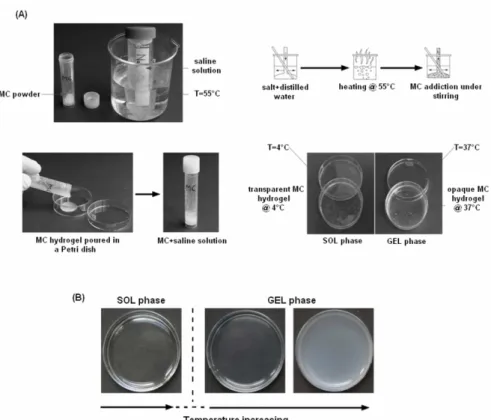
All the compositions of MC blended with saline solution changed from a clear solution at lower temperature (sol phase) to an opaque gel (gel phase) at elevated temperature (T ≥ 37°C). As a representative example (Fig. 3B), the 4SO0.2 hydrogel appears as a clear solution (sol phase, Fig. 3B, left panel) at low temperature (T = 4°C), then the gelation of the solution becomes evident by the opacity of the formed gel up to 37°C (gel phase, Fig.3B, right panel). The inversion test performed for all the investigated compositions (Table 2) evidenced the influence on the gelation temperature of the MC and the salt concentration (Fig. 4). In general, hydrogels obtained using high MC concentration showed a slower flow rate for the higher viscosity. On the contrary, hydrogels with low MC concentration showed a lower viscosity, hence flowed faster. Besides, at the same concentration of MC, different gelation temperature can be detected varying the salt (Fig. 4A). In particular, comparing 4SO0.1 and 4PBS10, a higher gelation temperature value was found for the hydrogel prepared with PBS. For the 8% w/v MC concentration, no correlation with type of salt was evidenced. Considering higher MC concentration, PBS and Na2SO4 showed the same low gelation temperature (T=20°C), instead, the gel prepared with CaCl2 maintained a higher gelation temperature (T=37°C). For all the investigated salts (Fig. 4B) and PBS (Fig. 4C) concentrations blended with MC, a decrease in the gelation temperature below 37°C was observed increasing the salt concentration. In addition, the limit salt concentration value, corresponding to the gelation temperature of 37°C, is not the same for all the investigated salt. This is due to a different ionic interaction between the salt ion and the MC. As an example, for 10SOx and 12SOx the limit concentration is 0.05 M, while for 12Clx is 0.1 M. Moreover, for PBS, the limit concentration is 10 g/l for 2PBSx and 8PBSx, while for 10PBSx and 12PBSx corresponds to 5g/l. These qualitative data indicated that the temperature at which the gelation starts can be varied by changing either the MC concentration and the formulation of the saline solution.
14
Figure 3 A-B. Schematic representation of the 3-steps hydrogel production process (A). Example of a sol-gel phase transition of a hydrogel spotted into a Petri dish (B).
Figure 4 A-C. Gelation temperature of MC blended with salt: (A) effect of concentration of MC blended with 0.1 M Na2SO4 or CaCl2, or 10 g/l PBS; (B) effect of Na2SO4 or CaCl2 concentration; (C) effect of PBS concentration. (x indicates the different MC concentration in the hydrogel).
1.3.2. Rheological characterization.
In Figure 5, the storage shear modulus (G’) and the loss shear modulus (G’’) obtained from the temperature sweep rheological analysis are reported for the compositions that showed a gelation temperature about 37°C in the inversion test. In fact, this value appears the optimal one for a possible use as smart hydrogel in regenerative tissue applications. For all the selected MC solutions, at low temperature (approximately in the range 5-10°C), G’ (Fig. 5 A-C) was lower than G’’ (Fig. 5
15
D-F) due to the viscous/liquid-like behavior of the MC solution, i.e. sol phase. Increasing the temperature, G’ first showed a decrease, reaching a minimum, then it rapidly increased for the sol/gel transition, as a soft elastic gel is formed. In addition, increasing the MC concentration higher values of G’ can be detected. Besides, increasing the salt concentration, at the same MC concentration, G’ increased and a slight shifting towards lower temperature (T<37°C) was evidenced for the gelation temperature, confirming the inversion test results.
Figure 5 A-F. Shear storage modulus (G’) and shear loss modulus (G’’) versus temperature for the solutions prepared varying MC and salt concentration: A-D) MC blended with Na2SO4; B-E) MC blended with CaCl2; C-F) MC blended with PBS. Curves represent the average behavior of G’ ad G’’ obtained by the rheological data collected at every 0.5°C temperature interval at a frequency of 1 Hz.
In the heating and cooling tests (Fig. 6), a hysteresis is observed for all the investigated solutions. In particular, a higher hysteresis is detected when MC concentration increases for MC blended with salts (Fig. 6A, e.g. 12SO0.02, 8SO0.1, Fig. 6B, e.g. 10Cl0.2, Fig. 6C, e.g. 12PBS5). The high hysteresis detected for some MC compositions could suggest that the solution did not reach the thermal equilibrium in the gelled state and that the dissociation in the MC gel-sol transition (during the cooling run) is not an exact reversal of the association process (during the heating run) [17]. MC based hydrogels prepared in this work exhibited reversible thermo-responsive properties. Phase transition was confirmed both by physical and rheological characterizations. Interesting, it was easily observed how temperature determined variations to hydrogel mechanical properties. When samples were heated, mechanical parameters increased confirming that polymer structure became
16
more and more ordinate and compact. Conversely, immediately after cooling down temperature, samples showed a decreasing of mechanical parameters which indicated that polymers were losing their compact structure to come back to the liquid phase. These properties are certainly related with MC chemical structure. In fact, it is characterized by the presence of both hydrophobic and hydrophilic groups. Methoxy groups (-CH3) represent the hydrophobic regions while hydroxy groups (-OH) represent the hydrophilic ones. At low temperature (< 10ºC) hydrophilic interactions between –OH groups and solvent are predominant so MC molecules are hydrated and the polymer structure is held together by simple entanglements. As temperature increase, hydrogels absorb energy and gradually lose their hydration water. Polymer-polymer interactions take place between – CH3 groups, forming a gel-network structure.
Figure 6 A-C. Temperature dependence of the shear storage modulus (G’) for the solution prepared varying either MC and salt concentration: A) MC blended with Na2SO4; B) MC blended with CaCl2; C) MC blended with PBS. Straight curves represent the average behavior of G’ during the heating up to 40°C, the dash curves during the cooling down to 5°C.
Rheological characterizations showed that MC and salts concentration strongly influenced hydrogels mechanical properties. In fact, the increasing of MC and salts concentration determined an increasing in mechanical parameters. Those are expected results because by adding more MC the final polymer will be composed by a higher number of hydrophobic regions that will form a more structured and compact gel-network. Increasing salts concentration, water molecules are forced to locate around them, reducing the intermolecular hydrogen-bond formation between water molecules and the hydroxyl groups of MC. Not only concentration but also the type of salts strongly influenced hydrogels mechanical properties. In fact usually salts have greater affinity for water than polymers molecules resulting from removing hydration water from the polymer and thus dehydrating the polymer. This phenomena is termed “salt-out” (or salt-assisted) effect and means that water is dropped out from polymer structure [4, 5, 14].
The ability of a salt to salt-out a polymer generally follows the salts order in the lyotropic series [14]. Cations follow the order Li+ > Na+ > K+ > Mg2+ > Ca2+ > Ba2+, and more common anions
17
follow the order CNS- < I- < Br- < NO3- <Cl- < tartrate < SO42- < PO43- [14]. These effects depend mostly on the anions. Accordingly, with the same MC and salts concentrations, more water molecules were removed from MC hydrogels when Na2SO4 was used than CaCl2.
Evaluating G’ and η* values obtained during cooling down phase, it was noticed that they were higher than heating ones. In contrast to the sharp increase of both the parameters from the heating process in the temperature range from 30 to 40°C, the gradual decrease of the parameters with temperature in the cooling process showed an outstanding deviation from the heating curve. This clearly indicates that the thermally induced hydrophobic dissociation is not an exact reversal of the hydrophobic association in the heating process. The hysteresis between the heating and cooling processes may be due to the existence of some associated aggregates or weak connections that have not completely disassociated [17]. Differences may be also due to a different dynamic in forming and destruction of the chain to chain interactions related to the different dynamic in absorbing and realising water. Only under 7ºC the values became again comparable.
Another factor that may affect the destruction of the polymeric structure is the time for which the system has equilibrated at the highest temperature before cooling. In this work, cooling process initiated immediately after the heating process. However, if the sample is allowed to equilibrate for some time before cooling starts, the time should have effect on the hydrogels behaviour during cooling process. Samples should be more difficult to dissociate because of the more perfect network formed, compared to a freshly formed gel at the same temperature and a different trend should be recorded. The aforementioned results indicated that the temperature at which gelation is initiated and the mechanical proprieties can be altered by the concentration of MC and the formulation of the aqueous solvent. In addition, MC hydrogel’s composition can be easily manipulated to obtain specific physic-chemical behaviours for different use.
1.3.3. In vitro cytotoxicity evaluation.
In vitro cytotoxicity tests were performed on extracts of complete medium previously put in contact
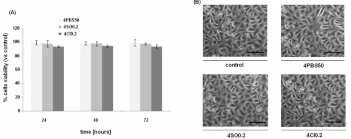
with 4SO0.2, 4Cl0.2, and 4PBS50 MC hydrogels for 7 days in order to exclude the release of toxic compounds. These hydrogels have been selected for the in vitro test for their higher content of salt used in the preparation of the hydrogels. In fact, a possible cytotoxic effect would be more evident for these hydrogels compared to the ones with a lower salt concentration. The MTT assay showed at each considered time-point (Fig. 7A), no cytotoxic effect caused by the possible release of toxic compounds from the tested MC hydrogels. Indeed, cells viability resulted in a range of 94-99% comparable to the control values for all the time-points with no significant statistically differences
18
(p>0.05) between the controls and the eluates. Besides, MTT results were also supported by the light microscopy observation (Fig. 7B). Cells cultivated for 72 h in contact with the MC hydrogel eluates showed a morphology, spread and density comparable when seeded in presence of the tested eluates and in fresh medium, confirming that no toxic compounds were released into the culture medium.
Figure 7 A-B. In vitro cytotoxicity evaluation results. (A) Cells viability after 24, 48 and 72 h after seeding in contact with hydrogel eluates; at each time-point, viability values were comparable between control and tested samples and no statistically significant differences were noticed (p>0.05); (B) optical microscopy images of L929 cells 72 h after seeding in contact with hydrogel eluates; a comparable cell morphology, spread and density was observed, supporting cell viability assay results. Bars represent means and standard deviations; bar scale = 200 µm.
According to the in vitro preliminary biological characterization results, the selected MC hydrogels showed to be not cytotoxic for mouse fibroblasts. In fact, the viability of cells cultivated in medium previously put in contact with the different hydrogels was comparable to the results obtained for cells cultivated with fresh medium. These data suggest that up to 7 days of hydrogel-medium direct contact, no toxic products were released into the medium. Since the hydrogels containing the highest amount of salts were tested, these findings could be extended also to the other compositions containing a lower salt amount. Besides, MC itself could not be considered as a possible source of toxicity since the one selected is a commercial MC powder largely used for different purposes such as for the food industry and it is well established its cytocompatibility.
1.3.4. In vivo immune response evaluation.
Mice implanted with hydrogel did not reported any macroscopic inflammatory reactions such as fibrosis (data not shown) also after a long period after operations. The collected spleens were comparable between control and hydrogel implanted mice (Figure 8 C) revealing the absence of macroscopic evidences of inflammatory processes. When the SI index was calculated at each
time-19
point, the hydrogel-coated samples score was always < 1 confirming that no inflammatory reactions were raised toward hydrogel. On the opposite, lymphocytes stimulated with the mitogen conA fast reacted reporting SI score always > 1. After 1, 3 and 6 weeks after hydrogel specimens implantation, comparing the SI scores, a statistical significant differences was always noticed between hydrogel and conA groups (Figure 8 D). These findings suggest that the collected primary lymphocytes were competent but reactive towards hydrogel, confirming the polymer biocompatibility.
Figure 8 A-D. In vivo immunological reaction. Hydrogel cylindrical samples (A) were implanted in a subcutaneous pocket realized in the mice dorsal skin (B); at each time-point spleens resulted as comparable with controls (C, representative for the 6 weeks) and the SI index revealed that no immune reaction was caused by the hydrogel implants (D) whereas lymphocytes reacted towards conA (D). Bars represent means and standard deviations.
1.4. CONCLUSIONS.
Capitolo 1In the first part of the Thesis, the salt concentrations were selected to investigate the influence on MC-hydrogels properties, in particular focusing on the thermo-reversible characteristic. It has been demonstrate that some of the prepared MC hydrogels have not only thermo-responsive properties but also reversible thermo-responsive properties. Preliminary in vitro cytotoxicity assay confirmed that hydrogels were compatible with mouse fibroblasts and no toxic compounds were released from them. Between the different tested compositions, the 8% MC Na2SO4 0.05M was selected and used for in vivo biocompatibility evaluation. The very encouraging results obtained with the biocompatibility evaluation suggest that the selected hydrogels represent a
20 very promising materials for biomedical applications.
Acknowledgments.
A. Cochis would like to thanks Dr. Andrea Carletta for the excellent technical support for the hydrogel preparation protocol development. Thanks to Dr. Elena Varoni for the help for the in vivo surgical procedures. A special thanks to Prof. Silvia Farè and Dr. Lina Altomare from the Polytechnics of Milan for their fundamental help in the mechanical tests accomplishment.
References.
1. Peppas NA. Hydrogels, biomaterial science: an introduction to materials in medicine. 2004;100-107.
2. Berger J, Reist M, Mayer JM, et al. Structure and interactions in covalently and ionically crosslinked chitosan hydrogels for biomedical applications. Eur J of Pharm Biopharm. 2004;57:19-34.
3. Slaughter BV, Khurshid SS. Hydrogels in regenerative medicine. Adv Mater. 2009;21:3307-29.
4. Chen CH, Tsai CC, Chen W, et al. Novel living cell sheet harvest system composed of thermoreversible methylcellulose hydrogels. Biomacromolecules. 2006;7:736-43.
5. Chen CH, Chang Y, Wang CC, et al. Construction and characterization of fragmented mesenchymal-stem-cell sheets of intramuscular injection. Biomaterials. 2007;28:4643-51.
6. Hoffman AS. Application of “smart polymers” as biomaterials. Biomaterial science: an introduction to materials in medicine. 2004;107-15.
7. Cooper SL, Visser SA, Hergenrother RW, et al. Polymers biomaterial science: An introduction to materials in medicine. 2004; 67-79.
8. Kumar A, Srivastava A, Galaev IY, et al. Smart Polymers: Physical forms and bioengineering applications. Prog Polym Sci. 2007;32:1205-37.
9. Yang J, Yamato M, Shimizu T, et al. Reconstruction of functional tissues with cell sheet engineering. Biomaterials. 2007;28:5033-43.
10. Honeyman J. Recent advances in the chemistry of cellulose and starch. J Polym Sci. 1960;45:552. 11. Endler A, Persson S. Cellulose synthesis in arabidopsis. Molecular Plan. 2011;4:199-211.
12. Xu Y, Li L, Zheng P, et al. Controllable gelation of methylcellulose by salt mixture. Langmuir. 2004;20:6134-38. 13. Liang HF, Hong MH, Ho RM, et al. Novel method using a temperature-sensitive polymer (methylcellulose) to
thermally gel aqueous alginate as a pH-sensitive hydrogel. Biomacromolecules. 2004;5:1917-25.
14. Li L, Thangamathesvaran PM, Yuc CY, et al. Gel network structure of methylcellulose in water. Langmuir. 2001;17:8062-68.
15. Tate MC, Shear DA, Hoffman SW, et al. Biocompatibility of methylcellulose-based constructs designed for intracerebral gelation following experimental traumatic brain injury. Biomaterials. 2001;22:1113-23.
16. Vandevord PJ, Matthew H, DeSilva SP, et al. Evaluation of the biocompatibility of a chitosan scaffold in mice. J Biomed Mater Res. 2002;59:585-90.
17. Chatterjee T, Nakatani AI, Adden R, et al. Structure and properties of aqueous methylcellulose gels by small-angle neutron scattering. Biomacromolecules. 2012;13:3355-69.
21
Chapter 2.
MC hydrogel as 3D matrix for the
bioreactor-guided in vitro production of artificial cartilage.
In collaboration with AO Research Institute of Davos
Platz, Switzerland.
Laboratory of Musculoskeletal Regeneration.
This work was supported by an European COST-NAMABIO grant to
A. Cochis for the visiting period in Davos.
22 2.1 INTRODUCTION.
2.1.1. Mesenchymal stem cells (MSCs) as possible source for cartilage repair.
Cartilage defects present a challenging reconstructive problem due to the tissue’s limited intrinsic capacity for self-repair. Currently, the only FDA-approved cellular-based therapy for cartilage defects involves autologous chondrocyte implantation (ACI), in which chondrocytes harvested from low-contact areas are expanded in culture and then re-injected into a defect [1]. This technique has shown promising results in early clinical studies [1], but is restricted by limited expansion of chondrocytes ex vivo, difficulty maintaining chondrocyte phenotype in vitro, and donor site morbidity [2, 3]. Alternative cellular therapies have turned to progenitor cell populations such as bone marrow derived stem cells (BMSCs), which have the ability to differentiate into several con-nective tissue cells types, including cartilage [4]. Clinically, autologous BMSCs have been used to repair articular cartilage defects by surgically transplanting collagen-embedded BMSCs [5-7] and by intra-articular injections of BMSCs [8]. Both techniques have yielded promising results with noted improvements in clinical symptoms such as pain and walking ability.
Adipose derived stem cells (ADSCs) have also been investigated as a less invasive source of chondrocyte progenitors that can be differentiated into chondrocytes in vitro [9]. Important considerations in this process include the use of appropriate growth factors, primarily those in the TGF-β superfamily [10], as well culture in a 3-dimensional environment by utilizing cellular scaffolds [11]. These preconditioned ADSCs are then capable of forming cartilage tissue in vivo [12]. In addition, uninduced ADSCs transplanted into hyaline cartilage defects in patellofemoral joints [13] and ear auricle defects [14] in animals have completely restored the native cartilage structure and fully repaired the defects at six months and three months, respectively.
2.1.2. The influence of mechanical forces into stem cells chondrogenesis.
Mechanical stimuli are of crucial importance for the development and maintenance of articular cartilage. Many forces are involved in the cartilage microenvironment: hydrostatic pressure, tension, compression and shear (Figure 1 A). Studying in vitro models for cartilage regeneration using mesenchymal stem cells (MSCs), it is very interesting to notice how all of these forces are able to shape cells fate when applied. During loading of the joint, water from the synovial fluid is retained within the cartilage matrix by the presence of charged proteoglycans, resulting in increased hydrostatic pressure (HP). The collagen network functions to prevent swelling of the tissue. Experimental studies indicate intermittent application of HP in the physiological range of 7 to 10 MPa over longer time periods promotes matrix synthesis, whereas constant pressure seems
23
unsuitable [15]. Tensile loading is not generally regarded as physiologically relevant for articular cartilage and has therefore attracted little attention. However, is evidence that articular cartilage in vivo is under a degree of static pretension: therefore, the effect of intermittent static biaxial tensile strains on cartilaginous constructs was studied [16]. Results showed that average magnitudes of 3.8% radial and 2.1% circumferential tensile strains for 30 minutes lead to the greatest increase in proteoglycan content. By far the greatest number of studies involving mechanical load has been performed using bioreactors that apply uniaxial compression. Direct compression results from direct contact between joint surfaces and has been simulated in a diverse number of reactor systems. For articular cartilage of the major weight bearing joints in the hip and the knee, average loadings of approximately 0.5 to 7.7 MPa and average compression amplitudes of more than 13% have been measured during normal daily movements [17]. A wide range of loading frequencies (0.001–5 Hz) and amplitudes has been applied to TE cartilaginous constructs. A number of studies involving dynamic compression suggest a beneficial effect of load for chondrogenesis of MSCs, resulting in an increase in collagen II and aggrecan. Most of these studies used a frequency of 1 Hz and either 10% [18] or 15% [19] compression. These are similar magnitudes to those that lead to the greatest increase in chondrogenic gene expression and GAG synthesis in chondrocytes [20]. Finally, shear stress is a potent modulator of the amount and type of extracellular matrix synthesized, suggesting that the best way to envision the typical loading processes affecting articular cartilage is to recognize them as a rolling movement of direct compression in concert with a generation of shear and tensile forces and high hydrostatic pressure [21].
2.1.3. Bioreactor-guided chondrogenesis.
Many different biomaterial scaffolds have been used to study the effect of dynamic loading on chondrocytes and MSCs. There is a strong dependency of the mechanical signal sensed by the cells on the material mechanical properties as shown by the wide range of compressive load applied. Tailored synthetic crosslinked poly(ethylene glycol) hydrogels have been prepared to study the interactions between chondrocytes and material in compressive static and dynamic culture systems [22]. Over short culture periods, dynamic loading of 15% strain at 1 Hz did not affect considerably the chondrocyte extracellular matrix gene expression (Type I and II collagen and aggrecan) compared with the static mode when encapsulated in a non degradable crosslinked pure poly(ethylene glycol) hydrogel [22]. At first glance, this seems to contradict others’ findings that demonstrate the importance of dynamic loading on chondrocyte gene expression and differentiation of MSCs encapsulated in a three-dimensional matrix. However, chondrocytes cultured in a similar
24
poly(ethylene glycol) hydrogel decorated with RGD peptides, which can act as a binding site for chondrocytes, showed substantial gene expression upregulation under mechanical loading compared with static culture [23]. Even if the presence of RGD peptides is not beneficial to the conservation of chondrocyte gene expression, this demonstrates the importance of the interaction/binding between the encapsulating matrix (ie, poly[ethylene glycol]) and the chondrocytes for conveying mechanical signals [23]. The importance of scaffold binding sites or ‘‘bioactivity’’ to transmit mechanical signals to seeded chondrocytes has also been demonstrated for other matrices [24]. MSC survival and differentiation are markedly dependent on their ability to attach on the substrate they have been seeded on. Therefore, similarly as for chondrocytes, the ability of the cells to bind the material is critical to convey mechanical signals. This is one reason for the frequent use of fibrin gel, a favorable substrate for cell attachment with mechanical properties easily tuned by variation of concentration and gelling mechanism. The effect of the hydrogel stiffness on viability could be clearly demonstrated using fibrin gels of different concentrations [25]. Taken together these data suggest culture in a scaffold material that allows for cell attachment, combined with greater than 10% compression at a frequency of 1 Hz, may be a suitable starting point for the physical stimulation of both MSCs and chondrocytes (Figure 1 B-D).
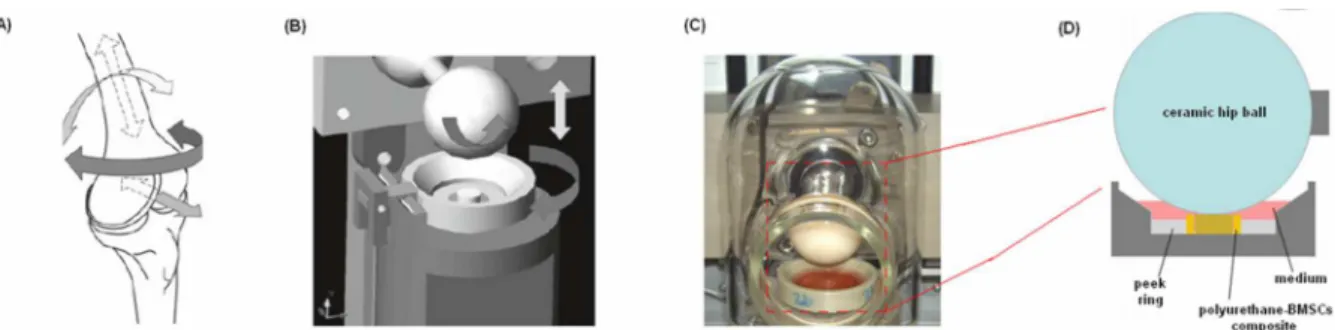
Figure 1 A-D. Schematic showing the directions of movement found within a human knee and (A) the directions of movement that can be applied using a multiaxial bioreactor (B). One of the six stations composing the bioreactor developed by Alini et al. (C) that is featured by a ceramic hip ball that compress and oscillate against a polyurethane-fibrin-BMSCs composite as represented in D.
(Modified from [26])
2.1.4 Aim of the work.
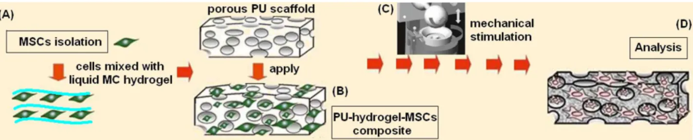
In this second part of the Thesis, the biocompatible MC-based hydrogel was tested as 3D matrix for the bioreactor-guided chondrogenesis of BMSCs. The cells were suspended into the liquid hydrogel that was used to fill a porous polyurethane (PU) scaffold. The PU-hydrogel composite was used for the cells mechanical stimulation exploiting a bioreactor developed by Alini et al. (AO Research Instutite, Davos Platz, Switzerland). This particular bioreactor (Figure 1 B-D) was developed in order to apply with a ceramic hip ball a defined compression and shear force against a porous PU
25
scaffold (Figure 1 C-D). After a 21 days period of stimulation, BMSCs chondrogenesis was evaluated by PCR and immunohystochemical analysis and the suitability of the MC-based hydrogel as 3D matrix for cells development was verified.
2.2 MATERIALS and METHODS.
2.2.1. Isolation and expansion of human mesenchymal stem cells.
Fresh human bone-marrow aspirates were obtained after full ethical approval (Freiburg, EK-326/08) and informed patient consent. Bone marrow stromal cells were isolated from 4 donors (Male/Female 19-49 years old) by standard density gradient procedure (Histopaque-1077) and selection by plastic adherence. Mesenchymal stromal cells (MSCs) were cultured in polystyrene flasks (TPP) at 37°C, 5% CO2 (for a 90% humidity atmosphere) in α-modified minimal essential medium (α-MEM, Sigma), 10% human MSC qualified foetal bovine serum (FBS-Hyclone) with 5 ng/mL fibroblast growth factor 2 (HGF, Fitzgerald Industries, Acton, MA, USA). Cells were detached with trypsin-EDTA solution at subconfluence and seeded into the required number of flasks. Thereafter, the medium was changed every 2 days. After the cells reached 70-80% confluence, they were harvested and used for the experiment at passage 2-3. Each experiment was performed separately in quadruplicate for each donor and the data collated for statistics.
2.2.2. Methylcellulose based hydrogel-polyurethane composite culture of MSCs.
Cylindrical (8 mm diameter x 4 mm height) porous polyurethane scaffolds (pore size of 90-300 µm) were prepared as described elsewhere [27]. MSCs were suspended in a 8% w/v methylcellulose (MC) 0.05M Na2SO4 based hydrogel before seeding them into the polyurethane scaffolds. Hydrogel was prepared as previously described in Chapter 1; cells were suspended into 160 µl of the liquid hydrogel at a density of 3 x 10⁶ per specimens. Hydrogel containing cells were used to fill the PU scaffold pores by compressing it several times until all the hydrogel was absorbed by PU pores (Figure 2 A-B). Constructs were then incubated for 2 hours at 37°C, 5 % CO2 and 95 % humidity to allow MC hydrogel transition from gel to solid phase before adding growth medium (DMEM, with 4.5 g/L glucose and 2.2 g/L NaHCO3, non-essential amino acids, containing 11.5 mg/L L-proline (Invitrogen/Life Technologies, Carlsbad, CA, USA), 50 µg/mL ascorbic acid 2-phosphate sesquimagnesium salt hydrate (Sigma-Aldrich, Buchs SG, Switzerland), ITS+1 (10 µg/mL insulin from bovine pancreas, 5.5 µg/mL human transferrin (substantially iron-free), 5 ng/mL sodium selenite, 0.5 mg/mL bovine serum albumin and 4.7 µg/mL linoleic acid; Sigma-Aldrich), 100 U/mL penicillin + 100 µg/mL streptomycin (Invitrogen)). After 2 days of pre-culture in 12-well plates,
26
constructs were transferred in polyether ether ketone (PEEK) holders. The experiments were carried out at 37°C, 5 % CO2, 95 % humidity. Medium was changed every 2 days and collected for further analysis.
2.2.3. Bioreactor.
Mechanical conditioning of the cell-scaffold constructs was performed using a pin-on-ball bioreactor system. Briefly, a ceramic ball 32 mm in diameter was pressed onto the scaffold [28]. Interface shear motion was generated by oscillation of the ball about an axis perpendicular to the scaffold axis. Superimposed compressive strain was applied along the cylindrical axis of the scaffold. Samples were exposed to unconfined dynamic compression at 1 Hz with 0.4 mm sinusoidal strain, superimposed on a 0.4 mm static offset strain, resulting in a strain amplitude of 10-20 % of the scaffold height at the centre of the construct. Simultaneously samples were also exposed to ball oscillation of ±25° at 1 Hz, superimposed on a 0.4 mm static compression offset strain. Mechanical load was applied during 1 hour a day for 21 consecutive days over 3 weeks (Figure 2 C-D). Cell-scaffold constructs not loaded into the bioreactor were used as controls. Experiments were carried out in quadruplicate for each donors for both loaded and not loaded samples.
Figure 2 A-D. Schematic representation of the experimental procedures. Stem cells (MSCs) were isolated from human bone marrow, expanded and seeded into the liquid hydrogel (A); afterwards cells-hydrogel solution were used to fill a porous polyurethane (PU) scaffold (B) that was mechanically stimulated for 21 days by bioreactor (C). Finally, scaffolds were collected and used for analysis (D).
2.2.4. Analysis.
After 3 weeks of culture and 21 loading cycles, cells scaffold constructs were vertically cut in two halves; 3 scaffold halves were processed for biochemical analysis, 3 for gene expression analysis and 2 for histological and immuno-hystochemical analysis.
27 2.2.5. Gene expression.
Scaffolds used for gene expression analysis were homogenized in 1mL TRI reagent and 5µL Polyacryl Carrier (both Molecular Research Center, Cincinnati, OH, USA) per scaffold, using a Tissue-Lyser (Retsch & Co., Haan, Germany) and centrifuged (Eppendorf, Basel, Switzerland) at 4 °C for 10 min at 12000g. RNA isolation was carried out according to the protocol from the manufacturer. RNA was reverse transcribed with TaqMan reverse transcription kit (Applied Biosystems, Foster City, CA, USA) using random hexamers. For real time PCR TaqMan Gene Expression Assays (Applied Biosystems) or custom designed primer-probe sets (from Microsynth, Balgach, Switzerland) were used on a GeneAmp 7500 Real Time PCR System (Applied Biosystems). The endogenous control gene was 18S rRNA. Chondrogenic markers (collagen type-II (Col 2), SRY (sex determining region Y) – box 9 (Sox9), osteogenic marker (collagen type-I (Col 1) and hypertrophic markers (collagen type-X (Col 10)) were analyzed. The primers and probes used are listed in Table 1.
Gene expression was analyzed according to the ∆∆Ct method, with expression levels normalized to the corresponding day 0 sample (day of cell seeding into scaffolds) of each donor.
2.2.6. Biochemical analysis.
Scaffolds used for biochemical analysis were digested with 0.5mg/mL proteinase K at 56°C overnight and used for DNA and glycosaminoglycan (GAG) measurement. DNA concentrations were determined with the Hoechst method using calf DNA as a standard. Fluorescence intensity was measured with an HTS 7000 Perkin Elmer Bio Assay Reader (Norwalk, CT, USA). The amount of glycosaminoglycan (GAG) was determined by the dimethylmethylene blue dye method, using bovine chondroitin sulphate as the standard. Proteinase K digests were used to measure the GAG content of the scaffolds. The total GAG content of the culture media, collected every 2 days, was also measured to assess the release of matrix molecules from the sample into the media. Absorbance was measured with a Victor3 Perkin Elmer (Waltham, MA, USA) 1420 multilabel counter. GAG values were normalized to the DNA content.
28 2.2.7. Histology and Immunohystochemistry.
For immunohystochemical analysis scaffolds were fixed in 70 % methanol at 37°C (to prevent hydrogel solid-gel phase transition) and incubated in 5% D(+) sucrose (Sigma-Aldrich, St. Louis, MO, USA) solution in phosphate buffered saline (PBS, pH 7.4) for 12 h at 37°C before embedding them in Jung tissue freezing compound and cryosectioning at 10µm (Microm HM560 CryoStar, Thermo Scientifi c, Waltham, MA, USA). The presence of glycosaminoglycans (GAG) was investigated by immunohystochemistry using safranin-O GAG specific marker (Sigma). The deposition of collagen types I and II was determined by immunofluorescence staining. After enzyme pre-treatment (0.5 U/mL Hyaluronidase for collagen types I and II staining), sections were blocked with 5% goat serum. Then sections were incubated using primary antibodies raised against collagen I (COL 1, 1:150) and collagen II (COL 2, 1:50). The antibody against type I and II collagen were from Abcam (Abcam, UK). Primary antibody was applied overnight at 4°C; afterwards, sections were washed 3 times with PBS and the appropriate secondary antibody was applied (1:500 in PBS). Samples were visually investigated by fluorescent microscope (Leica DM5500 B, Leica Microsystems, IL, USA).
2.2.8. Statistical analysis.
Statistical analysis was performed using the software package SPSS (Version 20, SPSS Inc, Chigaco, IL, USA). Data were analyzed with Wilcoxon’s test. The significance level was defined at
p< 0.05.
2.3. RESULTS and DISCUSSION. 2.3.1. PCR Analysis.
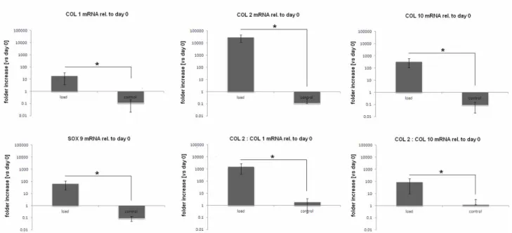
Polyurethane-hydrogel-MSCs composites gene expression after 21 days of mechanical stimulation with the bioreactor are reported in Figure 3. In general, loaded samples showed a higher expression of the selected genes if compared with the not loaded control ones. Therefore, it is possible to state that the mechanical forces of compression and shear were crucial to affect cells fate during the 21 days of stimulation.
29
Figure 3. Relative mRNA expression of human mesenchymal stem cells after 21 days of stimulation. Loaded samples showed a clear over-expression of chondrogenic markers collagen 2 (COL 2), collagen 10 (COL 10). Particular, the COL 2 : COL 1 ratio was significant increased by the mechanical load, confirming MSCs chondrogenesis. Bars represent means and standard deviations; all data were normalized to day 0 values.
Collagen 2 (COL 2) and collagen 10 (COL 10) expression are of crucial interest as they are reported in literature as suggestive for cartilage. Considering that the data are normalized to the day 0 expression values, a clear over-expression of COL 2 was noticed after the loading period. The stimulated specimens reported a 2.5x10⁴ folder increase compared to day 0 values, while to controls didn’t report same significant results. This represent a very important finding since COL 2 is selective expressed by chondrocytes, suggesting the successful differentiation of MSCs guided by the bioreactor induced forces. Another important result is related to box 9 (SOX 9) expression; it represent and important chondrogenic transcription factor peculiar of cells that are undergoing to the chondrocytes phenotype. As noticed for COL 2, also SOX 9 expression was raised by the mechanical loading; conversely, the control samples expressed very low levels of SOX 9. These two data represent an important evidences that the loaded MSCs were successfully guided towards chondrogenesis. The only gene that was not reported as over-expressed after mechanical loading was collagen 1 (COL 1). This represent an interesting result as COL 1 is strongly related with osteogenesis. Since it was previously demonstrated that is possible to induce osteogenesis by applying compression force alone, what it is possible to assume is that the combination with surface shear is satisfactory to refrain from progressing towards bone. This is not a very surprising finding since it is well established that surface shear forces are strongly involved in the amount and type of matrix synthesis. Therefore, the oscillating movement of the bioreactor was able to successfully
30
mimic the tensile forces and high hydrostatic pressure that in natural cartilage derive from the synovial fluid water that is retained within the cartilage matrix by the presence of charged proteoglycans. Since the collagen network functions to prevent swelling of the tissue, in the 3D microenvironment of the PU-hydrogel composite, cells were probably induced to produce a strong matrix to face the shear forces generated by the bioreactor. Moreover, the COL 2 : COL 1 ratio value suggested as this matrix was progressively composed mainly by the cartilage specific COL 2. An opened question still remain regarding the data obtained for collagen 10 (COL 10). In fact, COL 10 is representative not only for chondrogenesis but also for hypertrophy. In the literature is not rare to observe enhanced level of hypertrophy related to the use of mechanical forces inductive bioreactors; this fact remain as unclear since one potential reason in vitro MSCs chondrogenesis commonly leads to terminal hypertrophy is because the developing tissue is not stimulated. So, it is possible to suppose that the forces applied in this study were able to induce cells towards chondrogenesis but not enough to completely skip hypertrophy. However, regarding this, the COL 2 : COL 10 ratio value showed how the cells were probably differentiate to a chondrocyte phenotype than to the hypertophic one. In conclusion, PCR analysis proved as the mechanical stimulation was able to induce MSCs chondrogenesis in a MC hydrogel matrix.
2.3.2. Biochemical analysis.
The amount of glycosaminoglycans (GAG) per DNA into the medium was calculated every changes (2 days) while the scaffold amount was calculated at the end of the loading (21 days) (Figure 4 A-C). In general, the observed trend revealed a progressive increase of GAG in the stimulated samples. This data are particularly noticeable looking at the GAG release into the medium (Figure 4 A); the load samples values steadily increased during the 21 days of loading. On the opposite, the GAG accumulated in the medium of the control samples showed only a small increase over the 21 days. These data were also confirmed when the total amount of scaffold GAG accumulation was considered; the difference between loaded and control samples resulted as significantly different (p<0.05).
31
Figure 4 A-C. Accumulated GAG released in the medium over 3 weeks of culture (A); the total amount resulted as increased over the loading period. After 21 days the scaffold GAG amount was calculated and the total amount (medium + scaffold) compared between load and control (B). The total GAG amount was significant different between loaded and control samples (p<0.05, indicated by the star). These data were confirmed by the GAG staining using safranin-O (C); the matrix into the loaded samples scaffolds reported a massive amount of GAG while only a small amount was noticed in the control samples. Bar scale=200µm.
From a biochemical point of view, the production of GAG is an important parameter indicating a chondrogenic differentiation. Once MSCs acquire a chondrogenic phenotype, the challenge is to prevent them from becoming hypertrophic. Amongst others, this step is associated with a decrease in GAG secretion. Looking at the time course of GAG secretion in the medium, it was not possible to notice any values decrease indicating a possible hypertrophy appearance. Thus, it is possible to speculate that the loaded cells, even if expressed COL 10, were not undergoing an hypertrophic phenotype. The samples staining with GAG-specific marker safranin-O confirmed the previous results (Figure 4 C); in fact, microscope images showed a massive amount of GAG in the matrix present in the loaded samples pores. On the opposite, the matrix of control samples showed only a small amount of GAG, confirming the crucial role of the mechanical stimulation for chondrogenesis.
32 2.3.3. Histology.
Loaded and controls samples were investigated towards collagen I and collagen II using immunofluorescence (IF) in order to confirm PCR results. Collagen I staining are reported in Figure 5. PCR analysis revealed a collagen I expression in loaded samples (about 10 folder increase compared to day 0); IF confirmed the presence of collagen I in small amount (lower panel in green) for loaded samples while no signals were detected for the control samples. Thus the IF investigations seems to confirm PCR analysis data.
Figure 5. IF collagen I staining. Control samples images did not showed collagen I into the matrix (upper panel), while a small amount was found into the pores containing the matrix of loaded cells (lower panel, green signals).
A very interesting result is related to the collagen II staining, reported in Figure 6. According to PCR analysis, collagen II reported the highest folder increase compared to day 0 of all the investigated genes (more than 2x10⁴ folder increase). The IF staining confirmed the presence of collagen II into the cells matrix of loaded sample (lower panel, in green). The same results was not verified for the control samples that did not showed the presence of collagen II as previously indicated by the PCR analysis. So, also IF staining confirmed PCR analysis data, providing another important proof to the successful bioreactor-guided chondrogenesis.
33
Figure 6. IF collagen II staining. Control samples did not exhibit collagen II (upper panel) while the loaded ones clearly showed the collagen II presence (lower panel, in green).
However, according to the PCR analysis, it was expected to detect a higher amount of collagen II in the scaffolds. A possible explanation of this discordance is that Col 2 up-regulation on the protein level in MSCs is known to be very poor. Even under optimal medium composition, it can take several weeks, and one needs to consider that TGF-β, the most potent factor inducing chondrogenesis, is lacking in the culture medium. Second, as the matrix produced is relatively immature a large proportion is likely to have been released into the medium.
2.4. CONCLUSIONS.
The aim of the work was to evaluate (I) the efficiency of the mechanical induction of chondrogenesis toward MSCs and (II) the suitability of the MC hydrogel as 3D matrix supporting cells development. The data obtained suggested a positive response for both the answers. Cells successfully expressed chondrogenic genes and the GAG quantification confirmed the differentiation route of the MSCs. Histological analysis confirmed the presence of the cells into the PU scaffold pores and the presence of a surrounding matrix of collagen, confirming the suitability of MC-based hydrogel as cells carrier.
Acknowledgments.
A. Cochis would like to thanks Prof. Mauro Alini and Dr. Sibylle Grad of the AO Foundation Research Center of Davos Platz (Switzerland) for all the help during the exchange period and for giving the possibility to use the Muscoloskeletal Regeneration Laboratory bioreactor for the mechanical induction experiments. A special thanks to the European Cooperation in Science and