Indications and results of
osteosynthesis for proximal
humerus fragility fractures in
elderly patients
Luigi Murena,1Gianluca Canton,1
Chiara Ratti,1Bramir Hoxhaj,1
Gioia Giraldi,1Michele Francesco
Surace,2Federico Alberto Grassi 3 1Orthopedics and Trauma Unit – ASUGI,
Department of Medicine, Surgery and Health Sciences, University of Trieste;
2Interdisciplinary Research Centre for
Pathology and Surgery of the
Musculoskeletal System, Department of Biotechnology and Life Sciences, University of Insubria, Varese;
3Orthopedic and Trauma Unit, University
Hospital “Maggiore della Carità”, Department of Health Sciences, University of East Piedmont, Novara, Italy
Abstract
Proximal humerus fractures (PHF) are common injuries in the elderly population. Conservative treatment is indicated for undisplaced and stable fractures, which account for almost 80% of the cases. More complex fracture patterns might need sur-gery, with a wide variety of indication crite-ria and surgical techniques described in the literature. Surgical treatment should be reserved for patients in good clinical condi-tions, autonomous in daily living activities and able to adhere to postoperative rehabil-itation protocols. In the elderly population with severe osteoporosis, cognitive impair-ment and clinical comorbidities, the risk of surgical failures is high. In these patients, the choice between surgical and conserva-tive treatment, as well as for the type of pro-cedure, is even more difficult, with no gen-eral consensus in the literature. Final indi-cation is usually conditioned by surgeon’s experience and preference. Two independ-ent reviewers (B.H and G.G) independindepend-ently extracted studies on proximal humeral frac-tures. All selected studies were screened independently (B.H and G.G) based on title and abstract. Then the full text of any article that either judged potentially eligible was acquired and reviewed again. Any disagree-ment was resolved by discussing the full text manuscripts. Aim of the present paper is to review the literature about indications and results of osteosynthesis for proximal humerus fragility fractures in the elderly population.
Introduction
Proximal humerus fractures (PHFs) are the third most common fragility fractures of the appendicular skeleton, after proximal femur and distal radius fractures. These fractures are typically related to low energy trauma in elderly patients affected by osteo-porosis.1,2
Currently, the ideal treatment for these fractures is debated in the literature. Several indications and treatment options have been described without a clear evidence about outcome.3
Nearly 80% of PHFs have a stable con-figuration. In these cases, excellent results may be achieved with conservative treat-ment, especially in elderly patients with low functional demands. Conversely, absolute surgical indication for PHFs treatment is rare, representing less than 1% of cases. The remaining cases may benefit from surgical intervention. Whether reduction and fixa-tion or primary shoulder arthroplasty may be better indicated in these cases is still mat-ter of debate.4
Conservative treatment is generally pre-ferred also for displaced or comminuted PHFs in patients older than 85 years of age affected by severe osteoporosis, cognitive impairment or significant comorbidities.4-7
Proper indication for treatment should take into account different factors: the expected outcome (with and without surgi-cal treatment), patient’s functional demand and compliance to treatment protocol, sur-geon’s training and experience.8 Ideally,
fracture reduction and fixation should be preferred to arthroplasty because of the bet-ter clinical results achieved in uncomplicat-ed cases with anatomic reconstruction.9,10
On the other hand, especially in fragility osteoporotic fractures, osteosynthesis is jeopardized by a high rate of complications, that are frequently related to insufficient understanding of risk factors for humeral head avascular necrosis (AVN) and failure of fixation.11,12Humeral head vascular
sup-ply relays on the ascending branches of the axillary artery, with the posterior circumflex artery demonstrated to be dominant (about 64%).13 Hertel et al. identified some PHFs
patterns and characteristics strongly related with the risk of AVN of the humeral head. These include a disrupted medial hinge, a calcar segment shorter than 8 mm and a fracture of the anatomical neck. These crite-ria combined have a predictive positive value of humeral head AVN of 0.97 accord-ing to the authors.14 Even though it’s not
possible to quantify exactly the risk of fail-ure, internal fixation is not recommended for head splitting fractures or in presence of
multiple articular fragments in elderly patients: in these cases, shoulder arthroplas-ty should be preferred.15
According to the literature, osteosynthe-sis can be considered as a treatment option for Neer 2-part fractures, Neer 3 and 4-part frac-tures with tuberosities displaced more than 5 mm, false head split fractures (minimal part of the humeral head attached to the tuberosity fragment), Neer 2-part fractures of the surgi-cal neck, Neer 2, 3, 4-part fractures with angulation of the humeral head in varus or valgus > 30, and for fracture-dislocations with impaction fracture of the humeral head with retained soft-tissue attachment.1,4On the
other hand, the low inteobserver and intraob-server reliability of Neer classification ren-ders the latter criteria of limited clinical value.1Aim of the present paper is to review
the literature about indications and results of osteosynthesis for proximal humerus fragility fractures in the elderly population.
Correspondence: Bramir Hoxhaj, Orthopaedics and Trauma Unit – ASUGI, Department of Medicine, Surgery and Health Sciences, University of Trieste, Strada di Fiume 447, 34149 Trieste, Italy.
Tel.: +39.040.3994799.
E-mail: [email protected]
Key words: proximal humerus fractures, osteosynthesis, osteoporosis, fragility frac-tures, elderly.
Contributions: The authors contributed equally. Conflict of interests: Federico A. Grassi is consultant for DePuy Synthes. The other authors declare no conflict of interest. Funding: None.
Ethical approval: All procedures performed in studies involving human participants were in accordance with the ethical standards of the institutional and/or national research commit-tee and with the 1964 Helsinki declaration and its later amendments or comparable ethical standards.
Informed consent: Informed consent was obtained from all individual participants included in the study.
Received for publication: 29 March 2020. Accepted for publication: 13 April 2020. This work is licensed under a Creative Commons Attribution NonCommercial 4.0 License (CC BY-NC 4.0).
©Copyright: the Author(s), 2020
Licensee PAGEPress, Italy Orthopedic Reviews 2020;12:8559 doi:10.4081/or.2020.8559
Non-commercial
Peculiarities of proximal
humerus fragility fractures
fixation in the elderly
When facing a patient with a fragility PHF, the intrinsic difficulty of anatomically reducing and fixing with adequate stability the osteoporotic fragments must be consid-ered, beside the general features of the patient and the fracture.
The main technical difficulties arise from the combination of a weak and brittle bone (the humeral head can be conceived as an eggshell) with a comminuted fracture pattern.8
Closed reduction internal fixation (CRIF) and open reduction internal fixation (ORIF) techniques have been developed and commonly used for normal and healthy bone.
Ageing and osteoporosis affect the mechanical properties of the bone, altering both elastic and strength properties.
With osteoporosis cortices become thin-ner and the trabecular network is altered, making bone prone to mechanical failure particularly in the metaphyseal region.16,17
Conservative versus operative
treatment in the elderly patient
Fracture pattern is the first factor that is usually considered to define treatment strat-egy.18Most PHFs are undisplaced or
mini-mally displaced, involving the surgical neck and the greater tuberosity.19,20In the elderly
population, conservative treatment of these fractures is a well consolidated practice, that guarantees a high rate of successful outcomes.3,6,21
There are different immobilization tech-niques proposed for non-operative treat-ment of PHFs as a Gilchrist or a Velpeau bandage. Regardless the non-operative method, a close radiological and clinical follow-up is required in these patients.
There is no consensus on non-operative treatment modalities. However, early mobi-lization is generally recognized as the main-stay of conservative treatment. In unstable fracture patterns with a high risk of dis-placement progression, a period of immobi-lization from 3 to 4 weeks (until soft callus formation) is usually preferred.
The choice of the optimal treatment becomes more difficult when PHFs are dis-placed and more complex. In these cases, fracture morphology, patients’ demands and surgeons experience should be consid-ered.4,7Although a large number of studies
support operative treatment for displaced
2-part and 3-2-part PHFs, the most recent liter-ature is not prejudicial to conservative treat-ment.6There is lack of well-designed
com-parative studies for 2-part displaced PHFs, which account for about 30 – 44% of all PHFs and usually have acceptable clinical outcomes with non-operative treatment. In these fracture patterns conservative treat-ment should be considered in patients with low demands and poor bone quality, where-as operative treatment should be considered in patients with high demands and good bone quality.3,19-21
Isolated great tuberosity fractures, espe-cially with postero-superior displacement > 5mm, usually require operative treatment in order to avoid subacromial impingement and loss of external rotation.22Surgical
indi-cation is usually considered for young patients, who more often present with iso-lated displaced greater tuberosity fractures. Clinical data about this specific fracture in the elderly population is lacking, with no evidence about how displacement criteria applied to young patients should be trans-ferred to the elderly population.
Lesser tuberosity fractures are rare injuries produced by muscle contraction (as in seizures), or when the subscapularis mus-cle forcefully contracts to resist external forces in abduction and external rotation on the shoulder. These fractures typically affect young patients, with significant dis-placement in most cases. According to these epidemiological and biomechanical data, these injuries should not be considered as fragility fractures.23-25
Three and 4-part fractures account for nearly 11 – 20% of PHFs.20Operative
treat-ment is usually indicated for young patients with 3 and 4-part PHFs, but conservative management should be considered in elder-ly patient. In a retrospective study on 125 elderly patients with 4-part valgus impacted PHF, Court-Brown et al. reported good to excellent outcomes with conservative treat-ment.26A meta-analysis of randomized
con-trolled trials (RCT) on operative versus conservative treatment in displaced 3 and 4-part PHFs in the elderly concluded that functional improvement was not significant and complication rate was higher in the operative group.27Handoll et al. reported no
significant difference in the clinical out-come (Oxford Shoulder Score) between conservative and surgical treatment in 3 and 4-part PHFs.28Several reviews and studies
support these data.29-31A Cochrane review
on proximal humeral fractures evaluated 31 randomized controlled trials, of which only 8 studies (involving 567 elderly patients) compared conservative to operative treat-ment. The authors reported no significant difference in clinical result and quality of
life in patient-reported shoulder and upper-limb function at 1 and 2-year follow-up. Even though there was moderate evidence of a higher risk of complications after sur-gery, the 95% confidence intervals reveled a greater risk of potential complications in the conservative treatment group.3
Conversely, Olerud et al. reported better functional outcomes with locking plate fix-ation compared to conservative treatment in the elderly population with displaced 3-part PHFs. However, a 30% higher risk of addi-tional surgery in the operative group was noted.32
Many studies suggest that PHFs in the elderly may be treated conservatively with acceptable outcomes, but there are specific fracture patterns and patients that benefit from operative treatment.33Sabharwal et al.
conducted a meta-analysis on 528 patients detected in 7 RCTs comparing operative to conservative treatment of PHFs. Despite there were no differences in functional out-comes comparing conservative and opera-tive treatment of displaced PHFs, some dif-ferences in clinical outcome emerged when analyzing specific fracture patterns and sur-gical techniques.34 The four-part fractures
treated surgically had better clinical out-comes and were less likely to result in osteoarthritis, osteonecrosis and malu-nion.34However, these data were
heteroge-neous and did not reach any statistical sig-nificance. Studies aimed on specific frac-ture patterns and surgical procedures should be performed in order to identify the patients that may benefit from surgery and the type of the procedure that is more ade-quate in these cases.
Patient’s characteristics like age and associated injuries consistently influence decision making between operative or con-servative treatment of displaced PHFs in elderly patients. Many authors argue that conservative treatment in displaced or com-minuted PHFs should be preferred in patients older than 85 years old, with cogni-tive impairment or clinical comorbidity, severe osteoporosis, non-associated frac-tures, rheumatoid arthritis and concurrent neoplasm.4-7Clement et al. sustain that
fac-tors associated with social independence are more predictive of functional outcomes than age.21Despite the lower functional
out-comes reported with conservative treat-ment, subjective perception of outcome can be satisfactory if no residual pain is present.4Although the risk of non-union, malunion and osteonecrosis after conserva-tive treatment is generally accepted, the sur-geon should consider that salvage surgery in these cases is more likely to result in worse outcomes compared to primary operative treatment.4,35,36
Non-commercial
Percutaneous fixation in the
eld-erly patient
Closed reduction and percutaneous pin-ning (CRPP) of PHFs was described for the first time in 1962 by Bohler. CRPP may be considered as an alternative to ORIF in spe-cific fracture patterns and in selected patients. Stability achieved with CRPP is mechanically inferior to intramedullary nail and plate fixation.37,38Pin configuration is a
significant factor in order to enhance mechanical stability, that is improved by biplanar fixation and by increasing the number of pins engaging the cortex.
Indications for CRPP include 2-part fractures of the surgical neck, isolated greater tuberosity fractures, 3-part fractures of the surgical neck with involvement of the greater tuberosity and 4-part valgus impact-ed fractures.1,39,40 Use of CRPP in PHFs is
less invasive, allowing respect of soft tis-sues and blood supply during the surgical procedure. Compared to open reduction and internal fixation (ORIF), CRPP potentially has lower rates of avascular necrosis (AVN), higher union rates, less scar forma-tion at the scapulo-thoracic joint and better cosmetics.39Different studies reported good
outcomes with CRPP.41-43In a study on 113
patients with 2-part PHFs, Tamimi et al. reported better outcomes with CRPP in patients of all ages and better functional results compared to intramedullary nailing in elderly patients.43
Gupta et al. published a review on 4500 patients, reporting a considerably higher complication rate with CRPP when com-pared to ORIF, hemiarthroplasty (HA) and reverse shoulder arthroplasty (RSA). Complications observed with CRPP includ-ed humeral head necrosis (11.7%), pin migation/breakage (4.1%), superficial infection (4.1%), malunion (3%), neurolog-ic injuries (1.5%), nonunion and deep infec-tion (1%).44
Resch et al., in a study on 27 patients with 3 and 4-part PHFs treated with CRPP, reported that all 3-part fractures had very good functional results and no complica-tions. The valgus impacted 4-part fractures had good functional outcomes and 11% of avascular necrosis (2/18).45
In the study performed by Keener et al. on a total of 27 PHFs (7 two-part, 8 three-part and 12 four-three-part valgus impacted), a 100% rate of fracture healing was reported, together with good functional outcomes (mean Constant score of 73.9 points) and a low rate of complications after CRPP. Fracture type, age, malunion and osteoarthritis had no influence on outcomes.39It must be highlighted that
clin-ical series reporting on CRPP do not focus specifically on geriatric osteoporotic frac-tures.
Ideally, CRPP should be performed when a stable closed reduction can be achieved and in presence of minimal frac-ture comminution, an intact medial calcar, a good bone stock and a compliant patient.1,39,40 CRPP should not be used in
non-optimally reduced PHFs since the qual-ity of fracture reduction directly influences construct stability, hence anatomical and clinical outcomes.40 Fracture comminution
and poor bone quality are a relative con-traindication to CRPP, that consequently may hardly apply to osteoporotic elderly fractures. Pin migration, loosening and loss of reduction are associated with these fac-tors in different studies.1,39,40,46On the other
hand, some technical advances on the clas-sical CRPP technique, especially with threaded wires engaging the outer cortex and connected as an external fixator, may by-pass these limitations and become a good choice for elderly patients.40,47Blonna
et al. suggested an alternative to the
con-ventional pinning technique, consisting in the use of full threated pins augmented by an external frame. The authors introduced the term “hybrid technique” to describe how this technique includes features of both external and internal fixation. The authors prospectively studied 42 patients treated with conventional pinning (2.5 mm termi-nally threaded pins) and 49 patients treated with the hybrid technique (2.5 mm pins characterized by a 7 cm thread augmented with external fixator). They observed a sig-nificant reduction in complication and revi-sion rates in the hybrid group.48
Intramedullary nail (IMN)
fixa-tion in the elderly patient
The goal of nail fixation in PHFs is to provide stability in order to allow early motion of the shoulder and improve func-tional outcomes compared to conservative treatment.49From the first nail described by
Rush, humeral nailing has undergone important improvements and innova-tions.49,50While first generation nails had no
ability to control rotation, second genera-tion nails had the major disadvantage of fre-quent migration of the proximal interlock-ing screws.49 In fact, functional outcomes
reported with first and second generation nails were disappointing and threatened by frequent complications such as nonunion, hardware migration and chronic shoulder pain.42Third generation nails have a better
locking mechanism of proximal screws, allowing more stable constructs and a
medi-al entry point on the humermedi-al head to pre-serve the rotator cuff footprint. These improvements have led to better functional outcomes and decreased complication rates.1,42(Figure 1)
Different authors have reported good clinical outcomes in 2 and 3-part PHFs treated with third generation humeral nails.51-54 In a retrospective study on 38
patients with 2-part surgical neck PHFs treated with locked angular stable intramedullary nail, Hatzidakis et al. report-ed 100% primary healing, a mean Constant score of 71 points and a mean forward flex-ion of 132° with little residual shoulder pain.55 However, Nolan et al. reported a
high complication rate in 18 patients with 2 and 3-part PHFs treated with Polarus nail.56
In a systematic review including 2155 patients (66 studies) treated with different modalities for PHFs, Lanting et al. reported 11.9% complication rate for IMN. The inci-dence of nonunion or malunion was 5%, implant loosening or migration 3.2% and osteonecrosis 4.5% (19.2% in 3 and 4-part fractures).57
Intramedullary locked nails for PHFs compared to locked plates should provide the theoretical advantage of improved con-struct stability, even in case of osteopenic or osteoporotic 3 and 4-part PHFs.58 These
data are in discordance with cadaveric bio-mechanical comparative studies between IMN and locking plates, reporting lower resistance at bending and torsion for nails. The authors hypothesized that the early rate of failure of IMN is correlated to the moment transmitted to the screw-bone interface in the humeral head; this aspect is matter of concern for early postoperative mobilization in osteoporotic bone.59
Recent studies assert that results achieved with IMN (third generation implants) in 2, 3 and 4-part PHFs are com-parable to those reported with locking plates.60,61 Furthermore, in a comparative
randomized study of 2-part PHFs treated with IMN or plates, Zhu et al. reported complication rates of 4% and 31%, respec-tively.62
Age, osteoporosis and 3 or 4-part PHFs appear to affect clinical outcome of IMN.63
Patients older than 65 years have an aug-mented risk of worse outcome, most likely due to osteoporosis and the consequent decreased grip on bone of the implant. In this scenario, proximal screw cut out, greater tuberosity migration and varus dis-placement are the most common complica-tions.64 According to some authors, third
generation nails partially overcame these drawbacks.65Mihara et al. reported
satisfac-tory functional outcomes and no screw cut out with a “pin lock nail system’’ in 19
geri-Non-commercial
atric patients.66 Boileau et al. proposed a
new locked nail design to optimize tuberos-ity fixation and stable support for the humeral head. The preliminary functional results in 24 patients with a mean age of 64 years were good, with no need for further surgery.67
Some technical tips should be observed when nailing a PHF, especially in osteo-porotic bone. The supraspinatus should be split at the lateral edge of the articular sur-face through the muscle belly instead of splitting the tendon. The entry point must be at the center of the humeral head, so that the stability of the fixation doesn’t rely exclu-sively on the proximal screws, but is favored by the subchondral bone-nail inter-face, where the bone remains of better qual-ity in comparison with other areas of the humeral head. The superior resistance to varus forces, obtained with the interference of the nail with the subchondral bone, is particularly important in osteoporotic fragility fractures, in which the weak can-cellous bone of the medial calcar cannot ensure a reliable stability of the proximal screws.5,68 If these principles are not
respected, a high failure rate should be expected. (Figure 2)
Locking plate osteosynthesis in
the elderly patient
According to the literature, locking plate fixation (LPF) is the most widely used osteosynthesis technique for proximal humeral fractures. LPF is indicated for almost all fracture patterns, including humeral head fractures amenable to recon-struction. However, the burden of complica-tions, especially in elderly patients with osteoporosis, is often unacceptably high.4,69
In a study on 27.017 patients surgically treated for PHFs, Zhang et al. reported a higher readmission rate for ORIF (29%) when compared to reverse shoulder arthro-plasty (20 %) and hemiarthroarthro-plasty (16 %).70
In literature, the mean complication rate reported for ORIF is 30%, with articular screw penetration (primary or secondary to reduction loss) and avascular necrosis of the humeral head accounting for half of cases. The average re-operation rate is about 14%.71-73In interpreting these data, it must
be underlined that LPF is the gold standard for the treatment of more complex fracture patterns and figures about complications and re-operations may be affected by a selection bias.
Jung et al., reporting on 252 patients treated with locking plates, observed a
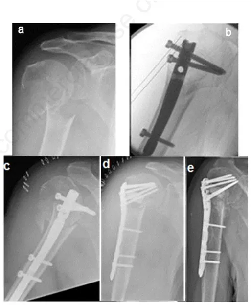
sig-Figure 1. Right proximal humeral 3-part fracture in a 79-year-old woman treated with IMN: (a) preoperative x-rays; (b) preoperative CT scans respectively in the coronal, axial and sagittal plane; intraoperative fluoroscopy (c) and postoperative x-rays (d) showing a correct medial entry point and proximal screws stabilization of the greater tuberosity; (e) x-rays 6 months after surgery showing uneventful healing without secondary displacement.
Figure 2. Right proximal humeral 2-part fracture in a 76-year-old woman affected by moderate dementia: (a) preoperative x-rays; (b) intraoperative fluoroscopy showing a wrong entry point (too anterior and lateral) and an excessively buried nail; (c) x-rays showing secondary displacement 4 days after surgery; (d) revision surgery with a locking plate and synthetic bone substitute to fill the antero-lateral bone defect resulting from nail migration; (e) x-rays 4 months after surgery.
Non-commercial
nificant correlation with loss of reduction in case of osteoporosis, varus displacement, medial comminution and insufficient medi-al support.74
There are several concepts to keep in mind when planning an ORIF with locking plate in osteoporotic bone, because the unreliable fixation of the implant is a major concern. With respect to conventional plates, locking plate systems can stabilize fracture fragments without friction between plate and bone, providing more stability in case of osteoporosis. The correct position of the plate, just inferior to the flare of the greater tuberosity and lateral to the bicipital groove, is important to avoid lateral impingement.75,76
An anatomic reduction is essential to achieve a stable fixation and contributes to increase its longevity. As suggested by Krappinger et al., correct alignment of the medial cortices and anatomic reduction are the most important prognostic factors to avoid secondary displacement.77(Figure 3)
The reconstruction of the calcar in case of disruption is the first step to achieve. This can be obtained by indirect manipula-tion or directly through the fracture line. If intact or partially preserved, the medial periosteum allows indirect reduction using ligamentotaxis. Fractures with medial com-minution are technically difficult to man-age: in this cases, an intended impaction of the humeral head may be the solution.75,78
As suggested by Gardner et al., achiev-ing a mechanical support of the inferomedi-al region of the proximinferomedi-al humerus for main-taining reduction is fundamental, and lock-ing plate alone are unable to support the humeral head from a lateral position, espe-cially if medial comminution is present.79
Fractures of the proximal humerus with medial comminution treated with locking plates are at risk of varus collapse. In their study on cadaveric humerus fixed with locking plate, Ponce et al. observed that medial comminution decreased the mean load to failure and the mean energy to fail-ure by 48 % and 44 %, respectively.80
When reduction of the medial cortex is performed or a stable impaction of the humeral head is achieved, the placement of a superiorly directed screw in the inferome-dial region of the proximal fragment is helpful to support the calcar, increasing the mean load to failure and the mean energy to failure by 31% and 44 %, respectively.80
As suggested by Padegimas et al., the calcar screw should be positioned < 12 mm from the apex of the arc of the calcar or within the bottom 25% of the humeral head. Within these cut-offs, the incidence of fixa-tion failures was significantly reduced in their clinical series.81(Figure 4)
Reduction of tuberosities is fundamen-tal to lie down the humeral head on a solid cortico-cancellous rim, as stated by Hertel.8
Accepting a non-anatomic reduction or over-reducing the greater tuberosity signifi-cantly reduces the stability of the construct. Placing tension band sutures within the rotator cuff is moreover strongly recom-mended to counteract the traction forces on the tuberosities, to augment their reduction and improve fracture fixation.8,76,82
Another important consideration is that the quality of cancellous bone in the humer-al head is heterogeneous, thus influencing proximal screws stability. The medial and
superior part of the humeral head should be considered the best location for screw placement, with a divergent or parallel ori-entation of the screws. This construct has the highest axial pull-out strength compared with convergent orientation.83-88
Other possible tips - not yet supported by strong evidence in literature - to gain bet-ter primary stability in osteoporotic bone are represented by cement augmentation (for the head fragment itself, for the head screws or to fill metaphyseal defects) and bone grafts.
Locking plates implanted with cement augmentation are associated with decreased
Figure 3. Right proximal humeral 3-part (surgical neck and greater tuberosity) fracture in a 75-year-old woman treated with MIPO technique through a transdeltoid approach: (a) preoperative x-rays; (b) intraoperative fluoroscopy; (c) postoperative x-rays 13 months after surgery, (d) clinical result 13 months after surgery respectively in forward elevation, internal and external rotation.
Figure 4. Right proximal humeral 3-part fracture in a 72-year-old woman treated with locking plate through a deltopectoral approach: preoperative x-rays (a) and axial CT scan (b) showing the valgus impacted 3-part fracture pattern and a lesser tuberosity undis-placed fracture; (c) postoperative x-rays 18 months after surgery; (d) clinical results 18 months after surgery respectively in forward elevation, internal and external rotation.
Non-commercial
interfragmentary motion, higher failure loads and increased stiffness values com-pared with locking plates alone. The early experiences with this procedure did not highlight any risk of chondral or osseous damage with cement.75,89
A cortico-cancellous bone graft can be considered if the reduction of a comminut-ed calcar cannot be achievcomminut-ed; this option should contribute to increase stiffness and varus failure load of the plate-bone com-plex.75
Conclusions
Surgical indication for proximal humer-al fragility fractures is still matter of debate. Surgical treatment should be reserved for patients in good clinical conditions, autonomous in daily living activities and able to adhere to postoperative rehabilita-tion protocols. On the other hand, the sur-geon should be able to choose the most suit-able procedure, favoring shoulder replace-ment in patients at high risk for fixation fail-ure. When considering osteosynthesis, spe-cific technical features must be respected in the osteoporotic bone to obtain satisfactory results. CRPP, IMN and LPF are all possi-ble options, with different indications and specific advantages and disadvantages.
References
1. Kancherla VK, Singh A, Anakwenze OA. Management of acute proximal humeral fractures. J Am Acad Orthop Surg 2017;25:42-52.
2. Passaretti D, Candela V, Sessa P, Gumina S. Epidemiology of proximal humeral fractures: a detailed survey of 711 patients in a metropolitan area. J Shoulder Elb Surg 2017;26:2117-24. 3. Haldoll HH, Brorson S. Intervention for
treating proximal humeral fractures in adult. Cochrane Db Syst Rev 2015; CD000434.
4. Murray IR, Amin AK, White TO, Robinson CM. Proximal humeral frac-tures: current concepts in classification, treatment and outcomes. J Bone Jt Surg Br 2011;93:1-11.
5. Schumaier A, Grawe B. Proximal humerus fractures: evaluation and management in the elderly patient. Geriatr Orthop Surg Rehabil 2018;9:2151458517750516.
6. McLaurin TM. Proximal humerus frac-tures in the elderly are we operating on too many? Bull Hosp Jt Dis N Y 2004;62:24-32.
7. Okike K, Lee OC, Makanji H, et al.
Factors associated with the decision for operative versus non-operative treat-ment of displaced proximal humerus fractures in the elderly. Injury 2013;44:448-55.
8. Hertel R. Fractures of the proximal humerus in osteoporotic bone. Osteoporos Int 2005;2:S65-72. 9. Jobin CL, Galdi B, Anakwenze OA, et
al. Reverse shoulder arthroplasty for the management of proximal humerus frac-tures. J Am Acad Orthop Surg 2015; 23:190-201.
10. Gomberawalla MM, Miller BS, Coale RM, et al. Meta-analysis of joint preser-vation versus arthroplasty for the treat-ment of displaced 3- and 4-part fractu-res of the proximal humerus. Injury 2013;44:1532-9.
11. Haasters F, Siebenburger G, Helfen T, et al. Complications of locked plating for proximal humeral fractures - are we get-ting any better? J Shoulder Elb Surg 2016;25:e295-303.
12. Boileau P, Pennington SD, Alami G. Proximal humeral fractures in younger patients: fixation techniques and arthro-plasty. J Shoulder Elb Surg 2011;20:S47-60.
13. Hettrich CM, Boraiah S, Dyke JP, et al. Quantitative assessment of the vascula-rity of the proximal part of the humerus. J Bone Jt Surg Am 2010;92:943-8. 14. Hertel R, Hempfing A, Stiehler M,
Leunig M. Predictors of humeral head ischemia after intracapsular fracture of the proximal humerus. J Shoulder Elb Surg 2004;13:427-33.
15. Berkes MB, Little MTM, Lorich DG. Open reduction internal fixation of pro-ximal humerus fractures. Curr Rev Musculoskelet Med 2013;6:47–56. 16. Von Ruden C, Augat P. Failure of
frac-ture fixation in osteoporotic bone. Injury 2016;S3-10.
17. Yaacobi E, Sanchez D, Maniar H et al. Surgical treatment of osteoporotic frac-tures: an update on the principles of management. Injury 2017;S34-40. 18. Neer CS. Displaced proximal humeral
fractures. I: classification and evalua-tion. J Bone Jt Surg 1970;52-A:1077-89.
19. Bergdahl C, Ekholm C, Wennergren D, et al. Epidemiology and patho-anatomi-cal pattern of 2,011 humeral fractures: data from the swedish fracture register. BMC Musculoskelet Disord 2016;17:159.
20. Court-Brown CM, Garg A, McQueen MM. The epidemiology of proximal humeral fractures. Acta Orthop Scand 2001;72:365-71.
21. Clement ND, Duckworth AD,
McQueen MM, Court-Brown CM. The outcome of proximal humeral fractures in the elderly: predictors of mortality and function. Bone Joint J 2014;96-B:970-7.
22. Platzer P, Kutscha-Lissberg F, Lehr S, et al. The influence of displacement on shoulder function in patients with mini-mally displaced fractures of the greater tuberosity. Injury 2005;36:1185-89. 23. Neer CS II, Craig EV, Fukuda H.
Cuff-tear arthropathy. J Bone Jt Surg Am 1983;65:1232-44.
24. Neer CS 2nd. Four-segment classifica-tion of proximal humeral fractures: pur-pose and reliable use. J Shoulder Elb Surg 2002;11:389-400.
25. Tosun B, Kesemenli CC. Isolated avul-sion fracture of lesser tuberosity of the humerus: review of the literature and report of two cases. Int J Shoulder Surg 2011;5:50-3.
26. Court-Brown CM, Cattermole H, McQueen MM. Impacted valgus fractu-res (B1.1) of the proximal humerus: the results of non-operative treatment. J Bone Jt Surg Br 2002;84:504-8. 27. Fu T, Xia C, Li Z, Wu H. Surgical
ver-sus conservative treatment for displaced proximal humeral fractures in elderly patients: a meta-analysis. Int J Clin Exp Med 2014;7:4607-15.
28. Handoll HH, Keding A, Corbacho B, et al. Five-year follow-up results of the PROFHER trial comparing operative and non-operative treatment of adults with a displaced fracture of the proxi-mal humerus. Bone Joint J 2017;99-B:383-92.
29. Huttunen TT, Launonen AP, Pihlajamäki H, et al. Trends in the sur-gical treatment of proximal humeral fractures - a nationwide 23-year study in Finland. BMC Musculoskelet Disord 2012;13:261.
30. Rabi S, Evaniew N, Sprague SA, et al. Operative vs non-operative manage-ment of displaced proximal humerus fractures in the elderly: a systematic review and meta-analysis of randomi-zed controlled trials. World J Orthop 2015;6:838-46.
31. Song JQ, Deng XF, Wang YM, et al. Operative vs. nonoperative treatment for comminuted proximal humeral frac-tures in elderly patients: a current meta-analysis. Acta Orthop Traumatol Turc 2015;49:345-53.
32. Olerud P, Ahrengart L, Ponzer S, et al. Internal fixation versus nonoperative treatment of displaced 3-part proximal humeral fractures in elderly patients: a randomized controlled trial. J Shoulder Elb Surg 2011;20:747-55.
Non-commercial
33. Ghert M, McKee M. To operate or not to operate, that is the question: the pro-ximal humerus fracture. Bone Joint Res 2016;5:490-1.
34. Sabharwal S, Patel NK, Griffiths D, et al. Trials based on specific fracture con-figuration and surgical procedures like-ly to be more relevant for decision making in the management of fractures of the proximal humerus. Bone Joint Res 2016;5:470-80.
35. Poeze M, Lenssen AF, Van Empel JM, Verbruggen JP. Conservative manage-ment of proximal humeral fractures: can poor functional outcome be related to standard trans-scapular radiographic evaluation? J Shoulder Elb Surg 2010;19:273-81.
36. Hanson B, Neidenbach P, de Boer P, Stengel D. Functional outcomes after non-operative management of fractures of the proximal humerus. J Shoulder Elb Surg 2009;18:612-21.
37. Wheeler D, Colville MR. Biomechanical comparison of intrame-dullary and percutaneous pin fixation for proximal humeral fracture fixation. J Orthop Trauma 1997;11:363-7. 38. Koval K, Blair B, Takei R, et al.
Surgical neck fractures of the proximal humerus: a laboratory evaluation of ten fixation techniques. J Trauma 1996;40:778–83.
39. Keener JD, Parsons BO, Flatow EL, et al. Outcomes after percutaneous reduc-tion and fixareduc-tion of proximal humeral fractures. J Shoulder Elb Surg 2007:16:33-8.
40. Calvo E, de Miguel I, de la Cruz JJ, Lopez-Martin N. Percutaneous fixation of displaced proximal humeral fractu-res: indications based on the correlation between clinical and radiographic results. J Shoulder Elb Surg 2007;16:774-81.
41. Magovern B, Ramsey ML. Percutaneous fixation of proximal humerus fractures. Orthop Clin North Am 2008;39:405-16.
42. Aaron D, Shatsky J, Paredes JC, et al. Proximal humeral fractures: internal fixation. J Bone Jt Surg Am 2012;94:2280-8.
43. Tamimi I, Montesa G, Collado F, et al. Displaced proximal humeral fractures: when is surgery necessary? Injury 2015;46:1921-9.
44. Gupta AK, Harris JD, Erickson BJ, et al. Surgical management of complex proximal humerus fractures: a systema-tic review of studies including 4500 patients. J Orthop Trauma 2015;29:54-9.
45. Resch H, Povacz P, Frohlich R,
Wambacher M. Percutaneous fixation of three- and four-part fractures of the proximal humerus. J Bone Jt Surg Br 1997;79:295–300.
46. Fenichel I. Oran A, Bursteln G. Perry Pritsch M. Percutaneous pinning using threaded pins as a treatment option for unstable two- and three-part fractures of the proximal humerus: a retrospective study. Int Orthop 2006:30:153-7. 47. Carbone S, Tangari M, Gumina S, et al.
Percutaneous pinning of three- or four-part fractures of the proximal humerus in elderly patients in poor general con-dition: MIROS® versus traditional pin-ning. Int Orthop 2012;36:1267–73. 48. Blonna D, Castoldi F, Scelsi M, et al.
The hybrid technique: potential reduc-tion in complicareduc-tions related to pins mobilization in the treatment of proxi-mal humeral fractures. J Shoulder Elb Surg 2010;19:1218-29.
49. Dilisio MF, Nowinski RJ, Hatzidakis AM, Fehringer EV. Intramedullary nai-ling of the proximal humerus: evolu-tion, technique, and results. J Shoulder Elb Surg 2016;25:e130–8.
50. Rush LV, Rush HC. Intramedullary fixation of the fracture of the humeral shaft by longitudinal pin. Surgery 1950;27,268.
51. Blum J, Hansen M, Rommens PM. Angle-stable intramedullary nailing of proximal humerus fractures with the proximal humeral nail. Oper Orthop Traumatol 2009;21:296-311.
52. Hessmann MH, Nijs S, Mittlmeier T, et al. Internal fixation of fractures of the proximal humerus with the MultiLoc nail. Oper Orthop Traumatol 2012;24:418-31.
53. Popescu D, Fernandez-Valencia JA, Rios M, et al. Internal fixation of proxi-mal humerus fractures using the T2-proximal humeral nail. Arch Orthop Trauma Surg 2009;129:1239-44. 54. Freynik F, Freynik S, Zenker W,
Pflugmacher R. Angular and sliding sta-ble internal fixation of proximal hume-rus fractures using the “Varion” intra-medullary nail. Z Orthop Unfall 2013;151:343-9.
55. Hatzidakis AM, Shevlin MJ, Fenton DL, et al. Angular-stable locked intra-medullary nailing of two-part surgical neck fractures of the proximal part of the humerus: a multicenter retrospective observational study. J Bone Jt Surg Am 2011;93:2172-9.
56. Nolan BM, Kippe MA, Wiater JM, Nowinski GP. Surgical treatment of displaced proximal humerus fractures with a short intramedullary nail. J Shoulder Elb Surg 2011;20:1241-7.
57. Lanting B, MacDermid J, Drosdowech D, Faber KJ. Proximal humeral fractu-res: a systematic review of treatment modalities. J Shoulder Elb Surg 2008;17:42-54.
58. Misra A, Kapur R, Maffulli N. Complex proximal humeral fractures in adults - a systematic review of management. Injury 2011;32:363-72.
59. Edwards SL, Wilson NA, Zhang LQ, et al. Two-part surgical neck fractures of the proximal part of the humerus: a bio-mechanical evaluation of two fixation techniques. J Bone Jt Surg Am 2006;88:2258-64.
60. Konrad G, Audigé L, Lambert S, et al. Similar outcomes for nail versus plate fixation of three-part proximal humeral fractures. Clin Orthop Relat Res 2012;470:602-9.
61. Lekic N, Montero NM, Takemoto RC, et al. Treatment of two-part proximal humerus fractures: intramedullary nail compared to to locked plating. HSS J 2012;8:86-91.
62. Zhu Y, Lu Y, Shen J, et al. Locking intramedullary nails and locking plates in the treatment of two-part proximal humeral surgical neck fractures: a pro-spective randomized trial with a mini-mum of three years of follow-up. J Bone Jt Surg Am 2011;93:159-68. 63. Gradl G, Dietze A, Kaab M, et al. Is
loc-king nailing of humeral head fractures superior to locking plate fixation? Clin Orthop Relat Res 2009;467:2986-93. 64. Karataglis D, Stavridis SI, Petsatodis G
et al. New trends in fixation of proximal humeral fractures: a review. Injury 2011;42:330-8.
65. Kitson J, Booth G, Day R. A biomecha-nical comparison of locking plate and locking nail implants used for fractures of the proximal humerus. J Shoulder Elb Surg 2007;16:362-6.
66. Mihara K, Tsutsui H, Suzuki K et al. New intramedullary nail for the surgical neck fracture of the proximal humerus in elderly patients. J Orthop Sci 2008;13:56–61.
67. Castoldi F, (Eds.). Simple and complex fractures of the humerus. a guide to assessment and treatment. Springer-Verlag Italia 2015.
68. Euler SA, Petri M, Venderley MB, et al. Biomechanical evaluation of straight antegrade nailing in proximal humeral fractures: the rationale of the "proximal anchoring point". Int Orthop. 2017;41:1715-21.
69. Maier D, Jajer M, Strohm PC, Sudkamp NP. Treatment of proximal humeral fractures - a review of current concepts enlightened by basic principles. Acta
Non-commercial
Chir Othop.et Traum. Čechosl 2012, p.307–16.
70. Zhang AL, Schairer WW, Feeley BT. Hospital readmissions after surgical tre-atment of proximal humerus fractures: is arthroplasty safer than open reduction internal fixation? Clin Orthop Relat Res 2014;472:2317-24.
71. Thanasas C, Kontakis G, Angoules A, et al. Treatment of proximal humerus frac-tures with locking plates: a systematic review. J Shoulder Elb Surg 2009;18:837-44.
72. Sproul RC, Iyengar JJ, Devcic Z, Feeley BT. A systematic review of loc-king plate fixation of proximal humerus fractures. Injury 2011;42:408-13. 73. Kavuri V, Bowden B, Kuman N,
Cerynik D. Complications associated with locking plate of proximal humerus fractures. Indian J Orthop 2018;52:108-16.
74. Jung SW, Shim SB, Kim HM, et al. Factors that influence reduction loss in proximal humerus fracture surgery. J Orthop Trauma 2015;29:276-82. 75. Laux CJ, Grubhofer F, Werner CML, et
al. Current concepts in locking plate fixation of proximal humerus fractures. J Orthop Surg Res 2017;12:137. 76. Shukla DR, Mc Anany S, Pean C, et al.
The results of tension band rotator cuff suture fixation of locked plating of displaced proximal humerus fractures.
Injury 2017;48:474-80.
77. Krappinger, Bizzotto N, Riedmann S, et al. Predicting failure after surgical fixa-tion of proximal humerus fractures. Injury. 2011; 42:1283–8.
78. Kralinger F, Unger S, Wambacher M, et al. The medial periosteal hinge, a key structure in fractures of the proximal humerus – a biomechanical cadaver study of its mechanical properties. J Bone Jt Surg Br 2009;91:973-6. 79. Gardner MJ, Weil Y, Barker JU et al.
The Importance of medial support in locked plating of proximal humerus fractures. J Orthop Trauma 2007;21:185–91.
80. Ponce BA, Thompson KJ, Raghava P et al. The role of medial comminution and calcar restoration in varus collapse of proximal humeral fractures treated with locking plates. J Bone Jt Surg Am 2013;95:e113.
81. Padegimas EM, Zmistowski B, Lawrence C et al. Defining optimal cal-car screw positioning in proximal humerus fixation. J Shoulder Elb Surg 2017;26:1931-37.
82. Badman B, Frankle M, Keating C et al. Results of proximal humeral locked pla-ting with supplemental suture fixation of rotator cuff. J Shoulder Elb Surg 2011;20:616-24.
83. Frich LH, Jensen NC. Bone properties of the humeral head and resistance to
screw cutout. Int J Shoulder Surg 2014;8:21-6.
84. Bekler H, Bulut G, Usta M, et al. The contribution of locked screw-plate fixa-tion with varying angle configurafixa-tions to stability of osteoporotic fractures: an experimental study. Acta Orthop Traumatol Turc 2008;42:125-9. 85. Tingart MJ, Lehtinen J, Zurakowski D,
et al. Proximal humeral fractures: regio-nal differences in bone mineral density of the humeral head affect the fixation strength of cancellous screws. J Shoulder Elb Surg 2006;15:620-4. 86. Brianza S, Roderer G, Schiuma D, et al.
Where do locking screws purchase in the humeral head? Injury, Int. J. Care Injured 2012;43:850-5.
87. Schiuma D, Plecko M, Kloub M, et al.; Influence of peri-implant bone quality on implant stability. Med Eng Phys. 2013; 35:82-7.
88. Wahnert D, Windolf M, Brianza S. et al. A comparison of parallel and diverging screw angles in the stability of locked plate constructs. J Bone Jt Surg Br 2011;93:1259-64.
89. Schliemann B, Wahnert D, Theisen C. et al. How to enhance the stability of locking plate fixation of proximal humerus fractures? An overview of cur-rent biomechanical and clinical data. Injury 2015;46:1207-14.