Abstract Nine patients with laryngeal polyps, four with Reinke’s edema, three with leukoplakia, one with papil-loma and one with malignant tumor were studied by means of laryngeal contact endoscopy during microlaryn-goscopy. This technique allowed in vivo and in situ visu-alization of the superficial layer of the laryngeal epithe-lium after staining with methylene blue. Cell structures evaluated were the size and color of the nuclei, the nu-cleus/cytoplasm ratio, nuclear and cytoplasmic contours, the presence of nucleoli, mitoses and keratoses, as well as the microvascular network of the mucosa and superficial cellular changes from normal to pathological. The normal squamous epithelium of the vocal cord showed a homoge-neous cellular population with regular nuclear and cyto-plasmic morphological characteristics and a uniform nu-cleus-to-cytolasm ratio. Specific cellular epithelial pat-terns and several alterations of the vascular distribution were found in different pathological conditions. Cytologi-cal pictures obtained at contact endoscopy were consistent with histological findings in all the patients studied. Key words Vocal cord pathology · Contact endoscopy · Cytological diagnosis
Introduction
Laryngeal contact endoscopy was reported by Andrea and Dias [2] in 1994. With this technique “in vivo” findings of the vocal cord ephitelium and microvascular network of
the laryngeal mucosa were described during microlaryn-goscopy [1, 3] using the Hamou microcolpohysteroscope and staining the vocal cords with methylene blue, which is non-toxic and has a short-term effect [9–12]. Our pre-sent aim was to use this technique to investigate different laryngeal diseases and review its diagnostic usefulness. Materials and methods
Contact endoscopy during microlaryngoscopy was performed in 18 patients (11 men, 7 women) whose ages ranged between 25 and 53 years (mean, 40 years). Informed consent was obtained in all cases. Pathologies were laryngeal polyps (n = 9), Reinke’s edema (n = 4), leukoplakia (n = 3), papilloma (n = 1), and in situ carci-noma (n = 1). Contact endoscopy was performed by means of a microcolpohysteroscope (Hamou I-Karl Storz 26156B), which was a rigid endoscope 24 cm long and 4 mm in diameter with a 30˚ angle (Fig. 1). A panoramic view of the larynx was provided by × 20 magnification, reaching × 60 and × 150 when in contact with the tissues. During examinations with the × 150 magnifica-tion it was necessary to lock the instrument for better control of movements along the cord surface. Video images and pictures were obtained by using a computerized flash connected to a Karl Storz 615 xenon light source and an Olympus photographic cam-era with Ektachrome 100 ASA 35 mm film. During microlaryn-goscopy, a Kleinsasser tube was positioned and any secretions pre-sent were carefully removed. The vocal cords were then stained with methylene blue and the contact endoscope was gently posi-tioned on the epithelial surface. Repeated stainings were needed for prolonged observation. All images were evaluated together with the histological findings obtained from tissue biopsies and/or from the excised tissues. All tissue specimens were fixed in 10% formaldehyde, embedded in paraffin and stained with hematox-ylin-eosin. In all patients with unilateral lesions the healthy oppo-site side was also evaluated in order to obtain a normal reference pattern.
Results
Images obtained by contact endoscopy on healthy mucosa were first evaluated. By gently moving the tip of the en-doscope along the longitudinal axis of the vocal cord a uniform cell distribution was observed within the epithe-lium. Nuclei appeared easily distinguishable and had reg-ular dimensions and contours,with a round shape and uni-Elena Carriero · Jacopo Galli · Guido Fadda ·
Stefano Di Girolamo · Fabrizio Ottaviani ·
Gaetano Paludetti
Preliminary experiences with contact endoscopy of the larynx
Eur Arch Otorhinolaryngol (2000) 257 : 68–71 © Springer-Verlag 2000
Received: 26 May 1998 / Accepted: 27 April 1999 L A RY N G O L O G Y
E. Carriero · J Galli · S. Di Girolamo · F. Ottaviani · G. Paludetti (!)
Department of Otolaryngology, Catholic University of the Sacred Heart, Largo A. Gemelli 8, 00168 Rome, Italy Tel.: +39 06 30 15 44 39, Fax: +39 063 05 11 94 G. Fadda
Department of Pathology,
Catholic University of the Sacred Heart, Largo A. Gemelli 8, Rome, Italy
69
form dark and intense staining. Cytoplasm appeared light blue. The nucleus/cytoplasm ratio was regular (Fig. 2). Cell structures such as nucleoli or mitoses were not observed in any healthy tissue examined. Accurate observation al-lowed the transition zone from the squamous epithelium of the cord surface to be distinguished from the ciliated epithelium covering most of the larynx. Longitudinal blood vessels of different caliber distributed on various layers and cross-linked in an anastomotic network were also identified (Figs. 3, 4). The vascular circulation appeared to be variously directed and red blood cells were detect-able at × 150 magnifications, although good-quality im-ages were difficult to obtain at this magnification. Fig. 1 Hamou microcolpohysteroscope
Fig. 2 Vocal cord epithelium observed after methylene blue stain-ing. Homogeneous cellular pattern can be seen with regular char-acteristics of cell shape and dimension. The nuclei are dark blue while the cytoplasm is light blue (× 60)
Fig. 3 The vascular network of the cord surface visualized by con-tact endoscopy (× 60)
Fig. 4 Blood vessels are visible on different tissue levels and are interlinked by anastomoses (× 60)
Fig. 5 Chronic laryngitis (× 60). A higher cellular density and an increased nucleus/cytoplasm ratio are shown. The nuclei have ir-regular staining. In the right side of the optical field, the transition zone can be seen from the squamous epithelium of the cord surface to the ciliated epithelium (ventricular zone)
1
2 3
Several alterations of the vascular distribution pattern were found in different pathological conditions. The most common were vascular ectasias, thrombosed vessels, red cell conglomerates, spiral vessels and generic vascular network reinforcement with increased number of detect-able anastomosis.
Cytological pictures obtained at contact endoscopy were in agreement with the histological evaluation in all patients. In Reinke’s edema the epithelial pattern was ho-mogeneous, while nuclei appeared modified, larger in size and had an increased nucleus-to-cytoplasm ratio. Because of the relative reduction in the cytoplasm, the epithelium showed a characteristically increased nuclear density. The cordal surface appeared entirely covered by squamous ep-ithelium, and the transition zone was detectable only at the ventricular level (Fig. 5).
Laryngeal polyp epithelium appeared normal, and the cells showed regular sizes and distribution, while some of
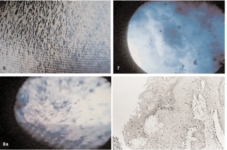
the alterations were superimposable to those observed in chronic edema patients. In leukoplakia several abnormali-ties were observed, especially a heterogeneity of cellular populations with nuclei different for size, color and shape. The heterogeneity of the cellular pattern corresponded to the histopathological findings of hyperkeratosis and dys-plasia (Fig. 6). The alterations were strictly proportional to the severity of the dysplasia.
Cell changes, such as increased nuclear density, dys-chromia, dyskaryosis and even mitoses, were observed in the most severe cases of dysplasia. Laryngeal papillomas were characterized by a convoluted aspect of the epithe-lium, with every convolution showing an autonomous vascular axis (Fig. 7). Findings in the carcinoma in situ during contact endoscopy revealed marked abnormalities of the cellular pattern, with a noticeable heterogeneity in nuclear size, shape, density and staining (Fig. 8 a, b).
Discussion
Contact endoscopy allows for both in vivo and in situ ob-servations of pathology in the superficial layer of the en-dolaryngeal epithelium because of the dynamic migration of deeper, underlying cells [16].
Our present study supports this technique and confirms the need for a better definition of normal cytological pa-rameters and subsequent pathological patterns. Our expe-70
Fig. 6 Leukoplakia (× 60). Heterogeneity of the cellular popula-tion is shown with nuclei different in size, shape and color Fig. 7 Papilloma (× 60). The lesion shows convolutions with an autonomous vascular axis
Fig. 8 a Vocal cord in situ carcinoma. An irregular pattern and nu-clear abnormalities in size and shape are identified (× 60). b Histo-patology of the vocal cord in situ squamous cell carcinoma with hyperkeratosis. Nuclear atypia of the keratocytes in the superficial layer is readely apparent. H & E, × 125
6
8a 88bb
71 rience has shown that contact endoscopy permits mapping
and cytological evaluation of the entire mucosal epithe-lium of the vocal cord due to the mobility of the endo-scope. This technique could be very useful in Reinke’s edema, in which an inflammatory state leads to an in-creased cellular turnover. As a consequence, cells with a lower degree of maturation (usually represented within the medium layer of the cord) and characterized by nuclei regular in shape and staining but increased in size become detectable on the epithelial surface. Contact endoscopy also allows for a good documentation in leukoplakias, as the detection of isolated cells containing keratin agglom-erates or plaques with complete disappearance of the cel-lular limits permits the assessment of different stages of keratinization. Other alterations, such as increased nuclear density, dyskromia, dyskariosis, and even cellular mi-toses, can be observed only in severe dysplasia [6, 7, 14]. Such cases require biopsy for tissue histology.
Contact endoscopy has allowed us to visualize the vas-cular network of the vocal cord surface and to document the distribution and dynamics of the microcirculation pre-sent [4, 5, 8]. Further improvements of the diagnostic ac-curacy of this technique should be possible due to the im-provement of imaging techniques and the use of new cell markers.
Contact endoscopy at present appears to be useful in the diagnosis of laryngeal pathologies, and could substi-tute for histological examination in some cases. Further studies are needed to clarify the penetration of methylene blue within the cord mucosa. However, our comparison of histological sections with images obtained by contact en-doscopy has shown that contact enen-doscopy only allows for evaluation of the upper layers of the epithelium, which is probably due to poor penetration of the colorant in the deeper layers. In fact, some authors state that contact en-doscopy would not permit the detection of mild (grade 1) dysplasias because most of the cellular abnormalities are limited to the basal layer of the epithelium [15, 17].
In general, methylene blue stains pathological cells more than normal ones [13], and even mild grades of dys-plasia will lead to cellular alterations in the surface ep-ithelium, with a gradual enlargement in nuclear size and an increased nucleus-to-cytoplasm ratio.
Nevertheless, overcoming the poor penetration of meth-ylene blue is expected to solve the main limit of this method. We should also point out that adequate use of this technique requires close cooperation between the
clini-cian and pathologist. Should the technical limits of the method be solved, contact endoscopy could become the method of choice for the screening and follow-up of la-ryngeal pathology and could improve current knowledge about the pathophysiology of endolaryngeal lesions [8]. References
1. Andrea M (1975) Arterial supply to the larynx: macro and mi-cro vascular distribution. Tese de Doutoramento, Lisboa 2. Andrea M, Dias O (1994) Rigid and contact endoscopy
associ-ated to microlaryngeal surgery. Arq Portugeses ORL Pat Cerv-Fac, Lisboa [Suppl 2]
3. Andrea M, Dias O, Paco J (1994) Endoscopic anatomy of the larynx. Curr Opin Otolaryngol Head Neck Surg 2:271–275 4. Andrea M, Dias O, Santos A (1995) Contact endoscopy during
microlaryngeal surgery. A new technique for endoscopic ex-amination of the larynx. Ann Otol Rhinol Laryngol 104:333– 339
5. Andrea M, Dias O, Santos A (1995) Contact endoscopy of the vocal cord. Normal and pathological patterns. Acta Otolaryn-gol (Stockh) 115:314–316
6. Crissman J (1991) Pathology of the upper aerodigestive tract mucosa. In: Paparella MM, Shumrick DA, Gluckmann JL, Meyeroff WL (eds) Otolaryngology, 3rd edn. Saunders, Phil-adelphia, pp 495–508
7. Ferlito A (1993) Neoplasms of the larynx. Churchill Living-stone, Edinburgh
8. Folkman J, Klagsbrun M (1987) Angiogenic factors. Science 235:442–447
9. Hamou JE (1983) Microendoscopy and contact endoscopy. Brevet Francais 79, 04168 Paris 1979. International patent. PCT/FR 80/0024, Paris, 1980. US patent 4, 385, 810, Wash-ington, DC
10. Hamou JE (1986) Hysteroscopie et micro-colpo-hysteroscopie. Atlas et traité. Masson, Paris
11. Hamou JE, Salat Baroux J, Coupex F, De Brux J (1984) Mi-crohysteroscopy: a new approach to the diagnosis of cervical intraepithelial neoplasia. Obstet Gynecol 63:567–574
12. Hamou JE, Salat Baroux J, Henrion R (1985) Hysteroscopie et micro-colpo-hysteroscopie. EMC 72, B10, Ed Technique, Paris 13. Lundgren J, Olofsson J, Hellquist H (1979) Toluidine blue. An aid in the microlaryngoscopic diagnosis of glottic lesion? Arch Otolaryngol Head Neck Surg 105:169–174
14. McGee J (1992) Oxford textbook of pathology. Oxford Uni-versity Press, London
15. Olofsson J (1984) Histologic grading of early lesions. In: Wigand E, Steiner W, Stell P (eds) Functional partial laryngec-tomy. Springer, Berlin Heidelberg New York, pp 37–38 16. Vancaille (1988) Manual of microcolposcopy. Elsevier,
Ams-terdam
17. Weiland L (1984) Histopathology of early laryngeal carci-noma. In: Wigand E, Steiner W, Stell P (eds) Functional partial laryngectomy. Springer, Berlin Heidelberg New York, pp 31– 36