A
A
l
l
m
m
a
a
M
M
a
a
t
t
e
e
r
r
S
S
t
t
u
u
d
d
i
i
o
o
r
r
u
u
m
m
–
–
U
U
n
n
i
i
v
v
e
e
r
r
s
s
i
i
t
t
à
à
d
d
i
i
B
B
o
o
l
l
o
o
g
g
n
n
a
a
DOTTORATO DI RICERCA
BIOLOGIA E FISIOLOGIA CELLULARE
Ciclo XXSettore/i scientifico disciplinari di afferenza: BIO/18 GENETICA
TITOLO TESI
ANALISI FUNZIONALE DEI RECETTORI PER LE
NEUROTROFINE p75NTR E Trka IN NEUROBLASTOMA
Presentata da:
ANTONELLA PAPA
Coordinatore Dottorato Relatore
Prof. Michela Rugolo Chiar.mo Prof.
Giuliano Della Valle
INDEX
Introduction
5
1.0 Neurotrophins and Neurotrophin Receptors
81.1 Trk receptors
101.2 The low affinity Neurotrophin receptor p75NTR
111.2.1 p75NTR as a positive regulator of cell survival and differentiation 14
1.2.2. p75NTR as a constitutively active pro-apoptotic receptor 15
1.2.3. Ligand-dependent p75NTR cell death 16
2.0 Downstream cell death signalling of p75NTR
183.0 p75NTR and the neurodegeneration
193.1 A
β
binds to p75NTR
22Result I
24p75NTR signals cell-death upon binding to the A
ββββ
peptide and recruiting
TNFR1–associated death domain (TRADD)
1.1 p75
NTR/TNFR1 conserved death domain aminoacidic residues are
required to trigger Abeta peptides induced cell death
251.2 TRADD dominant negative is able to interfere with p75NTR-dependent
A
β−
peptide induced cell death
301.3 p75NTR and TRADD interact mainly through the p75NTR/TNFR1
conserved residues required for A
β
-induced cell death
34Discussion I
39
Result II
Functional cooperation between TrkA and p75NTR accelerates
neuronal differentiation by increased transcription of GAP-43 and
p21(CIP/WAF) genes via ERK1/2 and AP-1 activities
2.1 p75NTR cooperates with TrkA to accelerate NGF-mediated neuronal
differentiation
422.2 p75NTR enhances TrkA autophosphorylation
462.3 p75NTR contributes to TrkA signaling by prolonging ERK1/2
activation
472.5 Activation of ERKs is required for NGF-mediated neuronal
differentiation
52Discussion II
58
Materials and Methods
60Introduction
Growth factor receptors define the linkage point between extracellular events and the intracellular responses, activating a plethora of signalling pathways. Transmembrane receptors proteins play critical roles in either development and maintenance of the cellular biology and are strongly correlated with the occurrence of disease (Shor NF, 2005). More than other, the brain tissue is the most sensitive to the presence of growth factors and, the differential expression of their specific receptors, amplify the complexity of the system. Neurotrophin are the specific growth factors in the nervous system. They were first identified as survival factors for sympathetic and sensory neurons but it is appreciated that they regulate many aspects of neuronal development and function, including synapse formation and synaptic plasticity (Lewin & Barde 1996; Bibel & Barde 2000; Kaplan &
Miller 2000; Huang et al. 2001; Poo 2001; Shooter 2001; Sofroniew et al. 2001; Dechant &
Barde 2002; Chao 2003; Huang & Reichardt 2003; Lu et al. 2005). The first Neurotrophin
identified was the Nerve Growth Factor (NGF), searching for survival factors that could explain the deleterious effects of deletion of target tissues on the subsequent survival of motor and sensory neurons (Levi-Montalcini 1987; Shooter 2001). The second neurotrophin to be characterized was the Brain-Derived Neurotrophic Factor (BDNF) purified from pig brain as a survival factor for several neuronal populations not responsive to NGF (Barde et
al. 1982). Given the conserved features of the sequences, it has been easier to clone the
other members of the family. Together with the NGF (Nerve Growth Factor) and BDNF (Brain Derived Neurotrophin Factor) other two factors are reported so far in mammals, the NT-3 and the NT-4 (Neurotrophin-3 and 4). It is known that neurothrophins have an important role in the development and function of neurons either in the central and in the peripheral nervous systems, including precursor proliferation and commitment, cell survival, axon and dendrite growth, membrane trafficking, synapse formation and function, as well as glial differentiation and interactions with neurons. Biological effects of each of the four mammalian neurotrophins are mediated through activation of one or more of the three members of the tropomyosin-related kinase (Trk) family of receptor tyrosine kinases (TrkA, TrkB and TrkC). In addition, all neurotrophins activate the p75 neurotrophin receptor p75NTR, a member of the tumour necrosis factor receptor superfamily.
NGF is the best characterised member of the family and is known to signal for opposite effects through interaction with two distinct families of receptors: activation of cell survival by TrkA and induction of cell death by binding to p75NTR. Engagement of Trk receptors leads to activation of well characterized pathway like Ras, phosphatidylinositol 3-kinase, phospholipase C-gamma1 and the mitogen-activated protein kinases. On the other side, the molecular mechanisms mediated by p75NTR still remain to define even though the JNK, NF-κB and ceramide have been implicated (Huang and Reichardt, 2003). Preclinical studies point to the therapeutic potential of neurotrophic factors in preventing or slowing the progression of neurodegenerative conditions. Given the difficulties inherent with a protein therapeutic approach to treating central nervous system disorders, increasing attention has turned to the development of alternative strategies related to the signalling pathways involved in the neurodegenaration (Reichardt LF, 2006).
1.0
Neurotrophins and Neurotrophin Receptors
The neurotrophins and their genes share homologies in sequence and structure and the organization of the genomic segments adjacent to these genes also resulted to be similar. Together, these observations provide evidence that the neurotrophin genes have arisen through successive duplications of a portion of the genome derived from an ancestral chordate (Hallbook 1999). Their genes share many similarities, including the existence of multiple promoters. The protein product of each gene includes a signal sequence and a prodomain, followed by the mature Neurotrophin sequence (Hempstead B. L. 2006). Thus, each gene product must be processed by proteolysis to form a mature protein. A recent work has demonstrated that regulation of their maturation is an important post-transcriptional control point that limits and adds specificity to their actions (Lee et al. 2001). The mature neurotrophin proteins are non-covalently associated homodimers. Although some neurotrophin monomers are able to form heterodimers with other neurotrophin monomers in
vitro, there is no evidence that these heterodimers exist at significant concentrations in vivo.
Each of these four proteins shares a highly homologous structure with features tertiary fold and cysteine knot that are present in several other growth factors, including transforming growth factor-β (TGF-β) and platelet derived growth factor (PDGF). Additional neurotrophins have been isolated from fish, where NT-6 and NT-7 have been characterized but they do not have orthologues in mammals or birds. The biological effect of the neurotrophins is mediated by interaction with two distinct classes of receptors:
• Trk receptors (tropomyosin- related kinase)
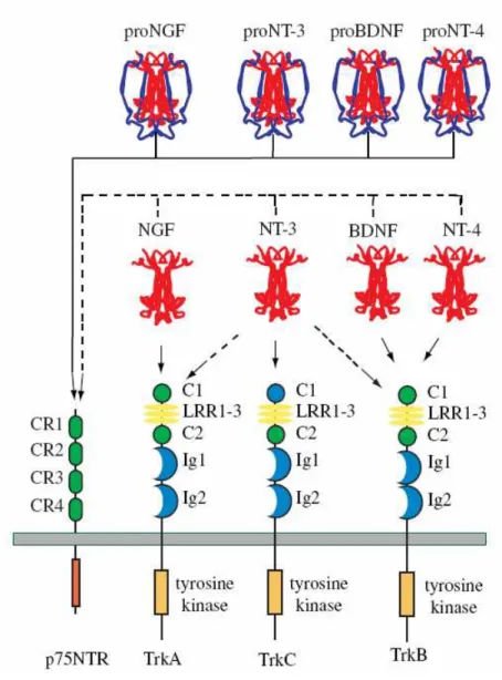
Fig. 1. Neurotrophin and Neurotrophin receptors. This illustrates the major interactions of each of the four
mammalian neurotrophins. Each proneurotrophin binds p75NTR, but not the Trk receptors. Following maturation through proteolysis of the proneurotrophins, each mature Neurotrophin is able to bind and activate p75NTR, but exhibits more specific interactions with the three Trk receptors. NGF binds specifically TrkA; BDNF and NT4 recognize TrkB; NT3 activates TrkC. In some cellular contexts, NT3 is also able to activate TrkA and TrkB with less efficiency. Differential splicing generates isoforms of TrkB and TrkC that have truncated cytoplasmic domains lacking a tyrosine kinase motif. Splicing also generates an isoform of TrkC with a small insert in the kinase domain that affects substrate specificity. Splicing of exons that generate the extracellular domain of each Trk receptor results in the expression of receptors with small peptide inserts between the second immunoglobin and transmembrane domains that affect ligand-binding specificity. Ligand-binding specificity is also affected by the presence of p75NTR.
1.1
The Trk receptors
TrkA was originally characterized as a transforming oncogene in which tropomyosin was fused to an unknown tyrosine kinase (Martin-Zanca et al., 1989). The corresponding proto-oncogene was shown to be a member of a highly related family of transmembrane tyrosine kinases which were expressed in discrete neuronal populations and which bound and were activated by specific neurotrophins, with TrkA preferentially binding NGF, TrkB preferring BDNF and NT-4/5, and TrkC interacting with NT-3 (Klein et al., 1990, 1991a,b; Cordon-Cardo et al., 1991; Kaplan et al.,1991a,b; Lamballe et al., 1991; Soppet et al., 1991; Squinto et al., 1991; Ip et al., 1993b). In the absence of p75NTR, high concentrations of NT-4/5 can activate TrkA and likewise, NT-3 can activate TrkA and TrkB. NT-3 is therefore a non-preferred ligand for TrkA and TrkB, and NT-4 is a non-non-preferred ligand for TrkA (Segal and Greenberg, 1996). All Trk receptors are Type I transmembrane proteins that are members of the receptor tyrosine kinase superfamily (Martin-Zanca et al., 1989; reviewed in Ip and Yancopoulos, 1994; Barbacid, 1995). The extracellular domains (ECDs) of the Trk receptors contain two cysteine-rich regions (domains 1 and 3) flanking a leucine-rich repeat (domain 2), followed by two immunoglobulin (IgG)-like domains in the juxtamembrane region (domains 4 and 5; Windisch et al., 1995). Binding and deletion studies on TrkA, TrkB and TrkC indicate that domain 5 is responsible for neurotrophin binding (Urfer et al., 1995, 1998; Perez et al., 1995; Ultsch et al., 1999), with the second leucine-rich domain having a modulatory, perhaps indirect, role in ligand interaction (Windisch et al., 1995).
1.2
The low affinity Neurotrophin receptor p75NTR
The first Neurotrophin receptor discovered was p75 neurotrophin receptor (p75NTR), initially identified as a low-affinity receptor for NGF, subsequently has been shown to bind each of the neurotrophins with a similar affinity (Rodriguez-Tebar et al. 1990; Frade and
Barde 1998; Chao and Hempstead, 1995). Based on the primary sequence and secondary
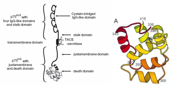
structure, p75NTR belongs to the Fas-tumor necrosis factor (TNF) receptor superfamily with an extracellular domain that includes four cysteine-rich motifs, a single transmembrane domain and a cytoplasmic domain that includes a juxtamembrane domain and a ‘death’ domain similar to those present in other members of this family (Liepinsh et al. 1997; He &
Garcia 2004).
Fig. 2. Schematic representation of p75NTR. Extracellular, transmembrane and intracellular domains of the receptor
on the left. Solution structure of the death domain of rat p75ICD on the right. Ribbon representation of residues 336– 416. The positions of the N-terminal residues of the helices are labelled. (Blochl and Blochl, 2007; Liepinsh et al, 1997)
Recent studies have identified new mechanisms able to process the receptor with subsequent generation of soluble peptides supposed to exert new cytoplasmic or nuclear functions (Kanning KC et al, 2003; Podlesniy et al, 2006).
Since p75NTR does not contain a catalytic motif, the signal mediation depends on the interaction with cytoplasmic proteins important for regulating neuronal survival and
differentiation as well as synaptic plasticity. The complex signalling of p75NTR makes it difficult to formulate a unified functional model for this receptor. Indeed, many trophic and apoptotic activities are integrated with that of other receptors and mechanisms. Synergistic and antagonistic interactions of p75NTR and Trk receptors such as complex formation, mutual inhibition, or negative control of Trk effects by p75NTR have been extensively investigated without defining a general model (Dechant 2001; Huang and Reichardt 2003; Teng and Hempstead 2004).
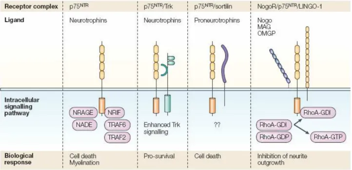
Fig. 3. p75 neurotrophin receptor and interactor receptors. P75NTR has been referred to as a ‘low-affinity’
Neurotrophin receptor, but this definition should be avoided because proNGF binds p75NTR with an affinity similar to that of nerve growth factor (NGF) binding to TrkA. Although it lacks a kinase domain, p75NTR can cooperate with many different protein partners and form multimeric receptor complexes to produce a number of cellular responses, including apoptosis, neurite outgrowth and myelination111–113. So far, sortilin61, LINGO-1, Nogo-66 (NgR)114 and Trk receptors115 have been identified as co-receptors. In addition to extracellular interactions that yield multimeric receptor complexes, the intracellular domain of p75NTR can also interact with many different adaptor and signalling proteins. These include neurotrophin-receptor-interacting MAGE (melanoma-associated antigen) homologue (NRAGE)116, neurotrophin-associated cell death executor (NADE)117, TNF (tumour necrosis factor)-receptor-associated factors 2 and 6 (TRAF2 and TRAF6)118,119, and neurotrophin-receptor-interacting factor (NRIF)120,121. GDI, guanine-nucleotide dissociation inhibitor; MAG, myelin-associated glycoprotein; OMGP, oligodendrocyte myelin glycoprotein; RhoA, small G protein.
Beside the more characterized interaction with the Trk receptors, p75NTR can also interact with other different cytoplasmic receptors. p75NTR can be influenced by co-receptors like NgR and Lingo1, which cooperate to prevent p75NTR activation, or sortilin, which shares the pro-neurotrophin ligands with p75NTR and directs p75NTR activity towards apoptosis (Nykjaer et al. 2004; reviews: Bronfman and Fainzilber 2004; Ceni and Barker 2005).
Taking together, all these studies suggest that rather than being just a co-receptor, all the identified interactors for p75NTR can modulate, inhibit or enhance its activities. The results is that the actions of the receptor may be explained by considering three different biological outcomes:
1. positive regulation of cell survival and differentiation 2. constitutive pro-apoptotic activation
1.1.1 p75NTR as a positive regulator of cell survival and differentiation
In neurotrophin-responsive neuronal populations, p75NTR and Trk receptor members are frequently co-expressed, particularly in the vertebrate peripheral nervous system. P75NTR provides a positive modulatory influence on TrkA function by increasing the number of high affinity binding sites (Mahadeo et al, 1994). Regulation of high-affinity site formation by co-expression of TrkA and p75NTR provides an explanation for how these receptors may cooperate to increase Neurotrophin responsiveness during development. Indeed, neuronal cell lines express both receptors have an enhanced autophosphorylation of TrkA, leading to a faster differentiative response with NGF (Verdi et al, 1994). The general feeling proposes that the cell survival is enhanced by a higher ratio of p75NTR to Trk receptors. Indeed, considering p75NTR null mice it has been found a selective losses in sensory and sympathetic innervations. From neonatal p75NTR null mice, sympathetic neurons required higher concentrations of NGF to survive than neurons from normal mice at earlier developmental stages (Lee et al, 1994). To explain how p75NTR can modulate Trk receptors functions two models have been proposed so far. The first is a ligand passing mechanism, which predicts that the high-affinity state is the result of ligand presentation by p75NTR to the TrkA receptor (Barker and Shooter, 1994). The second model predicts that p75NTR and TrkA are capable of a ligand-independent association, which produces a high affinity binding interaction. (Chao and Hempstead, 1995). The second model would predict that conformational changes would occur to facilitate ligand binding. (Ross et al, 1996) All of these observations are consistent with the hypothesis that p75NTR can serve as a positive influence upon TrkA function.
1.1.2. p75NTR as a constitutively active pro-apoptotic receptor
In the1993 Bredesen and colleagues hypothesized that p75NTR was a death receptor. Based on the observation that immortalized neural cells overexpressing p75NTR display an higher rate of apoptosis in response to serum withdrawal, they proposed a mechanism of ligand-indipendent death (Rabizadeh et al, 1993). According to this model, apoptosis promoted by p75NTR can be rescued after binding to NGF. How NGF gives a survival signal through binding to p75NTR has not been established. However, the correlation between high levels of p75NTR expression and susceptibility to apoptosis after growth factor withdrawal has been proved also in PC12 cells supporting this mechanism of cell death (Barrett and Georgiou, 1996). Furthermore, down-regulation of p75NTR expression in neonatal dorsal root sensory neurons, using an antisense strategy, reveals enhanced survival (Barrett and Bartlett, 1994). The phenotype of the p75NTR null mice supports a role in neuronal survival (Davies et al., 1993; Lee et al., 1994a,b) but an apoptotic function as well. Analysis of mice has revealed a significantly higher number of cholinergic neurons in the basal forebrain in p75NTR -/- mice compared to wild type controls (Van der Zee et al., 1996; Yeo et al., 1997). These observations indicate that the absence of p75NTR resulted in enhanced survival, similar to the antisense effects observed in postnatal sensory neurons (Barrett and Bartlett, 1994).
1.1.2. Ligand-dependent p75NTR cell death
Several lines of evidence now firmly support that neurotrophins can actively kill cells through direct engagement of its p75NTR receptor. Both in vitro and in vivo evidence has indicated that neurotrophins and p75NTR are required for apoptosis of selective cell populations. In contrast to the model of NGF rescue through p75NTR, other studies demonstrate that cultured trigeminal neurons at embryonic age E10 are killed by NGF through binding to p75NTR (Davey and Davies, 1998). The cell death role of p75NTR is elicited upon injury or traumatic conditions. Similarly, cultured glial cells, such as fully differentiated oligodendrocytes, express elevated levels of p75NTR are effectively killed by NGF (Casaccia-Bonnefil et al., 1996). The apoptotic effects of p75NTR are not only enhanced by NGF binding but, in certain conditions, also by high concentrations of BDNF and other neurotrophins. Postnatal sympathetic neurons express p75NTR and TrkA, but are killed by BDNF-mediated activation of p75 (Bamji et al., 1998). BDNF for survival are killed by NT-4 through binding to p75NTR (Agerman et al., 1999). Primary cell culture experiments demonstrated that p75NTR was necessary for neuronal cell survival promoted by BDNF, but NT-4 binding to p75 induced cell death. This remarkable set of observations indicates that p75NTR and Trk receptor can simultaneously influence life-and-death decisions in neurons depending upon which ligands are available. An issue raised by these experiments is that although all neurotrophins (NGF, BDNF, NT-3, and NT-4) bind to p75NTR with similar affinity, each neurotrophin may exert different effects on cell function and viability through p75NTR. For example, the effect of NGF on oligodendrocyte cultures could not be reproduced by similar concentrations of BDNF or NT-3. Furthermore, in PC12 cells treated with antisense oligonucleotides to downregulate TrkA expression, BDNF but not NGF can rescue cells from serum-withdrawal (Taglialatela et al., 1996). A very likely explanation for the effect of distinct neurotrophins in the oligodendrocyte system is the presence of TrkB and TrkC receptors in these cells (Cohen et al., 1996).An alternative explanation for cells not expressing other Trk receptors is the differential ability of neurotrophins to activate distinct signal transduction pathways (Carter et al., 1996; Carter and Lewin, 1997). This hypothesis is also supported by striking differences in the kinetics of binding and the degree of positive cooperativity of each neurotrophin to p75NTR
(Rodriguez-Tebar et al., 1990, 1992). A related explanation may involve different adaptor molecules that are associated with the receptor.
2. Downstream cell death signalling of p75NTR
Since the many effects modulated by p75NTR, it is reasonable the identification of as such high number of interactors that bind the intracellular domain of the receptor. Several different p75NTR interacting molecules, with and without catalytic activity, have been identified to date (Gentry et al, 2004). Non-catalytic interactors include a series of scaffolding and adaptor-like molecules, such as caveolin-1 (Bilderback et al, 1997), Bex3/NADE (Mukai et al, 2000) and TRAF6 (Khursigara et al, 1999; Ye et al, 1999); larger proteins containing zinc-finger domains with some degree of nuclear localization, such as NRIF1/2 (Casademunt et al, 1999) and SC-1 (Chittka and Chao, 1999); and members of the MAGE homology domain family, such as NRAGE (Salehi et al, 2000) and necdin (Tcherpakov et al, 2002) with proposed roles in the regulation of apoptosis. p75NTR interactors with catalytic activity include serine–threonine kinases involved in interleukin and NF-kB signaling, such as IRAK (Mamidipudi et al, 2002) and RIP2 (Khursigara et al, 2001); a protein tyrosine phosphatase (FAP-1) (Irie et al, 1999); and the small GTPase RhoA (Yamashita et al, 1999). How these p75NTR-interacting proteins connect to downstream signaling pathways and cellular responses is still not clear. Some of the principal downstream events characterized in p75NTR signalling include ceramide production (Dobrowsky et al, 1994) and activation of the transcription factor NF-kB (Carter et al, 1996) and the c-Jun kinases JNK1–3 (Casaccia-Bonnefil et al, 1996; Friedman, 2000; Harrington et al, 2002; Costantini et al, 2005).
3. p75NTR and the neurodegeneration
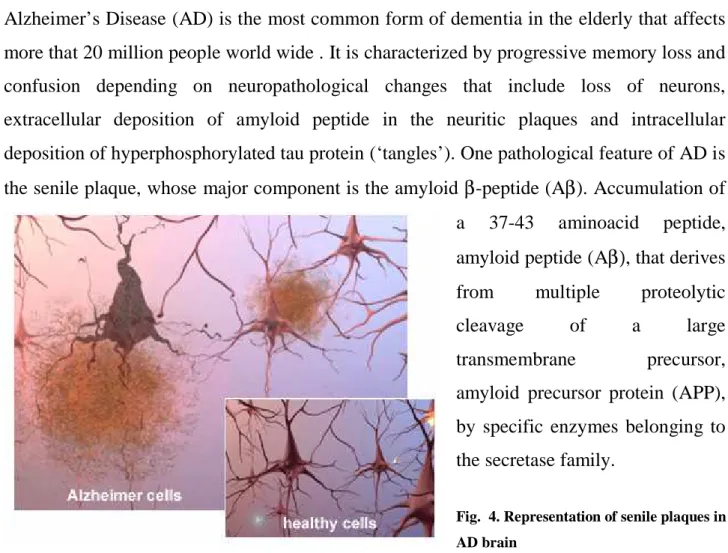
Alzheimer’s Disease (AD) is the most common form of dementia in the elderly that affects more that 20 million people world wide . It is characterized by progressive memory loss and confusion depending on neuropathological changes that include loss of neurons, extracellular deposition of amyloid peptide in the neuritic plaques and intracellular deposition of hyperphosphorylated tau protein (‘tangles’). One pathological feature of AD is the senile plaque, whose major component is the amyloid β-peptide (Aβ). Accumulation of a 37-43 aminoacid peptide, amyloid peptide (Aβ), that derives from multiple proteolytic
cleavage of a large
transmembrane precursor,
amyloid precursor protein (APP), by specific enzymes belonging to the secretase family.
Fig. 4. Representation of senile plaques in AD brain
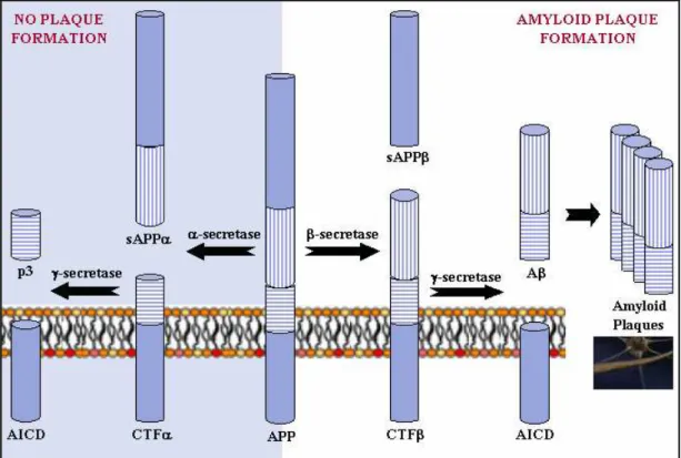
The generation of Aβ peptide is a physiological peptide derives from an amyloid precursor protein that can be processed in different ways by different sets of enzymes. One pathway leads to amyloid plaque formation (amyloidogenic), while another does not (non-amyloidogenic). Usually about 90% of APP enters the non-amyloidogenic pathway, and 10% the amyloidogenic one, but these ratios can change due to mutations, environmental factors, as well as the age of the individual. Cleavage products from both these pathways may play important roles in neural development and function.
Fig. 5. APP Processing α-secretase and γ-secretase produce non-plaque forming p3, while β-secretase and γ-secretase produce amyloid plaque-forming Aβ. The different regions of the APP protein are indicated.
In the non-plaque-forming pathway, APP is cleaved first by α-secretase to yield a soluble
N-terminal fragment (sAPPα) and a C-terminal fragment (CTFa). sAPPα may be involved in the enhancement of synaptogenesis, neurite outgrowth and neuronal survival, and are considered to be neuroprotective. CTFα is retained in the membrane, where it is acted upon by presenilin-containing γ-secretase to yield a soluble N-terminal fragment (p3) and a membrane-bound C-terminal fragment (AICD, or APP intracellular domain). AICD may be involved in nuclear signalling via transcriptional regulation as well as axonal transport through its ability to associate with a host of different proteins.
In the plaque-forming pathway, APP is cleaved first by a different enzyme, β-secretase (a
transmembrane aspartic protease), yielding a soluble N-terminal fragment (sAPPβ) and a membrane-bound C-terminal fragment (CTFβ). This cut is made closer to the N-terminal end of APP than the cut with α-secretase, making CTFβ longer than CTFα. CTFβ is then acted upon by γ-secretase (as occurred in the previous pathway), yielding a membrane-bound C-terminal fragment (AICD) the same as before, and a soluble N-terminal fragment (amyloid-β, or Aβ) that is longer than p3.
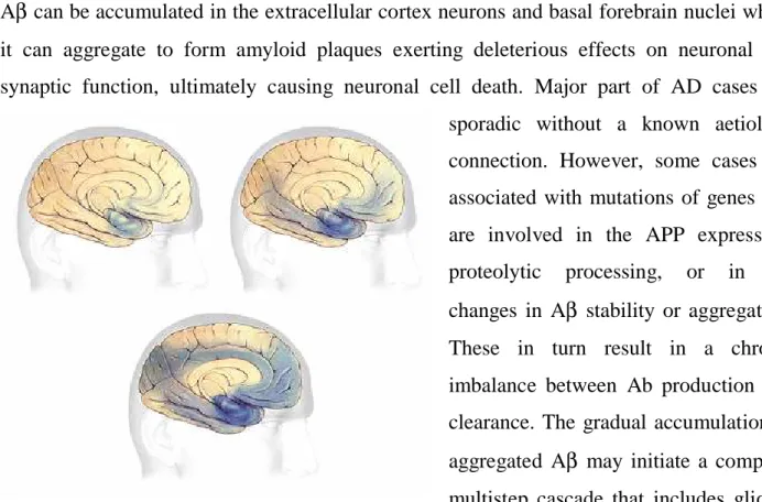
Aβ can be accumulated in the extracellular cortex neurons and basal forebrain nuclei where it can aggregate to form amyloid plaques exerting deleterious effects on neuronal and synaptic function, ultimately causing neuronal cell death. Major part of AD cases are sporadic without a known aetiology connection. However, some cases are associated with mutations of genes that are involved in the APP expression, proteolytic processing, or in the changes in Aβ stability or aggregation. These in turn result in a chronic imbalance between Ab production and clearance. The gradual accumulation of aggregated Aβ may initiate a complex, multistep cascade that includes gliosis,
Fig. 6. Progression of neurodegeneration in brain in AD inflammatory changes and formation of
neurofibrillary tangles.
In this scenario, an hypothesis that has been put forward to explain the aetiology of AD arises the connection between the occurance of the pathology with the Neurotrophic factors,
suggesting a causal link (Rabizadeh et al, 1994).
An early indicator of Alzheimer’s disease is the degeneration of the cholinergic basal forebrain neurons, which express the highest levels of the pan neurotrophin receptor (p75NTR) in the adult brain (Gibbs et al. 1989). These neurons are dependent on NGF-mediated survival signalling through TrkA which, unlike p75NTR, is reduced in Alzheimer’s disease patients. In addition, p75NTR is also expressed in the Trk-negative, degenerating cortical neurons of Alzheimer’s disease sufferers, a situation which is not reflected in healthy elderly subjects (Mufson et al. 1992). Furthermore, the neurotrophins, acting differentially through Trk receptors and p75NTR, appear to regulate the expression and cleavage of the amyloid protein precursor (APP; Costantini et al. 2005a), potentially regulating the generation of Aβ with ageing. Given that up-regulation and ligand activation of p75NTR have been widely shown to mediate neural cell death in animal models of neurodegenerative disease (Coulson et al. 2000a; Dechant and Barde 2002; Roux and
Barker 2002) and that the pro-form of NGF, which selectively binds to p75NTR to promote neuronal death (Lee et al. 2001), is increased in Alzheimer’s disease (Fahnestock et al. 2001; Peng et al. 2004), the receptor is a strong candidate for independently mediating the degeneration occurring in Alzheimer’s disease.
3.1 A
ββββ
binds to p75NTR
Further support to the hypothesis that p75NTR could be a mediator of the cytotoxic effect induced by deposition of Aβ derives from the study of Mina Yaar in which they proved the direct binding. In 1997 Yaar et al. reported that Aβ 1–40 binds to and immunoprecipitates with p75NTR (Yaar et al. 1997). In the brain, this protein is expressed at the highest level by the cholinergic neurons of the basal nuclear complex, which are sensitive to Aβ neurotoxicity, and undergo degeneration in AD. In contrast, the neurons of other cholinergic complexes in the brain (pedunculoponine and lateral tegmental nuclei) neither express p75NTR nor undergo degeneration in AD, suggesting that the vulnerability of basal nuclear neurons and their projections may be related to their high-level expression of p75NTR (Rabizadeh et al,1994). The use of rat cortical neurons and a cell line engineered to express p75NTR has demonstrated that P75NTR binds specifically fAβ, and that this binding is followed by apoptosis (Yaar et al, 1997; 2002). The binding of Aβ to P75NTR activates NF-kB in a time- and dose-dependent manner. Blockade of the interaction between Aβ and p75NTR with nerve growth factor or inhibition of NFKB activation by curcumin or NF-κB SN50 attenuated or abolished Aβ-induced apoptotic cell death (Kuner et al, 1998). Other studies have shown that P75NTR may be present in a trimer form that binds Aβ to induce receptor activation, and that Aβ binds to both the p75NTR trimer and the P75NTR monomers. In neuronal hybrid cells, it has been confirmed that p75NT mediates Aβ toxicity, and that the p75NTR-mediated Aβ neurotoxicity involves Go, c-jun kinase, reduced nicotinamide adenine dinucleotide oxidase and caspases 9/3 (Tsukamoto et al, 2003). However, it has been reported that, in human primary neurons in culture, p75NTR protects against extracellular Aβ-mediated apoptosis. This neuroprotection might occur through a P13Kdependent pathway. The reason for this difference may be explained by differential activation of a signal transduction pathway in primary neurons versus tumour
cell lines, a cell-type or species-specific effect of Aβ, or a differential expression of the other neurotrophic receptors (Zhang et al, 2003). Along with this hypothesis, other studies focused their attention on the possible mechanism that can be activated after binding of Aβ to p75NTR. In the 2002 Perini et al identified a specific cytoplasmic domain that resulted to be essential for the mediation of the Aβ cell death (Perini et al, 2002). To this purpose, Aβ has been tested for the neuronal toxicity on a SK-N-BE neuroblastoma cell line devoid of all Neurotrophin receptors, and on several SK-N-BE derived cell clones either expressing the full-length or truncated forms of p75NTR . It has been found that p75NTR plays a direct role in Aβ cell death through the signalling function of the death domain (DD). Furthermore, it has been showed that the cell death is caspase dependent, in particular through activation of caspase-8 and oxidative stress.
p75NTR signals cell-death upon binding to the A
ββββ
peptide and
recruiting TNFR1–associated death domain (TRADD)
1.1 p75
NTR/TNFR1 conserved death domain aminoacidic residues are
required to trigger Abeta peptides induced cell death
In the past years it has been shown that p75NTR signals cell death upon binding to the β -amyloid peptide (Aβ) (Yaar et al,1997; Kuner et al,1998; Costantini et al,2005).
In a previous study published from this laboratory, it has been proposed that p75NTR activates apoptosis through the Death Domain (DD) that characterizes the cytoplamic sequence of the receptor (Perini et al,2002). In this further study, we deeper analyze the mechanism by which p75NTRDD signals cell death putting forward a more detailed map of the domain. Furthermore, we describe one of the possible initial steps of the cytotoxic effects induced by Aβ correlating p75NTR with the changes that occur in Alzheimer’s disease.
In our studies the cell system of reference is a Neuroblastoma cell line (SK-N-BE) that does not express any neurotrophic receptors at detectable level and that allow us to better define the unique role of the p75NTR in the signal transduction induced by Aβ (Bunone et al., 1997)
Using derived Neuroblastoma cell pools of SK-N-BE engineered to express full-length or various truncated forms of p75NTR, we have already demonstrated that p75NTR is involved in the direct signaling of cell death A dependent through activation of caspases-8 and 3, inducing the production of reactive oxygen intermediates and formation of oxidative stress (Perini et al,2002).
To better understand the molecular mechanism by which p75NTR mediates this cell death, we reasoned on the assumption that p75NTR is devoid of any intrinsic enzymatic activity so it should signal, across the cell membrane, through protein-protein interaction, likely involving aminoacidic residues placed on the surface of the intracellular p75NTR-DD. Starting from this observation, the first aim of our experiments was to characterize which residues were more responsible or more involved in the transduction of the Aβ dependent cell death. Previous NMR and solvent accessibility studies have analyzed the conformation of the p75NTR DD highlighting the amino acidic residues putatively located on the outer
surface. (Liepinsh et al,1997) Knowing the tridimensional structure, we decided to mutagenize the identified outer residue by site-specific mutagenesis through Alanine-substitution. (Cunningham & Wells,1989).
We targeted different residues listed as follow:
• D355 (Interhelical loop I)
• H359 and E363 (Helix II) Q367 (Interhelical loop II)
• P368 and E369 (Helix II) A378 (Interhelical loop III)
• D392 and S393 (Interhelical loop IV)
• D397 (Helix V)
• R404, R405, Q407 and R408 (Interhelical loop VI)
• D410 and E413 (Helix VI)
The specific residues are numbered following the order in the original paper (Liepinsh et al,1997)
All the residues found by NMR to be located at the surface of the p75NTR DD have been substituted to A but the A378 substituted to D. (Residues in the helix I have been excluded from this analysis because they were not deleted in the p75NTR∆DD truncated mutant used in the previous study Perini et al,2002). For all the PCR-mediated site-specific mutagenesis we used the rat p75NTR cDNA cloned in the pcDNA3 expression vector (p75NTR/pcDNA). The mutated plasmids have been used to generate stable pools of SK-N-BE. Subsequentially, cells expressing wild-type or mutant p75NTR, carrying either single or multiple substitutions (Table 1 and Fig 1A) have been analyzed to determine if and in which way they could be involved in the mediation of cell death induced by Aβ. Pools of SK-N-BE have been exposed to the aggregated Aβ(25-35) as well as to the Aβ(35-25) reverse peptides as negative control. The treatment has been performed for 48 hours the levels of cell death have been evaluated by microscopic analysis after TUNEL labeling.
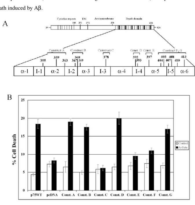
Analyzing the cell death levels after Aβ treatment, it was evident that not all the substituted residues were involved at the same way in mediating cytotoxicity. Expression of the Rp75NTR-C, Rp75NTR-E and Rp75NTR-F mutant receptors decreases the level of Aβ peptide induced cytotoxicity (fig 1B) whereas the Rp75NTR-A, B, D and G did not show any changes
in the levels of cell death. Therefore, our first conclusion was that only the most C-terminal half of p75NTR-DD (between interhelical region III and the helix VI) is required for the cell death induced by Aβ.
Fig. 1. Role of the p75NTR death domain outer aminoacidic residues in Aββββ–induced cytotoxicity. (A) Schematic
representation of the rat p75NTR receptor with details of the death domain. The aminoacidic residues target of the site directed mutagenesis in the different mutant p75NTR expression vectors (constructs A to G) are indicated (construct A: aa 355-359-363; construct B: aa 367-368-369; construct C: aa 378; construct D: aa 392-393; construct E: aa 397; construct F: aa 404-405-407-408-410-413; construct G: aa 407). The mutated amino acid are putatively located at the death domain surface and their position in the death domain alpha-helix (α) or interhelical loop (I) are indicated. The different expression vectors has been transfected in neuroblastoma SK-N-BE cells and stably transfected pools have been selected. (B) Aβ cytotoxicity analysis by TUNEL labelling and Ethidium Bromide nuclear counterstaining. SK-N-BE cell pools expressing either wild type p75NTR (p75WT) or the indicated mutant p75NTR were exposed to A (25-35) 20 M (A-Beta) or A (35-25) 20 M (control) for 48 hrs, then cell death level has been evaluated. Plotted cell death results are shown. SK-N-BE cells stably transfected with the empty pcDNA vector are used as negative control. Data are means + ES of three experiments.
These first data gave us a new insights and new interests to better define the protein conformation of the p75NTRDD. Indeed, we considered that, since its primary sequence
and secondary structure, p75NTR belongs to the Superfamily of the Tumor Necrosis Factor Receptors, which contains also the TNFRI itself and Fas receptors.
The similarity of the structures and the homology of the sequences between the death domains of p75NTR, TNFRI and Fas receptor have been previously analyzed in a paper by Barbara Chapman (Chapman et al, 1995). In that study the comparison of DDs sequences from p75NTR, TNFR1 and Fas from human, mouse and rat using a matrix procedure to detect low level of aminoacid identity, has let to evaluate 32,8% identity between p75NTR and TNFR1 and 25,4% between TNFR1 and Fas (Chapman,1995). Notably, some specific residues were found almost completely conserved and more importantly four of them have been previously demonstrated to be necessary for TNFα-induced TNFR1-mediated cell death (Tartaglia et al,1993). In detail, the residues E369, W378, D390, I408 are highly conserved between the DD of p75NTR and TNFRI and correspond to the following p75NTR residues: E377, W388, D397 and I411. (figure Rasmol!)
Interestingly, our first screening revealed that mutations in conserved residues or very close to them (A378D, D397A, and residues substituted in the construct p75NTR-F) are sufficient to block almost completely the cytotoxic signal (Fig.1B).
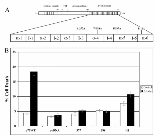
To extend our analysis we also mutated the E377, W388 and I411 residues into Ala (Fig. 2A). Interestingly, as shown for D397 (Fig. 1B) also E377, W388 and I411 seem to be crucial for Aβ triggered cytotoxicity.
Fig. 2 Role of the p75NTR/TNFR1 conserved Aminoacidic residues in Aββββ–induced cytotoxicity (A) Schematic
representation of the rat p75NTR receptor with details of the death domain. Mutant of p75NTR cDNA were generated by PCR introducing substitution to A of the indicated amino acid residues conserved with TNFR1 (aa E377, W388, and I411). The different expression vectors has been transfected in neuroblastoma SK-N-BE cells and stably transfected pools have been selected. (B) Aβ cytotoxicity analysis by TUNEL labelling and Ethidium Bromide nuclear counterstaining. SK-N-BE cell pools expressing either wild type p75NTR (p75WT) or the indicated mutants p75NTR (377, 388, 411) were exposed to A (25-35) 20 M (A-Beta) or A (35-25) 20 M (control) for 48 hrs, then cell death has been evaluated. SK-N-BE cells stably transfected with the empty pcDNA vector are used as negative control. Data are means + ES of three experiments.
We found that indeed single point mutations are enough to abate almost to the background level the percentage of TUNEL positive nuclei after 48 hours of Aβ exposure.
All together these data suggest that, upon Aβ peptide activation, p75NTR can signal cell death through some specific areas of its DD and importantly these areas include conserved residues shared with TNFR1 receptor.
1.2 TRADD dominant negative is able to interfere with p75NTR-dependent
A
β−
peptide induced cell death.
The first set of experiments we performed clarified that, after exposure to the A peptide, p75NTR signals cell death in Neuroblastoma cell line and we identified specific Death Domain residues required in mediating cell death.
The identification of the conserved residues between p75NTR and TNFR1 represented a moment of discussion that allow us to speculate about the possible mechanisms by which p75NTR can transduce the signal into the cell. Since the involvement of the shared residues seemed to be crucial for Aβ-induced cell death, we postulated that at least in this contest, p75NTR could signal cell death through apathway similar to the one already characterized for TNFR1.
In support of our idea many reports have showed that Aβ is able to trigger death in different cell types in which p75NTR is expressed: rat primary cortical neurons, NIH-3T3 and neuroblastoma cell stably expressing p75NTR. It is also known that Aβ activates different signaling pathways involving JNKs, p38/SAPK and the transcription factor NFkB (Kuner et al,1998;Yaar et al,2002;Costantini et al,2005; Yao et al,2005). Interestingly, all of these pathways are known to be activated in various cell context through the TNFR1-DD (PER JNK: De Smaele et al,2001: Tang et al,2001; Deng et al,2003; Li et al,2005; Kamata et al, 2005). Furthermore, it has been recently reported that in MCF7 breast cancer cells the Tumor Necrosis Factor Receptor1-associated Death Domain protein (TRADD), the most proximal TNFR1 cytoplasmic adaptor protein, is able to interact with the p75NTR-DD (El
Yazidi-Belkoura et al,2003).
To address the question if TRADD can have a role in Aβ induced p75NTR-mediated cytotoxicity, we generated a dominant negative (DN) for TRADD. The HA-TRADD∆DD mutant lacks the Death Domain of the protein at the C-terminal end and is HA-tagged at the N-terminal end.
A TRADD deleted of the C-terminal domain cannot anymore interact with the TNFRI DD but can still interact with downstream factors through the N-terminal region. A similar mutant has been successfully used to block the activation of TNFR1- or p75NTR-dependent signaling pathways such as the Nf-κB, JNKs and p38/SAPK pathways through a dominant negative effect most likely elicited by titration of downstream factors, such as
TNFR-associated factor-2 (TRAF2) (Kieser et al,1999; El Yazidi-Belkoura et al,2003). To validate the effectiveness of this reagent, we proceeded verifying if the TRADD∆DD mutant was actually able to interfere with a TNFR1-dependent cell death process.
To do so we induced TNFRI overexpression in HEK293 cells, leading to the formation of trimeric receptor complex that mimic the activation induced by ligand. Thus, we performed experiments of co-transfection to study the role of the dominant negative, TRADD∆DD. To label the transfected cells we included a vector to express GFP in every transfection experiment.
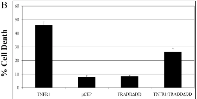
24 hours after transfection, cells have been fixed and stained with Hoechst 33342 and observed at the microscope to evaluate the number of transfected cell showing normal or apoptotic morphology. The transfected cells have been scored for the general cell morphology made evident by the ubiquitous expressed GFP, looking at the presence of the typical membrane blebbings and for the nuclear morphology, looking at the bright and condensed Hoechst staining. Indeed, overexpressed TNFR1 was able to trigger high level of cell death in HEK293 whereas co-transfection with the TRADD∆DD vector significantly reduced the number of dead cells. (fig. 2A) This experiment indicates that the TRADD∆DD is able to interfere with a TNFR1/TRADD dependent cell death process making it a suitable reagent to test if TRADD is also involved in Aβ induced p75NTR-mediated cytotoxicity in Neuroblastoma SK-N-BE cells.
Fig. 3A Role of cytoplasmic adaptor TRADD in mediation of TNFRI depending cell death. The dominant negative
effect of the TRADD truncated mutant (TRADD∆DD) has been validated in a known cell-death paradigm as TNFR1 overexpression in transiently transfected HEK293 cells. HEK293 cells have been transiently co-transfected with a
plasmid to express EGFP to label transfected cells and an empty vector (pCEP) or plasmids to express TRNFRI alone (TNFR1), TRADD∆DD alone (TRADD∆DD) or TNFR1 and TRADD∆DD (TNFR1/TRADD∆DD). After 24 hrs cells have been fixed and stained with Hoechst 33342 and cell death has been assessed looking for nuclear condensation in GFP positive cells displaying round shape and membrane blebbing. Data are means + ES of three experiments.
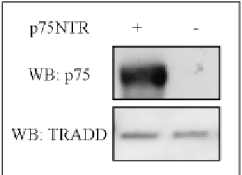
After validation of our dominant negative, we tested whether we could reproduce the same effect observed with TNFRI/TRADD DD also co-transfecting TRADD∆DD in cells expressing p75NTR. To this purpose SK-N-BE derived pools, selected after transfection of either p75NTR/pcDNA or pcDNA empty vectors, were further transfected with either TRADD∆DD/pCEP4 or pCEP4, the latter one used as negative control. After Hygromycin selection, pools of double transfected cells were collected, characterized both for p75NTR and for TRADD∆DD expression and cell pools expressing comparable level were analyzed for the cell death. No changes in the TRADD expression level have been detect with the overexpression of p75NTR. (Figure2B)
Fig. 3B Expression level of SK-N-BE derived pools. Western-blotting analysis of total protein extracts obtained from
SKNBE cell pools stably transfected with a pCDNA empty vector or a p75NTR/pCDNA vector to express wild type p75NTR. After SDS-PAGE of equal amount of total protein extract and transfer to nitrocellulose membrane, the blot has been probed with anti-p75NTR or anti-TRADD antibody as indicated. The panel shows that both cells expressing p75NTR and control cells express equal amount of endogenous TRADD protein.
As described above, cells were exposed to either Aβ peptide or to the control peptide for 48 hours and cell death has been evaluated after TUNEL labeling. As shown by the histogram in figure 3C, the treatment with Aβ in SK-N-BE expressing wild type p75NTR arose a 22% of dead cell.
Expression of TRADD∆DD by itself does not affect in any way the level of cell death. The co-transfection p75NTR WT/TRADD∆DD shows a level of cytotoxicity comparable with the one observed in the control.
Fig. 3C Role of the cytoplasmic adaptor TRADD in Aββββ–induced p75NTR mediated cytotoxicity. A TRADD
truncated mutant lacking the death domain (TRADD∆DD)generate a dominant negative effect in neuroblastoma cell line. A-SKNBE derived cell pools stably transfected with empty vectors (pcDNA), or plasmids to express p75NTR alone (p75NTR), TRADD∆DD alone (TRADD∆DD), p75NTR and TRADD∆DD (p75/TRADD∆DD), were selected and exposed to Aβ 25-35 (A-Beta) or to the control peptide A 35-25 (control). After 48 hrs cells were analyzed to assess cell death using TUNEL labelling and Ethidium Bromide nuclear counterstaining. Data are means + ES of three experiments.
Indeed, TRADD∆DD is able to interfere with the cytotoxic signal and almost to block Aβ -induced p75NTR-mediated cell death. (fig 3C) These data suggest that, at least upon Aβ binding, p75NTR could signal cell death through a protein complex that involves a p75NTR/TRADD pathway, resembling in this way the pathway used by TNFR1.
1.3 p75NTR and TRADD interact mainly through the p75NTR/TNFR1
conserved residues required for A
β
-induced cell death..
The reduced cell death levels observed in SK-N-BE pools expressing p75NTR-WT and TRADD∆DD, pushed us to verify whether TRADD could also be a direct interactor for p75NTR. The best way to address this question was to set up a Co-Immunoprecipitation Assay after transient transfection of HEK293 cells. Since it is known that TRADD is able to interact directly with TNFRI Death Domain but not with Fas-associating protein with death domain (FADD), the direct Fas cytoplasmic adaptor protein, we used these two death domain containing receptors respectively as positive and negative control.
We generated expression vectors to express HA tagged versions of FADD (HA-FADD) and TRADD (HA-TRADD) and a FLAG tagged version of TNFR1 (FLAG-TNFR1) (Fig. 3A).
Fig. 4 Physical interaction between p75NTR and TRADD. Co-Immunoprecipitation Assays after transient transfection
of HEK293 cells have been performed to compare the ability of p75NTR and TNFR1 to interact with the cytoplasmic adaptors: TRADD and FADD. (A) Schematic drawings of the transfected tagged-proteins: 3XFLAG-TNFR1, HA-TRADD and HA-FADD. (B) After 24 hrs of transfection of indicated vectors, 1.5 mg of total protein extract has been used to immunoprecipitate HA-tagged TRADD or FADD. Western-blotting analysis has been done probing film with antibodies raised against the indicated epitopes: i) p75NTR antibody against p75NTR, flag antibody against FLAG-TNFR1; HA antibody against HA-TRADD and HA-FADD. The shown autoradiographs are representative of at least three independent experiments.
Indeed, the Co-Immunoprecipitation Assay showed that in the double transfection p75NTR/TRADD experiment, pulling down TRADD by HA antibody, we could co-immunoprecipitate p75NTR, reproducing for p75NTR the same interaction that occurs between TNFR1 and TRADD.
Notably, the importance and specificity of this results was highlighted by the absence of coimmunoprecipitation signal from the cotransfection of p75NTR and FADD expressing vectors.
As already known and reproduced by our experiment in Fig. 4B, FADD does not interact with TNFRI even if it possesses a Death Domain. The fact that we did not find interaction between FADD and p75NTR gives more specificity and relevance to the observation of a phisical interaction between p75NTR and TRADD. In other words, the presence of a Death Domain is a required condition but not necessary to guarantee a protein-protein interaction
through Death Domain containig proteins and so the Co-Immunopracipitation TRADD/p75NTR likely reflects the existence of a real protein complex.
Indeed, to investigate the possible role of a p75NTR/TRADD interaction in mediating the cytotoxic signal of Aβ, we tested if the p75NTR single substitution mutants could affect in some way the complex assembly.
HEK293 cells have been co-transfected with plasmid to express HA-TRADD alone or together with either wild type p75NTR or the mutants p75E377A and p75W388A. As shown in Fig. 5, the p75E377A and p75W388A mutants have not been efficiently co-immunoprecipitated by TRADD-HA as we observed for p75NTR WT. This could suggest that p75NTR mutants could fail to transduce the cytotoxic signal most likely because they cannot interact efficiently with TRADD. In agreement with our results, Telliez and collegues showed that the mutation E369A in TNFR1-DD (corresponding to p75NTR E377A) not only blocks TNFR1-dependent cell-death but also affects TNFR1-TRADD interaction (Telliez et al,2000). All together these data suggest that a p75NTR/TRADD protein complex could mediate the Aβ cytotoxic signal.
Fig. 5. The p75NTR mutants that do not signal cytotoxicity have reduced ability to co-immunoprecipitate with TRADD. Transient transfection of HEK293 cells has been performed to compare the level of the
Co-Immunoprecipitation efficiency between wild type p75NTR (p75WT) and p75NTR mutants (p75E377A, p75W388A). After 24 hrs transfection of above indicated vectors, 1.5 mg of total protein extract has been used to immunoprecipitate HA-tagged TRADD followed by Western-blotting probed with antibodies raised against the indicated epitopes: i) p75NTR antibody against p75NTR ; HA antibody against HA-TRADD. The shown autoradiographs are representative of at least three independent experiments.
1.4 A
β
peptides modulates p75
NTR-TRADD interaction.
To assess the role of the p75NTR/TRADD interaction in a more physiologic setting, we investigated if the protein complex formation could be modulated by Aβ peptide exposure. We engineered SK-N-BE neuroblastoma cells to express both p75NTR and HA-tagged full length TRADD (HA-TRADD). Transfected cells have been selected to generate pools that we characterized for the p75NTR and HA-TRADD expression levels (data not shown). The selected cell pool has been exposed to Aβ peptides, or to control reverse peptides, for 1, 2, 3, 4, 7, 8 hours. The time course has been done trying to reproduce the possible kinetic required to allow the binding of the Aβ to the receptor. After exposures, cells have been scraped and equal amount of crude cell protein extracts have been incubated on anti-HA agarose-beads. Following the Co-Immunoprecipitation protocol, complex have been eluted from the beads and separated on SDS-PAGE then transferred to a membrane that has been probed sequentially with anti-p75NTR and anti-HA antibodies to quantify the co-immunoprecipitated p75NTR and the immunoprecipitated TRADD. As results from Fig 6, after 1 hour of treatment we could not detect p75NTR protein pulled down by HA-TRADD. Proceeding with a longer exposure of 2 and 4 hours, we noticed an increased amount of p75NTR co-immunoprecipitated with HA-TRADD. Prolonged treatments at 6-8 hours do not show anymore interaction between the proteins.
Fig 4A
Fig. 6. Aββββ peptide modulates p75NTR/TRADD interaction. Time course of p75NTR/TRADD interaction: SKNBE cell
pools stably transfected to express p75NTR and HA-TRADD or stably transfected with a pCEP4 empty vector have been exposed to Amyloid Beta peptide (Aβ) for 1, 2, 4, 6, 8 hrs. 1,5 mg total protein extract for each time course point has been used to immunoprecipitate HA-TRADD followed by Western-blotting probed with antibodies raised against the
indicated epitopes: i) p75NTR and HA-TRADD. The shown autoradiographs are representative of at least three independent experiments.
Indeed, this result indicates that Aβ peptides can modulate and activate the interaction between p75NTR and TRADD. As showed, this activation occurs in a specific time-dependent manner that peaks after 4 hours of exposure.
Finally, we define a possible mechanism by which p75NTR can mediate cell death in an Aβ dependent manner, recruiting TRADD at the level of the membrane and transferring the signal to the cytoplasm.
Discussion
In our study we discuss the role of the Neurotrophin Receptor p75NTR in mediating cell death induced by exposure to the Aβ peptide. The aim of our project is to investigate the molecular mechanisms that follow the binding of the peptide to the receptor peptide and mainly to identify the cytoplasmic pathway that are involved in the cell death signal. By deleting specific sequences in the intracellular domain of p75NTR, we already defined the essential role of the death domain (DD) as responsible for the signal transduction (Perini et al., 2002). Furthermore, in the present work we address in details why the DD is essential and in which way mediates cell death. We show for the first time that the membrane receptor p75NTR, upon binding to β-Amyloid (Aβ) peptide, is able to transduce a cytotoxic signal through a mechanism very similar to the one adopted by Tumor Necrosis Factor Receptor 1 (TNFR1), when activated by TNFα.
Our first approach was based on the analysis of the tridimensional conformation of the receptor. This allowed us to identified specific residues in the DD that, when mutated to A, are no longer able to signal cell death. p75NTR belongs to the family of Tumor Necrosis Factors Receptor 1, this suggested us the possibility of a common mechanism to signal cell death. To test our hypothesis we considered the sequence homology between the death domains of p75NTR and TNFR1 and we identified few almost complete identical residues. By exposing to Aβ peptide neuroblastoma cell stably expressing p75NTR mutated versions we could indeed verify that those aminoacids (E377, W388, A397 and I411) are involved in mediating cell death. Since the residues shared between p75NTR and TNFRI seemed to be important for both the receptors for transducing the death signal (Tartaglia et al 1993) we further speculated about the putative cytoplasmic interactors involved in this process. The Neurotrophin receptor p75NTR is known to mediate different effects, from cell survival to cell death, into the cell most likely depending on which is the extracellular stimulus and on which are, among the many identified, the cytoplasmic interactors expressed in the considered cell type . A recent paper reported that in MCF7 cells, p75NTR binds the Tumor Necrosis Factor Receptor1-associated Death Domain protein (TRADD), one of the first player in the TNFRI depending pathway. (Kieser et al,1999; El Yazidi-Belkoura et al,2003). Considering all together these insights, we decided to test if TRADD could be involved in
mediating Aβ-induced p75NTR-dependent cell death. For this purpose we generated a TRADD dominant negative mutant lacking the death domain required for the interaction with the receptor. Coexpressing p75NTR and TRADD∆DD in Neuroblastoma cells we assessed that also in this contest, TRADD is involved in the mediating the cell death upon Aβ exposure. To confirm these preliminary indications we set up a Co-Immunoprecipitation assays in transiently transfected HEK293 cells that indeed proved the direct interaction between p75NTR and TRADD. The p75NTR and TRADD forming complex resembles the same initial step of TNFR1 dependent signal transduction pathway even though in a different contest. Furthermore, we verified that the Fas-associating protein with death domain (FADD), the direct Fas cytoplasmic adaptor protein, does not interact with p75NTR, although displaying a death domain. This can suggest the presence of a death domain could be a feature necessary but not sufficient to generate a protein-protein interaction. So we proved that TRADD is a good candidate to initiate the p75NTR-dependent death signal transduction into the cytoplasm.
Once assessed the involvement of TRADD in the p75NTR dependent signaling pathway, we tried to clarify whether it could also be an important player in mediating Aβ induced cell death. As first approach, we challenged by Co-Ip the efficiency of the interaction between TRADD and two p75NTR mutants: p75E377A and p75W388A. In this case, we found that these two p75NTR mutants, that more than the others failed to signal Aβ cell death, have been co-immunoprecipitated with a lower efficiency compared to wild type p75NTR. This suggests that p75NTR mutants could be less efficient to transduce the cytotoxic signal most likely because they cannot interact efficiently with TRADD. These findings arise a possible involvement of TRADD as a mediator of the cell death signal induced by Aβ peptide. To deeper prove that TRADD contributes to the Aβ induced cell death of neuronal cells, we performed a time-course treatment on SK-N-BE pools expressing p75NTR to see if the Aβ peptide binding to p75NTR can modulate p75NTR physical interaction to TRADD.
To this purpose, we monitored the efficiency of the interaction between p75NTR and TRADD at different time point after exposures to Aβ and we could assess that the complex formation is indeed modulated by the peptide since we could observe a time-dependent kinetic.
We observed that TRADD interacts with p75NTR raising a peak between 2 to 4 hrs after peptide exposure. This observation possibly correlates with the kinetic of the Aβ sedimentation and the activation of the receptor. Indeed, the relatively slow kinetic of the p75NTR/TRADD interaction we observed can be explained by the low solubility level of aggregated Aβ peptide and is consistent with the activation of the downstream signaling pathways, JNK and p38/SAPK, previously described using our cell system (Costantini et
al,2005). Collectively, our study proposes a new mechanism that underlies the neurotoxicity
induced by Aβ peptide and mediated by the Neurotrophin receptor p75NTR. In this contest, multiple proteins have been so far claimed to be candidates for the Aβ depending cell death (Hashimoto Y et al., 2004), perhaps depending on the different neuronal cell populations considered. In this respect, it has been reported that neuronal cell death in AD is mediated in part by the interaction of Aβ with p75NTR. (Rabizadeh et al 1994). Aβ binding to p75NTR results in c- JUN N-terminal kinase (JNK) activation and apoptotic cell death in p75NTR expressing cells (Yaar et al 1997; Bhakar et al,2003; Becker et al, 2004; Ham et al,2005). Aβ activates nuclear factor- kB (Nf-κB) by binding to p75NTR in neuroblastoma cells (Kuner and Hertel, 1998). Furthermore, we proved that the p75NTR DD is responsible for the mediation of Aβ neurotoxicity (Perini et al, 2002). Finally, reflecting some similarity with the TNFR1, we map specific area in the DD of p75NTR responsible for the mediation of the Aβ cell death. This signal transduction recruits the new player, TRADD, to the membrane and activates the apoptotic pathway. In this study, we confirm the essential role of p75NTR in the mediation of the neurotoxicity depending on Aβ, even though we still have to verify the downstream pathway that ultimately activates the programmed cell death (Costantini et al, 2005).
With our study we try to elucidate the role of p75NTR in the Aβ neurotoxicity, suggesting a possible molecular mechanism. In this respect, still a lot remains to be proved since the even higher complexity of the neurodegeneration observed in the Alzheimer’s Disease.
Functional
cooperation
between
TrkA
and
p75NTR
accelerates neuronal differentiation by increased transcription
of GAP-43 and p21(CIP/WAF) genes via ERK1/2 and AP-1
activities
2.1 p75NTR cooperates with TrkA to accelerate NGF-mediated neuronal
differentiation
To investigate how the expression of both p75NTR and TrkA can affect neuronal differentiation, we took advantage of SK-N-BE, a human neuroblastoma cell line which expresses neither receptors [67] although it can be committed to differentiate into sympathetic neurons upon treatment with retinoic acid or TPA [68,69]. SK-N-BE cells represent an interesting neuronal model in which it is possible to re-establish the expression of both receptors and study their reciprocal influence on the activation of specific signal transduction pathways. In the specific case, SK-N-BE cells were transfected with the appropriate vectors and stable cell pools (BEp75, BETrkA and BEp75/TrkA) were isolated and maintained in the appropriate selective medium (see Materials and methods for further details). As a control, cell pools carrying the empty vector (BE vector) were also generated. Expression of either TrkA or p75NTR was monitored by immunoblotting (Fig. 1) and receptor membrane localization was verified by fluorescent immunolabeling with specific antibodies (data not shown). Cell pools expressing comparable levels of the receptors were chosen for further analyses.
Fig. 2.1 – Expression of TrkA and p75NTR in SK-N-BE derived cells. Total protein extracts from each cell pool (50 µg) were separated by SDS-PAGE, transferred onto a nylon membrane and
probed with either anti-Trk (C-14, Santa Cruz) or anti-p75NTR antibodies (9992). The position of protein molecular weight markers (kDa) is indicated.
First, we studied whether NGF can induce neuronal differentiation in the selected cell pools. BEvector, BEp75, BETrkA and BEp75/TrkA cells were seeded at a density of 2.5×10⁴/cm2 and treated with NGF. Neurite outgrowth, taken as a marker of neuronal differentiation, was monitored at different time points (0, 24, 48, 72 h). As shown in Fig. 2A, 100 ng/ml NGF can only induce differentiation of cells expressing the TrkA receptor. A similar experiment was also conducted using 10 ng/ml NGF to show that NGF, even at very low concentrations, can stimulate neuronal differentiation (data not shown).
Fig. 2 – NGF dependent differentiation of the cells. Cells were seeded at a density of 2.5×10⁴/cm2 in complete
medium. (A) After 24 h cells were washed with 1× PBS and maintained in a 0.1% FBS medium containing 100 ng/ml of NGF. Pictures (200× magnification) of the differentiating cells were taken at an interval of 24 h. (B, C) Quantification of the rate of differentiation is expressed as the percentage of cells that display neurite outgrowth upon exposure to 100 ng/ml NGF (B) or 10 ng/ml NGF (C) Specifically neurite outgrowth was considered positive when the length of the neuritis was at least twice the cell body diameter. For each tested condition, 500 cells from 10 independent fields were counted and the standard error was calculated.
Quantification of the differentiation state, expressed as a percentage of cells displaying neurite outgrowth, is described in Fig. 2B (100 ng/ml NGF) and Fig. 2C (10 ng/ml NGF). Neurite outgrowth is significantly more pronounced in BEp75/TrkA than in BETrkA cells at 24 and 48 h after NGF exposure (Figs. 2B, C), suggesting that coexpression of both receptors may contribute to increase and accelerate the rate of NGF-mediated neuronal differentiation.
