Correlation between tumor necrosis factor-alpha
and
D
-dimer levels in non-small cell lung
cancer patients
F. Guadagni
a, P. Ferroni
b,*
, S. Basili
c, F. Facciolo
d, S. Carlini
d,
M. Crecco
e, F. Martini
b, A. Spila
a, R. D’Alessandro
a, S. Aloe
a,
V. Cerasoli
d, G. Del Monte
a,f, S. Mariotti
f, T.C. Mineo
g, M. Roselli
faLaboratory of Clinical Pathology, Regina Elena Cancer Institute, Via E. Chianesi 53, Rome 00144 , Italy bDepartment of Experimental Medicine and Pathology, University La Sapienza, Viale Regina Elena 324,
Rome 00161, Italy
cDepartment of Medical Therapy, University La Sapienza, Viale del Policlinico, Rome 00185, Italy dDivision of Thoracic Surgery, Regina Elena Cancer Institute, Via E. Chianesi 53, Rome 00144, Italy eDivision of Radiology, Regina Elena Cancer Institute, Via E. Chianesi 53, Rome 00144, Italy
fMedical Oncology, Department of Surgery, University ‘‘Tor Vergata’’, Via di Tor Vergata 135, Rome
00133, Italy
gThoracic Surgery, Department of Surgery, University ‘‘Tor Vergata’’, Via di Tor Vergata 135, Rome
00133, Italy
Received 24 July 2003 ; received in revised form 17November 2003; accepted 18 November 2003
KEYWORDS Non-small-cell lung cancer; Fibrinolysis; Coagulation; Tumor necrosis factor-alpha; D-Dimer; Thrombin—antithrombin complexes
Summary The present study was designed to investigate whether a correlation
ex-ists between IL-6, TNF-␣and coagulation (Thrombin—antithrombin, TATc) or fibrinol-ysis (D-dimer) activation in non-small cell lung cancer (NSCLC) patients. One
hun-dred thirty patients with NSCLC (n = 65, 53 males, mean age 65 ± 8, adenocarcinoma
n = 32, squamous cancer n = 33) or chronic obstructive pulmonary disease (COPD)
(n = 65, 51 males, mean age 67 ± 9) were studied. As control group 65 healthy donors (51 males, mean age 61 ± 14) were also evaluated. The results obtained showed that median D-dimer levels were higher in NSCLC patients (3.0g/ml) compared either
to COPD patients (1.1g/ml, P < 0.05) or controls (0.3g/ml, P < 0.0001). Posi-tive TNF-␣levels (>10 pg/ml) were found in 26% of NSCLC compared to 3% of COPD (P < 0.002) and 5% of controls (P < 0.0005). On the other hand, positive (>8.5 pg/ml) IL-6 levels were found in 53% of NSCLC and 21% of COPD patients, compared to 5% of control subjects (P < 0.001). Median TATc levels were elevated in either NSCLC (6.9g/l) or COPD (5.7g/l) patients compared to controls (1.8g/l, P < 0.0001). El-evatedD-dimer levels were significantly associated to positive TNF-␣levels in patients
without distant metastasis (F = 4.3, P < 0.05). Moreover, TNF-␣ levels (P < 0.01) were independently related to the presence of positiveD-dimer levels in patients with
non-metastatic NSCLC. These results suggest that increased levels of TNF-␣might be responsible for an activation of fibrinolysis in patients with NSCLC.
© 2003 Elsevier Ireland Ltd. All rights reserved.
*Corresponding author. Tel.: +39-06-4452955; fax: +39-06-4454820. E-mail address: [email protected] (P. Ferroni).
0169-5002/$ – see front matter © 2003 Elsevier Ireland Ltd. All rights reserved. doi:10.1016/j.lungcan.2003.11.009
1. Introduction
Haemostatic abnormalities can be found in more than 90% of cancer patients, regardless of the presence or absence of clinical evidence for throm-boembolic disease, and there is reason to believe that these abnormalities contribute substantially to solid tumor organization, demarcation of tumor from normal host tissues, regulation of inflamma-tory cell accumulation, tumor angiogenesis and tumor stroma generation[1].
Although a number of studies have found an asso-ciation between fibrinolysis and non-small cell lung cancer (NSCLC), this relationship is by no means completely understood. Indeed, significant eleva-tion in the blood concentraeleva-tions of the split prod-uct from cross-linked fibrin,D-dimer, was found in
lung cancer patients, with either extensive or lim-ited disease [2—6], suggesting that a sub-clinical activation of blood coagulation and fibrinolysis can occur in NSCLC from the early clinical stages of dis-ease. Furthermore, the increase of D-dimer levels
has been related to unresponsiveness to treatment [7] and unfavorable prognosis [7—10] in patients with NSCLC.
Despite these studies, the origin of the activation of fibrinolysis in NSCLC is still unclear. The elevated levels ofD-dimer may, in fact, indicate a host
com-pensatory response to the increased formation of cross-linked fibrin[5,11], as indicated by the find-ing of a concomitant activation of the coagulation pathway in NSCLC[2—6]. Indeed, in a recent study we have shown thatD-dimer levels are increased in
NSCLC concomitantly to thrombin/anti-thrombin III complexes (TATc), an activation marker of coagula-tion [5]. However, we did not find any correlation between D-dimer and TATc, suggesting that
mech-anism(s) other than the compensatory response to fibrin production might be operating in NSCLC, such as an accelerated activity of the tumor-derived plasmin-mediated activation of fibrinolysis. Yet, in a comprehensive study of systemic fibrinolysis in patients with NSCLC, Pavey et al. [6] demon-strated that significant activation of fibrinolysis occurs in NSCLC patients, although the origin of this activation could not be related to consistent changes in plasminogen activator and inhibitor levels. Thus, the question that remains to be an-swered is whether the increased fibrinolysis found in NSCLC is due to the presence of cancer and/or cancer-related products or represents a normal physiological process to balance the activation of coagulation.
It is well known that tumor cells and/or tumor-associated leukocytes may produce inflammatory cytokines, such as IL-6 and TNF-␣ [12], and it has
been recently demonstrated by in-situ hybridiza-tion that elevated TNF-␣ mRNA levels can be found in both non-small cell lung cancer cells and tumor infiltrating lymphocytes (TIL)[13]. Moreover, circu-lating levels of these cytokines have been associ-ated with the disease status of lung cancer patients
[14—16].
The release of inflammatory cytokines is involved in activation of the fibrinolytic/coagulative system. IL-6, for example, is thought to act over the ex-trinsic pathway of coagulation through tissue fac-tor expression [17], whereas TNF-␣ is thought to play a direct role in the activation pathway of the fibrinolytic system[18,19]. Thus, the present study was aimed at analyzing the possible association be-tween TNF-␣, IL-6 and D-dimer or TATc levels in
patients with different stages of NSCLC, to better characterize the possible link between these in-flammatory cytokines and fibrinolysis activation in NSCLC.
2. Patients and methods
Sixty-five patients with NSCLC, treated at our Ins-titutions, entered the study. Patients (53 males, 12 females; mean age 65 ± 8, ranging from 48 to 82 years; 22 current and 22 ex-smokers) were his-tologically diagnosed with lung adenocarcinoma (n = 32) or squamous cancer (n = 33). NSCLC was pathologically staged according to the tumor-nodes-metastasis classification (UICC–—Union In-ternational Contre le Cancer TNM classification of malignant tumors). Nineteen (29.2%) patients were classified as stage I, 6 (9.2%) as stage II, 23 (35.4%) as stage III, and 17(26.2%) as stage IV lung cancer. Sixty-five patients with chronic obstructive pulmonary disease (COPD) (51 males, 14 females; mean age 67 ± 9, ranging from 46 to 85 years; 11 current and 33 ex-smokers) and 65 healthy subjects (51 males, 14 females; mean age 61 ± 14, ranging from 47to 83 years; 12 current and 25 ex-smokers) were evaluated as control groups. The latter were chosen among healthy subjects with a family his-tory of cardiovascular disease participating in a prevention program for atherothrombotic disorders at the University of Rome La Sapienza. Selection of controls was performed by an independent in-vestigator, blind to laboratory data, in order to have a population similar for age, sex and smok-ing habit to the NSCLC and COPD groups. Diabetes mellitus (fasting blood glucose level >115 mg/dl or treatment with a hypoglycemic agent), acute inflammatory disease, history of alcohol or drug abuse, peripheral-, cardio- and cerebro-vascu-lar atherosclerotic diseases (by clinical history,
physical examination, and instrumental diagnosis) were considered as exclusion criteria. No subject was on non-steroidal anti-inflammatory drugs, an-ticoagulant or antiplatelet agents in the 2 weeks preceding the study. The study was performed un-der the appropriate institutional ethics approvals and in accordance with the principles embodied in the Declaration of Helsinki. Written informed con-sent was obtained from each participating subject.
2.1. Sample collection and immunoassays
Plasma samples from resectable lung cancer pa-tients were drawn within 1 week before surgery, while samples from patients with metastatic dis-ease were obtained at the time of clinical diag-nosis, and prior to any treatment. Blood samples from COPD patients were drawn at the time of clin-ical and/or instrumental diagnosis and prior to any treatment. After a rest period of at least 20 min, blood samples were withdrawn from each consent-ing subject, without stasis, from the antecubital vein using a 20 G needle, and anticoagulated in Na citrate 3.8% (1:9, v:v). Samples were immediately centrifuged at 1500 × g for 10 min to obtain plasma, aliquoted, coded and stored at −40◦C until theas-says were performed.
Plasma TNF-␣, IL-6 (both by R&D Systems, Min-neapolis, MN, USA) and TATc (Enzygnost TAT, Dade-Behring, Marburg, Germany) levels were measured by commercially available enzyme immunoas-says according to the manufacturers’ instructions.
D-Dimer concentration was determined on a STA
analyzer (Roche Diagnostics, Mannheim, Germany) using the STA LIATEST D-DI (Roche Diagnostics). Measurements were done blinded. All samples were assayed in duplicate and those showing val-ues above the standard curve were re-tested with appropriate dilutions. The cutoff limits for TNF-␣ and IL-6 plasma levels were calculated as the mean + 2S.D. of the values observed in the control population, and were set at 10 and 8.5 pg/ml, re-spectively. As previously reported, the cutoff limits for TATc andD-dimer levels were set at 3.7 g/l and
0.72 g/ml, respectively[5].
2.2. Statistical analysis
Statistical analysis was performed by χ2statistics,
Pearson’s correlation coefficient, ANOVA and/or unpaired t-test. When necessary, log transforma-tion was used to normalize the data, or appropri-ate non-parametric tests were employed (Spear-man correlation analysis, Kruskall—Wallis and/or Mann—Whitney U-tests). Data are presented as me-dian and interquartile range (IRQ; 25th percentile
to 75th percentile) or as percentages of positive patients. Only P-values lower than 0.05 were re-garded as statistically significant. All calculations were made using a computer software package (Statistica, StatSoft Inc., Tulsa, OK, USA).
3. Results
IL-6, TNF-␣, D-dimer and TATc levels were
de-termined in plasma samples obtained from 195 subjects, including 65 NSCLC patients with histolog-ically diagnosed lung adenocarcinoma (n = 32) or squamous cancer (n = 33), 65 patients with COPD and 65 control subjects. Median levels of all four variables did not significantly differ between cur-rent smokers, ex-smokers and never smokers in the three groups under evaluation (data not shown). As summarized in Table 1, median TNF-␣ levels were higher in NSCLC compared to either COPD patients (P < 0.05) or controls (P < 0.0001). Indeed, pos-itive (above the cutoff value) TNF-␣ levels were found in a higher percentage of patients with NSCLC (26%) compared to either COPD patients (3%,
P < 0.001) or healthy subjects (5%, P < 0.001).
Sim-ilarly, median IL-6 levels were higher in NSCLC com-pared to either COPD patients (P < 0.01) or controls (P < 0.0001). Indeed, positive (above the cutoff value) IL-6 levels were found in a higher percentage of patients with NSCLC (53%) compared to either COPD patients (21%, P < 0.001) or healthy subjects (5%, P < 0.001). Furthermore, COPD patients had IL-6 levels higher than controls (P < 0.0001).
On the other hand, both patients with COPD and NSCLC had higher median TATc levels com-pared with healthy subjects (P < 0.0001), but no difference was observed between the two former groups (Table 1). Median D-dimer plasma levels
were also significantly elevated in COPD patients compared to healthy subjects (P < 0.0001), but were significantly lower than in patients with NSCLC (P < 0.05). Positive D-dimer levels were
found in 86% of patients with NSCLC compared to 52% of COPD patients and 9% of healthy subjects (P < 0.0001) (Table 1).
Positive levels of either TNF-␣ or TATc, but not IL-6 orD-dimer, were found in a higher percentage
of patients with distant metastasis compared to those without. However, the measured levels of both analytical variables did not significantly differ in the two subgroups of patients (Fig. 1). Of in-terest, COPD patients had TATc levels significantly higher than those observed in non-metastatic pa-tients (P < 0.05) and quite similar to those found in Stage IV NSCLC (Fig. 1). No significant association was found between all laboratory variables and
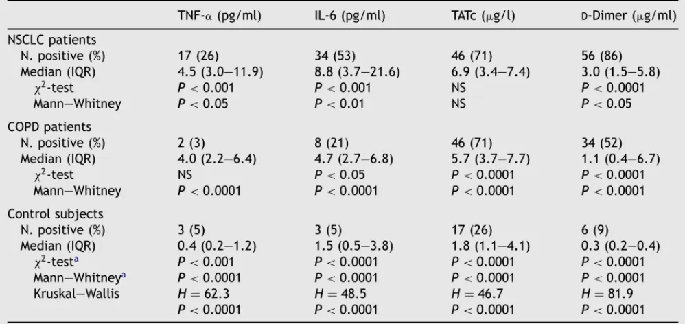
Table 1 Plasma TNF-␣, TATc andD-dimer levels in patients with non-small cell lung cancer (NSCLC) or chronic
obstructive pulmonary disease (COPD) and control subjects
TNF-␣(pg/ml) IL-6 (pg/ml) TATc (g/l) D-Dimer (g/ml)
NSCLC patients N. positive (%) 17(26) 34 (53) 46 (71) 56 (86) Median (IQR) 4.5 (3.0—11.9) 8.8 (3.7—21.6) 6.9 (3.4—7.4) 3.0 (1.5—5.8) χ2-test P < 0.001 P < 0.001 NS P < 0.0001 Mann—Whitney P < 0.05 P < 0.01 NS P < 0.05 COPD patients N. positive (%) 2 (3) 8 (21) 46 (71) 34 (52) Median (IQR) 4.0 (2.2—6.4) 4.7 (2.7—6.8) 5.7 (3.7—7.7) 1.1 (0.4—6.7) χ2-test NS P < 0.05 P < 0.0001 P < 0.0001 Mann—Whitney P < 0.0001 P < 0.0001 P < 0.0001 P < 0.0001 Control subjects N. positive (%) 3 (5) 3 (5) 17(26) 6 (9) Median (IQR) 0.4 (0.2—1.2) 1.5 (0.5—3.8) 1.8 (1.1—4.1) 0.3 (0.2—0.4) χ2-testa P < 0.001 P < 0.0001 P < 0.0001 P < 0.0001 Mann—Whitneya P < 0.0001 P < 0.0001 P < 0.0001 P < 0.0001 Kruskal—Wallis H = 62.3 H = 48.5 H = 46.7 H = 81.9 P < 0.0001 P < 0.0001 P < 0.0001 P < 0.0001 aNSCLC vs. control subjects.
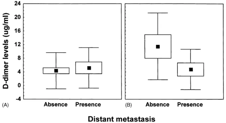
tumor size, lymph node involvement or histolog-ical diagnosis (Table 2). Thus, the interactions among TNF-␣, D-dimer levels and the presence
of distant metastases was analyzed by two-way
Fig. 1 Box-plot analysis of plasma TNF-␣(Panel A), IL-6 (Panel B), Thrombin—antithrombin complex (TATc) (Panel C) andD-dimer (Panel D) levels in 65 patients with chronic obstructive pulmonary disease (COPD) and 65 non-small cell
lung cancer (NSCLC). Solid lines indicate mean values, open circles indicate outlier values, open diamonds indicate extreme vales, bars indicate standard deviations, columns indicate standard errors of the mean.
Anova (Fig. 2). Of interest, elevated D-dimer
lev-els were significantly associated to positive TNF-␣ levels only in patients without distant metastasis (F = 4.3, P < 0.05).
Table 2 Association of positive levels of TNF-␣, TATc and D-dimer with different pathological variables in 65
non-small cell lung cancer patients
Variable Plasma levels above the cutoff value
N (%) TNF-␣
(>10 pg/ml) IL-6(>8.5 pg/ml) TATc(>3.7g/l) D(>0.72-Dimerg/ml) Tumor size
T1 15 2 (13) 5 (33) 11 (73) 11 (73)
T2 21 5 (24) 10 (48) 11 (52) 20 (95)
T3 25 10 (40) 17(68) 20 (80) 22 (88)
T4 4 0 (0) 2 (50) 4 (100) 3 (75)
Lymph node involvement
N0 31 7(23) 18 (58) 20 (65) 26 (84) N1 9 2 (22) 1 (11) 5 (56) 8 (89) N2 25 8 (32) 15 (69) 21 (84) 22 (88) Distant metastasis No 45 8 (18) 22 (49) 28 (62) 38 (84) Yes 20 9 (45)* 12 (69) 18 (90)* 18 (90)
Diagnosis of primary tumor
Adenocarcinoma 32 9 (28) 14 (44) 25 (78) 28 (88)
Squamous 33 8 (24) 20 (61) 21 (64) 28 (85)
*P < 0.05.
Correlation analysis among TNF-␣, IL-6,D-dimer
and TATc levels in the three groups of patients demonstrated thatD-dimer directly correlated with
TATc either in COPD patients (P < 0.001) or control subjects (P < 0.05), whereas the former signif-icantly correlated with TNF-␣ (P < 0.01) or IL-6 (P < 0.05) in NSCLC patients (P < 0.01). However, subgroup analysis of patients with or without dis-tant metastasis, showed that D-dimer and TNF-␣
correlated only in the subgroup of patients
with-Fig. 2 Two-way Anova analysis ofD-dimer levels in patients with or without distant metastasis of NSCLC and negative
(panel A) or positive (panel B) TNF-␣levels. Closed squares indicate mean values, bars indicate standard deviations, columns indicate standard errors of the mean.
out metastatic disease (P < 0.001) (Table 3). Thus, to further analyze the nature of this relation-ship a multiple regression analysis including age, sex, smoking status, histological diagnosis, stage, TNF-␣, IL-6 and TATc levels was carried out. The final model obtained by stepwise regression anal-ysis revealed that only TNF-␣ levels (regression coefficient = 0.36, P < 0.01) were independently related to the presence of elevated D-dimer
Table 3 Summary of Spearman rank correlation coefficients among the variables analyzed in NSCLC, COPD patients and control subjects
NSCLC patients All patients
(n = 65) Stages I through III(n = 45) Control subjects(n = 65) COPD patients(n = 65)
TNF-␣ IL-6 TATc TNF-␣ IL-6 TATc TNF-␣ IL-6 TATc TNF-␣ IL-6 TATc
D-Dimer 0.345* 0.261‡ 0.0270.522† 0.136 0.035 0.122 0.161 0.246‡ 0.112 0.166 0.444† TATc −0.046 −0.043 −0.085 −0.105 0.163 −0.033 0.233 0.166 IL-6 0.354* 0.352‡ 0.325* 0.179 *P < 0.01. †P < 0.001. ‡P < 0.05.
other hand, regression analysis of COPD patients including age, sex, smoking status, TNF-␣, IL-6 and TATc levels showed that only TATc levels (regression coefficient = 0.52, P < 0.001) were independently related to the presence of elevatedD-dimer levels.
4. Discussion
Several studies have demonstrated that pulmonary malignancy is frequently accompanied by alter-ations of fibrinolysis. The process of fibrinolysis involves an enzymatic cascade, which helps to break down cross-linked fibrin molecules. Fibrinol-ysis may be activated primarily–—and thus indepen-dently of activation of the coagulation cascade–—or secondarily in response to fibrin formation. How-ever, the complex biochemical mechanisms of the activation of fibrinolysis in cancer patients are not completely understood.
In the last few decades a series of in vivo and in vitro studies has provided more insight into the pathogenetic mechanisms and the role of cytokines in the activation and regulation of the coagulation/ fibrinolytic cascade, although the interactions are complicated, and the effects are time-dependent and transient [20]. To date, it is widely accepted that, on a cellular level, TNF-␣ may directly acti-vate the fibrinolytic system. Injection of TNF-␣ into healthy volunteers, in fact, results in a transient activation of fibrinolysis [21,22]. Furthermore, treatment with anti-TNF-␣ in the human and chim-panzee sepsis models inhibits the fibrinolytic system
[23,24], whereas coagulation is not affected,
show-ing that TNF plays an important role in regulation of the fibrinolytic response. Although this scenario has been derived largely from studies with purified endotoxin or gram-negative bacteria, we hypothe-sized similar alterations to occur in the activation
of fibrinolytic systems in NSCLC, because the key in-termediate cytokine TNF-␣ has been correlated to the disease status of lung cancer patients[14—16]. In the present study, plasma IL-6 and TNF-␣ lev-els were significantly elevated in NSCLC compared to either COPD patients or healthy controls. One limitation of the methodology used is that it does not allow distinguishing what is the main source of circulating cytokines in patients with NSCLC. These may be produced directly by the tumor, or may be the result of the host inflammatory response to cancer cells and/or their released products, al-though both phenomena can occur. Indeed, Li et al. recently showed by in-situ hybridization that, de-spite an immunosuppressive state, TNF-␣ mRNA levels were elevated in both tumor cells and TIL of NSCLC patients[13]. Whatever the source might be our finding that TNF-␣ was the only variable in-dependently associated toD-dimer in patients with
non-metastatic NSCLC suggests an involvement of this cytokine in the primary activation of fibrinoly-sis in this malignancy.
The finding of increased plasma levels of TATc (an activation marker of coagulation) and ofD-dimer (a
marker of fibrinolysis) in a substantial percentage of NSCLC patients is in agreement with the find-ings by other authors [2—4,6], although we could not confirm the correlation ofD-dimer levels with
tumor stage previously observed[6]. Furthermore, the lack of a significant correlation between TATc and D-dimer levels suggests that these two
ana-lytical variables represent different pathological mechanisms of activated coagulation and fibrinol-ysis in patients with NSCLC. Elevated TATc levels were also found in COPD, which is not surprising, because of the occurrence of a pro-thrombotic state in this clinical setting [25—28]. The per-centage of patients with positive TATc levels did not significantly differ between COPD and NSCLC
patients, and median TATc levels of COPD patients were similar to those measured in plasma samples obtained from metastatic NSCLC patients. On the other hand, medianD-dimer levels of COPD patients
were significantly lower than those of NSCLC. Fur-thermore, TATc levels were independently related toD-dimer levels in COPD patients, suggesting that
the activation of the fibrinolytic pathway observed in COPD might be secondary to thrombin generation and fibrin deposition.
The significant association found in NSCLC be-tween TNF-␣ andD-dimer levels, but not with TATc,
suggests that the host inflammatory response to cancer cells, and/or their released products, as well as tumor-derived cytokines, could be responsible for a primary activation of the fibrinolytic pathway, unrelated to the coagulation activation, at least in the early stages of disease. Indeed, the association between TNF-␣ andD-dimer levels was confined to
non-metastatic patients, despite the occurrence of elevated levels of TNF-␣ in Stage IV NSCLC. How-ever, we should consider that approximately 90% of metastatic patients also had elevated TATc levels, which is in agreement with the opinion currently accepted that the incidence, nature, and extent of coagulation abnormalities is related to the tumor burden [1]. Activation of the coagulation system and consequent thrombin generation could be due to increased production of tissue factor (TF) by lung cancer cells[29]. TF is the key factor in the acti-vation of the extrinsic pathway and a regulator of tumor angiogenesis [30]. Furthermore, TF expres-sion has been correlated with the clinical stage of other types of human cancer, with the highest per-centages found in metastatic disease[31]. Thus, we can hypothesize that in metastatic lung cancer a secondary activation of fibrinolysis might also oc-cur due to the increased thrombin generation and consequent fibrin production.
In conclusion, the present study confirms pre-viously published observation that NSCLC causes a profound alteration in fibrinolysis and suggests that TNF-␣ could be, at least in part, responsible for the increasedD-dimer levels found in this
ma-lignant disease. There have been no other reports of such an association between TNF-␣ andD-dimer
levels in NSCLC, but our results suggest that the increasedD-dimer levels might be accounted for a
primary activation of fibrinolysis by TNF-␣ in the early stages of NSCLC. Additional studies in a larger number of patients are required to better define the role of tumor-derived cytokines in NSCLC. Bet-ter knowledge of the biologic effects of TNF-␣ in human cancer will help to improve our under-standing of the pathophysiological significance of tumor-induced coagulopathies.
Acknowledgements
The authors wish to thank Simone Giommi, Alessia Cialfi and Alessandro Catalini for their excellent technical assistance. This work has been partially supported by Grant P.F. Ministero della Salute 1999.
References
[1] Rickles FR, Levine MN, Dvorak HF, Abnormalities of hemostasis in malignancy. In: Colman RW, Hirsh J, Marder VJ, Clowes AW, George JN, editors. Hemostasis and thrombosis. Basic principles and clinical practice, 4th ed. Philadelphia: Lippincott Williams & Wilkins; 2001. p. 1131—52.
[2] van Wersch JW, Tjwa MK. Coagulation/fibrinolysis balance and lung cancer. Haemostasis 1991;21:117—23.
[3] Gabazza EC, Taguchi O, Yamakami T, Machishi M, Ibata H, Suzuki S. Evaluating prethrombotic state in lung cancer using molecular markers. Chest 1993;103:196—200. [4] Matsuyama W, Hashiguchi T, Mizoguchi A, Iwami F,
Kawa-bata M, Arimura K, et al. Serum levels of vascular endothe-lial growth factor dependent on the stage progression of lung cancer. Chest 2000;118:948—51.
[5] Roselli M, Mineo TC, Basili S, Mariotti S, Martini F, Bel-lotti A, et al. Vascular endothelial growth factor (VEGF-A) plasma levels in non-small cell lung cancer: relationship with coagulation and platelet activation markers. Thromb Haemost 2003;89:177—84.
[6] Pavey SJ, Hawson GA, Marsh NA. Alterations to the fibri-nolytic enzyme system in patients with non-small cell lung carcinoma. Blood Coagul Fibrinolysis 1999;10:261—7. [7] Seitz R, Rappe N, Kraus M, Immel A, Wolf M, Maasberg M,
et al. Activation of coagulation and fibrinolysis in patients with lung cancer: relation to tumour stage and prognosis. Blood Coagul Fibrinolysis 1993;4:249—54.
[8] Taguchi O, Gabazza EC, Yasui H, Kobayashi T, Yoshida M, Kobayashi H. Prognostic significance of plasmaD-dimer lev-els in patients with lung cancer. Thorax 1997;52:563—5. [9] Ferrigno D, Buccheri G, Ricca I. Prognostic significance
of blood coagulation tests in lung cancer. Eur Respir J 2001;17:667—73.
[10] Pavey SJ, Hawson GA, Marsh NA. Impact of the fibrinolytic enzyme system on prognosis and survival associated with non-small cell lung carcinoma. Blood Coagul Fibrinolysis 2001;12:51—8.
[11] Gabazza EC, Taguchi O, Yamakami T, Machishi M, Ibata H, Tsutsui K, et al. Coagulation-fibrinolysis system and markers of collagen metabolism in lung cancer. Cancer 1992;70:2631—6.
[12] Balkwill F, Mantovani A. Inflammation and cancer: back to Virchow? Lancet 2001;357:539—45.
[13] Li R, Rüttinger D, Li R, Si LS, Wang YL, Analysis of the immunological microenvironment at the tumor site in pa-tients with non-small cell lung cancer, Langenbecks Arc Surg 2003;388:406—12.
[14] Ardizzoia A, Lissoni P, Brivio F, Tisi E, Perego MS, Grassi MG, et al. Tumor necrosis factor in solid tumors: increased blood levels in the metastatic disease. J Biol Regul Homeost Agents 1992;6:103—7.
[15] Matanic D, Beg-Zec Z, Stojanovic D, Matakoric N, Flego V, Milevoj-Ribic F. Cytokines in patients with lung cancer. Scand J Immunol 2003;57:173—8.
[16] Aleman MR, Santolaria F, Batista N, de La Vega M, Gonzalez-Reimers E, Milena A, et al. Leptin role in ad-vanced lung cancer. A mediator of the acute phase re-sponse or a marker of the status of nutrition? Cytokine 2002;19:21—6.
[17] Stouthard JM, Levi M, Hack CE, Veenhof CH, Romijn HA, Sauerwein HP, et al. Interleukin-6 stimulates coagulation, not fibrinolysis, in humans. Thromb Haemost 1996;76:738— 42.
[18] Zwaveling JH, Maring JK, Mulder AB, Bom VJ, van Ginkel RJ, Schraffordt Koops H, et al. Effects of hyperthermic isolated limb perfusion with recombinant tumor necrosis factor alpha and melphalan on the human fibrinolytic sys-tem. Cancer Res 1996;56:3948—53.
[19] Suharti C, van Gorp EC, Setiati TE, Dolmans WM, Djoko-moeljanto RJ, Hack CE, et al. The role of cytokines in activation of coagulation and fibrinolysis in dengue shock syndrome. Thromb Haemost 2002;87:42—6.
[20] ten Cate JW, van der Poll T, Levi M, ten Cate H, van Deventer SJH. Cytokines: triggers of clinical thrombotic disease. Thromb Haemost 1997;78:415—9.
[21] Van Deventer SJH, Buller HR, ten Cate JW, Aarden LA, Hack CE, Sturk A. Experimental endotoxinaemia in humans: analysis of cytokine release and coagulation, fibrinolytic, and complement pathways. Blood 1990;76:2520—6. [22] van der Poll T, Levi M, Buller HR, van Deventer SJ, de Boer
JP, Hack CE, et al. Fibrinolytic response to tumor necrosis factor in healthy subjects. J Exp Med 1991;174:729—32. [23] van der Poll T, Levi M, van Deventer SJH, ten Cate H,
Haagmans BL, Biemond BJ, et al. Differential effects of anti-tumor necrosis factor monoclonal antibodies on sys-temic inflammatory responses in experimental endotox-emia in chimpanzees. Blood 1994;83:446—51.
[24] van der Poll T, Coyle SM, Levi M, Jansen PM, Den-tener M, Barbosa K, et al. Effect of recombinant dimeric tumor necrosis factor receptor on inflammatory re-sponses to intravenous endotoxin in normal humans. Blood 1997;89:3727—34.
[25] Nakstad B, Lyberg T, Skjonsberg OH, Boye NP. Local acti-vation of the coagulation and fibrinolysis systems in lung disease. Thromb Res 1990;57:827—38.
[26] Alessandri C, Basili S, Violi F, Ferroni P, Gazzaniga PP, Cor-dova C. C.O.B.H. Group. Hypercoagulability state in pa-tients with chronic obstructive pulmonary disease. Thromb Haemost 1994;72:343—6.
[27] Dav`ı G, Basili S, Vieri M, Cipollone F, Santarone S, Alessan-dri C, et al. C.O.B.H. Group. Enhanced thromboxane biosynthesis in patients with chronic obstructive pulmonary disease. Am J Resp Crit Care Med 1997;156:1794—9. [28] Ferroni P, Basili S, Martini F, Vieri M, Labbadia G, Cordova
C, et al. Soluble P-selectin as a marker of platelet hy-peractivity in patients with chronic obstructive pulmonary disease. J Invest Med 2000;48:21—7.
[29] Keller T, Salge U, Konig H, Dodt J, Heiden M, Seitz R. Tissue factor is the only activator of coagulation in cul-tured human lung cancer cells. Lung Cancer 2001;31: 171—9.
[30] Rickles FR, Shoji M, Abe K. The role of the hemostatic sys-tem in tumor growth, metastasis, and angiogenesis: tissue factor is a bifunctional molecule capable of inducing both fibrin deposition and angiogenesis in cancer. Int J Hematol 2001;73:145—50.
[31] Nakasaki T, Wada H, Shigemori C, Miki C, Gabazza EC, Nobori T, et al. Expression of tissue factor and vascular endothelial growth factor is associated with angiogenesis in colorectal cancer. Am J Hematol 2002;69:247—54.