ISSN: 1524-4636
Copyright © 2009 American Heart Association. All rights reserved. Print ISSN: 1079-5642. Online
7272 Greenville Avenue, Dallas, TX 72514
Arteriosclerosis, Thrombosis, and Vascular Biology is published by the American Heart Association.
DOI: 10.1161/ATVBAHA.109.198622
published online Dec 3, 2009;
Arterioscler Thromb Vasc Biol
Sanguigni, Claudio Stefanutti, Stefania Basili and Francesco Violi
Pasquale Pignatelli, Roberto Carnevale, Roberto Cangemi, Lorenzo Loffredo, Valerio
Hypercholesterolemia
Circulating Levels in Patients With
phox
Atorvastatin Inhibits gp91
http://atvb.ahajournals.org
located on the World Wide Web at:
The online version of this article, along with updated information and services, is
http://www.lww.com/reprints
Reprints: Information about reprints can be found online at
[email protected]
410-528-8550. E-mail:
Fax:
Kluwer Health, 351 West Camden Street, Baltimore, MD 21202-2436. Phone: 410-528-4050.
Permissions: Permissions & Rights Desk, Lippincott Williams & Wilkins, a division of Wolters
http://atvb.ahajournals.org/subscriptions/
Biology is online at
Atorvastatin Inhibits gp91
Circulating Levels in Patients
With Hypercholesterolemia
Pasquale Pignatelli, Roberto Carnevale, Roberto Cangemi, Lorenzo Loffredo, Valerio Sanguigni,
Claudio Stefanutti, Stefania Basili, Francesco Violi
Objective—The inhibition of oxidative stress is among the most relevant pleiotropic effects of statins. The mechanism by
which statins exert their antioxidant effect in vivo is still undefined. NADPH oxidase is among the most important
sources of reactive oxygen species involved in atherosclerotic disease.
Methods/Results—We developed an ELISA to evaluate serum levels of soluble-gp91
phox, the catalytic core of phagocyte
NADPH oxidase. In a cross-sectional study performed in 30 hypercholesterolemic patients and in 20 controls, serum
soluble-gp91
phoxand urinary isoprostane, a marker of oxidative stress, were measured. The 2 variables were also
measured in hypercholesterolemic patients, randomized to diet (n
⫽15), or diet plus atorvastatin (10 mg daily, n⫽15)
and followed for 30 days. Compared to controls, hypercholesterolemic patients had higher and significantly correlated
(R
⫽0.71; P⬍0.001) serum soluble-gp91
phox(P
⬍0.001) and urinary isoprostanes (P⬍0.001). After follow-up, the
statin-allocated group showed a significant reduction of soluble-gp91
phox(
⫺33%, P⬍0.01), that paralleled that of
isoprostanes (
⫺37%, P⬍0.01) and cholesterol (⫺25%, P⬍0.01). The diet-allocated group showed only a weak
reduction of cholesterol.
Conclusion—Our study demonstrates that statins exert an antioxidant effect via inhibition of soluble gp91
phoxexpression. (Arterioscler Thromb Vasc Biol. 2010;30:00-00.)
Key Words: gp91
phox䡲 oxidative stress 䡲 hypercholesterolemia 䡲 NADPH oxidase 䡲 statins
P
rimary and secondary prevention trials with statins
clearly demonstrated that this drug category is able to
reduce cardiovascular events.
1,2Even if the principal
mech-anism of action of statins is to lower cholesterol, other effects,
the so-called pleiotropic effects, have been considered as
adjunctive properties potentially accounting for the
antiath-erosclerotic effect of statins.
3Inhibition of oxidative stress
may be considered an intriguing pleiotropic effect in view of
the fact that oxidative stress is thought to be a key event in the
initiation and progression of atherosclerotic disease.
4Reduc-tion of several markers of oxidative stress including
isopros-tanes, 8-hydroxydeoxyguanosine (8-OHdG), and
nitroty-rosine have been observed after statin treatment but the
underlying mechanism is still unclear.
5–9In a pilot study performed in patients with chronic
granu-lomatous disease (X-CGD), a very rare life-threatening
dis-ease secondary to hereditary deficiency of gp91
phox(the
catalytic subunit of phagocyte NADPH oxidase), we found a
significant reduction of urinary isoprostanes.
10Also, in
chil-dren with hypercholesterolemia, we observed a significant
correlation between platelet gp91
phoxexpression and urinary
isoprostanes.
11These previous observations suggest a role for
the phagocyte NADPH oxidase, one of the most important
cellular producers of superoxide anion (O
2. ),
12in the
forma-tion of this marker of oxidative stress. Because previous
studies provided in vitro evidence that statins inhibit the
expression and activation of NADPH oxidase,
6,13we sought
to analyze whether this occurs in vivo and ultimately
con-tributes to the reduction of oxidative stress. Thus, we
devel-oped a method to measure gp91
phoxin the circulation of
patients affected by hypercholesterolemia that is
character-ized by accelerated atherosclerosis and enhanced oxidative
stress.
14,15Then, we undertook an interventional trial to see
whether a statin was able to affect circulating gp91
phox. For
this purpose, 30 hypercholesterolemic patients were
random-ized to 1 month of treatment with standard diet or standard
diet plus atorvastatin. Herein, we provide the first evidence
that statin reduces circulating gp91
phoxin patients with
hypercholesterolemia.
Materials and Methods
Study Participants
We studied 30 consecutive hypercholesterolemic (HC) patients (16 males and 14 females; 52⫾4 years of age) and 20 healthy subjects (HSs) who were screened in the ambulatory of our division between September 2005 and February 2006. Subjects were excluded if they
Received October 8, 2009; revision accepted November 18, 2009.
From the Department of Experimental Medicine (P.P., R. Carnevale, R. Cangemi, L.L., S.B., F.V.), “Sapienza” University; Department of Internal Medicine (V.S.), University of Rome “Tor Vergata”; and Department of Clinical and Medical Therapy (C.S.), “Sapienza” University, Rome, Italy.
Correspondence to Prof Francesco Violi, Department of Experimental Medicine, Divisione I Clinica Medica, “Sapienza” University, Rome, Italy, Viale del Policlinico 155, Roma, 00161, Italy. E-mail [email protected]
© 2009 American Heart Association, Inc.
Arterioscler Thromb Vasc Biol is available at http://atvb.ahajournals.org DOI: 10.1161/ATVBAHA.109.198622
had liver insufficiency, serious renal disorders (serum creatinine, ⬎2.5 mg/dL), diabetes mellitus, arterial hypertension, a history or evidence of previous myocardial infarction or other atherothrombotic diseases, any autoimmune diseases, cancer, present or recent infec-tions or were taking nonsteroidal antiinflammatory drugs, drugs interfering with cholesterol metabolism, or vitamin supplements. Both patients and controls were recruited from the same geographic area, and they were all of white race. Informed consent was obtained from each participating subject and the protocol was approved by the “Sapienza” University, Rome, Italy Ethic Committee.
X-Linked Chronic Granulomatous Disease Patients
We studied 3 recently identified male patients (age, 27⫾2.5 years) with hereditary deficiency of gp91phox that was diagnosed as previously described.16X-CGD is a rare (prevalence 1:1 000 000 individuals) primary immunodeficiency affecting the innate immu-nologic system; X-CGD is characterized by life-threatening bacterial and fungal infections.17It is caused by mutations in any of the 4 genes encoding subunits of the O2. generating NADPH oxidase,
resulting in defective O2. generation and intracellular killing.18
Design of the Studies
In the first part of the study, we performed a cross-sectional analysis comparing urinary isoprostanes and serum soluble (s)gp91phox be-tween population of HC patients and HSs. Then, we performed a 30 day interventional study with atorvastatin. For this purpose, the 30 HC patients were openly randomized to a treatment with diet or diet plus atorvastatin (10 mg/d) to assess whether diet alone or diet plus atorvastatin were able to influence serum levels of sgp91phoxand/or urinary isoprostanes excretion. During the study, all participants followed a low-fat diet with mean macronutrient profiles that were close to the present Adult Treatment Panel III guidelines (7% energy from saturated fat and, 200 mg dietary cholesterol per day).19
A medical doctor not involved in the study assigned codes to the study treatments, randomly allocating the selected participants to a treatment with diet or diet plus atorvastatin, and kept the key in a sealed envelope. The randomization was carried out by a procedure based on a random numeric sequence. The authors were unaware of treatment allocation. The principal investigator performed the treat-ment allocation unblinded only after the study had ended and laboratory analyses were completed.
Blood Sampling and Laboratory Analysis
Between 8:00 and 9:00AM, subjects underwent routine biochemical evaluations including fasting total cholesterol and glucose as well as WBC count. After overnight fasting (12 hours) and supine rest for at least 10 minutes, blood samples were collected in vacutainers (Vacutainer Systems, Belliver Industrial Estate) and centrifuged at 300g for 10 minutes to obtain supernatant, which was stored at ⫺80°C until use. Fasting serum levels of total cholesterol and triglycerides were determined with enzyme-based methods (Dade Behring, Switzerland). High-density lipoprotein (HDL) cholesterol was measured after phosphotungstic acid/MgCl2 precipitation of fresh serum (Dade Behring, Switzerland). Low-density lipoprotein (LDL) cholesterol was calculated according to the Friedewald formula.
Urinary 8-isoprostaglandin F2␣ (PGF2␣-III) was measured by previously described and validated EIA assay method.20,21Briefly, 10-mL urine aliquots were extracted on a C-18 SPE column; the purification was tested for recovery by adding a radioactive tracer (tritiated PGF2a-III) (Cayman Chemical, Ann Arbor, Mich). The elutes were dried under nitrogen, recovered with 1 mL of buffer, and assayed in a PGF2␣-III–specific EIA kit (Cayman Chemical). PGF2␣-III concentration was corrected for recovery and creatinine excretion and expressed as picograms per milligrams of creatinine. Intra- and interassay coefficients of variation were 2.1% and 4.5%, respectively.
Immunoprecipitation of sgp91
phoxFrom
Human Serum
Gp91phoxwas immunoprecipitated from serum in denaturized
con-ditions. Serum was incubated overnight at 4°C with gp91phox antibody sc-74514 (mouse monoclonal antibodies raised against amino acids 231 to 290 of gp91phoxof human origin, Santa Cruz
Biotechnology Inc, Santa Cruz, Calif). Immunocomplexes were bound to ImmunoPure-immobilized protein A (sc-2003, Santa Cruz Biotechnology Inc), boiled in sample buffer, and separated by SDS-PAGE (Bio-Rad, Hercules, Calif) following a described proto-col. Negative controls involved a similar procedure using a goat IgG (Santa Cruz Biotechnology Inc).
After electroblotting of proteins to Immobilon membranes, the membranes were blocked and incubated with monoclonal mouse anti-gp91phox antibody. After incubation, the pure nitrocellulose
membranes (0.45m) were washed and incubated with goat anti-mouse IgG1– horseradish peroxidase (HRP) (sc-2004 Santa Cruz Biotechnology Inc) for 2 hours. Immune complexes were detected by enhanced chemiluminescence. (Bio-Rad).
Electrophoresis and Immunoblotting of sgp91
phoxPatient Serum
Equal amounts of serum protein (130g/lane) estimated by Bradford assay were solubilized in a 2⫻ Laemmli sample buffer containing 2-mercaptoethanol and loaded in a denaturing SDS/10% polyacryl-amide gel. Western blot analysis was performed with monoclonal anti-gp91phox(2g/mL) incubated overnight at 4°C.
After incubation, the pure nitrocellulose membranes (0.45 m) were washed and incubated with goat anti-mouse IgG1-HRP for 2 hours. Immune complexes were detected by enhanced chemilumi-nescence. The developed spots were calculated by densitometric analysis on a NIH Image 1.62f analyzer and the value was expressed as arbitrary units. Each sample was analyzed in triplicate.
ELISA of sgp91
phoxPatient Serum
Reagents
Reagents consisted of a coating buffer (0.05 mol/L carbonate-bicarbonate pH 9.6), a washing solution (0.05% Tween 20 in 50 mmol/L Tris-buffered saline at pH 8.0), a blocking buffer (50 mmol/L Tris-buffered saline at pH 8.0, 1% BSA), and a stopping solution for color development (1mol/L sulfuric acid).
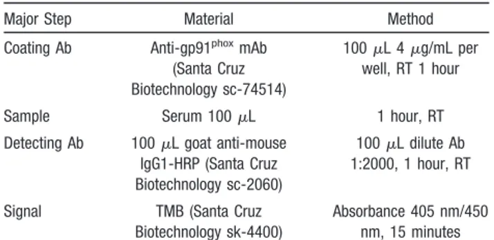
Procedure
ELISA conditions for serum sgp91phoxevaluation are outlined in
Table 1. Standard (50 L; gp91phox peptide from the sequence
LNFARKRIKNPEGGLC of gp91phox; New England Peptide, Gard-ner, Mass) or sample was added into each antibody-coated well and incubated for 60 minutes at room temperature while shaking. After washing 3 times with the washing buffer, 100 L of diluted HRP-conjugated detecting antibody (0.4 g/mL) was added and incubated at room temperature for 60 minutes with gentle agitation.
Table 1. ELISA Conditions for Serum sgp91phox
Evaluation
Major Step Material Method Coating Ab Anti-gp91phoxmAb
(Santa Cruz Biotechnology sc-74514)
100L 4 g/mL per well, RT 1 hour Sample Serum 100L 1 hour, RT Detecting Ab 100L goat anti-mouse
IgG1-HRP (Santa Cruz Biotechnology sc-2060)
100L dilute Ab 1:2000, 1 hour, RT Signal TMB (Santa Cruz
Biotechnology sk-4400)
Absorbance 405 nm/450 nm, 15 minutes Zero noise level was determined to be 0.015 pg/mL. Ab indicates antibody; mAb, monoclonal antibody; RT, room temperature.
Wells were again washed 3 times with washing buffer and enzyme substrate was added. After incubation for 20 minutes at room temperature, the reaction was stopped with 100 L of 1 mol/L H2SO4, and the absorbance was read at 405 nm/450 nm. Intra- and
interassay coefficients of variation were 5.2% and 6%, respectively.
Platelet Preparation
To obtain platelet-rich plasma samples (n⫽5, HSs) were centrifuged 15 minutes at 180g. To avoid leukocyte contamination, only the top 75% of the platelet-rich plasma was collected.22Pelleted platelets were washed and suspended in HEPES buffer, pH 7.4 (2⫻108
platelets/mL, unless otherwise noted).22 To evaluate the level of sgp91phox, platelet were stimulated with or without arachidonic acid (1 mmol/L), and the samples were analyzed by ELISA method as above reported.
Human Polymorphonuclear
Leukocyte Preparation
Polymorphonuclear leukocytes (PMNs) were isolated from freshly taken EDTA-blood from healthy volunteers (n⫽5, HSs) by dextran enhanced sedimentation of red blood cells, Ficoll-Histopaque density centrifugation, lysis of remaining erythrocytes with distilled water and washing of cells with Hank’s balanced salt solution (HBSS) in the absence of any divalent cations. Finally, the cell pellet was suspended in 1 mL of HBSS and stimulated with or without 10mol/L of phorbol 12-myristate 13-acetate (PMA). To evaluate sgp91phox in PMNs, the supernatant was analyzed by ELISA method as above reported.
Lymphocyte/Monocyte Preparation
Blood samples were collected in heparinized tubes (10 IU/mL). Lymphocytes/monocytes were isolated after centrifugation of the blood from healthy volunteers (n⫽5, HSs) with a polysucrose-sodium diatrizoate solution, 1.077 g/mL density and 280 mOsm osmolarity (Lymphoprep; Nycomed, Oslo, Norway) at 800g at 20°C. The lymphocyte/monocyte cell layer was collected and the cells were thus washed 2 times in a solution of cold PBS (pH 7.2), supple-mented with 1% FCS and 2 mmol/L EDTA (Sigma-Aldrich, Milano, Italy). The cell suspension was stimulated with or without lipopoly-saccharide (LPS) (100 ng/mL); sgp91 content in the supernatant was evaluated by ELISA method as above reported.
Circulating Blood Microparticle Preparation
Microparticles were isolated from serum. An aliquot of serum was thawed to 37°C and then filtered using a microtiter plate format 0.2-m vacuum filtration device (Ceveron MFU-500, Technoclone, Dorking, UK) that is designed to remove microparticles.23Cellular microparticles were measured using a functional assay (Zymuphen MP-Activity, Hyphen BioMed, Neuville-sur-Oise, France). The samples, either microparticle-free or microparticle-rich, were ana-lyzed by ELISA as above reported.
Statistical Analysis
Categorical variables were reported as counts (percentage) and continuous variables as means⫾SD unless otherwise indicated. Independence of categorical variables was tested by2test.
Com-parisons between HC patients and HSs were carried out by Student t test and were replicated as appropriate with nonparametric test (Kolmogorov–Smirnov [z] test) in the case of nonhomogeneous variances as verified by Levene’s test.
To account for the inflation of the experimentwise type 1 error attributable to multiple testing, Bonferroni correction was used.
The correlation analysis was performed with Spearman test. Interventional study data were analyzed for the assessment of treatment effect on sgp91phox, total cholesterol, and urinary
isopros-tanes performing a multivariate ANOVA with 1 between-subject factor (treatment group) and 1 within-subject factor (time at 2 levels: baseline, 30 days after the beginning of the treatment). As covariates, we considered the possible random differences in age, sex, body mass index (BMI), systolic, and diastolic blood pressure between the
2 groups (the one allocated to diet and atorvastatin and the other allocated to diet alone).
We recruited all the patients (n⫽30) who respected the inclusion/ exclusion criteria for the cross-sectional study of sample size determination, as above reported. The number of controls (n⫽20) was computed with respect to a 2-tailed Student t test for indepen-dent groups, considering (1) as relevant difference in serum sgp91phoxlevels to be detected between patients and controls␦ⱖ10
(pg/mL); (2) SDs between the groups, SD⫽17 (pg/mL); and (3) type 1 error probability␣⫽0.05 and power 1-⫽0.90; this resulted in n⫽12/group. With regard to the interventional crossover study, we computed the minimum sample size with respect to a 2-tailed 1-sample Student t test, considering (1) relevant difference serum sgp91phoxlevels to be detected between before and after treatments
␦ⱖ10 (pg/mL); (2) standard deviation of the paired differences SD⫽8 (pg/mL); and (3) type 1 error probability␣⫽0.05 and power 1-⫽0.90; this resulted in n⫽9/group.
For the in vitro experiments of cell stimulation, data were compared by paired Student t test. P⬍0.05 was considered as statistically significant. The statistical analysis was performed using the SPSS 13.0 software for Windows.
Results
Demonstration of sgp91
phoxin Human Serum by
Immunoprecipitation and Immunoblotting
Immunoblots of the immunoprecipitates from sera from 5
HSs separated by SDS-PAGE showed bands of 91 and 105
kDa. A 105-kDa band was present in the background lane in
all immunoblots of the electrophoresis, whereas the 91-KDa
band was recognized by the specific monoclonal
anti-gp91
phox(Figure 1A). We also showed the presence of
sgp91
phoxby serum immunoblotting in HSs (n
⫽20) and
hypercholesterolemic patients (n
⫽30) (Figure 1B). We also
immunoprecipitated as a positive control human PMNs from
HSs (n
⫽3) (Figure 1C) and PMNs from X-CGD patients
(n
⫽3) (Figure 1C).
ELISA Detection of sgp91
phoxAn ELISA method was developed to simplify the
method-ology of sgp91
phoxdetection. Serum samples were diluted
1:100 with coating solution. The standard curve was
constructed by different concentrations (62.5, 31.25, 15.8,
7.6, 3.8 pg/mL) of a gp91
phoxpeptide from the sequence
LNFARKRIKNPEGGLC of sgp91
phox. The curve was
con-structed by plotting the mean absorbance for each
concentra-tion on the y axis against the concentraconcentra-tion on the x axis, and
a best fit curve through the points on the graph were drawn
(Figure 2A). Intraassay was estimated from ten
determina-tions in the same plate; the interassay was estimated from ten
determinations in ten different plates.
The values of serum sgp91
phoxdetected by Western blot
analysis significantly (r
⫽0.79, P⬍0.001) correlated to that
evaluated by ELISA.
Source of sgp91
phoxTo evaluate the source of sgp91
phox, we isolated platelets,
PMNs, and lymphocytes/monocytes from the same blood
sample. Cells suspension in PBS was stimulated with
arachi-donic acid for platelets, PMA for PMNs, and LPS for
lymphocytes/monocytes as above reported; the supernatant
sgp91
phoxcontent was detected by ELISA. Sgp91
phoxwere
1.18
⫾0.84 pg/mL in unstimulated and 7.05⫾2.04 pg/mL in
arachidonic acid–stimulated platelets. In PMA-stimulated
PMNs, sgp91
phoxwere 11.85
⫾3.23 pg/mL versus 1.53⫾0.66
pg/mL in unstimulated PMNs. In LPS-stimulated
lympho-cytes/monocytes, sgp91
phoxwere 7.5
⫾2.64 pg/mL versus
1.11
⫾0.55 pg/mL in unstimulated samples (Figure 2B). The
sum of sgp91
phoxreleased from activated platelets, PMNs,
and monocytes was 31.8 pg/mL, which was
⬎90% of the
sgp91
phoxin the whole serum sample (35.42
⫾2.87 pg/mL;
Figure 2B).
Cross-Sectional Study
Table 2 shows demographic, laboratory, and clinical
charac-teristics of subjects included in the cross-sectional study. As
shown, age, sex, BMI, smoking habit, fasting blood glucose
levels, WBC count, and systolic and diastolic blood pressure
did not differ between HC and HSs. Serum total cholesterol,
LDL cholesterol, HDL cholesterol, and triglycerides were
significantly higher in patients with hypercholesterolemia
compared to HSs (P
⬍0.001).
HC patients had enhanced oxidative stress, as documented
by elevated urinary excretion of isoprostanes (Table 2 and
Figure 2C) and ELISA-evaluated sgp91
phoxserum levels
compared to controls (Table 2 and Figure 2D). At bivariate
analysis, serum sgp91
phoxlevels significantly correlated with
serum cholesterol (R
⫽0.52, P⬍0.001) and isoprostane
excre-tion (R
⫽0.71, P⬍0.001); isoprostane excretion significantly
correlated with serum cholesterol (R
⫽0.59, P⬍0.001).
Interventional Study
At baseline, patients randomized to diet alone (group A) and
those randomized to diet plus atorvastatin (10 mg daily)
(group B) had similar clinical and laboratory characteristics;
Figure 1. A1, Three representative gp91phoximmunoprecipitations of 5 performed in HSs. Serum was precipitated with anti-gp91phox (lanes 1, 3, and 5) and aspecific goat IgG (lanes 2, 4, and 6). Molecular weight markers (kDa) are indicated. A2, Quantitative analysis of sgp91phox (n⫽5; *P⬍0.001). B1, Representative Western blots of 10 of 30 performed in HC and 10 of 20 performed in HSs. B2, Quan-titative analysis of sgp91phox in HSs (n⫽20) and HC patients (n⫽30; *P⬍0.001). C1, Three Western blots of PMNs from HSs and X-CGD patients. C2, Quantitative analysis of sgp91phox in HSs (n⫽3) and X-CGD patients (n⫽3; *P⬍0.001).
also, no difference in markers of oxidative stress including
serum sgp91
phox, microparticle-bounded gp91
phox, and
uri-nary isoprostanes was observed (Table 3 and Figure 3A
through 3C).
After the 30 days of treatment, group B showed a
signifi-cant reduction of serum sgp91
phox(
⫺33%, from 36.6⫾5.6 to
24.5
⫾7.7 pg/mL, P⬍0.001) and microparticle-bounded
gp91
phox(
⫺27% from 9.3⫾2.1 to 6.8⫾2.8 pg/mL, P⫽0.01),
in conjunction with a reduction of urinary isoprostanes
(
⫺37%, from 383⫾51 to 241⫾58 pg/mg creatinine,
P
⬍0.001) and total cholesterol (⫺25%, from 276⫾46 to
208
⫾38 mg/dL, P⬍0.001). On the contrary, group A showed
only a weak reduction in total cholesterol (
⫺7%, from
280
⫾32 to 262⫾15 mg/dL, P⫽0.045) (Figure 3D). We did
not find any difference in the WBC count before and after the
interventions (7513
⫾1236 mm
3preintervention versus
7462
⫾1620 mm
3postintervention
in
group
A
and
7584
⫾1503 mm
3preintervention versus 7407
⫾1459 mm
3postintervention in group B). No significant correlations were
observed between serum total cholesterol and WBC count at
baseline as well as after 30 days of atorvastatin treatment.
To further define the effect of treatment on the variable
studied, we performed a multivariate ANOVA analysis that
showed a significant effect of the interaction between time
per group, indicating a significant effect of the different
treatments on serum sgp91
phox(F[1,21]
⫽5.6, P⫽0.02),
uri-nary isoprostanes (F[1,21]
⫽66.1, P⬍0.01) and total
choles-terol (F[1,21]
⫽9.6, P⬍0.01).
Conversely, we did not find any significant effect of time
and its interaction with covariates such as age, sex, BMI,
smoke, blood pressure, and WBC count on the
above-mentioned variables.
Discussion
This study shows that an intervention with a statin is able to
significantly reduce circulating sgp91
phox, indicating that this
drug category may impair oxidative stress via inhibition of
NADPH oxidase.
There is much evidence indicating that patients with
hypercholesterolemia have enhanced oxidative stress.
Exper-imental and clinical studies showed that
hypercholesterol-emia is associated with enhanced production of ROS in
several cell lines including endothelial cells and platelets.
24 –27Also, several circulating markers of oxidative stress,
includ-ing 8-OHdG, lipid peroxides, nitrotyrosine, and urinary
isoprostanes, are elevated in patients with either polygenic or
familial hypercholesterolemia.
5–9The enzymatic pathway
that may be potentially implicated in such a phenomenon has
not been fully elucidated. Although we confirm that oxidative
stress is enhanced in HC patients, as documented by the
Figure 2. A, gp91phoxcalibration curve. B, sgp91phox in serum, in arachidonic acid (AA)-stimulated and unstimulated platelets, in PMA-stimulated and unPMA-stimulated PMNs, and LPS-PMA-stimulated and unPMA-stimulated lymphocytes/monocytes (n⫽5; *P⬍0.001). C, Urinary iso-prostanes levels in 30 HC patients and 20 HSs. D, Serum sgp91phoxin 30 HC patients and 20 HSs evaluated by ELISA.
elevated values of urinary isoprostanes, the novelty of the
present study is the observation of high circulating serum
values of sgp91
phoxin HC patients compared to controls. Also
of note is that the circulating levels of sgp91
phoxcorrelated
with urinary isoprostanes, suggesting that it may be
respon-sible for the enhanced production of isoprostanes detected in
patients with hypercholesterolemia. This finding further
sup-ports our previous report showing a correlation between
platelet gp91
phoxupregulation and enhanced urinary
isopros-tanes in children with hypercholesterolemia.
11Previous studies have shown that statins exert an
antioxi-dant effect that seems to be only partly related to the
cholesterol lowering effect.
6The mechanism accounting for
such antioxidant effect is still unclear. Statins have been
shown in vitro and in animal models to reduce the expression
of several NADPH oxidase subunits such as rac1, p22
phox,
and gp91
phox.
26 –28However, evidence indicating an interplay
between statin treatment and NADPH oxidase expression is
still lacking in humans. Herein, we demonstrated that after 1
month of atorvastatin treatment the circulating levels of
sgp91
phoxsignificantly reduced and paralleled the decrease of
urinary isoprostanes. Such parallel behavior reinforce the data
of the cross-sectional study strongly indicating that the
activation of NADPH oxidase has a key role in the formation
of isoprostanes in the human body. Also, the data suggest that
downregulation of NADPH oxidase is likely to be a key
element of the statins’ antioxidant effect.
Even if in vitro studies suggested that statins directly
downregulate NADPH oxidase,
6,13we have no evidence in
support of this hypothesis. Thus, inhibition of sgp91
phoxcirculating levels was parallel to serum cholesterol reduction,
indicating that the downregulation of gp91
phoxexpression
was related to the statin property of lowering cholesterol.
Further study is necessary to see whether statins directly
interfere with NADPH oxidase expression in vivo.
NADPH oxidase is a key enzyme of the innate immune
system that is present not only in monocytes and leukocytes
but also in platelets and endothelial cells.
12,16,29Several
NADPH oxidases homologs, namely NOX1, NOX3, NOX4,
and NOX5,
30that are relevant for the production of oxidant
species have been recently detected in the cells of the arterial
wall. Experimental study performed with blood cells allowed
us to demonstrate that
⬎90% serum sgp91
phoxstemmed from
platelets, PMNs, and monocytes. A small amount of
sgp91
phoxwas also detected in microparticles, which is in
accordance with previous studies showing that NADPH
oxidase is expressed in microparticles.
31It is possible that
vascular NADPH oxidase can also contribute to sgp91
phox,
but further study is necessary to explore this issue.
The reduction of sgp91
phoxby atorvastatin was observed in
both bound and unbound gp91
phoxto microparticles,
suggest-ing that such effect would prevalently reflect downregulation
of gp91
phoxat level of blood cellular lines. We cannot
exclude, however, that such downregulation can also occur at
vascular cell lines such as endothelial cell; thus, the effect of
atorvastatin at level of vascular cell lines should be explored
in the future.
The study has potential implications and limitations. We
have only indirect evidence that serum sgp91
phoxreflects the
amount of the enzyme subunit released by blood cells; also,
the cellular source contributing to sgp91phox bound to
microparticles is unclear. Therefore, the contribution of each
cell line in increasing serum sgp91
phoxin
hypercholesterol-emia needs to be determined in the future. Furthermore, it is
Table 2. Baseline Characteristics of Hypercholesterolaemic Patients and Healthy Subjects
Variables Hypercholesterolemic Patients (n⫽30) Healthy Subjects (n⫽20) P Age (yr)* 52.5⫾3.8 52⫾3 0.277 Gender (male/female) 16/14 10/10 0.954 BMI (kg/m2)* 25.4⫾2.5 25.7⫾2.4 0.628 Systolic blood pressure
(mm Hg)*
127⫾12 125⫾11 0.924 Diastolic blood pressure
(mm Hg)* 75⫾9 75⫾10 0.928 Smokers 3 2 0.630 Total cholesterol (mg/dL)* 278⫾39 187⫾11 0.001 LDL cholesterol (mg/dL)* 187⫾13 98⫾14 0.001 HDL cholesterol (mg/dL)* 62⫾11 50⫾11 0.001 Triglycerides (mg/dL) 103⫾21 73⫾15 0.001 Fasting blood glucose
levels (mg/dL)*
84⫾12 84⫾12 0.961 Total leukocyte count
(mm3) 7549⫾1345 7396⫾1442 0.708 Urinary isoprostanes (pg/mg creatinine)* 366⫾63 210⫾38 0.001 sgp91phox(pg/mL)* 35⫾5.9 16.3⫾3.5 0.001
*Data are expressed as means⫾SD.
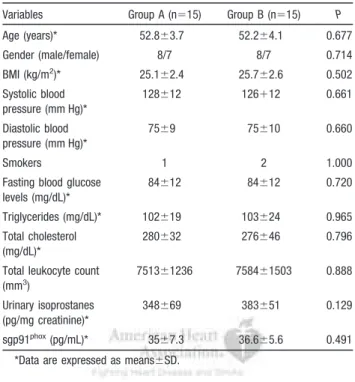
Table 3. Interventional Study: Baseline Characteristics of Hypercholesterolemic Patients Randomized to Diet Alone (Group A) or Diet Plus Atorvastatin (Group B)
Variables Group A (n⫽15) Group B (n⫽15) P Age (years)* 52.8⫾3.7 52.2⫾4.1 0.677 Gender (male/female) 8/7 8/7 0.714 BMI (kg/m2)* 25.1⫾2.4 25.7⫾2.6 0.502 Systolic blood pressure (mm Hg)* 128⫾12 126⫹12 0.661 Diastolic blood pressure (mm Hg)* 75⫾9 75⫾10 0.660 Smokers 1 2 1.000
Fasting blood glucose levels (mg/dL)* 84⫾12 84⫾12 0.720 Triglycerides (mg/dL)* 102⫾19 103⫾24 0.965 Total cholesterol (mg/dL)* 280⫾32 276⫾46 0.796 Total leukocyte count
(mm3) 7513⫾1236 7584⫾1503 0.888 Urinary isoprostanes (pg/mg creatinine)* 348⫾69 383⫾51 0.129 sgp91phox(pg/mL)* 35⫾7.3 36.6⫾5.6 0.491
*Data are expressed as means⫾SD.
likely that analysis of gp91phox only partly reflects the
NADPH oxidase expression because other subunits and/or
catalytic cores of NADPH oxidase homologs could be
de-tected in the human serum.
32Analysis of oxidative stress in humans is essentially based
on the measurement of markers of oxidative stress or
mole-cules that modulate antioxidant status such as antioxidant
vitamins or enzymes implicated in ROS scavenging or
degradation.
4,33,34Analysis of molecules implicated in the
generation of ROS such as NADPH oxidase may open new
avenues in understanding the role of oxidative stress in
several clinical settings including atherosclerosis. In this
latter context, measurement of sgp91
phoxin vivo could be
useful to explore the role of this ROS-generating pathway in
the progression of atherosclerosis.
The fact that atorvastatin inhibits sgp91
phoxin vivo is novel
and provides further insight in the mechanisms through which
statins could halt the progression of atherosclerotic disease.
Thus, studies conducted in human atherosclerotic plaque
demonstrated that NADPH oxidase is overexpressed and
predominantly contributes to vascular oxidative stress
35; also,
experimental studies demonstrated that the functional
defi-ciency of NADPH oxidase is associated with reduced
inflam-mation and atherosclerotic lesion.
12The exact mechanism by which atorvastatin reduces
NADPH oxidase, however, is unclear. Recent study showed
that statin treatment inhibits leukocyte ROCK activity, a
protein kinase implicated in the activation of NADPH
oxi-dase,
36with a mechanism that seems to be independent from
lowering cholesterol
37; further study is necessary to
deter-mine whether gp91
phoxdownregulation by statin is
ROCK-mediated.
In conclusion, we provide evidence that in
hypercholester-olemia, atorvastatin inhibits oxidative stress via gp91
phoxdownregulation. The inhibition of circulating sgp91
phoxby
atorvastatin represents a novel mechanism potentially
ac-counting for the antiatherosclerotic effect of statins.
Sources of Funding
This study was supported by a grant from the University of Rome “La Sapienza” (Ateneo 2006) (to F.V.).
Disclosures
None.
References
1. Mills EJ, Rachlis B, Wu P, Devereaux PJ, Arora P, Perri D. Primary prevention of cardiovascular mortality and events with statin treatments: a network meta-analysis involving more than 65,000 patients. J Am Coll Cardiol. 2008;52:1769 –1781.
2. Baigent C, Keech A, Kearney PM, Blackwell L, Buck G, Pollicino C, Kirby A, Sourjina T, Peto R, Collins R, Simes R. Cholesterol Treatment Trialists’ (CTT) Collaborators. Efficacy and safety of cholesterol-Figure 3. Serum sgp91phox(A), microparticle-bound gp91phox (B), urinary isoprostane (C), and total cholesterol (D) levels in hypercho-lesterolemic patients randomized to diet alone (group A) or diet plus atorvastatin (group B) at baseline and after 30 days of treatment. Box plots indicate median and 95% confidence intervals; whiskers, minimum and maximum values.
lowering treatment: prospective meta-analysis of data from 90,056 par-ticipants in 14 randomised trials of statins. Lancet. 2005;366:1267–1278. 3. Calabro` P, Yeh ET. The pleiotropic effects of statins. Curr Opin Cardiol.
2005;20:541–546.
4. Valko M, Leibfritz D, Moncol J, Cronin MT, Mazur M, Telser J. Free radicals and antioxidants in normal physiological functions and human disease. Int J Biochem Cell Biol. 2007;39:44 – 84.
5. Martino F, Pignatelli P, Martino E, Morrone F, Carnevale R, Di Santo S, Buchetti B, Loffredo L, Violi F. Early increase of oxidative stress and soluble CD40L in children with hypercholesterolemia. J Am Coll Cardiol. 2007;49:1974 –1981.
6. Cangemi R, Loffredo L, Carnevale R, Perri L, Patrizi MP, Sanguigni V, Pignatelli P, Violi F. Early decrease of oxidative stress by atorvastatin in hypercholesterolaemic patients: effect on circulating vitamin E. Eur Heart J. 2008;29:54 – 62.
7. Haramaki N, Ikeda H, Takenaka K, Katoh A, Sugano R, Yamagishi S, Matsuoka H, Imaizumi T. Fluvastatin alters platelet aggregability in patients with hypercholesterolemia: possible improvement of intraplatelet redox imbalance via HMG-CoA reductase. Arterioscler Thromb Vasc Biol. 2007;27:1471–1477.
8. Pereira EC, Bertolami MC, Paludi AA, Sevanian A, Abdalla DS. Anti-oxidant effect of simvastatin is not enhanced by its association with alpha-tocopherol in hypercholesterolemic patients. Free Radic Biol Med. 2004;37:1440 –1448.
9. Shishehbor MH, Brennan ML, Aviles RJ, Fu X, Penn MS, Sprecher DL, Hazen SL. Statins promote potent systemic antioxidant effects through specific inflammatory pathways. Circulation. 2003;108:426 – 431. 10. Violi F, Sanguigni V, Loffredo L, Carnevale R, Buchetti B, Finocchi A,
Tesauro M, Rossi P, Pignatelli P. Nox2 is determinant for ischemia-induced oxidative stress and arterial vasodilatation: a pilot study in patients with hereditary Nox2 deficiency. Arterioscler Thromb Vasc Biol. 2006;26:131–132.
11. Martino F, Loffredo L, Carnevale R, Sanguigni V, Martino E, Catasca E, Zanoni C, Pignatelli P, Violi F. Oxidative stress is associated with arterial dysfunction and enhanced intima-media thickness in children with hy-percholesterolemia: the potential role of nicotinamide-adenine dinucleo-tide phosphate oxidase. Pediatrics. 2008;122:648 – 655.
12. Cave AC, Brewer AC, Narayanapanicker A, Ray R, Grieve DJ, Walker S, Shah AM. NADPH oxidases in cardiovascular health and disease. Anti-oxidRedox Signal. 2006;8:691–728.
13. Hong H, Zeng JS, Kreulen DL, Kaufman DI, Chen AF. Atorvastatin protects against cerebral infarction via inhibition of NADPH oxidase-derived superoxide in ischemic stroke. Am J Physiol Heart Circ Physiol. 2006;291:H2210 –H2215.
14. Carmena R, Duriez P, Fruchart JC. Atherogenic lipoprotein particles in atherosclerosis. Circulation. 2004;15(suppl III):III-2–III-7.
15. Ferroni P, Basili S, Falco A, Davi G. Oxidant stress and platelet activation in hypercholesterolemia. Antioxid Redox Signal. 2004;6:747–756. 16. Pignatelli P, Sanguigni V, Lenti L, Ferro D, Finocchi A, Rossi P, Violi F.
gp91phox-dependent expression of platelet CD40 ligand. Circulation. 2004;110:1326 –1329.
17. Martire B, Rondelli R, Soresina A, Pignata C, Broccoletti T, Finocchi A, Rossi P, Gattorno M, Rabusin M, Azzari C, Dellepiane RM, Pietrogrande MC, Trizzino A, Di Bartolomeo P, Martino S, Carpino L, Cossu F, Locatelli F, Maccario R, Pierani P, Putti MC, Stabile A, Notarangelo LD, Ugazio AG, Plebani A, De Mattia D. IPINET. Clinical features, long term follow-up and outcome of a large cohort of patients with chronic granu-lomatous disease: an Italian multicenter study. Clin Immunol. 2008;126: 155–164.
18. Segal BH, Leto TL, Gallin JI, Malech HL, Holland SM. Genetic, bio-chemical, and clinical features of chronic granulomatous disease. Medicine. 2000;79:170 –200.
19. Executive Summary of The Third Report of The National Cholesterol Education Program (NCEP) Expert Panel on Detection, Evaluation, And Treatment of High Blood Cholesterol In Adults (Adult Treatment Panel III). JAMA. 2001;285:2486 –2497.
20. Hoffman SW, Roof RL, Stein DG. A reliable and sensitive enzyme immunoassay method for measuring 8-isoprostaglandin F2 alpha: a marker for lipid peroxidation after experimental brain injury. J Neurosci Methods. 1996;68:133–136.
21. Wang Z, Ciabattoni G, Cre´minon C, Lawson J, Fitzgerald GA, Patrono C, Maclouf J. Immunological characterization of urinary 8-epi-prostaglandin F2 alpha excretion in man. J Pharmacol Exp Ther. 1995;275:94 –100. 22. Carnevale R, Pignatelli P, Lenti L, Buchetti B, Sanguigni V, Di Santo S,
Violi F. LDL are oxidatively modified by platelets via GP91(phox) and accumulate in human monocytes. FASEB J. 2007;21:927–934. 23. Lawrie AS, Albanyan A, Cardigan RA, Mackie IJ, Harrison P.
Micro-particle sizing by dynamic light scattering in fresh-frozen plasma. Vox Sang. 2009;96:206 –212.
24. Blair P, Rex S, Vitseva O, Beaulieu L, Tanriverdi K, Chakrabarti S, Hayashi C, Genco CA, Iafrati M, Freedman JE. Stimulation of Toll-like receptor 2 in human platelets induces athromboinflammatory response through activation of phosphoinositide 3-kinase. Circ Res. 2009;104: 346 –354.
25. Pignatelli P, Sanguigni V, Lenti L, Loffredo L, Carnevale R, Sorge R, Violi F. Oxidative stress-mediated platelet CD40 ligand upregulation in patients with hypercholesterolemia: effect of atorvastatin. J Thromb Haemost. 2007;5:1170 –1178.
26. Rueckschloss U, Galle J, Holtz J, Zerkowski HR, Morawietz H. Induction of NAD(P)H oxidase by oxidized low-density lipoprotein in human endothelial cells: antioxidative potential of hydroxymethylglutaryl coenzyme A reductase inhibitor therapy. Circulation. 2001;104: 1767–1772.
27. Vecchione C, Brandes RP. Withdrawal of 3-hydroxy-3-methylglutaryl coenzyme A reductase inhibitors elicits oxidative stress and induces endothelial dysfunction in mice. Circ Res. 2002;91:173–179.
28. Wassmann S, Laufs U, Mu¨ller K, Konkol C, Ahlbory K, Ba¨umer AT, Linz W, Bo¨hm M, Nickenig G. Cellular antioxidant effects of atorvastatin in vitro and in vivo. Arterioscler Thromb Vasc Biol. 2002;22:300 –305. 29. Cathcart MK. Regulation of superoxide anion production by NADPH
oxidase in monocytes/macrophages: contributions to atherosclerosis. Arterioscler Thromb Vasc Biol. 2004;24:23–28.
30. Violi F, Basili S, Nigro C, Pignatelli P. Role of NADPH oxidase in atherosclerosis. Future Cardiol. 2009;5:83–92.
31. Janiszewski M, Do Carmo AO, Pedro MA, Silva E, Knobel E, Laurindo FR. Platelet-derived exosomes of septic individuals possess proapoptotic NAD(P)H oxidase activity: A novel vascular redox pathway. Crit Care Med. 2004;32:818 – 825.
32. Brown DI, Griendling KK. Nox proteins in signal transduction. Free Radic Biol Med. 2009;47:1239 –1253.
33. Pryor WA. Vitamin E and heart disease: basic science to clinical inter-vention trials. Free Radic Biol Med. 2007;28:141–164.
34. Tsimikas S. Oxidative biomarkers in the diagnosis and prognosis of cardiovascular disease. Am J Cardiol. 2006;98:9P–17P.
35. Griendling KK, Sorescu D, Ushio-Fukai M. NADPH oxidase: role in cardiovascular biology and disease. Circ Res. 2000;86:494 –501. 36. Rivera P, Ocaranza MP, Lavandero S, Jalil JE. Rho kinase activation and
gene expression related to vascular remodeling in normotensive rats with high angiotensin I converting enzyme levels. Hypertension. 2007;50: 792–798.
37. Liu PY, Liu YW, Lin LJ, Chen JH, Liao JK. Evidence for statin pleiotropy in humans: differential effects of statins and ezetimibe on rho-associated coiled-coil containing protein kinase activity, endothelial function, and inflammation. Circulation. 2009;119:131–138.